Self-etching adhesive (SEA) systems were re-cently introduced to simplify the bonding proce-dure, reducing the number of operative steps, and were initially considered a good replacement for the etch-and-rinse adhesive system that uses
or-thophosphoric acid (H₃PO₄) (PA) for enamel and dentin etching [1]. However, the recent literature underlines that the effectiveness of the enamel bonding with SEA is questionable [2]. This tech-nique in fact provides a bond strength of
compos-ORIGINAL PAPERS
Giuseppe Lo Giudice
1, A, F, Roberto Lo Giudice
2, D, Angelo Sergio Lizio
1, B,
Giuseppe Pantaleo
2, D, Frank Lipari
1, C, Michele Simeone
2, C,
Alessandra Valletta
2, B, Carlo Rengo
2, EEffects of Pre-Etching in Class V Cavities Restored
with Silorane and Methacrylate-Based Composites
Efekty wstępnego wytrawiania ubytków klasy V wypełnionych
materiałami kompozytowymi na bazie siloranów i metakrylanów
1 Department of Medical-Surgery and Odontostomatologic Experimental Sciences, University of Messina, Italy 2 Department of Neurosciences, Reproductive and Odontostomatological Sciences, University of Naples
“Federico II”, Italy
A – research concept and design; B – collection and/or assembly of data; C – data analysis and interpretation; D – writing the article; E – critical revision of the article; F – final approval of article
Abstract
Background. Self-etching adhesive (SEA) systems were recently introduced to simplify the bonding procedure and
were initially considered a good replacement for the etch-and-rinse adhesive system that uses orthophosphoric acid (PA) for enamel and dentin etching. Siloranes, a new class of ring opening monomers, were synthesized to overcome the problems related to polymerization shrinkage. This new type of monomer is obtained from the reac-tion of oxirane and silorane molecules with a volumetric shrinkage determined to be 0.99%.
Objectives. To assess the influence of preliminary phosphoric acid etching on the sealing ability of silorane- and
methacrylate-based composites.
Material and methods. Standard class V cavities were prepared on the buccal side of 48 extracted, sound,
human premolars. The specimens were randomly divided in two groups: A) Silorane System®/Filtek Silorane®;
B) Scotchbond Universal®/Filtek Supreme®. Each group was divided in two subgroups. A1) and B1): no
pre-etching was performed. A2) and B2): selective, enamel pre-pre-etching was performed. The interfacial sealing ability of the materials was evaluated by scoring the depth of methylene blue penetration through optical microscope observations. The differences in infiltration scores recorded for the tested materials were evaluated for statistical significance (Kruskal-Wallis ANOVA, Mann-Whitney U test, p < 0.05).
Results. No groups showed a “0” score, and group B2 had the lowest individual score, reaching at least a “2” score.
In the silorane groups, pre-etching decreased the infiltration score but the result was not statistically significant. No statistically significant differences emerged among the tested materials except for the Bis-GMA composite restored group where the pre-etching significantly reduced the interfacial leakage (p < 0.05).
Conclusions. Selective enamel pre-etching significantly reduced the marginal infiltration in class V cavities,
restored with Bis-GMA composite and a self-etching adhesive system. The low-shrinking silorane composite achieved better sealing ability (Dent. Med. Probl. 2016, 53, 3, 365–372).
Key words: adhesion, microleakage, acid-etching, silorane, self-etch.
Słowa kluczowe: adhezja, mikroprzeciek, wytrawianie kwasem, silorany, samowytrawianie.
Dent. Med. Probl. 2016, 53, 3, 365–372
ite to enamel significantly lower when compared with the etch-and-rinse system, due to its lower etching capability [3].
The better result achieved with the total-etch system seems to be correlated with the particular morphology of the interface obtained using 34–37% PA for enamel etching [4].
Self-etching primers are less aggressive than PA, do not form a proper and defined acid etch-ing pattern and the conditionetch-ing effects are also reduced on intact enamel surfaces [5].
Selective etching of enamel with PA is a poten-tial technique to improve the SEA system’s capabil-ity to achieve a higher bond strength of composite to enamel. It has been shown that pre-etching could provide a more retentive bonding substrate and more uniform resin-enamel bonding interface [6].
The combined action of PA and acidic self-etching monomers could cause a sufficient hy-bridization of enamel, which results in a reliable bond strength on enamel surface [7].
The majority of composites used in restorative dentistry are chemically based on the polymer-ization reaction of methacrylates. The conversion of methacrylate monomers into a polymer net-work, through the formation of covalent bonds, leads to a reduction of molecular distances and vol-ume reduction generating polymerization stress [8]. Negative clinical outcomes of composite resto-rations have often been associated with polymeriza-tion shrinkage and contracpolymeriza-tion stress, which may cause micro cracks, debonding, post-operative sen-sitivity, secondary caries and marginal gaps [9–12]. Siloranes, a new class of ring opening mono-mers, were synthesized to overcome the problems related to polymerization shrinkage. This new type of monomer is obtained from the reaction of oxirane and silorane molecules with a volumet-ric shrinkage determined to be 0.99%. The com-pensating mechanism for stress in this new sys-tem is achieved by opening and extending the ox-irane ring, during polymerization, to compensate for volume reduction [13, 14].
In order to ensure the best possible bonding between the silorane composites and hard den-tal tissues, a special adhesive has been developed. Silorane adhesive compositions include a philic one-step self-etching primer and a hydro-phobic viscous bond coating resin [15]. Accord-ing to the manufacturer, the silorane primer pres-ents a pH of 2.7, which provides mild etching and demineralization of the tooth structure, as well as a strong and durable bond.
Several laboratory and clinical studies on self-etch adhesives have shown that while ade-quate bonding could be achieved on dentin, the same approach does not ensure the same results
on enamel [16, 17]. Thus, preliminary phosphoric acid etching has been suggested to improve the ad-hesion to enamel [18–20].
The purpose of this experimental study was to analyze the effects of pre-etching in two differ-ent SE systems.
Material and Methods
Sample Preparation
Two commercially available composites were used in this study. The first composite (Filtek Silorane® 3M, 3M Italy, Milan, Italy) is based
on a siloranic resin, while the second composite (Filtek Supreme® XTE 3M, 3M Italy, Milan, Italy)
is a Bis-GMA nano-composite.
Forty-eight sound teeth extracted for peri-odontal reasons were used, stored in a 1% chlo-ramine solution at room temperature (20°C) for maximum 72 h.
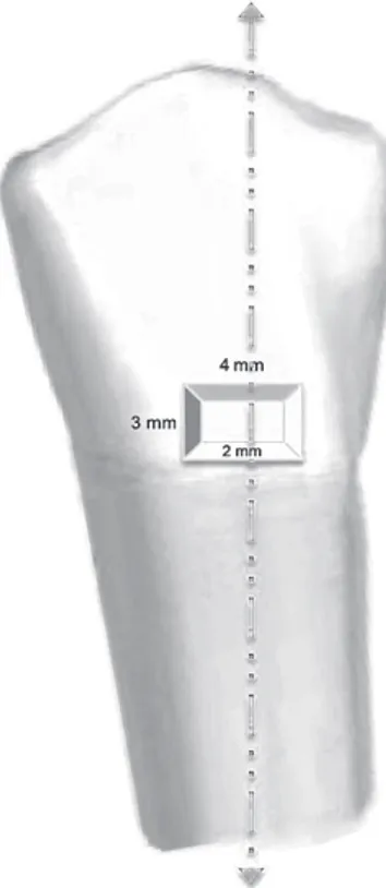
The radicular apexes of the teeth were sealed with epoxy resin and class V cavities were pre-pared with restoration margins placed in enamel and the bottom of the cavity in dentin. Cavities, with dimensions of 4 × 3 mm and 2 mm of depth were prepared on the buccal surface of each tooth, forming, in section, an axial wall and two per-pendicular (cervical and coronal) walls. Variation of ± 1 mm was considered acceptable [21] (Fig. 1).
The 2 mm depth was chosen in order to as-sure that the resinous materials could be totally
Fig. 1. Cavity
scheme with section line
placed in one single increment. The cervical mar-gin of the cavity was placed about 1.5 mm coro-nal from the cement-enamel junction. The cavity preparation was carried out by a single operator, using diamond drills (Komet ISO 233012, Komet Italy, Milan, Italy) mounted on a high-speed hand piece (Silent Power Gold CASTELLINI, Castellini, Bologna, Italy) under abundant water cooling [22].
The specimens were randomly divided into two groups (24 teeth each) in relation to the adhe-sive and the material used for the restorative phase. In the first group (A), a two-step self-etching adhe-sive (Silorane System 3M ESPE) and a low-shrink-ing composite (Filtek Silorane 3M ESPE) were used. In the second group (B), a one-step self-etching ad-hesive (Scotchbond® Universal 3M ESPE, 3M
Ita-ly, Milan, Italy) and a nano-filled composite (Filtek Supreme XTE 3M ESPE) were used.
Before the adhesive procedure, each group (A and B) was divided into two subgroups of twelve teeth each (A1–A2 and B1–B2). In the specimens of subgroups A1–B1, no pre-etching was per-formed. In the subgroups A2–B2, selective enamel pre-etching (15 s) with 37% PA (Scotchbond Uni-versal Etch 3M ESPE) was performed. The cavities were rinsed and dried according to the wet tech-nique [23].
In groups A1–A2, the adhesive used was a two-step Silorane Adhesive System (3M ESPE).
According to the manufacturer’s instructions, the primer was applied for 15 s, and cured for 10 s, then bonding was applied and light-cured for 20 s.
The restorative composite resin was placed in bulk and light cured for 40 s. Light-polymeri-zation was performed using a LED-curing device (LED Anthos T, CEFLA S.C., Imola, Italy) at 1000 mW/cm2.
In groups B1–B2, the self-etching adhesive was applied and rubbed on the cavity walls for 20 s, and then light cured for 10 s. The composite resin used in this group was light cured for 20 s.
All the restorations were finished with flexi-ble polishing disks (Sof-Lex, 3M ESPE, 3M Italy, Milan, Italy), Medium (40 µm), Fine (24 µm), and Super fine (8 µm), mounted on a slow speed hand piece operating at 5000 rev/min for 20 s, according to the manufacturer’s instructions.
To control the marginal adaptation, all op-erating procedures were carried out using
a head-mounted loupe (EyeMag Pro S Zeiss, Carl Zeiss S.p.A., Milan, Italy) at 4× magnification [24].
After 1 week of water storage at 37°C, the teeth were thermo-cycled (1.800 cycles at 5°C and 55°C with 60 s dwell time and 10 s transfer time).
Subsequently, the entire outer surface was cov-ered with a nail varnish within 1 mm of the resto-ration margins, and, following the manufacturer instructions, stored for 24 h to allow the varnish to dry. Subsequently, the teeth were immersed in a 7% methylene blue solution at room temperature for 3 days. The specimens were then water rinsed and sectioned longitudinally at the center of the restoration, obtaining two sections.
Optical Microscopy
and Microleakage Assessment
The tracer infiltration was evaluated using an optical microscope (OPMI PROergo S7B Zeiss, Carl Zeiss S.p.A., Milan, Italy) with a magnifica-tion of 12.5×.
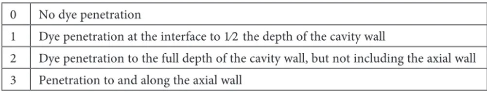
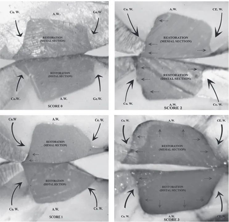
Infiltration assessment and measurement fol-lowed the scheme proposed by Osorio et al. [25], which assigns a progressive score in accordance with the infiltration extent (Table 1) (Fig. 2).
Two different operators separately carried out the evaluation. In case of disagreement, the high-est score was used for statistical analysis when the score was assigned.
Statistical analysis
A Kruskal-Wallis Non-Parametric Analyses of Variance (ANOVA) was applied to assess the significance of the differences in infiltration scores recorded for the tested materials (Table 4). For post hoc comparisons, a series of Mann-Whit-ney U tests was used (Tab. 5). In all the tests, the level of significance was set at p < 0.05, and calcu-lations were handled by IBM SPSS Statistics soft-ware (IBM Corporation, New York, USA).
Results
The descriptive statistics and score distribu-tion of this experimental in vitro study are shown in Tables 2 and 3.
Table 1. Score system to quantify marginal infiltration 0 No dye penetration
1 Dye penetration at the interface to 1⁄2 the depth of the cavity wall
2 Dye penetration to the full depth of the cavity wall, but not including the axial wall 3 Penetration to and along the axial wall
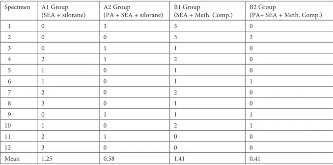
The Kruskal-Wallis test showed the presence of statistically significant differences between the tested materials (p = 0.028) (Table 4). No groups showed a mean overall “0” score. Moreover, the pre-etching technique improved the overall out-come, increasing the mean number of “0” scores found in each group. Both A1 and A2 groups showed a score of “3”. The B2 group had the low-est individual score, reaching at least a “2” (Table 2 and 3).
The Mann-Whitney U test showed that pre- -etching had a significant effect in the Bis-GMA composite restored group (Table 5).
The comparison between the two silorane subgroups (A1, A2) showed that the pre-etching
technique decreases the average infiltration value, from a mean value of 1.25 (A1) to 0.58 (A2), how-ever this outcome was not statistically significant (Table 5).
In the evaluation of the pre-etching procedure on the two different restorative materials, silorane and methacrylate, a similar performance was ob-served, with infiltration mean values respective-ly of 0.58 and 0.41, which were not significantrespective-ly different (Table 5). When the pre-etching tech-nique was not performed, in the silorane com-posite group an average grade of infiltration lower than in the Bis-GMA composite (1.25 vs 1.5) was observed (Table 2). Even this last comparison was not statistically significant (Table 5).
Fig. 2. Score examples: arrows indicate blue colorant solution infiltration. Ce.W. – cervical wall, A.W. – axial wall,
Table 2. Descriptive statistics of infiltration score data Specimen A1 Group
(SEA + silorane) A2 Group(PA + SEA + silorane) B1 Group(SEA + Meth. Comp.) B2 Group(PA+ SEA + Meth. Comp.)
1 0 3 3 0 2 0 0 3 2 3 0 1 1 0 4 2 1 2 0 5 1 0 1 0 6 1 0 1 1 7 2 0 2 0 8 3 0 1 0 9 0 1 1 1 10 1 0 2 1 11 2 1 0 0 12 3 0 0 0 Mean 1.25 0.58 1.41 0.41
Table 3. Score distribution in the different groups
Table 4. Kruskal-Wallis analysis
Adjusted H d.f. p-value
9.108 3 0.028
Discussion
Different authors have compared the influence of the pre-etching technique to many other chem-ical or mechanchem-ical techniques on the effective-ness of self-etching adhesives. Fitzgerald et al. [26] demonstrated how a pre-etching step with ortho-phosphoric acid (37%) for 5 s is preferable to a me-chanical preliminary treatment. Nazari et al. [7] recommended the pre-etching prior to application of the adhesive instead of grinding.
Lee et al. [27] showed that the additional ac-id etching both on enamel and dentin improves bond strength when low acidic one-step self-etch-ing adhesives are used, but adverse effects of ad-ditional acid etching, particularly on dentin, were reported.
These conclusions are linked to the possibil-ity of overly-aggressive dentin etching that might cause an excessive removal of the dentinal smear
layer, hindering a complete monomer infiltration into the collagen network [28].
Moreover, several studies have reported a low-er etching capability of self-etching adhesives on enamel, which can be caused by their relatively lower acidity compared to the 37% phosphoric ac-id used in most of the total-etch systems [29, 30]. This additional acid etching step should work se-lectively on the enamel and determines increased bond strength values. It could be speculated that the better adhesive performances are linked to a micro-mechanical retention determined by enhanced enamel porosity [28].
This effect is not the only parameter to fo-cus on. Carvalho et al. [12] and Osorio et al. [25] have reported that it is possible that the efficacy of a self-etching primer is not related only to pH lev-el, but also may be caused by the presence, after curing, of unpolymerized acid monomers.
Therefore, in the present study, the authors chose the self-etching adhesive approach because it is clinically most promising, since it elimi-nates the etching and the rinsing phases, reduc-es operative application time and greatly decreas-es the sensitivity of the technique or the possibility of making mistakes [3, 29]. Self-etch adhesive sys-tems create a wider protected collagen fiber area, due to the simultaneous infiltration of monomers that incorporates the smear layer into the hybrid layer [7].
When the self-etching adhesive was used without pre-etching, we observed that siloranes ensured a better seal, while, with pre-etching, the better adhesive effectiveness compensated for the different shrinking degree, and the infiltration score decreased significantly in the methacrylic-resin-restored group, which showed the best per-formance.
The silorane group without pre-etching achieved the best results and this could be linked to the ring shape polymerization reaction and the consequently low-shrinkage [13, 14].
The results from this study are consistent with those of Krifka et al. [31], who analyzed micro-leakage in class V comparing methacrylates and siloranes. They observed that silorane-based com-posites exhibited the best marginal seal.
Furthermore, other studies have underlined the advantages of the use of siloranes compared to other restorative materials. Silorane-based com-posites were more efficient for high C-factor cavi-ties [8].
The etching patterns of silorane primer are considered mild (pH 2.7) and may cause a reduced demineralization of the intact enamel surfaces. In our study, the limited thickness of the restora-tion and the low shrinking behavior of the com-posites may have determined lower infiltration scores [5].
The quality of marginal and internal adapta-tion of the Filtek Silorane composite was recently evaluated by Gregor et al. [22], who compared dif-ferent protocols of adhesive application.
The parameter analyzed was the continu-ous margin, defined as the adaptation of the lut-ing agent to enamel. Selective enamel etchlut-ing prior to the application of the adhesive showed a signifi-cantly higher number of infiltration-free specimens compared to the “non-etched” groups (p > 0.05).
Many authors have investigated the associa-tion between a silorane system adhesive and differ-ent pre-etching techniques too. Ustunkol et al. [32] compared it to laser etching and to a control group without any preliminary treatment. As with our results, they revealed that PA treatment seems the most promising surface treatment for increasing the enamel bond strength of silorane adhesive sys-tems.
Poureslami et al. [33] analyzed the effect of pre- -etching on the marginal adaptation of silorane resins in primary teeth too, concluding that this technique, used with different composite resins, provides the lowest marginal micro-leakage in the primary teeth.
In our research, in the methacrylate res-in group, pre-etchres-ing significantly improved the quality of the seal compared to the specimens where only self-etching was used.
The results achieved in the Bis-GMA restored group, when PA was applied, showed the best performances, even when compared to the low-shrinking composite group too.
In this study, independently of the restora-tion material used, the pre-etching groups showed Table 5. Statistical significance of between-group differences. Different capital letters label
statistically significant differences in infiltration scores
Group N Mean (SD) p-value Significance (p < 0.05)
A1 12 1.25 (1.139 0.15854 A
A2 12 0.58 (0.90) 0.0164 B
B1 12 1.41 (0.99) 0.70394 A
a better quality of seal than groups without pre-etching. This data is evidenced considering both the average infiltration scores and the number of specimens in each group with no infiltration (Ta-ble 3). Furthermore our microscopic reliefs con-firmed the low number of specimens with high-est infiltration.
The current experimental study demonstrat-ed that the pre-etching of enamel with orthophos-phoric acid significantly reduced the degree of marginal leakage in class V cavities restored with Bis-GMA composite and a self-etching adhesive system. In addition, the low-shrinking silorane composite demonstrated the ability to reduce mar-ginal infiltration.
According to this data, it can be concluded that the enamel pre-etching technique guarantees a good seal when self-etching adhesive systems are used, and is especially indicated when Bis-GMA resins are used.
Because of the reduced polymerization shrinkage of the silorane composite compared to the traditional methacrylate composite, the inter-face is exposed to significantly less stress, so the need for a pre-etching phase is reduced. Howev-er, pre-etching is still a step that the dental clini-cian should take into consideration to obtain bet-ter clinical performances even for these new low-shrinking silorane composites.
References
[1] Sheets J.L., Wilcox C.W., Barkmeier W.W., Nunn M.E.: The effect of phosphoric acid pre-etching and thermo-cycling on self-etching adhesive enamel bonding. J. Prosthet. Dent. 2012, 107, 102–108.
[2] Erikson R.L., Barkmeier W.W., Latta M.A.: The role of etching in bonding to enamel: A comparison of self-etching and etch-and-rinse adhesive systems. Dent. Mat. 2009, 25, 1459–1467.
[3] Osorio R., Monticelli F., Moreira M.A., Osorio E., Toledano M.: Enamel-resin bond durability of self-etch and etch & rinse adhesives. Am. J. Dent. 2009, 22, 371–375.
[4] Hegde M.N., Hegde P., Chandra C.R.: Morphological evaluation of new total etching and self etching adhesive system interfaces with dentin. J. Conserv. Dent. 2012, 15, 151–155.
[5] Devarasa G.M., Subba Reddy V.V., Chaitra N.L., Swarna Y.M.: Self-Etching adhesive on intact enamel, with and without pre-etching. Microsc. Res. Tech. 2012, 75, 650–654.
[6] Guilherme Erhardt M.C., Assad Cavalcante L.M., Freire Pimenta L.A.: Influence of phosphoric acid pre-treatment on self-etching adhesives. J. Esthet. Restor. Dent. 2004, 16, 33–41.
[7] Nazari A., Shimada Y., Sadr A., Tagami J.: Pre-etching vs. grinding in promotion of adhesion to intact enamel using self-etch adhesives. Dent. Mater. J. 2012, 31, 394–400.
[8] Ferracane J.L.: Developing a more complete understanding of stresses produced in dental composites during po-lymerization. Dent. Mater. 2005, 21, 36–42.
[9] Lo Giudice G., Lo Giudice A., Isola G., Fabiano F., Artemisia A., Fabiano V., Nucera R., Matarese G.: Evaluation of bond strength and detachment interface distribution of different bracket base designs. Acta Med. Mediterr. 2015, 31, 585–590.
[10] Braga R.R., Ballester R.Y., Ferracane J.L.: Factors involved in the development of polymerization shrinkage stress in resin composites: A systematic review. Dent. Mater. 2005, 21, 962–970.
[11] Kleverlaan C.J., Feilzer A.J.: Polymerization shrinkage and contraction stress of dental resin composites. Dent. Mater. 2005, 21, 1150–1157.
[12] Carvalho R.M., Pereira J.C., Yoshiyama M., Pashley D.H.: A review of polymerization contraction: The in-fluence of stress development versus stress relief. Oper. Dent. 1996, 21, 17–24.
[13] Weinmann W., Thalackar C., Guggenberger E.: Siloranes in dental composites. Dent. Mater. 2005, 21, 68–74. [14] Boaro L.C., Gonçalves F., Guimarães T.C., Ferracane J.L., Versluis A., Braga R.R.: Polymerization stress,
shrinkage and elastic modulus of current low shrinkage restorative composites. Dent. Mater. 2010, 26, 1144–1150. [15] Duarte S., Phark J.H., Varjao F.M., Sadan A.: Nanoleakage, ultramorphological characteristics, and microtensile
bond strengths of a new low-shrinkage composite to dentin after artificial aging. Dent. Mater. 2009, 25, 589–600. [16] Van Meerbeek B., Yoshihara K., Yoshida Y., Mine A., De Munck J., Van Landuyt K.L.: State of the art of
self-etch adhesives. Dent. Mater. 2011, 27, 17–28.
[17] Van Meerbeek B., Van Landuyt K.L., De Munck J., Hashimoto M., Peumans M., Lambrechts P., Yoshida Y., Inoue S., Suzuki K.: Technique-sensitivity of contemporary adhesives. Dent. Mater. 2005, 21, 1–13.
[18] Lo Giudice G., Centofanti A., Artemisia A., Bramanti E., Militi A., Rizzo G., Favaloro A., Alessia I., Lo Giudice R., Cutroneo G.: Dentin morphology of root canal surface: A quantitative evaluation based on a scan-ning electronic microscopy study. BioMed. Res. Int. 2015, 1–7, ID 164065.
[19] Van Landuyt K., Kanumilli P., De Munck J., Peumans M., Lambrechts P., Van Meerbeek B.: Bond strength of a mild self-etch adhesive with and without prior acid-etching. J. Dent. 2006, 34, 77–85.
[20] Goracci C., Rengo C., Eusepi L., Juloski J., Vichi A., Ferrari M.: Influence of selective enamel etching on the bonding effectiveness of a new “all-in-one” adhesive. Am. J. Dent. 2013, 26, 99–104.
[21] Manhart J., Chen H.Y., Mehl A., Weber K., Hickel R.: Marginal quality and microleakage of adhesive class V restorations. J. Dent. 2001, 29, 123–130.
[22] Gregor L., Bortolotto T., Feilzer A.J., Krejci I.: Effect of different bonding strategies on the marginal adap-tation of class 1 silorane restorations. Am. J. Dent. 2013, 26, 127–131.
[23] Araújo J.F., Barros T.A., Braga E.M., Loretto S.C., Silva e Souza Pde A., Silva e Souza M.H.: One-year eval-uation of a simplified ethanol-wet bonding technique: A randomized clinical trial. Braz. Dent. J. 2013, 24, 267–272. [24] Lo Giudice G., Lo Giudice R., Matarese G., Isola G., Cicciù M., Terranova A., Palaia G., Romeo U.:
Eval-uation of magnification systems in restorative dentistry: An in vitro study. Dent. Cadmos, 2015, 85, 296–305. [25] Osorio R., Toledano M., de Leonardi G., Tay F.: Microleakage and interfacial morphology of self-etching
ad-hesives in class V resin composite restorations. J. Biomed. Mater. Res. Part B. Appl. Biomater. 2003, 66, 399–409. [26] Fitzgerald I., Bradley G.T., Bosio J.A., Hefti A.F., Berzins D.W.: Bonding with self-etching primers-pumice
or pre-etch? An in vitro study. Eur. J. Orthod. 2012, 34, 257–261.
[27] Lee I.S., Son S.A., Hur B., Kwon Y.H., Park J.K.: The effect of additional etching and curing mechanism of com-posite resin on the dentin bond strength. J. Adv. Prosthodont. 2013, 5, 479–484.
[28] Ikeda M., Kurokawa H., Sunada N., Tamura Y., Takimoto M., Murayama R., Ando S., Miyazaki M.: Influ-ence of previous acid etching on dentin bond strength of self-etch adhesives. J. Oral Sci. 2009, 51, 527–534. [29] Lo Giudice G., Cicciù M., Cervino G., Lizio A., Visco A.M.: Flowable resin and marginal gap on tooth third
me-dial cavity involving enamel and radicular cement. SEM evaluation of two restoration techniques. Indian J. Dent. Res. 2012, 23, 763–769.
[30] Lo Giudice G., Lipari F., Lizio A., Cervino G., Cicciù M.: Tooth fragment reattachment technique on a pluri traumatized tooth. J. Conserv. Dent. 2012, 15, 80–83.
[31] Krifka S., Federlin M., Hiller K.A., Schmalz G.: Microleakage of silorane and methacrylate-based class V com-posite restorations. Clin. Oral Investig. 2012, 16, 1117–1124.
[32] Ustunkol I., Yazici A.R., Gorucu J., Dayangac B.: Influence of laser etching on enamel and dentin bond strength of Silorane System Adhesive. Lasers Med. Sci. 2015, 30, 695-700.
[33] Poureslami H.R., Sajadi F., Sharifi M., Farzin Ebrahimi S.: Marginal microleakage of low-shrinkage compos-ite silorane in primary teeth: An in vitro study. J. Dent. Res. Dent. Clin. Dent. Prospects 2012, 6, 94–97.
Address for correspondence:
Roberto Lo Giudice Via Dei Mille n. 272 98123, Messina Italy
Tel: +39 393 439 91 97
E-mail: [email protected] Conflict of interest: None declared Received: 9.03.2016
Revised: 3.04.2016 Accepted: 5.04.2016