Angiotensin II type 1/adenosine A
2A
receptor oligomers: a novel target
for tardive dyskinesia
Paulo A. de Oliveira
1, James A. R. Dalton
2, Marc López-Cano
3,4, Adrià Ricarte
2, Xavier
Morató
3,4, Filipe C. Matheus
1, Andréia S. Cunha
1, Christa E. Müller
5, Reinaldo N. Takahashi
1,
Víctor Fernández-Dueñas
3,4, Jesús Giraldo
2, Rui D. Prediger
1,6& Francisco Ciruela
3,4Tardive dyskinesia (TD) is a serious motor side effect that may appear after long-term treatment with neuroleptics and mostly mediated by dopamine D2 receptors (D2Rs). Striatal D2R functioning may be
finely regulated by either adenosine A2A receptor (A2AR) or angiotensin receptor type 1 (AT1R) through
putative receptor heteromers. Here, we examined whether A2AR and AT1R may oligomerize in the
striatum to synergistically modulate dopaminergic transmission. First, by using bioluminescence resonance energy transfer, we demonstrated a physical AT1R-A2AR interaction in cultured cells.
Interestingly, by protein-protein docking and molecular dynamics simulations, we described that a stable heterotetrameric interaction may exist between AT1R and A2AR bound to antagonists (i.e.
losartan and istradefylline, respectively). Accordingly, we subsequently ascertained the existence of AT1R/A2AR heteromers in the striatum by proximity ligation in situ assay. Finally, we took advantage
of a TD animal model, namely the reserpine-induced vacuous chewing movement (VCM), to evaluate a novel multimodal pharmacological TD treatment approach based on targeting the AT1R/A2AR complex.
Thus, reserpinized mice were co-treated with sub-effective losartan and istradefylline doses, which prompted a synergistic reduction in VCM. Overall, our results demonstrated the existence of striatal AT1R/A2AR oligomers with potential usefulness for the therapeutic management of TD.
Angiotensin II (AII) is a peptidic hormone that causes vasoconstriction through activation of angiotensin recep-tor type 1 (AT1R). Indeed, it is a key component of the renin-angiotensin system (RAS), which regulates blood
pressure1. Accordingly, blocking AT
1Rs with selective antagonists (i.e. losartan) constitutes the first-line therapy
to deal with hypertensive patients2. Interestingly, AII is also synthesized in the brain, where its levels are much
higher than those observed in plasma3. In addition, AT
1Rs are expressed both in neurons and glial cells4. Thus,
the existence of an endogenous brain angiotensin system has been postulated, which may respond to AII synthe-sized in and/or transported into the brain (for review see ref. 5). The function of AII in the brain has still not been fully elucidated. However, a role in the control of stress reaction and cerebral circulation, and in the mechanisms leading to brain ischemia, neuronal injury and inflammation has been demonstrated5. In addition, AT
1R blockade
reduced brain inflammation responses6 and had beneficial effects in processes involving microglial activation
and neuroinflammation (such as animal models of Alzheimer’s disease, brain ischemia and multiple sclerosis) (for review see ref. 7). Similarly, in animal models of parkinsonism induced by neurotoxins 6-hydroxydopamine (6-OHDA) and 1-methyl-4-phenyl-1,2,3,6-tetrahydropyridine (MPTP), an increase in AII levels, with concomi-tant AT1R overactivation, has been observed8–10. On the other hand, the presence of RAS components in the basal
ganglia in general and in the nigrostriatal system in particular has also been reported. Altogether, it has been
1Departamento de Farmacologia, Universidade Federal de Santa Catarina, Trindade, 88049-900, Florianópolis,
SC, Brazil. 2Institut de Neurociències and Unitat de Bioestadística, Universitat Autònoma de Barcelona, Network
Biomedical Research Center on Mental Health (CIBERSAM), Bellaterra, Spain. 3Unitat de Farmacologia, Departament
de Patologia i Terapèutica Experimental, Facultat de Medicina, IDIBELL-Universitat de Barcelona, L’Hospitalet de Llobregat, Spain. 4Institut de Neurociències, Universitat de Barcelona, Barcelona, Spain. 5PharmaCenter Bonn,
Pharmaceutical Institute, Pharmaceutical Chemistry I, University of Bonn, Bonn, Germany. 6Programa de
Pós-graduação em Neurociências, Centro de Ciências Biológicas, Universidade Federal de Santa Catarina, Trindade, 88049-900, Florianópolis, SC, Brazil. Paulo A. de Oliveira and James A. R. Dalton contributed equally to this work. Correspondence and requests for materials should be addressed to J.G. (email: [email protected]) or R.D.P. (email: [email protected]) or F.C. (email: [email protected])
Received: 12 January 2017 Accepted: 6 April 2017 Published: xx xx xxxx
postulated that brain RAS may be involved in dopaminergic degeneration, especially when the dopaminergic system is impaired, thus contributing to the pathogenesis and progression of dopaminergic-related pathologies such as Parkinson’s disease (PD).
The concept that cell surface receptors may physically interact forming oligomers appeared early in the eight-ies, while characterizing G protein-coupled receptors (GPCRs) for neurotransmitters11, 12. Notably, striatal
dopa-minergic receptors in general, and the dopamine D2 receptor (D2R) in particular, constitute the archetypal GPCR
capable of forming receptor-receptor complexes. Indeed, the potential impact of these oligomers in pathophysi-ological conditions involving dopaminergic dysfunction has been extensively studied. Interestingly, the D2R has
been shown to oligomerize with several GPCRs13, including the adenosine A
2A receptor (A2AR)14. The D2R-A2AR
heteromer is expressed in GABAergic striatopallidal neurons and a reciprocal negative allosteric receptor-receptor interaction is defined as its “biochemical fingerprint”15. Noteworthy, the D
2R-A2AR heteromer has been defined
as a potential pharmacological target for pathologies associated with dysfunctional dopaminergic signaling, such as PD and schizophrenia. Indeed, A2AR antagonists (i.e. istradefylline) are currently used for PD treatment in
Japan16. On the other hand, the D
2R has also been shown to oligomerize with the AT1R in the striatum17, thus the
potential use of AT1R ligands to modulate dopaminergic signaling has been postulated. Interestingly, early studies
also indicated interactions between the adenosinergic and the angiotensinergic systems, for instance the antino-ciceptive effect of AII was related to that produced by adenosine A1 receptor agonists18. In addition, an A2AR- and
AT1R-mediated synergistic interaction in the peripheral RAS was described19, 20. Thus, while adenosine was able
to reverse the stimulatory effect of AII on Na+-ATPase activity in the renal proximal tubules via A
2AR activation21,
A2AR blockers reduced AII-mediated ROS formation via Nox2 (NADPH complex enzyme) in endothelial cells20.
Conversely, AII potentiated the adenosine-induced contraction of afferent arterioles22, while losartan-mediated
AT1R blockade abolished the adenosine-mediated reflex sympatho-excitatory response in the brachial artery23.
Altogether, the aforementioned evidence highlights the need for a better understanding of the adenosinergic system-RAS interaction. Furthermore, this interaction may be relevant not only in the periphery but also in the brain, where a functional interplay with the dopaminergic system may occur.
Here, we study the possible existence, both in cultured cells and in mouse striatum, of a physical AT1R-A2AR
interaction, which may be a potential target for managing dopaminergic-related disorders (i.e. tardive dyskinesia, TD). Also, we seek to characterize the most likely heteromeric receptor arrangement through protein-protein docking and long-timescale molecular dynamics (MD) simulations. Finally, we propose a novel multimodal treat-ment for TD based on the use of AT1R and A2AR antagonists at sub-effective doses, and test it in a mouse TD
model, namely the reserpine-induced vacuous chewing movement (VCM).
Results
AT
1R and A
2AR form heteromers in cultured cells.
Based on the existence of AT1R/D2R heteromers17,we aimed to elucidate whether AT1R is also able to oligomerize with the A2AR, a well-known D2R partner24. To
this end, we first assessed the co-distribution of AT1R and A2AR in cultured cells through the fluorescence
detec-tion of CFP/YFP tagged receptors. Thus, by means of confocal microscopy analysis of HEK-293T cells transiently expressing AT1RCFP and A2ARYFP, a high overlapping in the distribution of the former receptors was observed
(Fig. 1a). Next, we examined the possible physical interaction of AT1R and A2AR in living cells by means of the
BRET approach. Thus, cells were transiently transfected with receptor constructs carrying the appropriate fluoro-phore pairs (A2ARRluc and AT1RYFP). A positive and saturable BRET signal was observed in cells co-transfected
with a constant concentration of the A2ARRluc and increasing concentrations of AT1RYFP (Fig. 1b). Of note, as the
control pair GABAB2RRluc and AT1RYFP led to a low and linear distribution, the specificity of the saturation
(hyper-bolic) assay for the A2ARRluc and AT1RYFP pair could be established (Fig. 1b). Overall, these results demonstrate
that AT1R and A2AR form heteromers in living HEK-293T cells.
Structure of AT
1R/A
2AR heteromer.
Computational modeling, protein-protein docking, and MD simu-lations were used to probe the interaction between AT1R and A2AR, and determine their most likely heteromericarrangement. Initially, AT1R and A2AR antagonists (losartan and istradefylline, respectively), were docked into
their respective inactive-state receptor crystal structure using Autodock4.225. The corresponding best docked
AT1R-losartan and A2AR-istradefylline complexes were then embedded in lipid bilayer membranes and
sub-jected to MD simulations of 250 ns and 500 ns, respectively, where both bound antagonists were observed to stabilize. In particular, in AT1R, ARG167, located on extracellular loop 2 (ECL2) above the orthosteric pocket,
was observed to make H-bonds with losartan at both ends of the ligand (see SI Fig. 1) in a similar manner to that observed in the AT1R crystal structure containing bound olmesartan26. Likewise, in A2AR, ASN253
(ASN6.55 in Ballesteros-Weinstein numbering27) made an H-bond with istradefylline in a similar manner to other
co-crystallized A2AR xanthine antagonists28 (Fig. S1).
As both AT1R and A2AR are thought to form functional homodimers at the cell surface29–35, we investigated the
likely structure and behavior of these respective homodimers with bound antagonists, prior to investigating het-eromeric interactions. In order to do this we utilized the A2AR homodimer crystal structure with co-crystallized
antagonist36 as a structural guide for initializing AT
1R-losartan and A2AR-istradefylline homodimer models. This
dimeric crystal structure is observed to contain an interface between TM4 and TM5 helices of each monomer, with TM4 of one monomer interacting with TM5 of the other, and vice versa36. Initial AT
1R and A2AR
homod-imer models were refined with protein-protein docking using the ROSIE webserver37, each consisting of two
antagonist-bound receptors in the same MD-generated conformation (see above). Following protein-protein docking, the A2AR and AT1R homodimers were subjected to further MD simulations of 1.5 μs and 750 ns,
respectively. During these simulations, both AT1R and A2AR homodimers were seen to form significant
inter-actions via their TM4 and TM5 helices, respectively, with considerable contact between monomers, indicative of energetically stable dimers (Fig. S2). In addition, the respective bound antagonists remained stably bound in
each participating monomer, with all receptor subunits maintaining an inactive state. From these results, it was inferred that the antagonist-bound homodimeric states of AT1R and A2AR are stable in silico, and likely form the
minimum constituents that participate in cross-receptor heteromeric interaction.
As other described heteromeric interactions involving A2AR fit a heterotetramer model38, 39, and as MD
simu-lations of AT1R and A2AR homodimers suggest their respective stability, we investigated heterotetrameric
interac-tions between the two receptor homodimers. As there is no crystal structure for GPCRs in tetrameric formation, we performed extensive protein-protein docking with ROSIE to identify the highest possible scoring interaction of AT1R and A2AR homodimers (see Methods). The “best” conformation identified a tetramer with cross-receptor
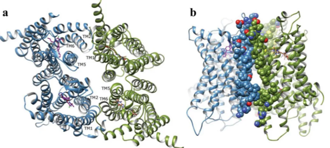
interfaces involving TM5 and TM6 of one receptor with TM1 and TM2 of the other, and vice versa (Fig. 2). In order to assess the stability of the proposed interaction, the heterotetramer complex was subjected to an MD sim-ulation in a membrane for 2 µs. Results show the receptors progressively stabilized (RMSD curve in Fig. S3) and enhanced their interaction, whilst maintaining the original tetrameric configuration (Fig. 2). Furthermore, the respective AT1R and A2AR homodimers remained stable and unperturbed within the tetramer, maintaining their
respective inactive states. In conclusion, stable heterotetrameric interaction between AT1R and A2AR is plausible
at a molecular level and compatible with bound antagonists, losartan and istradefylline.
Functional consequences of the AT
1R and A
2AR oligomerization.
The formation of AT1R-A2ARcomplexes in transfected cells suggests that there might exist a functional coupling between these two receptors. Thus, we assessed the impact of A2AR expression on AT1R-mediated intracellular Ca2+ mobilization from internal
stores by means of Fluo4 determinations. Thus, in Fluo4 loaded cells expressing AT1R alone, the activation with
angiotensin II increased intracellular Ca2+ (Fig. 3a, red trace), as expected. Interestingly, in cells co-expressing
AT1R and A2AR, the angiotensin II-mediated intracellular Ca2+ mobilization was boosted (Fig. 3a, blue trace).
Indeed, in cells expressing only A2AR, a residual and not significant effect of angiotensin II was observed,
prob-ably because of the endogenous expression of AT1R in HEK-293T cells (Fig. 3a, black trace). Quantification of
the results (Fig. 3c) demonstrated a significant [F (2,6) = 8.40 (P < 0.05)] difference between the experimental groups assessed, thus a significant (P < 0.05) increase in the AT1R-mediated intracellular calcium accumulation
Figure 1. AT1R and A2AR physically interact in HEK-293T cells. (a) Co-distribution of AT1R and A2AR in
HEK-293T cells. Transiently transfected HEK-293T cells with AT1RYFP (red) and A2ARCFP (green) were fixed
and observed by confocal microscopy. Co-distribution (yellow) is shown in the merge image. Scale bar: 10 µm. (b). Direct interaction between AT1 and A2A receptors. BRET saturation curves in HEK-293T cells expressing
A2ARRluc and AT1RYFP (blue) or GABAB2RRluc and AT1RYFP (red). Plotted on the x-axis is the fluorescence value
obtained from the YFP, normalized with the luminescence value of Rluc-tagged vectors 10 min after benzyl-coelenterazine incubation. Results are expressed as mean ± SEM (n = 3, in triplicate).
in AT1R-A2AR cells was observed (Fig. 3c). These results suggest that a functional interplay between AT1R and
A2AR might exist upon expression in heterologous cells.
AT
1R and A
2AR heteromers are expressed in mouse striatum.
Once demonstrated that AT1R andA2AR assemble into functionally interacting complexes in living cells, we aimed to determine the existence of
AT1R/A2AR heteromers in native tissue, namely the striatum. To this end, we first conducted
immunofluores-cence experiments to assess the expression levels and distribution of both AT1R and A2AR in mouse striatum.
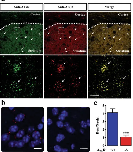
Interestingly, both receptors showed a high degree of co-distribution throughout the striatal neuropil (Fig. 4a, upper panels) and eventually within the medium spiny neurons (MSN) cell bodies (Fig. 4a, lower panels). Importantly, the myelinated fiber bundles that penetrate the striatum were visible as dark (not stained) structures within the stained neuropil (Fig. 4a, upper panel). These results give rise to the possibility that these two receptors might be forming heteromers under native conditions. Subsequently, to confirm the existence of AT1R/A2AR
heteromers in the striatum we implemented the P-LISA approach, a well described technique providing enough Figure 2. Conformational arrangement of AT1R/A2AR heterotetramer. Model generated by protein-protein
docking and 2 μs MD simulation. (a) Top view of tetramer (AT1R in blue, A2AR in green, losartan in purple and
istradefylline in brown). (b) Side view of interaction between A2AR and AT1R.
Figure 3. A2AR expression potentiates AT1R functioning. (a) Representative Angiotensin II-mediated
intracellular Ca2+ accumulation determined by Fluo4 assay. HEK-293T cells were transiently transfected with
A2AR (black trace), AT1R (red trace) and A2AR + AT1R (blue trace). Cells were loaded with Fluo4-NW dye and
challenged with Angiotensin II (50nM). The [Ca2+]
i dynamics is shown as change in fluorescence of the Fluo4
signal (F) expressed as percentage of the maximal Ca2+ influx elicited by ionomycin (F
i) in each experimental
conditions. (b) Quantification of the AT1R-mediated [Ca2+]i accumulation measured by Fluo4. The integrated
area under the curve (AUC) of the normalized AT1R-mediated Fluo4 signal (F) is expressed as percentage of
the corresponding ionomycin signal (Fi) for each transfection. The data are expressed as the mean ± SEM of three independent experiments performed in triplicate. The asterisk indicates statistically significant differences (**P < 0.01, ***P < 0.001; 1-way ANOVA with a Newman-Keuls post-hoc test).
sensitivity to evaluate receptor’s close proximity within a named GPCR oligomer in native conditions40. Thus, by
using proper antibody combinations, the AT1R/A2AR heteromer expression in mouse striatum was addressed by
P-LISA assays. Indeed, red dots reflecting a positive P-LISA signal was observed in the striatum of wild-type mice (Fig. 4b), thus allowing the visualization of the AT1R/A2AR receptor-receptor interaction. Interestingly, in striatal
slices from the A2AR-KO mice the P-LISA signal was negligible (Fig. 4b), thus reinforcing the specificity of our
P-LISA assay. Indeed, when the P-LISA signal was quantified the wild-type animal showed 4 ± 0.5 dots/nuclei while the A2AR-KO displayed only 1 ± 0.2 dots/nuclei under the same experimental conditions. Thus, a marked
and significant (P < 0.005) reduction in the P-LISA signal was observed in the A2AR-KO striatal slices. Taken
together, data gathered from our P-LISA experiments strongly support the existence of AT1R/A2AR heteromers
in the mouse striatum.
Functional interplay between AT
1R and A
2AR in an animal model of TD.
A2AR-containingoli-gomers, including A2AR/D2R41, are thought to be involved in the control of locomotor function both in normal
Figure 4. Detection of AT1R and A2AR proximity in mice striatal sections. (a) Immunohistochemistry
detection of AT1R and A2AR in mice striatum. Representative confocal microscopy images of AT1R (red) and
A2AR (green) immunoreactivities in the striatum are shown. Lower panels show a magnification of the square
area shown in the upper panel. Arrows indicate potential location of medium spiny neurons (MSN) cell bodies. Superimposition of images revealed a high receptor co-distribution in yellow (merge). Scale bars: 350 μm (upper panels) and 10 μm (lower panels). (b) Photomicrographs of dual recognition of AT1R and A2AR with P-LISA.
Representative images from wild-type (left) and A2AR-KO (right) mice striatum. (c) Quantification of P-LISA
signals for AT1R and A2AR proximity confirmed the significant difference of P-LISA signal density between wild
type and A2AR-KO mice (***P < 0.001). Values in the graph correspond to the mean ± SEM (dots/nuclei) of at
and pathological conditions42, 43. However, although A
2AR has been linked to neuroleptic-induced TD44, 45, its
impact on this syndrome is still ambiguous46. Consequently, we sought to investigate whether the AT
1R/A2AR
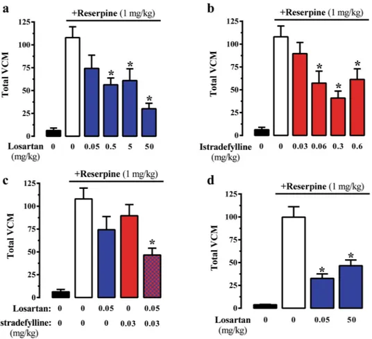
heteromer might play a role in TD. We took advantage of the vacuous chewing movement (VCM) model of TD in mice. Interestingly, administration of the AT1R antagonist losartan dose-dependently reduced reserpine-induced
VCM (Fig. 5a). Similarly, administration of the A2AR antagonist istradefylline dose-dependently reduced
reserpine-induced VCM (Fig. 5b). Subsequently, we investigated whether co-treatment at sub-effective low doses of AT1R and A2AR antagonists would elicit a significant reduction of VCM in our reserpine-induced TD
ani-mal model. Therefore, for combination treatment, 0.05 mg/kg of losartan and 0.03 mg/kg of istradefylline were selected as they were not effective in reducing VCM. Noteworthy, the combined treatment produced a significant (P < 0.05) reduction in VCM (Fig. 5c), thus demonstrating a synergistic interaction between both drugs. Overall, these results suggest that co-treatment with AT1R and A2AR antagonists at sub-effective low doses is a useful
ther-apeutic approach for TD management.
Finally, in an attempt to ascertain the role of AT1R/A2AR oligomers in the synergistic effect observed upon
receptor antagonist co-treatment, we assessed the efficacy of the VCM sub-effective losartan dose in mice lack-ing the A2AR (i.e. A2AR-KO mice). Interestingly, the low dose of losartan (0.05 mg/kg) was able to significantly
(P < 0.05) reduce the number of VCM in the A2AR-KO mice (Fig. 5d). Hence, in the absence of A2AR the
effi-cacy of losartan was higher, thus indicating that AT1R/A2AR heteromers are crucial for finely modulating TD.
Collectively, these results suggest that AT1R and A2AR functionally interact in vivo and that this functional
inter-play may be provided by the existence of AT1R/A2AR oligomers.
Discussion
TD is a serious motor side effect associated to long-term treatment with neuroleptics47. Notably, D
2R
occu-pancy and its transience to occupation have been identified as a potential mechanistic substrate to develop antipsychotic-induced TD48. Indeed, D
2R-mediated control of motor function has been related to the ability of
this receptor to oligomerize with other GPCRs in general49 and with the A
2AR in particular42, 43, 50. Also, in the
brain, dopaminergic neurotransmission can be modulated by AII through AT1R. Thus, AT1R blocking precludes
AII-mediated dopamine release51, 52. Furthermore, a functional interaction between angiotensin and dopamine
Figure 5. Effect of AT1R and A2AR blocking in the TD animal model. The effect of different doses of losartan
(a) or istradefylline (b) on total vacuous chewing movements (VCM) in the reserpine-based animal model of TD in mice was monitored during 10 min. (c) Effects of sub-effective dose co-administration (i.p.) of losartan (0.05 mg/ml) and istradefylline (0.03 mg/ml) in the VCM of TD animal model. (d) Effect of losartan in the VCM of TD animal model performed in A2AR-KO mice. Results are represented as the mean ± SEM (n = 10 animals).
receptors in the striatum and substantia nigra53, 54, together with the formation of D
2R and AT1R heteromers in the
striatum has been described17. Based on these data, we decided to explore a possible direct interaction between
AT1R and A2AR, and revealed for the first time the existence of AT1R/A2AR oligomers in the striatum and its
implications in TD.
Our experimental data shows that AT1R and A2AR form heteromers both in co-transfected cells and in mouse
striatum. This feature is especially strengthened by our in-silico analysis, which has predicted a heterotetrameric receptor arrangement that was stable during 2 μs of MD simulation. The “best” receptor-receptor interface identified for the AT1R/A2AR heterotetramer involves TM5 and TM6 of one receptor with TM1 and TM2 of the other, and
vice versa, while in the respective homodimers the TM4 of one monomer interactac with TM5 of the other, and vice versa. Interestingly, the D2R/A2AR heterodimeric interface has been postulated to be formed by the TM4 and TM5
of D2R interacting with TM4 and TM5 of the A2AR49, 55. Therefore, when considering a putative AT1R/D2R/A2AR
oligomer new in-silico analysis will be needed to accurately determine TM-TM contacts and receptor rearrange-ment defining AT1R/D2R/A2AR oligomer stoichiometry. Overall, this information will be extremely valuable when
assessing potential multimodal TD pharmacotherapeutic interventions based on drugs targeting these receptors. The AT1R/A2AR oligomerization was shown to elicit functional consequences, since co-expression with A2AR
boosted AT1R signaling. This AT1R gain of function may most likely result from an A2AR-mediated AT1R increased
cell surface targeting, as was previously reported56. Alternatively, an A
2AR-mediated direct trans-activation of
AT1R could not be excluded, as has been described for other A2AR-containing oligomers41. Thus, further work
is needed to elucidate the precise molecular mechanism behind this AT1R/A2AR oligomer-dependent AT1R gain
of function. Nevertheless, our main purpose consisted of ascertaining the in vivo implications of the AT1R/A2AR
oligomer formation, which is the cornerstone when describing a new GPCR oligomer57. Indeed, our P-LISA data
strongly supported the existence of AT1R/A2AR heteromers in the mouse striatum, thus warranting the need to
assess the impact of this oligomer in behaving animals. Accordingly, we demonstrated an unprecedented syner-gism of AT1R and A2AR antagonists on the control of involuntary mandibular movements induced by reserpine
in an animal model of TD. Thus, co-treatment with AT1R and A2AR antagonists at sub-effective low doses robustly
(>60%) reduced reserpine-mediated VCM. Certainly, this makes this multimodal pharmacological approach an attractive solution for TD management.
The striatum is considered a pivotal brain region, since it receives projections from other basal ganglia areas and from many other brain regions involved in motor and non-motor functions, such as the motor cortex, the pre-frontal cortex and the hippocampus58, 59. Indeed, both the renin-angiotensin and the adenosinergic systems play an
important role in controlling the striatal function. Thus, the ability of AT1R and A2AR to heteromerize in the
stria-tum might constitute a way of fine-tuning multiple receptor-signaling pathways harmonizing dopaminergic neuro-transmission. Therefore, the AT1R/A2AR oligomer could be envisaged as a potential drug target for striatum-related
adverse motor dysfunctions associated to therapy, including TD and L-DOPA induced dyskinesia (LID). Indeed, A2AR antagonists have been postulated and licensed as antiparkinsonian drugs60 and eventually studied in the
man-agement of LID61. Furthermore, A
2AR has been linked to neuroleptic-induced TD44, 45, although with some debate46.
Similarly, preclinical studies have demonstrated that blockade of AT1R reduces LID62. It is assumed that these A2AR-
and AT1R-mediated anti-LID effects are related to their ability to heteromerize with D2R17, 24 and thus controlling
dopaminergic neurotransmission. However, it could be speculated that AT1R and A2AR might control D2R function
through functional AT1R/D2R/A2AR-containing complexes in GABAergic striatopallidal neurons. A number of facts
support this last statement: i) the high and selective co-expression of AT1R, D2R and A2AR in these particular cells;
ii) the demonstration of A2AR/D2R, AT1R/D2R and AT1R/A2AR heteromers; and iii) the existence of strong multiple
interactions between the three receptors. In conclusion, the demonstration of their simultaneous physical interac-tion may constitute a novel and very attractive target for developing new drugs in the management of pathologies in which these receptors play a key role, such as TD.
Methods
Reagents.
The primary antibodies used were: rabbit anti-AT1R polyclonal antibody (Abcam, Cambridge,UK), and mouse anti-A2AR monoclonal antibody (Millipore, Billerica, MA, USA). The secondary antibodies
were: horseradish peroxidase (HRP)-conjugated goat anti-rabbit IgG (Pierce Biotechnology, Rockford, IL, USA) and Cy3-conjugated donkey anti-mouse IgG antibody (Jackson ImmunoResearch Laboratories, West Grove, PA, USA). The ligands used were: losartan (Abcam); angiotensin II, istradefylline (KW-6002), reserpine and ionomy-cin from Sigma-Aldrich (St. Louis, MO, USA).
Plasmid constructs.
To perform co-localization and BRET experiments, the A2AR constructs containinga cyan fluorescent protein (CFP; A2ARCFP), or the Renilla luciferase (Rluc; A2ARRluc) were used. The AT1R and
GABAB2 receptor constructs containing a yellow fluorescent protein (YFP; AT1RYFP, GABAB2RYFP) were cloned,
as previously described63.
Animals.
CD-1 mice (Charles River Laboratories and from the central animal facility of Federal University of Santa Catarina) and A2AR-KO mice developed in a CD-1 genetic background64 (animal facility of Universityof Barcelona) weighing 20–25 g were used. The University of Barcelona and Federal University of Santa Catarina Committee on Animal Use and Care approved the protocol. Animals were housed and tested in compliance with the guidelines described in the Guide for the Care and Use of Laboratory Animals65 and following the European Union
directives (2010/63/EU). All efforts were made to minimize animal suffering and the number of animals used. All animals were housed in groups of five in standard cages with ad-libitum access to food and water and maintained under 12 h dark/light cycle (starting at 7:30 AM), 22 °C temperature, and 66% humidity (standard conditions).
Cell culture.
Human embryonic kidney (HEK)-293T cells were grown in Dulbecco’s modified Eagle’s medium (DMEM) (Sigma-Aldrich) supplemented with 1 mM sodium pyruvate, 2 mM L-glutamine, 100 U/mL streptomycin, 100 mg/mL penicillin and 5% (v/v) fetal bovine serum at 37 °C and in an atmosphere of 5% CO2.HEK-293T cells growing in 25 cm2 flasks or six-well plates containing 18 mm coverslips were used for western
blot and fluorescence imaging, respectively. They were transiently transfected with the cDNA encoding the spec-ified proteins using Polyethylenimine (Polysciences, Inc. Warrington, PA, USA).
Fixed brain tissue preparation.
Mice were anesthetized and perfused intracardially with 100–200 ml ice-cold 4% paraformaldehyde (PFA) in phosphate-buffered saline (PBS; 8.07 mM Na2HPO4, 1.47 mM KH2PO4,137 mM NaCl, 0.27 mM KCl, pH 7.2). Brains were post-fixed in the same solution of PFA at 4 °C during 12 h. Coronal sections (25 μm) were processed using a vibratome (Leica Lasertechnik GmbH, Heidelberg, Germany). Slices were collected in Walter’s Antifreezing solution (30% glycerol, 30% ethylene glycol in PBS, pH 7.2) and kept at −20 °C until processing.
Bioluminescence resonance energy transfer measurements.
Bioluminescence resonance energy transfer (BRET) experiments in HEK-293T cells were performed as previously described66. In brief, HEK-293Texpressing the indicated constructs were rapidly washed, detached, and resuspended in HBSS buffer (137 mM NaCl, 5 mM KCl, 0.34 mM Na2HPO4, 0.44 mM KH2PO4, 1.26 mM CaCl2, 0.4 mM MgSO4, 0.5 mM MgCl2, 10 mM
HEPES, pH 7.4) containing 10 mM glucose. Cell suspensions (20 μg of protein) were distributed in 96-well microplate plates, 5 μM h-coelenterazine (NanoLight Technology, Pinetop, AZ, USA) was added and BRET determined in a POLARstar Optima plate-reader (BMG Labtech, Durham, NC, USA) as previously described66.
Intracellular calcium determination.
The AT1R-mediated intracellular Ca2+ accumulation was assessedby Fluo4-NW Calcium Assay Kit (Invitrogen, Carlsbad, CA, USA). Thus, transiently transfected HEK-293T cells were lifted and plated in 96-well black plates with transparent bottoms. Cells were incubated with the Fluo4-NW following the instructions of the manufacturer and washed with HBSS. Fluorescence signals were measured at 530 nm during 60 s while injecting Angiotensin II (50 nM) and ionomycin (5 μM) at seconds 5 and 40 respectively, using a POLARstar Optima plate-reader (BMG Labtech). The specific Angiotensin II-induced Fluo4 signal (F) was expressed as percentage of the signal elicited by ionomycin (Fi) in each set of experimental conditions67.
Immunohistochemistry.
Previously collected slices were washed three times in PBS, permeabilized with 0.3% Triton X-100 in PBS for 2 hours and rinsed back three times more with wash solution (0.05% Triton X-100 in PBS). The slices were then incubated with blocking solution (10% NDS in wash solution; Jackson ImmunoResearch Laboratories, Inc., West Grove, PA, USA) for 2 h at R.T. and subsequently incubated with the primary antibodies overnight at 4 °C. After two rinses (10 min each) with 1% NDS in wash solution, sections were incubated for 2 h at R.T. with the appropriate secondary antibodies conjugated with Alexa dyes (Invitrogen, Carlsbad, CA, USA), then washed (10 min each) two times with 1% NDS in wash solution and two more times with PBS, and mounted on slides. Fluorescence striatal images were obtained using a Leica TCS 4D confocal scanning laser microscope (Leica Lasertechnik GmbH).Proximity ligation in situ assay.
Duolink in situ PLA detection Kit (Olink Bioscience, Uppsala, Sweden) was performed in a similar manner as immunohistochemistry explained above until the secondary antibody incubation step. The following steps were performed following the manufacturer’s protocol, as previously described24, 40. Fluorescence images were acquired on a Leica TCS 4D confocal scanning laser microscope (LeicaLasertechnik GmbH) using a 60x N.A. =1.42 oil objective from the selected area. High-resolution images were acquired as a z-stack with a 0.2 μm z-interval with a total thick of 5 μm. Nonspecific nuclear signal was eliminated from PLA images by substracting DAPI labeling. Analyze particle function from Image J (NIH) was used to count particles larger than 0.3 μm2 for PLA signal and larger than 100 μm2 to discriminate neuronal from glia
nuclei68. For each image a number of oligomer particles and neuron nuclei was obtained and ratio among them
was calculated.
Computational modeling.
Ligand docking. Crystal structures of AT1R (PDB id: 4ZUD) and A2AR (PDBid: 4EIY) were converted into apo wt forms by removing co-crystallized ligands and non-native fusion proteins i.e. cytochrome b562, building missing intracellular and extracellular loop sections with MODELLER v9.1469, and
energy minimizing in the AMBER14SB force-field70 with CHIMERA v1.10.271. The AT
1R antagonist losartan and
A2AR antagonist istradefylline were downloaded from PubChem72, energy-minimized in AMBER14SB force-field
with CHIMERA and docked into respective receptor structures with Autodock4.225 using default parameters.
Grid points were generated to cover total orthosteric pocket volumes. The final docking conformations of losartan and istradefylline represented top hits identified by best predicted affinity (nM). These were checked to be con-sistent with previously reported binding modes of relevant co-crystallized antagonists26, 28. In particular, losartan
was docked to interact with Arg167 and istradefylline was docked to interact with Asn253. Subsequent energy minimization of docked structures was performed with CHIMERA in the AMBER-14SB force-field.
Protein-protein docking. For generating homodimers of respective receptors: AT1R and A2AR with bound
antag-onists, two molecular dynamics (MD)-generated receptor-ligand monomers (see MD methods) of either AT1R
or A2AR, in each case, were superimposed onto the A2AR homodimer crystal structure (PDB id: 4EIY), yielding
an initial homodimer model, which was then submitted to the ROSIE webserver37 for protein-protein docking.
For both AT1R and A2AR, the best docked homodimer was identified by three factors: best possible ROSETTA
orientation. For construction of an AT1R-A2AR heterotetramer, two initial tetrameric arrangements were
man-ually generated by combining respective MD-generated AT1R and A2AR homodimers (see MD section) in
alter-native ways: (i) where homodimers are arranged side-to-side in a rectangular-like configuration, where each homodimer subunit interacts with a subunit of the other homodimer (by respective TM1/2–5/6 helices), (ii) where homodimers are partially displaced with respect to one another creating a parallelogram-like configura-tion, where both subunits of one homodimer interact with a single subunit of the other homodimer (by respective TM4/5 helices). Both these alternative configurations were submitted to the ROSIE webserver for identifica-tion of the best possible tetrameric arrangement according to the same criteria implemented previously. For all protein-protein docking runs executed on the ROSIE webserver, default local parameters were used, i.e. perturba-tion of 3 Å between proteins, 8° of tilt, and 360° rotaperturba-tion around protein centers, with generaperturba-tion of 1000 docking solutions per case.
Molecular dynamics system setup. Five different systems were generated using the CHARMM-GUI web-based
interface73, each in a POPC membrane and solvated with TIP3P water molecules: AT
1R monomer with bound
losartan, A2AR monomer with bound istradefylline, AT1R homodimer with bound losartan, A2AR homodimer
with bound istradefylline, and AT1R-A2AR heterotetramer with bound antagonists. All receptor structures were
orientated according to the OPM database74 entry: 4eiy. Charge neutralizing ions (0.15 M KCl) were introduced
to each system. Parameters of membrane, water and protein were automatically generated by CHARMM-GUI73
according to CHARMM36 force-field75 with ligand parameters automatically generated according to
CHARMM36 General Force Field76–78.
Molecular dynamics simulations. Molecular dynamics (MD) simulations of AT1R and A2AR were performed
using the CHARMM36 force-field75 with ACEMD79 on specialized GPU-computer hardware, totaling 5 μs across
systems. In detail, monomer AT1R/A2AR systems were equilibrated for 20 ns at 300 K and 1 atm, while AT1R/A2AR
homodimers and heterotetramer systems were equilibrated for 50 ns under same conditions. During equilibra-tion, positional harmonic restraints on protein and antagonist heavy atoms were progressively released over the first 8 ns and then continued without constraints. After equilibration, AT1R and A2AR monomers were subjected
to unbiased production runs of 250 ns and 500 ns under same conditions, respectively. Likewise, AT1R and A2AR
homodimers were subjected to unbiased production runs of 750 ns and 1.5 μs, respectively. The AT1R/A2AR
het-erotetramer was subjected to an unbiased production run of 2 μs. Simulation trajectories were analyzed using VMD software v1.9.280.
Reserpine-induced vacuous chewing movements.
The VCM model of TD48 was induced in micethrough two subcutaneous (s.c.) reserpine injections (1 mg/kg) administered with an interval of 48 h. Twenty-four hours after the last reserpine administration, mice were treated by intraperitoneal (i.p.) route with losartan (0.05– 50 mg/kg) and/or istradefylline (0.03–0.06 mg/kg). VCM parameters were evaluated as previously described81 but
with some modifications. Thus, the evaluation of VCM frequency consists of a manual counting of continuous single mouth openings in a vertical plane, not directed to a physical material. Mirrors were placed on the table and behind the glass cylinder (Ø 19 cm and 22 cm height) to allow observation of the orofacial movements when mice were not facing the observer. The evaluation of this parameter during 10 min was performed by a blind observer, 30 min after the pharmacological treatments administered 24 h after the second reserpine injection81.
Statistics.
The number of samples (n) in each set of experimental conditions is indicated in figure legends. Statistical analysis was performed by one-way ANOVA followed by Newman-Keuls post-hoc test or Student’st-test when appropriate. Statistical significance was considered at P < 0.05.
References
1. Brunner, H. R., Chang, P., Wallach, R., Sealey, J. E. & Laragh, J. H. Angiotensin II vascular receptors: their avidity in relationship to sodium balance, the autonomic nervous system, and hypertension. J. Clin. Invest. 51, 58–67, doi:10.1172/JCI106797 (1972). 2. Goa, K. L. & Wagstaff, A. J. Losartan potassium: a review of its pharmacology, clinical efficacy and tolerability in the management of
hypertension. Drugs 51, 820–45, doi:10.2165/00003495-199651050-00008 (1996).
3. Hermann, K., McDonald, W., Unger, T., Lang, R. E. & Ganten, D. Angiotensin biosynthesis and concentrations in brain of normotensive and hypertensive rats. J. Physiol. (Paris). 79, 471–80 (1984).
4. Garrido-Gil, P., Valenzuela, R., Villar-Cheda, B., Lanciego, J. L. & Labandeira-Garcia, J. L. Expression of angiotensinogen and receptors for angiotensin and prorenin in the monkey and human substantia nigra: an intracellular renin-angiotensin system in the nigra. Brain Struct. Funct. 218, 373–88, doi:10.1007/s00429-012-0402-9 (2013).
5. Saavedra, J. M. Brain Angiotensin II: New Developments, Unanswered Questions and Therapeutic Opportunities. Cell. Mol. Neurobiol. 25, 485–512, doi:10.1007/s10571-005-4011-5 (2005).
6. Saavedra, J. M. Angiotensin II AT(1) receptor blockers ameliorate inflammatory stress: a beneficial effect for the treatment of brain disorders. Cell. Mol. Neurobiol. 32, 667–81, doi:10.1007/s10571-011-9754-6 (2012).
7. Labandeira-Garcia, J. L. et al. Brain renin-angiotensin system and dopaminergic cell vulnerability. Front. Neuroanat. 8, 67, doi:10.3389/fnana.2014.00067 (2014).
8. Grammatopoulos, T. N. et al. Angiotensin type 1 receptor antagonist losartan, reduces MPTP-induced degeneration of dopaminergic neurons in substantia nigra. Mol. Neurodegener. 2, 1, doi:10.1186/1750-1326-2-1 (2007).
9. Zawada, W. M. et al. Generation of reactive oxygen species in 1-methyl-4-phenylpyridinium (MPP+) treated dopaminergic neurons occurs as an NADPH oxidase-dependent two-wave cascade. J. Neuroinflammation 8, 129, doi:10.1186/1742-2094-8-129 (2011). 10. Sonsalla, P. K. et al. The angiotensin converting enzyme inhibitor captopril protects nigrostriatal dopamine neurons in animal
models of parkinsonism. Exp. Neurol. 250, 376–83, doi:10.1016/j.expneurol.2013.10.014 (2013).
11. Agnati, L. F., Fuxe, K., Zini, I., Lenzi, P. & Hokfelt, T. Aspects on receptor regulation and isoreceptor identification. Med. Biol. 58, 182–187 (1980).
12. Fuxe, K. et al. Evidence for the existence of receptor–receptor interactions in the central nervous system. Studies on the regulation of monoamine receptors by neuropeptides. J. neural Transm. 18, 165–179 (1983).
13. Gomes, I. et al. G Protein–Coupled Receptor Heteromers. Annu. Rev. Pharmacol. Toxicol. 56, 403–425, doi: 10.1146/annurev-pharmtox-011613-135952 (2016).
14. Ciruela, F. et al. Combining Mass Spectrometry and Pull-Down Techniques for the Study of Receptor Heteromerization. Direct Epitope–Epitope Electrostatic Interactions between Adenosine A 2A and Dopamine D 2 Receptors. Anal. Chem. 76, 5354–5363, doi:10.1021/ac049295f (2004).
15. Ferre, S. et al. An Update on Adenosine A2A-Dopamine D2 receptor interactions. Implications for the Function of G Protein-Coupled Receptors. Curr. Pharm. Des. 14, 1468–1474, doi:10.2174/138161208784480108 (2008).
16. Müller, T. The safety of istradefylline for the treatment of Parkinson’s disease. Expert Opin. Drug Saf. 14, 769–75, doi:10.1517/14740 338.2015.1014798 (2015).
17. Martínez-Pinilla, E. et al. Dopamine D2 and angiotensin II type 1 receptors form functional heteromers in rat striatum. Biochem. Pharmacol. 96, 131–142, doi:10.1016/j.bcp.2015.05.006 (2015).
18. Pechlivanova, D. M. & Georgiev, V. P. Interaction of angiotensin II and adenosine A1 and A2A receptor ligands on the writhing test in mice. Pharmacol. Biochem. Behav. 72, 23–8, doi:10.1016/S0091-3057(01)00707-9 (2002).
19. Tchekalarova, J., Kambourova, T. & Georgiev, V. Long-term theophylline treatment changes the effects of angiotensin II and adenosinergic agents on the seizure threshold. Brain Res. Bull. 52, 13–16, doi:10.1016/S0361-9230(99)00254-3 (2000).
20. Thakur, S., Du, J., Hourani, S., Ledent, C. & Li, J. M. Inactivation of adenosine A2A receptor attenuates basal and angiotensin II-induced ROS production by Nox2 in endothelial cells. J. Biol. Chem. 285, 40104–40113, doi:10.1074/jbc.M110.184606 (2010). 21. Gomes, C. P. et al. Crosstalk between the signaling pathways triggered by angiotensin II and adenosine in the renal proximal tubules:
Implications for modulation of Na+ -ATPase activity. Peptides 29, 2033–2038, doi:10.1016/j.peptides.2008.07.004 (2008).
22. Lai, E. Y. & Patzak, A. Persson, a. E. G. & Carlström, M. Angiotensin II enhances the afferent arteriolar response to adenosine through increases in cytosolic calcium. Acta Physiol. 196, 435–445 (2009).
23. Rongen, Ga, Brooks, S. C., Ando, S. I., Abramson, B. L. & Floras, J. S. Angiotensin AT1 receptor blockade abolishes the reflex sympatho- excitatory response to adenosine. J. Clin. Invest. 101, 769–776, doi:10.1172/JCI480 (1998).
24. Fernández-Dueñas, V. et al. Untangling dopamine-adenosine receptor-receptor assembly in experimental parkinsonism in rats. Dis. Model. Mech 8, 57–63, doi:10.1242/dmm.018143 (2015).
25. Morris, G. M. et al. AutoDock4 and AutoDockTools4: Automated docking with selective receptor flexibility. J. Comput. Chem. 30, 2785–91, doi:10.1002/jcc.v30:16 (2009).
26. Zhang, H. et al. Structural Basis for Ligand Recognition and Functional Selectivity at Angiotensin Receptor. J. Biol. Chem. 290, 29127–29139, doi:10.1074/jbc.M115.689000 (2015).
27. Ballesteros, J. A. & Weinstein, H. Integrated methods for the construction of three-dimensional models of structure–function relations in G protein-coupled receptors. Methods Neurosci 25, 366–428, doi:10.1016/S1043-9471(05)80049-7 (1995).
28. Doré, A. S. S. et al. Structure of the Adenosine A2A Receptor in Complex with ZM241385 and the Xanthines XAC and Caffeine. Structure 19, 1283–1293, doi:10.1016/j.str.2011.06.014 (2011).
29. Thévenin, D., Lazarova, T., Roberts, M. F. & Robinson, C. R. Oligomerization of the fifth transmembrane domain from the adenosine A 2A receptor. Protein Sci. 14, 2177–2186, doi:10.1110/ps.051409205 (2005).
30. Canals, M. et al. Homodimerization of adenosine A2A receptors: qualitative and quantitative assessment by fluorescence and bioluminescence energy transfer. J. Neurochem 88, 726–734, doi:10.1046/j.1471-4159.2003.02200.x (2004).
31. Karip, E., Turu, G., Supeki, K., Szidonya, L. & Hunyady, L. Cross-inhibition of angiotensin AT1 receptors supports the concept of receptor oligomerization. Neurochem. Int. 51, 261–267, doi:10.1016/j.neuint.2007.05.018 (2007).
32. AbdAlla, S., Lother, H., Langer, A., el Faramawy, Y. & Quitterer, U. Factor XIIIA Transglutaminase Crosslinks AT1 Receptor Dimers of Monocytes at the Onset of Atherosclerosis. Cell 119, 343–354, doi:10.1016/j.cell.2004.10.006 (2004).
33. Hansen, J. L., Theilade, J., Haunso, S. & Sheikh, S. P. Oligomerization of Wild Type and Nonfunctional Mutant Angiotensin II Type I Receptors Inhibits G q Protein Signaling but Not ERK Activation. J. Biol. Chem. 279, 24108–24115, doi:10.1074/jbc.M400092200
(2004).
34. Fanelli, F. & Felline, A. Dimerization and ligand binding affect the structure network of A2A adenosine receptor. Biochim. Biophys. Acta - Biomembr. 1808, 1256–1266, doi:10.1016/j.bbamem.2010.08.006 (2011).
35. Gracia, E. et al. A2A adenosine receptor ligand binding and signalling is allosterically modulated by adenosine deaminase. Biochem. J. 435, 701–709, doi:10.1042/BJ20101749 (2011).
36. Liu, W. et al. Structural Basis for Allosteric Regulation of GPCRs by Sodium Ions. Science (80-.). 337, 232–236, doi:10.1126/ science.1219218 (2012).
37. Lyskov, S. et al. Serverification of Molecular Modeling Applications: The Rosetta Online Server That Includes Everyone (ROSIE). PLoS One 8, e63906, doi:10.1371/journal.pone.0063906 (2013).
38. Bonaventura, J. et al. Allosteric interactions between agonists and antagonists within the adenosine A2A receptor-dopamine D2 receptor heterotetramer. Proc. Natl. Acad. Sci. USA 112, E3609–18, doi:10.1073/pnas.1507704112 (2015).
39. Navarro, G. et al. Quaternary structure of a G-protein-coupled receptor heterotetramer in complex with Gi and Gs. BMC Biol. 14, 26, doi:10.1186/s12915-016-0247-4 (2016).
40. Taura, J., Fernández-Dueñas, V. & Ciruela, F. Visualizing G Protein-Coupled Receptor-Receptor Interactions in Brain Using Proximity Ligation In Situ Assay. Curr. Protoc. Cell Biol. 67, 17.17.1–17.17.16 (2015).
41. Ciruela, F. et al. Adenosine receptor containing oligomers: their role in the control of dopamine and glutamate neurotransmission in the brain. Biochim. Biophys. Acta 1808, 1245–1255, doi:10.1016/j.bbamem.2011.02.007 (2011).
42. Ferre, S. et al. Adenosine A2A-dopamine D2 receptor-receptor heteromers. Targets for neuro-psychiatric disorders. Parkinsonism Relat. Disord. 10, 265–271, doi:10.1016/j.parkreldis.2004.02.014 (2004).
43. Ferre, S. et al. Adenosine A2A receptors and A2A receptor heteromers as key players in striatal function. Front. Neuroanat. 5 (2011). 44. Parsons, B., Togasaki, D. M., Kassir, S. & Przedborski, S. Neuroleptics up-regulate adenosine A2a receptors in rat striatum:
implications for the mechanism and the treatment of tardive dyskinesia. J. Neurochem. 65, 2057–64, doi:10.1046/j.1471-4159.1995.65052057.x (1995).
45. Bishnoi, M., Chopra, K. & Kulkarni, S. K. Involvement of adenosinergic receptor system in an animal model of tardive dyskinesia and associated behavioural, biochemical and neurochemical changes. Eur. J. Pharmacol. 552, 55–66, doi:10.1016/j.ejphar.2006.09.010
(2006).
46. Ivanova, S. A. et al. No involvement of the adenosine A2A receptor in tardive dyskinesia in Russian psychiatric inpatients from Siberia. Hum. Psychopharmacol. Clin. Exp. 27, 334–337, doi:10.1002/hup.v27.3 (2012).
47. Andreassen, O. A. & Jørgensen, H. A. Neurotoxicity associated with neuroleptic-induced oral dyskinesias in rats. Implications for tardive dyskinesia? Prog. Neurobiol. 61, 525–41, doi:10.1016/S0301-0082(99)00064-7 (2000).
48. Turrone, P., Remington, G. & Nobrega, J. N. The vacuous chewing movement (VCM) model of tardive dyskinesia revisited: is there a relationship to dopamine D(2) receptor occupancy? Neurosci. Biobehav. Rev. 26, 361–80, doi:10.1016/S0149-7634(02)00008-8
(2002).
49. Fuxe, K. et al. Moonlighting proteins and protein-protein interactions as neurotherapeutic targets in the G protein-coupled receptor field. Neuropsychopharmacology 39, 131–55, doi:10.1038/npp.2013.242 (2014).
50. Fuxe, K., Marcellino, D., Genedani, S. & Agnati, L. Adenosine A(2A) receptors, dopamine D(2) receptors and their interactions in Parkinson’s disease. Mov. Disord. 22, 1990–2017, doi:10.1002/mds.v22:14 (2007).
51. Mendelsohn, F. A., Jenkins, T. A. & Berkovic, S. F. Effects of angiotensin II on dopamine and serotonin turnover in the striatum of conscious rats. Brain Res. 613, 221–9, doi:10.1016/0006-8993(93)90902-Y (1993).
52. Brown, D. C., Steward, L. J., Ge, J. & Barnes, N. M. Ability of angiotensin II to modulate striatal dopamine release via the AT1 receptor in vitro and in vivo. Br. J. Pharmacol. 118, 414–20, doi:10.1111/bph.1996.118.issue-2 (1996).
53. Villar-Cheda, B. et al. Nigral and striatal regulation of angiotensin receptor expression by dopamine and angiotensin in rodents: Implications for progression of Parkinson’s disease. Eur. J. Neurosci 32, 1695–1706, doi:10.1111/j.1460-9568.2010.07448.x (2010). 54. Villar-Cheda, B. et al. Aging-related dysregulation of dopamine and angiotensin receptor interaction. Neurobiol. Aging 35,
1726–1738, doi:10.1016/j.neurobiolaging.2014.01.017 (2014).
55. Borroto-Escuela, D. O. et al. Characterization of the A2AR-D2R interface: focus on the role of the C-terminal tail and the transmembrane helices. Biochem. Biophys. Res. Commun. 402, 801–807, doi:10.1016/j.bbrc.2010.10.122 (2010).
56. Moriyama, K. & Sitkovsky, M. V. Adenosine A2A Receptor Is Involved in Cell Surface Expression of A2B Receptor. J. Biol. Chem.
285, 39271–39288, doi:10.1074/jbc.M109.098293 (2010).
57. Ciruela, F. et al. G protein-coupled receptor oligomerization for what? J. Recept. Signal Transduct. Res. 30, 322–330, doi:10.3109/10 799893.2010.508166 (2010).
58. Ferré, S. et al. Adenosine-cannabinoid receptor interactions. Implications for striatal function. Br. J. Pharmacol. 160, 443–53, doi:10.1111/j.1476-5381.2010.00723.x (2010).
59. Yager, L. M., Garcia, A. F., Wunsch, A. M. & Ferguson, S. M. The ins and outs of the striatum: Role in drug addiction. Neuroscience
301, 529–541, doi:10.1016/j.neuroscience.2015.06.033 (2015).
60. Jenner, P. Istradefylline, a novel adenosine A2A receptor antagonist, for the treatment of Parkinson’s disease. Expert Opin. Investig. Drugs 14, 729–738, doi:10.1517/13543784.14.6.729 (2005).
61. Jenner, P. et al. Adenosine, adenosine A 2A antagonists, and Parkinson’s disease. Parkinsonism Relat. Disord. 15, 406–413, doi:10.1016/j.parkreldis.2008.12.006 (2009).
62. Muñoz, A., Garrido-gil, P., Dominguez-meijide, A. & Labandeira-garcia, J. L. Angiotensin type 1 receptor blockage reduces L -dopa-induced dyskinesia in the 6-OHDA model of Parkinson’ s disease. Involvement of vascular endothelial growth factor and interleukin-1 β. Exp. Neurol. 261, 720–732, doi:10.1016/j.expneurol.2014.08.019 (2014).
63. García-Negredo, G. et al. Coassembly and coupling of SK2 channels and mGlu5 receptors. J. Neurosci. 34, 14793–802, doi:10.1523/ JNEUROSCI.2038-14.2014 (2014).
64. Ledent, C. et al. Aggressiveness, hypoalgesia and high blood pressure in mice lacking the adenosine A2a receptor. Nature 388, 674–8, doi:10.1038/41771 (1997).
65. Clark, J. D., Gebhart, G. F., Gonder, J. C., Keeling, M. E. & Kohn, D. F. Special Report: The 1996 Guide for the Care and Use of Laboratory Animals. ILAR J. 38, 41–48, doi:10.1093/ilar.38.1.41 (1997).
66. Ciruela, F. & Fernández-Dueñas, V. GPCR oligomerization analysis by means of BRET and dFRAP. Methods Mol. Biol. 1272, 133–144, doi:10.1007/978-1-4939-2336-6_10 (2015).
67. Garcia-Negredo, G. et al. Coassembly and Coupling of SK2 Channels and mGlu5 Receptors. J. Neurosci. 34, 14793–14802, doi:10.1523/JNEUROSCI.2038-14.2014 (2014).
68. Matamales, M. et al. Striatal medium-sized spiny neurons: identification by nuclear staining and study of neuronal subpopulations in BAC transgenic mice. PLoS One 4, e4770, doi:10.1371/journal.pone.0004770 (2009).
69. Sali, A. Comparative protein modeling by satisfaction of spatial restraints. Mol. Med. Today 1, 270–7, doi: 10.1016/S1357-4310(95)91170-7 (1995).
70. Case, D. A. et al. The Amber biomolecular simulation programs. J. Comput. Chem. 26, 1668–1688, doi:10.1002/jcc.20290 (2005). 71. Pettersen, E. F. et al. UCSF Chimera–a visualization system for exploratory research and analysis. J. Comput. Chem. 25, 1605–12,
doi:10.1002/jcc.20084 (2004).
72. Kim, S. et al. PubChem Substance and Compound databases. Nucleic Acids Res. 44, D1202–D1213, doi:10.1093/nar/gkv951 (2016). 73. Jo, S., Kim, T., Iyer, V. G. & Im, W. CHARMM-GUI: a web-based graphical user interface for CHARMM. J. Comput. Chem. 29,
1859–65, doi:10.1002/jcc.20945 (2008).
74. Lomize, M. A., Lomize, A. L., Pogozheva, I. D. & Mosberg, H. I. OPM: Orientations of Proteins in Membranes database. Bioinformatics 22, 623–625, doi:10.1093/bioinformatics/btk023 (2006).
75. Huang, J. & MacKerell, A. D. CHARMM36 all-atom additive protein force field: Validation based on comparison to NMR data. J. Comput. Chem. 34, 2135–2145, doi:10.1002/jcc.23354 (2013).
76. Vanommeslaeghe, K. et al. CHARMM general force field: A force field for drug-like molecules compatible with the CHARMM all-atom additive biological force fields. J. Comput. Chem. 31, 671–90, doi:10.1002/jcc.21367 (2010).
77. Vanommeslaeghe, K., Raman, E. P. & MacKerell, A. D. Automation of the CHARMM General Force Field (CGenFF) II: Assignment of Bonded Parameters and Partial Atomic Charges. J. Chem. Inf. Model. 52, 3155–3168, doi:10.1021/ci3003649 (2012).
78. Vanommeslaeghe, K. & MacKerell, A. D. Automation of the CHARMM General Force Field (CGenFF) I: Bond Perception and Atom Typing. J. Chem. Inf. Model. 52, 3144–3154, doi:10.1021/ci300363c (2012).
79. Harvey, M. J., Giupponi, G. & Fabritiis, G. De. ACEMD: Accelerating Biomolecular Dynamics in the Microsecond Time Scale. J. Chem. Theory Comput. 5, 1632–9, doi:10.1021/ct9000685 (2009).
80. Humphrey, W., Dalke, A. & Schulten, K. VMD: visual molecular dynamics. J. Mol. Graph. 14, 33–8, 27–8 (1996).
81. Cunha, A. S. et al. Agmatine attenuates reserpine-induced oral dyskinesia in mice: Role of oxidative stress, nitric oxide and glutamate NMDA receptors. Behav. Brain Res. 312, 64–76, doi:10.1016/j.bbr.2016.06.014 (2016).
Acknowledgements
The authors are thankful to the Brazilian agencies CNPq, CAPES and FAPESC for the financial support. Also, supported by MINECO/ISCIII (SAF2014-55700-P, SAF2014-58396-R, 019-C03-03, PCIN-2013-018-C03-02 and PIE14/00034), the Catalan government (2014 SGR 1054), ICREA (ICREA Academia-2010), Fundació la Marató de TV3 (Grant 20152031) and FWO (SBO-140028) to FC. RNT and RDP are supported by research fellowship from CNPq.
Author Contributions
P.A.O. performed in cell and in vivo experiments and analysed animal behaviour. J.A.R.D. performed structural modelling experiments and wrote the paper. M.L.-C. performed in cell experiments and analysed the data. A.R. performed structural modelling experiments. X.M. performed in cell experiments. F.C.M. performed in
vivo experiments. A.S.C. performed in vivo experiments. C.E.M. synthesized the compound. R.N.T. analysed
pharmacological results. V.F.-D. wrote the paper. J.G. analysed data and wrote the paper. R.D.P. designed experiments. F.C. conceived and supervised the project, designed experiments, analysed data and wrote the paper.
Additional Information
Competing Interests: The authors declare that they have no competing interests.
Publisher's note: Springer Nature remains neutral with regard to jurisdictional claims in published maps and institutional affiliations.
Open Access This article is licensed under a Creative Commons Attribution 4.0 International License, which permits use, sharing, adaptation, distribution and reproduction in any medium or format, as long as you give appropriate credit to the original author(s) and the source, provide a link to the Cre-ative Commons license, and indicate if changes were made. The images or other third party material in this article are included in the article’s Creative Commons license, unless indicated otherwise in a credit line to the material. If material is not included in the article’s Creative Commons license and your intended use is not per-mitted by statutory regulation or exceeds the perper-mitted use, you will need to obtain permission directly from the copyright holder. To view a copy of this license, visit http://creativecommons.org/licenses/by/4.0/.


