SCUOLA DOTTORALE BIOLOGIA
DOTTORATO DI RICERCA IN SCIENZE BIOMOLECOLARI E CELLULARI
XXIV CICLO
(2008-2011)
Titolo della tesi
Inflammatory and immune reactions
in response to chemotherapy-induced cell death
Risposte immuni e infiammatorie indotte dalla chemioterapia
CANDIDATO: Antonella Sistigu
DOCENTE GUIDA: Prof. Elisabetta Affabris (Roma Tre, Università degli Studi, Roma)
CO-DOCENTI GUIDA: Dr. Laura Bracci (Istituto Superiore di Sanità, Roma)
Dr. Enrico Proietti (Istituto Superiore di Sanità, Roma)
INDEX
1. INTRODUCTION
1.1 CANCER DESPITE IMMUNOSURVEILLANCE:
IMMUNOEDITING AND IMMUNOSUBVERSION………...1-3 1.1.1 Cancer immunoediting: from immunosurveillance to tumor escape
1.1.2 Immunosubversion
1.2 CHEMOTHERAPY AND TUMOR IMMUNITY: AN UNEXPECTED COLLABORATION……….3-10 1.2.1 Immunogenic cancer cell stress and death
1.2.1.1 The key-lock paradigm
1.2.2 Immunostimulatory side effects of anticancer drugs: Cyclophosphamide as elected drug 1.3 IMMUNOTHERAPY………10-14
1.3.1 Cancer Vaccines 1.3.2 Monoclonal Antibodies 1.3.3 Adoptive Cell Transfer 1.3.4 Cytokines 1.3.4.1 Type I IFN
References………...15-19
2. PUBLICATIONS
2.1 Sistigu A, Schiavoni G, Valentini M, Mattei F, Sestili P, Spadaro F, Sanchez M, Lorenzi S, D’Urso MT, Belardelli F, Gabriele L, Proietti E and Bracci L. Cyclophosphamide synergizes
with type I interferons through systemic dendritic cell reactivation and induction of
immunogenic tumor apoptosis. Cancer Res. 2011 Feb 1;71(3):768-8………...…20-30
2.2 Lorenzi S, Mattei F, Sistigu A, Bracci L, Spadaro F, Sanchez M, Belardelli F, Gabriele L and Schiavoni G. Type I IFN promote antigen retention after uptake of tumor apoptotic cells by
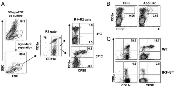
CD8α+ DC and stimulate cross priming. J Immunol. 2011 May 1;186(9):5142-50…………..…………31-39
2.3 Sistigu A, Viaud S, Chaput N, Bracci L, Proietti E and Zitvogel L. Immunomodulatory
effects of cyclophosphamide and implementations for vaccine design. Semin. Immunopath.,
2011 Jul;33(4):369-83………..……40-54
2.4 Hannani D, Sistigu A, Kepp O, Galluzzi L, Kroemer G, Zitvogel L. Prerequisites for the
antitumor vaccine-like effect of chemotherapy and radiotherapy. Cancer J. 2011
Sep;17(5):351-8………55-62
3. DISCUSSION……….…..63-65 References……….65-67
1. INTRODUCTION
A daunting diversity of distinct molecular etiologies gives rise to one class of life-threatening diseases — cancer [1, 2] — which affects half of the inhabitants of developed countries during their lifetime and kills one-third of them.
Cancer is widely considered a cell-autonomous genetic disease that results from epigenetic and genetic reprogramming in oncogenes, tumor-suppressor genes and genome-stability genes, all these being essential players in both oncogenesis and tumor progression.
As defined primarily by Hanahan and Weinberg, the tumorigenic process stems from six hallmark criteria i.e., growth signal self-sufficiency, resistance to growth-inhibitory signals, resistance to apoptosis, limitless growth potential, sustained angiogenesis, and metastasizing potential [1]. As an ancillary proposition tumors are more than insular masses of proliferating cancer cells. Instead, they are complex tissues of multiple distinct cell types (cancer cells, stromal cells, immune cells and the extracellular matrix) that participate, in a silent movie, in heterotypic interactions with one another. Such ensemble of cells is a main battleground during the neoplastic process, fostering proliferation, survival and migration of tumor cells. Indeed, for the development of full-blown neoplasia, cancer cells must overcome intrinsic (cell autonomous) and extrinsic (immune mediated) barriers to oncogenesis [3]. Only when tumor cells overreach immune control they can progress. As recently proposed by Schreiber and colleagues [4, 5], avoidance of immunosurveillance might be the seventh hallmark of cancer.
Comprehensive information on the tumor and the immune status of an individual could be expected to provide a precise picture of the ongoing evolution of the tumor (and therefore a useful tool for prognostic extrapolation), as well as to yield invaluable information about which strategy (surgery, chemotherapy, radiotherapy and/or immunotherapy) will result in an optimal therapeutic outcome.
1.1 CANCER DESPITE IMMUNOSURVEILLANCE: IMMUNOEDITING AND IMMUNOSUBVERSION Comments made decades ago by Burnet and Thomas, the architects of the ―cancer immunosurveillance hypothesis‖, that ―there is little ground for optimism about cancer‖ [6] and ―the greatest trouble with the idea of immunosurveillance is that it cannot be shown to exist in experimental animals‖ [7], reflect the problems that, until recently, fomented intense debate over whether natural immune defense mechanisms can protect the host against the development of cancers of non-viral origin. The difficulty was clear: if immunosurveillance of developing tumors in immunocompetent hosts was indeed successful, then how could such an apparently invisible process be experimentally revealed? With the development of mouse tumor models using inbred mice with molecularly defined immunodeficiencies, the notion that the immune system intimately regulates cancer development experienced a new resurgence.
It is now recognized that the immune system plays at least three distinct roles in preventing cancer: (i) it protects the host against viral infection and hence suppresses virus-induced tumors; (ii) it prevents the establishment of an inflammatory environment that facilitates tumorigenesis by eliminating pathogens and by prompt resolution of inflammation; and (iii) it eliminates tumor cells in certain tissues because nascent transformed cells often co-express ligands for activating receptors on innate immune cells and tumor antigens that are recognized by immune receptors on lymphocytes of the adaptive immune system.
Nonetheless, tumors can and do arise in the presence of a functional and intact immune system. A troubled relationship exists between tumors and the immune system. Cancer cells lull immune cells into a false sense of security, thus avoiding immunosurveillance. The known ploys cancer uses are immunoselection (i.e., selection of non-immunogenic tumor-cell variants, a process also known as immunoediting) and immunosubversion (i.e., active suppression of the immune response) [3, 8, 9].
1.1.1 Cancer immunoediting: from immunosurveillance to tumor escape
Approximately 10 years ago, Schreiber and colleagues newfound the dual host-protective and tumor-promoting actions of immunity. In the face of continuous immune pressure, cancer cells can be shaped to become immunologically silent or refractory, and then better suited to survive ultimately causing harm. The acknowledgement that the immune system controls not only tumor quantity but also tumor quality (immunogenicity), has led to the refinement of the cancer immunosurveillance theory into one now termed cancer immunoediting [5].
In its most complex embodiment, the cancer immunoediting process is envisaged to proceed sequentially through three distinct phases: ―elimination‖, ―equilibrium‖, and ―escape‖ (FIG. 1).
In the elimination phase, innate and adaptive immunity work together to destroy developing tumors long before they become clinically apparent. Many of the immune molecules (IFNγ; IFNαβ; IL-12; TNF; NKG2D; TRAIL; perforin) and cells (CD8 T, CD4 T, γδ T, NK, NKT, DC, Mф cells) that participate in the elimination phase have been identified, but more work is needed to determine their exact sequence of action. If this phase goes to completion, then the host
remains free of cancer, and elimination thus represents the full extent of the process. If, however, a rare cancer cell variant is not destroyed in the elimination phase, it may then enter the equilibrium phase, in which its outgrowth is prevented by immunologic mechanisms. T cells, IL-12, and IFNγ are required to maintain tumor cells in a state of functional dormancy, whereas NK cells and other effector cells or molecules are not required; this indicates that equilibrium is a function of adaptive immunity only. Editing of tumor immunogenicity occurs in the equilibrium phase. Equilibrium may also represent a second stable endpoint of cancer immunoediting and may restrain outgrowth of occult cancers for the lifetime of the host. However, as a consequence of constant immune selection pressure placed on genetically unstable tumor cells held in equilibrium, tumor cell variants may emerge that (i) are no longer recognized by adaptive immunity (antigen loss variants or tumor cells that develop defects in antigen processing or presentation); (ii) become insensitive to immune effector mechanisms; or (iii) induce an immunsuppressive state within the tumor microenvironment (immunosubversion) [3]. The end result is the generation via a Darwinian selection process of poorly immunogenic tumor cell variants that become ―invisible‖ to the immune system and thus acquire the capacity to grow progressively and emerge in clinically apparent disease [5, 10].
FIG.1 Extrinsic tumor suppression by the immune system. Transformed cells escaping intrinsic control are subjected to extrinsic
tumor suppressor mechanisms that detect and eliminate developing tumors before they become clinically apparent. This is known as the elimination phase of a broader process that has been termed cancer immunoediting. Cancer immunoediting takes into account the observation that the immune system both protects the host against tumor development and promotes tumor growth. Cancer
immunoediting is now considered a process composed of 3 phases: elimination, or cancer immune surveillance; equilibrium, a phase of tumor dormancy where tumor cells and immunity enter into a dynamic equilibrium that keeps tumor expansion in check; and escape, where tumor cells emerge that either display reduced immunogenicities or engage a large number of possible
immunosuppressive mechanisms to attenuate antitumor immune responses leading to the appearance of progressively growing tumors. These phases have been termed the 3 Es of cancer immunoediting. (Figure adapted from Swann JB and Smyth M, The
Journal of Clinical Investigation, 2007)
1.1.2 Immunosubversion
The molecular tricks by which tumor cells can subvert the immune system thus ‗paralyzing‘ immunosurveillance are the subject of intense investigation.
It was originally thought that the inefficiency of tumor-associated antigen (TAA)-specific immunity was due to intrinsic causes: (i) tumors simply did not present enough TAA; (ii) antigen-presenting cells (APC) did not have sufficient stimulatory capacity; or (iii) there were not enough effector cells or effector cytokines. On this basis, attempts were made to bolster TAA-specific immunity through administration of stimulatory cytokines (IL-2, IL-12 or IFNα) or TAA (peptides), or by using optimal APC (DC vaccines) [11-20]. In a different approach, TAA-specific effector T cells from cancer patients were expanded ex vivo followed by adoptive transfer [13, 16, 21, 22]. These approaches were met with
some success both in mouse models and in early clinical trials in humans, thus strengthening experimentally induced TAA-specific immunity as an efficacious approach to treat established tumors.
Recent studies have shown the other side of the coin: poor TAA-specific immunity is not due to a passive process whereby adaptive immunity is shielded from detecting TAA. On the contrary, there is an active process of ―tolerization‖ taking place in the tumor microenvironment [23]. In mouse models, advanced cancer invariably subverts immune function. Typically, tumor-specific CD8 T cells are activated at the stage of initiation of tumor growth, but these cells show a progressive loss of cytolytic function at the later stage of tumor expansion [24]. Similarly, tumor-specific CD4 T cells progressively lose their antitumor activity [25], whereas the number of regulatory T (Treg) cells increases. One possible explanation for how tumors subvert the immune response is to consider that the tumor is a ―false lymphoid organ; therefore, T-cell priming in the tumor microenvironment is defective as a result of the presence of dysfunctional or tolerogenic antigen-presenting cells. Indeed, some tumors overproduce various factors (such as vascular endothelia growth factor (VEGF), IL-6, IL-10, transforming growth factor (TGF)β, macrophage colony-stimulating factor (M-CSF), nitric oxide synthase (NOS)2, arginase-1, indoleamine 2,3 dioxygenase (IDO), prostaglandin (PG)E2, cyclooxygenase (COX)2 and gangliosides that can inhibit the differentiation, maturation and function of DC [26] as well as T-cell function [27]. Accordingly, local DC tend to mediate immunosuppressive, rather than immunostimulatory, effects and to promote IL-10 producing Treg-cell differentiation [26, 28]. Moreover, some human tumors (prostate, colon and pancreatic carcinomas) constitutively express IDO assigned for tryptophan degradation. This metabolic device blocks local proliferation of CD8 T cells [29] and promotes apoptosis of CD4 T cells, thus promoting resistance to immune-mediated rejection. Yet some other tumors can express CD95L still killing CD95-expressing tumor-specific T cells [30].
A series of recent studies have proposed new disadvantageous leukocytes to add to the list of suppressive cells to challenge. In an ultraviolet-irradiation-induced tumor model, irradiation-induced immunosuppression was found to be mediated by CD1d-restricted natural killer T (NKT) cells. These CD4 NKT cells produced IL-13, which suppressed CTL-mediated tumor rejection. Moreover, IL-13 from NKT cells activated myeloid suppressor cells to produce TGFβ, which also suppressed cytotoxic T lymphocytes
(
CTL) activity [31]. In addition, tumor-associated macrophages (TAM) mostly belong to the M2 class of macrophages, fully polarized to produce arginase-1, IL-10, TGFβ and PGE2, thus playing a key role in subversion of adaptive immunity and in inflammatory circuits that promote tumor growth and progression [32, 33] . Another possible explanation for tumor-mediated immunosubversion is based on a quantitative issue. Cancer traits that are immunostimulatory in small tumors can become immunosuppressive in large tumors. For example, the expression of NKG2D ligands (which stimulates an immune response at the initial stages of oncogenesis, as discussed earlier) seems to be immunosuppressive in larger tumors. NKG2D-ligand expressing tumor cells (as well as soluble NKG2D ligands that are shed from tumor cells) can downregulate NKG2D expression by CD8 T cells and NK cells or can uncouple NKG2D signaling from intracellular mobilization of Ca2+ or cell-mediated cytolysis, therebycontributing to suppression of the immune response [34]. Similarly, it could be argued that large tumors cause a general or specific downregulation of T-cell responses as a result of ‖high-dose tolerance‖ to TAA. Following successful systemic chemotherapy — for example, for ovarian carcinoma — CD8 T-cell function can recover [35], indicating that antitumor chemotherapies that have limited immunosuppressive side-effects can restore the normal immune response by abolishing tumor-mass-related immunosubversion.
Although ―black and white‖ signals have been identified in tumor immunity, from what described above, it is evident that this is an oversimplification and that interactions between tumor cells and immune cells would be represented by a multitude of colors. So, the ―bad news‖ is that cancer cells can strategically avoid immune attack. The ―good news‖ is that this newfound knowledge, comprehensive on the tumor and the immune status, is a powerful tool to which oncologists might capitalize aiming to the optimal management of the disease. As stated by Prendergast and Jaffee, to win the fight against cancer is necessary to stop ―segregating cancer immunology from cancer genetics and cell biology‖ [36].
1.2 CHEMOTHERAPY AND TUMOR IMMUNITY: AN UNEXPECTED COLLABORATION
Cancer therapy is continuously evolving in order to strategically optimize the chance of cure. The therapeutic approach to cancer today most frequently involves surgery (whenever possible) alone or in association with a single-agent or combinatorial treatment based on radio- or chemotherapy. Radiation therapy is used to achieve locoregional control, whereas systemic therapies (chemotherapy, endocrine therapy, molecularly targeted therapies, and adjunctive therapies – bisphosphonates -) are used to control diffuse disease (in hematologic malignancies) or disease that has spread beyond the primary site (in solid tumors).
A growing body of evidence suggests that conventional therapy for cancer may profit from the participation of the immune system whose contribution is elicited in two ways. On one hand, some therapeutic programs can tickle specific cellular responses — beyond the stereotypical apoptotic pathway — that render tumor-cell death immunogenic. On the other hand, some drugs may have side effects (beyond their effect on the tumor itself) that stimulate the immune system, through a transient lymphodepletion, the subversion of immunosuppressive mechanisms and the direct or indirect stimulatory effects of immune effectors. Moreover, vaccination against cancer-specific antigens can sensitize the tumor to subsequent chemotherapeutic treatment.
1.2.1 Immunogenic cancer cell stress and death
It has been generally assumed that most if not all chemotherapeutic agents induce cancer cell death by apoptosis and that apoptotic cell death would - by definition - lead to silent corpse removal and hence fail to induce an immune response against the dying cells. In apparent contrast with this idea, some chemotherapeutic agents do induce a type of cell death that is immunogenic, yet is accompanied by the all known biochemical and morphological hallmarks of apoptosis. Thus, tumor cells that have been killed in vitro with some chemotherapeutic agents such as anthracyclines, oxaliplatin or cyclophosphamide [3, 37, 38] (but not with others such as cisplatin) elicit a tumor-specific cytotoxic T lymphocyte response when they are injected subcutaneously into immunocompetent mice. This leads to the long-term protection of vaccinated mice against challenge with live tumor cells of the same type. In essence, a limited array of antineoplastic drugs induces immunogenic cancer cell death (ICD), which in turn provokes an anticancer immune response that allows the immune system to control (and possibly to eliminate) residual tumor cells. Such tumor-host productive dialogue involves the transfer of TAA to immune cells that stimulate a tumor-specific immune response. This is critical for the eradication of residual cancer (stem) cells as it operates irrespective of their resistance to therapy offering a possible explanation to how the anticancer immune response can contribute to the undeniable success of some antineoplastic regimens.
1.2.1.1 The key-lock paradigm
Cancer cells dying in an immunogenic fashion emit specific cell death-associated molecular patterns (CDAMP) that - in a correct spatial and temporal appearance - bear the ability to convert non-immunogenic corpse removal into an immunogenic reaction. Obviously, such a conversion also relies on the correct perception of CDAMP by dedicated sentinels of the host immune system. Thus, antigens from cancer cells succumbing to ICD inducers (like anthracyclines, oxaliplatin, cyclophosphamide and ionizing radiations) are efficiently taken up and processed by DC, which in turn cross-prime naïve T cells and drive the development of a tumor-specific immune response. The interaction between DC and dying cancer cells is controlled by the emission and/or release from the latter of the so-called ―eat me‖ and ―don‘t eat me‖ signals, i.e., membrane-bound or soluble molecules that stimulate or inhibit phagocytosis, respectively. The systematic analysis of surface proteome alterations in anthracycline-treated tumor cells revealed that ICD is associated with the ectopic co-exposure of the endoplasmic reticulum (ER) chaperones calreticulin (CRT) and ERp57 [39]. Ecto-CRT functions as an ―eat-me‖ signal for DC, thereby facilitating DC-mediated antigen uptake, and is an absolute requirement for the immunogenicity of dying tumor cells [39]. The co-exposure of CRT and ERp57 reportedly ensues the induction of an ER stress response that is associated with massive ultrastructural alterations of this organelle, and depends on the activation of (at least) three signaling modules. First, the ER-resident protein kinase R-like endoplasmic reticulum kinase (PERK) gets activated and couples ER stress signals to translation inhibition by phosphorylating the eukaryotic initiation factor 2α (eIF2α). Accordingly, the disruption of the eIF2α phosphatase complex PP1/GADD34 by small peptide inhibitors, resulting in increased phospho-eIF2α suffices to trigger CRT exposure in cancer cells [38, 40]. Second, an apoptotic module that involve the mitochondrion-permeabilizing proteins BAX and BAK (which also work at the interface between the ER and mitochondria to regulate calcium fluxes) [41, 42], caspase-8 and its substrate BAP31 - an ER sessile protein implicated in the lethal response to ER stress - is activated. Thus, the pan-caspase inhibitor Z-VAD-fmk, as well as genetic interventions whereby BAX, BAK and/or caspase-8 are removed or depleted, blocks CRT exposure and abolishes the tumor-vaccinating effect of cells undergoing ICD [39, 40]. Third, approximately 5-10 % of the endogenous CRT pool is exposed together with ERp57 at the surface of dying cells via SNAP and NSF attachment receptor (SNARE)-dependent exocytosis. This occurs well before plasma membrane permeabilization (which occurs as the final step of apoptosis), and also precedes the translocation of phosphatidylserine (PS) from the inner to the outer leaflet of the plasma membrane. PS is the
prototypic ―eat-me‖ signal of apoptotic cells (though it has been implicated also in non-apoptotic cell death) [43] and the kinetics of its exposure might affect the switch between the silent removal of dying cells by macrophages and the initiation of a cognate immune response by DC. The receptor that is responsible for antigen uptake by DC upon CRT binding remains to be determined. Possible candidates include the major CRT receptor CD91 as well as other CRT-interacting proteins like scavenger receptor A (SR-A), scavenger receptor expressed on endothelia cell I (SREC-I), CD40 ligand, tumor necrosis factor (TNF)-related apoptosis-inducing ligand (TRAIL) or CD95/FAS ligand. The CRT-driven uptake of tumor antigens by DC is per se insufficient to elicit an antitumor immune response as internalized antigens must be processed and re-exposed for the cross-priming of CD4 and CD8T lymphocytes. This implies that other signaling pathways are involved in ICD.
A systematic study of the response to CDAMP of distinct Toll-like receptors (TLR) on naïve T-cells revealed that TLR4 is both required and sufficient for efficient antigen presentation by DC [44]. Among other proteins, TLR4 binds the non-histone chromatin-binding nuclear protein high-mobility group box 1 (HMGB1), leading to the activation of the downstream effector myeloid differentiation primary response 88 (MYD88). This inhibits the fusion between lysosomes and antigen-containing phagosomes, thus facilitating antigen processing and presentation to T cells. HMGB1 also stimulates the neo-synthesis of pro-IL-1β but per se does not serve as a DC maturation signal. The release of HMGB1 from tumor cells succumbing to ICD manifests with a dual kinetics whereby HMGB1 first translocates from the nucleus to the cytoplasm and then, following the breakdown of the plasma membrane, gets released into the extracellular space [45].
The vaccine-like effect of ICD relies on the elicitation of an IFNγ-polarized T cell response, which in turn requires the function of the NLRP3 inflammasome, a multiprotein caspase-1-activating complex. Caspase-1 activation is critical for activating an antitumor immune response as it catalyzes the proteolytic maturation of IL-1β [46]. One of the most abundant factors that activate the NLRP3 inflammasome is ATP, and at least in DC, it does so by binding to the purinergic P2RX7 receptor on the cell surface. ATP also constitutes a CDAMP, as it gets released during the final steps of cell death, possibly via voltage-gated hemichannels of the pannexin 1 or connexin type [47]. Accordingly, the depletion of intracellular or extracellular ATP in cells succumbing to ICD abolishes the development of an IFNγ-polarized response, and P2RX7-deficient mice fail to mount an immune response against syngeneic cancer cells succumbing to ICD. Intriguingly, ATP also serves as a ―find me‖ signal for the attraction of immune cells. Altogether, these observations highlight the multifaceted and critical role of ATP for the vaccine-like effects of ICD inducers.
The spatially and temporally regulated emission of immunogenic factors from dying tumor cells accounts for the recruitment and activation of immune cells to tumor bed and governs the immune response to cancer cells undergoing ICD. Thus, the stress conditions that cancer cells confront during chemo- and radiotherapy determine whether the subsequent wave of cell death will elicit an antitumor immune response or rather will remain immunologically silent. Normally, cells attempt to cope with stress by arresting normal activities and by activating a series of cytoprotective mechanisms that aim at re-establishing homeostasis. This is accompanied by alterations of the surface proteome that, in the case of ICD, account for the recognition by immune cells, and by the emission of soluble mediators with chemotactic and anti-chemotactic properties. This is crucial for the ―selection‖ and differentiation/maturation of engulfing cells, which in turn dictates the immunogenic or tolerogenic outcome of cell death. In this sense, the exposure of the DC-specific ―eat me‖ signal CRT paralleled by the disclosure of other, hitherto uncharacterized, ―don‘t eat me‖ signals, facilitates the recognition and uptake of dying tumor cells by DC rather than by macrophages. Based on these observations, the spatially-restricted and temporally-ordered appearance of CRT, HMGB1 and ATP might constitute a ―key‖ that would precisely fit into a series of pattern recognition receptors (PRR) expressed by DC (the ―lock‖) for the conversion of non-immunogenic into immunogenic cell death and for the elicitation of an anticancer immune response [45] (FIG.2).
By shaping T cell responses, DC are the first-line decision makers of the innate immune system and their role in immunogenic chemotherapy has been deeply investigated. Experiments in transgenic mice that express the diphtheria toxin receptor (DTR) under the control of a DC-specific promoter (allowing for in vivo DC depletion) revealed the essential role of DC in the perception and decoding of ―come and get-me‖ signals emitted by dying tumor cells during ICD [44]. Similarly, the in vivo depletion of CD8 T cells with specific antibodies has been instrumental to highlight the critical role of this lymphocyte subset for the vaccine-like effect of chemo- and radiotherapy in a large panel of murine tumor models, including CT26 colon cancer, EL4 thymomas, TS/A mammary carcinomas, MCA205 fibrosarcomas and GOS osteosarcomas. In line with these observations, CD8 T cells have been shown to mediate potent anticancer immune effects in clinical settings, for instance in colorectal tumors, where immune infiltration might serve as a prognostic factor [48]. Moreover, it has recently been shown that a precise orchestration of the T cell response is required for immune effectors to eradicate tumors. In this context, the IL-1β-dependent recruitment of IL-17-secreting γ/δ T cells had to precede the infiltration of tumors by Tc1 lymphocytes for the efficacy of immunogenic chemotherapy in vivo [49]. Thus, a finely regulated crosstalk between components of the innate (DC) and cognate (γ/δ and CD8 T cells) immune system is required for cell death to be perceived as immunogenic, for the elicitation of an anticancer immune response, and for complete tumor eradication leading to therapeutic success.
FIG.2 Immunogenic signals emitted by dying cells form a spatiotemporal code unlocking DC to mount a potent immune response
toward tumor cells. (i) Early exposure of ecto-CRT by dying tumor cells, which facilitates engulfment by DC. (ii) HMGB1 released from dying cells binds to TLR4 on DC, thus favoring antigen cross-presentation and up-regulating pro-IL-1β. (iii) ATP liberated from dying cells binds to the purinergic receptor P2RX7 on DC, activates the NLRP3 inflammasome, and leads to the secretion of active IL-1β, which polarizes CD8+ T cells toward IFN-γ production. (iv) An additive DC maturation factor remains to be characterized. (Figure adapted from Hannani D et al.,Cancer J, 2011)
1.2.2 Immunostimulatory side effects of anticancer drugs: Cyclophosphamide as elected drug
Cytotoxic drugs can be used in unexpected ways to break systemic mechanisms of immune tolerance, or to alter the host environment in which the antitumor immune response develops.
Many chemotherapy drugs can have both positive and negative immunomodulating activity, with the type of influence depending on the drug dosing and the relative timing of administration [50].
Cyclophosphamide (CTX), the lead compound of alkylating agents, is one of the most potent and widely used drugs for the treatment of hematological and solid organ malignances, autoimmune disorders, and as a conditioning regimen for blood and marrow transplantation.
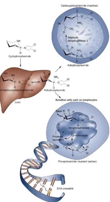
In order to achieve cytotoxic effects, CTX needs to be metabolically converted by the hepatic oxidase into its metabolites acrolein and phosphoramide mustard which are responsible for the cytostatic and urotoxic effects of the drug, respectively, through DNA-alkylating and cross-linking mechanisms. The metabolic activation of CTX is irreversibly compromised in cells with high levels of aldehyde dehydrogenase 1 (for example, hematopoietic stem cells). This enzyme converts aldophosphamide into carboxyphosphamide. Carboxyphosphamide does not decompose to phosphoramide mustard and therefore lacks alkylating and cytotoxic activity (FIG. 3).
Depending on the dosage and the timing of administration, CTX displays either immunosuppressive or immunopotentiating effects [51]. At high dosage, CTX exerts potent cytotoxic and lymphoablative effects, indispensable for dose intensity and immunosuppressive regimens in the oncological and internal medicine armamentarium. More recent work highlighted the immunostimulatory and antiangiogenic effects of low dosing or metronomic (i.e., frequent, repeated low doses) CTX, thus repositioning this drug in the field of cancer immunotherapy. It is interesting to note that a single CTX injection induces a transient myelo-lymphodepletion (whose intensity depends on the dose) that is followed by a recovery phase during which homeostatic mechanisms occur increasing leucocyte cell counts well above baseline levels. Following this ―rebound‖ event, cell counts slowly reach pre-treatment levels (FIG.4). This double-face activity accounts for many of the observed immunomodulatory effects ascribed to the drug and for the capability of synergizing with a series of immunotherapy approaches.
Great relevance was attributed to the reduction of cells that negatively modulate immune responses in vivo. Studies performed by Awwad and North in the 80s demonstrated that CTX administration can enhance the effectiveness of adoptive immunotherapy through the selective reduction of a suppressor T cell population, that was not clearly identified at that time [52]. Later work from Ghiringhelli‘s and Lutsiak‗s groups showed, indeed, that the administration of CTX reduces the number [53] and inhibits the functions of CD4 CD25Tregs in tumor-bearing animals by
downregulating the expression of the key functional markers forkhead box P3 (FOXP3) and glucocorticoid-induced TNF-receptor-related protein (GITR) [54]. This was a formal proof of how CTX treatment circumvents tumor-induced immune tolerance tipping the balance toward an effective antitumor response. Furthermore, mitigating the influence of Treg and stimulating IFNα/β production by host leukocytes [55], CTX might account for the augmented antibody responses and the persistence of memory T cells. All these effects contribute to the eradication of immunogenic tumors in synergy with specific immunotherapy approaches [56, 57]. As reported by Ercolini and colleagues, CTX pretreatment affects Treg/Teffector ratio enabling the vaccine-mediated recruitment of high avidity CD8 T cells to the antitumor response in tolerized neu transgenic mice [58]. Importantly, these findings correlate with tumor rejection, an outcome never seen with vaccine alone in this tolerized setting.
FIG. 3 Cyclophosphamide metabolism. After intravenous or oral administration, cyclophosphamide is rapidly distributed in the
body. In the liver, it is converted to hydroxycyclophosphamide, which stays in equilibrium with aldophosphamide.
4-hydroxycyclophosphamide and aldophosphamide readily cross the cell membranes by passive diffusion. In cells with high levels of aldehyde dehydrogenase 1 (for example, hematopoietic stem cells), aldophosphamide is irreversibly converted to
carboxyphosphamide. Carboxyphosphamide does not decompose to phosphoramide mustard and therefore lacks alkylating and cytotoxic activity. In the absence of a high concentration of aldehyde dehydrogenase (for example, lymphocytes), aldophosphamide spontaneously liberates phosphoramide mustard and acrolein. Phosphoramide mustard is a bifunctional DNA alkylating molecule and forms interstrand DNA crosslinks primarily at the guanine (G) sites. (Figure adapted from Emadi A et al., Nature Reviews, 2009)
In small clinical studies, the combination of low doses of intravenous CTX with vaccines has been shown to augment delayed type hypersensitivity (DTH) responses [59, 60], decrease the proportion of suppressor T cells [61] and prolong the survival of patients with metastatic cancer [59]. One month of metronomic program of CTX given to patients with end-stage cancer could suppress Treg-cell inhibitory functions, restore the proliferative capacity of effector T cells and restore the cytotoxicity of NK cells [62, 63]. However, the ablation of regulatory cells is likely to be of varying importance, depending on the tumor type, stage and location [64]. Further studies revealed that the strong therapeutic efficacy of combined chemoimmunotherapy stems from a bystander effect on host lymphocytes (as well as adoptive lymphocytes) occurring during the recovery phase following CTX-induced myelo/lymphodepletion [22, 65]. An early study from Proietti‘s group demonstrated that a single injection of CTX, followed by the adoptive transfer of antitumor immune cells, could eradicate established tumors and prevent metastases spreading. This effect depended on the production of a plethora of until then uncharacterized soluble factors, which may sustain the proliferation, survival and activity of transferred cells [65]
.
Of note, such antitumor efficacy was abolished when mice were treated with antibodies neutralizing type I IFN. A subsequent study clarified that CTX leads to the expression of type I IFN in vivo. Among the soluble factors coming out in the so called ―cytokine storm‖, type I IFN are obligate for the expansion of CD4 and CD8 T cells exhibiting a memory (CD44hi) phenotype [55].
CTX role in favoring memory T cells was laterconfirmed by the finding that prolonged metronomic schedule of chemotherapy, despite diminishing the number of proliferating tumor-specific CTL, preserved CD43low memory CD8 T cells [66].
FIG. 4 After CTX (100 mg/kg)-induced lymphodepletion, a ―rebound‖ phase occurs, during which a cytokine storm drives the
homeostatic proliferation, activation and trafficking of different lymphocyte pools. (Figure adapted from Proietti E et al., J Clinl
Invest, 1998)
More recent studies showed that CD4 T cells are responsible for the synergism between chemotherapy and adoptive immunotherapy and for the cooperation of transferred cells with the host immune system [67]. Furthermore, CTX promoted the migration of specific tumor-immune lymphocytes to the tumor bed and induced the homeostatic proliferation/activation of transferred B and T lymphocytes. A first characterization of the molecular mechanism underlying the immunomodulatory properties of CTX came from the gene expression analysis of a selected panel of cytokines by real-time PCR in the bone marrow (BM) and spleen of treated mice. Optimal therapeutic responses to the adoptive transfer of immune cells were found to be associated with the chemotherapy-mediated induction of a ―cytokine storm‖ occurring during the rebound phase after drug-induced myelo-lymphodepletion [67]. In a subsequent study, the transient upregulation of a variety of immunomodulatory factors, including danger signals, pattern recognition receptors, inflammatory mediators, growth factors, Th1-polarizing and homeostatic cytokines, chemokines, and chemokine receptors was observed by gene and protein expression analysis early after CTX injection [68]. These
factors are involved in sensing CTX myelotoxicity and activating repair mechanisms, which, in turn, stimulate immunoactivation events that promote chemotherapy efficacy. Notably, the relevance of the increased expression of homeostatic cytokines (IL-7 and IL-15) was also confirmed by the finding that the antitumor efficacy of the combination of a subletal total body irradiation (TBI) with the adoptive transfer of CD8 T cells was impaired in mice deficient of both cytokines [69]. Of great interest, Matar and colleagues observed a Th2 to Th1 shift in cytokine production in a rat metastatic lymphoma setting after treatment with low-dose of chemotherapy [70].In line with these observations, it has been demonstrated that low-dose, as well as metronomic CTX, promotes the expansion and differentiation of CD4 T producing IL-17A, in naïve and tumor bearing mice [68, 71]. These data agreed with the clinical observation of advanced cancer patients treated with non myeloablative and non lymphodepleting doses of CTX (3 week-oral treatment with 50 mg/day). Ex vivo IL-17 release by circulating peripheral blood mononuclear cells (PBMC) after T cell receptor (TCR) stimulation (anti-CD3/CD28 Ab cross-linking) were significantly enhanced following CTX regimen [71] .
An increasing number of studies underscored that also the innate arm of the immune response might be involved in the immunopotentiating activity of chemotherapy. According to Salem and colleagues, systemic CTX increases the relative number and the activation status of myeloid DC through the induction of high levels of inflammatory cytokines such as IFNα and IL-6 among others [72]. In accordance, Radojcic and colleagues found that a myelosuppressive dosage of CTX, perturbs DC homeostasis. This leads to the occurrence of tumor-infiltrating DC locally secreting IL-12, and therefore able to prime T-cell responses [73] (FIG.5).
FIG.5 Immunomodulatory effects of cyclophosphamide. (Figure adapted from Sistigu A et al.,Semin. Immunopath., 2011)
Fifty years after the US Food and Drug Administration (FDA) approval, CTX remains a safe and affordable compound endowed with multifaceted properties and a plethora of clinical indications. The acknowledgement of chemotherapy -in general- and CTX -in particular- immunomodulatory effects strengthens the need for a rational
combination of chemo- and immune- therapy, leveraging additive or even synergistic activity. The thoughtful combination of multiple treatment modalities should allow the full power of immunotherapy to be unleashed, resulting in increasing survival benefits and ultimately in the eradication and relapse prevention of malignant disease.
1.3 IMMUNOTHERAPY
Cancer immunotherapy consists of approaches designed to reconstruct host-tumor immunobiology, tipping the balance of immunologic homeostasis in favor of the host.
The concept of cancer immunotherapy goes back as far as the late nineteenth century, when Coley observed tumor shrinkage and even disappearance following the injection of bacterial products in and around tumors [74]. Since then, many observations, such as the rare but well-documented occurrence of spontaneous remissions, the higher incidence of cancer in patients who are immunosuppressed, and the identification of tumor-specific antigens and lymphocytes, have stimulated research on strategies that aim to induce specific antitumor responses. Over the past decades, considerable knowledge has been obtained on the components that are relevant in antitumor immune responses and immune escape mechanisms, yet the development of immunotherapy as a treatment modality for cancer has been hampered by several factors. These include difficulties in the selection of the optimal dose and schedule, the methods of evaluation, and financial support. Although durable clinical remissions have been observed with various immunotherapeutic strategies, the percentage of patients who benefited from these interventions has remained too small to justify the general use of such strategies. As a consequence, for many years, the clinical progress in the field of immunotherapy has been slow. However, the recent positive preclinical and clinical results with novel immunoactive drugs as well as the unexpected finding of a positive interaction between immunotherapy and chemotherapy may herald a new era for the immunotherapy of cancer. An additional great boost to immune-based therapy derived from improved ways of evaluating responses to treatment due to the progressive understanding of immunotherapy-induced responses.
Of great importance, immunotherapy is uniquely able to exert a durable therapeutic effect due to the induction of immunologic memory, minimizing systemic toxicity. This effect makes prolonged, repetitive cycles of therapy unnecessary and identifies immunotherapy as a promising modality for the secondary prevention of disease relapse and ultimately the prevention of primary tumor development.
Immune-based therapy can be broadly divided into approaches that employ the passive administration of immunologic effectors like monoclonal antibodies (mAb), cytokines and lymphocytes, and strategies that actively induce these immune effectors in vivo (i.e., vaccines).
1.3.1 Cancer Vaccines
Prevention or treatment with a cancer vaccine, or active specific immunotherapy, is a very attractive therapeutic option because the mechanism of action is eventually an enhanced endogenous immune response against the host‘s malignancy [50, 75]. Vaccine approaches utilize tumor antigens and antigen-presenting cells to enhance a preexisting antitumor immune response, or, perhaps in some cases, to induce an antitumor immune response that did not previously exist. There are many potential sources of tumor antigens including purified or synthesized tumor-cell surface molecules, which may be peptides or proteins, cells or lysates derived from fresh or cryopreserved autologous tumor samples (which is actually a mixture of normal and malignant cells), and cells or lysates of allogeneic or autologous tumor cell lines. There are a variety of methods by which tumor antigens can be presented including a purified antigen, via heat shock proteins, in viruses, or DNA, or by APC such as DC, or as the idiotypes of mAb that have been selected by their tumor antigen recognition. There are numerous molecules that might be useful as adjuvants to enhance the immunogenicity of a vaccine. There are also many routes by which vaccinations might be delivered including subcutaneous, intradermal, intramuscular, intravenous and intralymphatic.
As vaccines are minimally associated with side effects and invasive procedures, it is time to consider whether they can also be used to prevent tumor development. As microorganisms are the cause of 10-20% of all human tumors (reported in World Cancer Report – Stewart BW & Kleihues P – IARC Press, Lyon 2003), vaccines that reduce infection with viruses that cause cancer are of the utmost importance in primary cancer prevention. Vaccination against hepatitis B virus, for example, has reduced the incidence of hepatocellular carcinoma [76], whereas vaccines against human papilloma viruses are expected to greatly reduce the incidence of cervical carcinoma [77].
In animals, anti-tumor vaccines are effective in preventing a subsequent tumor challenge. This is a well-substantiated observation established through countless tumor-challenge experiments performed in immunized animals using many different fast-growing and aggressive mouse tumors [78]. In these experiments, immunization against a tumor antigen is followed by a subcutaneous, intramuscular or orthotopic challenge with a lethal dose of a transplantable tumor. In mice, effective immunity is often elicited and a successful pre-immunization against almost any kind of tumor seems to be feasible.
Unfortunately, there are usually several etiological agents for most human cancers and therefore instead of being prophylactic, vaccine strategies need to be therapeutic. In 2005, more than 200 clinical trials were in progress (reported in ClinicalTrials.Gov database of the National Cancer Institute & European Organization for the Research
and Treatment of Cancer Protocols Database websites). The results achieved so far, however, have been poor: partial responses were rare and complete responses extremely rare. Only in few patients has the progression of previously growing tumors been halted and prolonged survivals observed. As benefits that were due to vaccination have been sustained in no more than a handful of cases [79], new strategies are being explored [12, 79], including vaccines based on engineered viral vectors, various approaches with DC, and strategies that are aimed at inhibiting immunosuppressive cells of lymphoid or myeloid origin. Perhaps vaccination alone is not the solution for treating existing tumors, and evidence is emerging that shows that combining immunotherapy with chemotherapy, radiotherapy, anti-angiogenic therapy and other approaches could yield synergistic or additive results [80, 81]. Therapeutic vaccines are also poorly effective in mouse models of cancer, so the lack of benefit seen in clinical trials is perhaps not surprising. More knowledge on the schedules, routes, doses and adjuvants is required to optimally use these strategies. However after many years of hard work and negative trials, it was gratifying when, in 2010, the cell product Sipuleucel-T emerged as the first therapeutic vaccine approved by the US FDA for the treatment of prostate cancer. This cell-based vaccine consists of autologous PBMC, which include professional APC that have been activated with a fusion protein of the prostate antigen prostatic acid phosphatase and the immunostimulant GM-CSF. Sipuleucel-T was approved based on results from a placebo-controlled Phase III randomized trial. Despite showing a lack of benefit in progression-free survival and the fact that tumor regressions were rare, an overall survival benefit of 4.1 months was demonstrated compared to placebo [82]. Other examples of promising vaccine-based approaches have now entered Phase III clinical trials include an idiotype-based vaccination plus GM-CSF for follicular lymphoma, a combination of gp100 and high dose IL-2 for melanoma [83], a prostate-specific antigen-targeted poxviral vaccine in prostate cancer [84], and a melanoma-associated antigen-3-protein vaccine in non-small cell lung cancer [85].
It is expected that predictive oncology will assess the individual risk of cancer as a function of sex, age, family history, genetic makeup and lifestyle (reported in American Cancer Society Who is at Risk & your Disease Risk websites), whereas gene expression profiles and molecular biology will outline the probability that a particular onco-antigen will be expressed by the tumor for which the person is at risk. By combining this information, one can envisage to appeal to a custom-tailored preventive vaccination.
1.3.2 Monoclonal Antibodies
The concept of using antibodies to selectively target tumors was proposed by Ehrlich over a century ago (reported in Ehrlich, Collected studies on immunity. New York: J. Wiley & Sons, 1906).
The advent of hybridoma technology in 1975 enabled the production of immunoglobulins from a single clone of B-cells, and hence the term ―monoclonal antibodies‖ (mAb) [86]. The potential clinical application of such biological products in cancer therapy was quickly recognized and repeatedly emphasized [87-89]. However their applicability in the field of immunotherapy has been slow. Owing to their origins in mice, mAb were typically immunogenic in humans and had poor abilities to induce human immune effector responses. Later advances in antibody engineering provided flexible platforms for the development of chimeric, humanized and fully human mAb which satisfactorily addressed many of these problems (reported in Dillman, Principles of cancer biotherapy, 2009). The past decades have witnessed the evolution of mAb as ‗magic bullets‘ from concept to clinical reality [90], and their effectiveness in treating cancer patients has been increasingly recognized.
Humanized and chimeric mAb can be administered to block critical cancer signaling pathways, induce tumor cell apoptosis or promote antibody-dependent killing of cancer cells. In addition to antibodies that target tumor antigens, antibodies that target the tumor microenvironment slow tumor growth either by enhancing host immune responses to TAA or by curtailing pro-tumorigenic factors produced in the tumor stroma. These attributes of target specificity, low toxicity and the ability to activate the immune system suggest the continuing promise of therapeutic antibodies.
Trastuzumab and Cetuximab are mAb approved for clinical use that target the human epidermal growth factor receptor 2 (HER-2) and the epidermal growth factor receptor (EGFR), critically required for the progression of some tumors [90]. Moreover, the mAb against CD20 molecule on the surface of B cells, Rituximab, is used in the treatment of B cell lymphomas [90]. Among their interference with cancer cell signaling, mAb promote tumor cell death through antibody-dependent cellular cytotoxicity (ADCC) and complement-dependent cytotoxocity (CDC) [90]. Current studies are addressing the potential efficacy of mAb that target costimulatory molecules on immune cells such as OX40, member of the tumor necrosis factor receptor superfamily, and that antagonize inhibitory receptors such as the Cytotoxic T-Lymphocyte Antigen 4 (CTLA-4) and the Programmed Death 1 (PD-1) [90]. Although these mAb can induce disease regression, a dose-dependent toxicity, in terms of autoimmune or autoinflammatory side effects, has been reported [91, 92]. Monoclonal antibodies may also be used in combination with exogenously administered cytokines, as IL-2, GM-CSF or G-CSF[90]. Among all, the effects of the combination of G-CSF with Rituximab have been studied in a Phase I/II clinical trial in patients with low grade lymphoma [93]. Instead of an overall response rate similar to that reported for Rituximab alone, the period of remission was remarkably longer.
Of utmost interest, the next generation of mAb working as carriers to provide greater specificity for cytotoxic agents such as radioisotopes and toxins and of unconjugated antibody therapies will undoubtedly yield many effective
new treatments for cancer over the next decade. These advances will arise from the identification and validation of new targets, the manipulation of tumor–host microenvironment interactions, and the optimization of antibody structure to promote the amplification of antitumor immune responses. One could easily envision combining mAb with a variety of specificities and antitumor mechanisms for patient-specific ―cocktails‖ .
1.3.3 Adoptive Cell Transfer
Adoptive Cell Transfer (ACT) refers to the administration of huge numbers of tumor-specific T cells as anticancer therapy. These cells can be derived from the tumor environment (such as tumor-infiltrating lymphocytes (TIL)), from peripheral blood or they can be genetically modified to express a high affinity anti-tumor TCR.
ACT studies in experimental tumor models, dating back to the 1960‘s, provided the earliest evidence that splenocytes from immunized animals sustain a therapeutic effect when used to treat transplanted syngeneic tumors [94, 95]. Subsequent studies have been aimed at defining the phenotype of the transferred T cells determining the ACT efficacy. Early experiments in mice showed that T-cell populations that had been cultured with leukemia cells could eradicate an established leukemia in vivo [96, 97], thus suggesting the involvement of memory cellular immunity. Strengthening this observation, following studies have demonstrated on one hand that the antitumor efficacy of ACT is related to the transfer of CD8 and CD4 T cells [98, 99], and on the other hand that T differentiation status deeply influences ACT outcome. In fact, adoptively transferred cells with a CCR7+,CD27+,CD28+,CD62L+ phenotype, proper
of central memory T cells, were proved more effective than highly differentiated cells that lost these markers [100]. The clinical feasibility of this strategy was first shown in post-transplant lymphoproliferative disease, which is an Epstein-Barr virus (EBV)– associated B cell lymphoma that develops under conditions of severe T cell immunosuppression. Infusion of EBV-specific T cells led to the rejection of lymphomas and restored the severely hampered EBV-targeted immunity [101]. In 2002, the National Cancer Institute reported the outcome of a Phase II study of adoptive transfer of tumor-reactive TIL in pretreated patients with metastatic melanoma [22]. TIL obtained from a melanoma metastasis were grown in vitro with high dose IL-2 for overcoming the tumor-induced anergic state. Large amounts of cultured cells were re-infused into patients. Preconditioning the host with cyclophosphamide and fludarabine for inducing severe lymphocytopenia, was shown to be necessary for the survival and expansion of the infused TIL. Single-arm Phase II studies from separate institutions have shown objective response rates of 50% in patients with metastatic melanoma who are receiving this treatment [102, 103]. Approximately 10% of patients obtain a complete response, which may be durable. Side effects resulting from the infusion of TIL were mostly mild, consisting of vitiligo or uveitis in some patients, but both the non-myeloablative chemotherapy and high-dose IL-2 resulted in significant well-known toxicities.
As well as the cost and labor intensity, a major drawback of this approach is that TIL cannot be cultured from all patients. Moreover, the specificity of T cells within the TIL graft that are responsible for the clinical effects is unknown. This strategy could be refined by cloning high-affinity tumor-specific TCR from these cells that can serve as donor TCR to genetically modify unselected peripheral blood T lymphocytes for adoptive transfer [104]. This so-called TCR gene transfer is currently being tested in several clinical trials [105]. In order to use this strategy, the choice of target antigen is of utmost importance. Lethal toxicity can be induced by infusion of T lymphocytes genetically engineered to express TCR targeting antigens common to the tumor and the host [106, 107]. By contrast, antigens that have their expression restricted to tumor cells, such as cancer/testis antigen 1 (NY-ESO-1) in melanoma patients or CD19 in non-Hodgkin‘s lymphoma B cell patients [108], can be targeted by TCR-redirected T cells thus reducing the risk of inducing systemic toxicity.
Along with ACT progress it remains to be seen whether the morbidity associated with any resultant autoimmune disease outweighs the antitumor benefit.
1.3.4 Cytokines
Cytokines are proteins secreted by cells that affect the immune response, typically via effects on other cells through receptors. Anticancer behavior of cytokines is generally believed to be mediated by their action on immune cells [109]. It is not surprising that numerous cytokines have attracted interest in the context of cancer therapy, including hematopoietic colony-stimulating factors, interferons, at least 35 different interleukins, tumor-necrosis factor, and a number of other protein ligands (reported in Lewko, Principles of Cancer Bioth, 2009).
There are a number of cytokines that have received FDA approval, including erythropoietin (EPO), G-CSF, and GM-CSF. At this immunotherapeutic crossroad, the two cytokines specifically approved as cancer therapy are type I IFN and IL-2.
1.3.4.1 Type I IFN
Type I IFN emerge as exquisite candidates for anti-cancer therapy. The power of this cytokine family in the scenario of neoplasias is mainly related to their antiangiogenic and proapoptotic effects on the tumor side and the positive conditioning of innate and adaptive immunity on the host side.
Type I IFN are a large family of immune regulatory proteins consisting of 14 functional IFNα cytokines, and one IFNβ cytokine sharing the same receptor and exerting similar biological activities. The ligand binding to the heterodimeric receptor (IFNAR-1 and IFNAR-2) [110, 111] complex leads to the tyrosine phosphorilation and activation of IFNAR-1-associated Tyk2 and IFNAR-2-associated Jak1, which, in turn, phosphorilate cytosolic Stat1 and Stat2, giving rise to two types of activated transcription factors: a Stat1 homodimer and IFN-stimulated gene factor 3 (ISGF3), a tripartite complex of phosphorilated Stat1, Stat2, and the constitutive DNA binding protein IFN regulatory factor-9 (IRF-9). The activated transcription factors enter the nucleus, bind to specific enhancer elements sequences on certain genes, and by regulating the expression of these genes, induce the wide range of IFN-dependent effects on cellular function [112-115]. Comparative functional analyses of several members of the type I IFN family revealed that they induce a significantly overlapping array of biologic responses, although quantitative differences have been noted in the specific activities of each particular form [116, 117]. A small number of genes have been identified that are differentially regulated by IFNα vs IFNβ [118, 119].
Type I IFN were discovered in 1957 by Isaacs and Lindenmann as soluble proteins able to inhibit virus replication in cell cultures [120-122]. More than 50 years of research revealed a panoply of antitumor biologic effects. Indeed, type I IFN have a long record use in clinical oncology. Even though today some new anticancer drugs have somehow replaced such cytokines in the treatment of certain hematological malignancies (hairy cell leukemia and chronic myeloid leukemia), type I IFN are still widely used for the treatment of metastatic melanoma, renal cell carcinoma and Kaposi sarcoma [123, 124].
For a long time, it was thought that the direct inhibitory effects on tumor cell growth/functions were the major mechanisms involved in the antitumor response in IFN-treated patients. In fact, type I IFN can directly inhibit the proliferation of normal and tumor cells in vitro and in vivo, and can exert other direct effects on tumor cells, including downregulation of oncogene expression, induction of tumor-suppressor genes, and increase of major histocompatibility complex (MHC) class I expression [125].
In addition to the direct effects on tumor cells, type I IFN exert several effects on host immune cells that can play a pivotal role in the overall antitumor response [126]. In the early 1990s, Ferrantini and colleagues carried out an ensemble of studies characterizing the effect of local production of type I IFN at the tumor site and discovering their ability to switch a highly tumorigenic behavior into an immunogenic one. Highly metastatic Friend leukemia cells (FLC) genetically modified to secrete IFNα1 exhibited a marked loss of their tumorigenic potential when injected into syngeneic mice. Likewise, these genetically modified IFN-producing tumor cells inhibited the growth of metastatic parental cells in transplantation assays [127]. The findings that IFNα1-FLC immune rejection and subsequent immune protection against a rechallenge was mostly mediated by CD8 T cells [128], implied that memory T cells were generated upon exposure of mice to the IFNα1-producing cells. In fact, a marked proliferation of CD8 T lymphocytes, especially among memory-phenotype CD44hi cells, in both the spleen and the lymph nodes, was observed after injection
of viable tumor cells producing IFNα. Tumor cell-targeted cytokine gene therapy has been widely evaluated in animal models by many groups with different approaches, comprising both the use of genetically modified cells and the in vivo delivery of IFNα genes via injection of viral vectors or plasmid DNA [129].
Today, new attention is given to type I IFN as important factors bridging innate and adaptive immunity. Along with the understanding of the cytokine network regulating Th cell functions, several studies provided evidence on the importance of type I IFN in the differentiation of the Th1 subset, as well as in the generation and activity of CTL [124]. In particular, type I IFN are important for the in vivo expansion and long-term survival of CD8 T cells in response to specific antigens [130] and for the adjuvant activity on T cells induced by CpG DNA administration [131]. Type I IFN also prolong the survival of T cells in mice and the expression of anti-apoptotic genes in human primary T lymphocytes [132]. Similarly, a new interest in type I IFN as a ―bridge system‖ linking innate and adaptive immunity stemmed from the identification of ―natural IFN-producing cells‖ (the rare blood cell population that produces 200-1000 times more IFN than other blood cells after microbial challenge) also defined as plasmacytoid DC [133].Type I IFN also affect monocyte and/or macrophage function and differentiation. Thus, these cytokines markedly support the differentiation of monocytes into partially mature DC with high capacity for Ag presentation [145], stimulate macrophage antibody-dependent cytotoxicity, and positively or negatively regulate the production of various cytokines (e.g., TNF, IL-1, IL-6, IL-8, IL-12, and IL-18) by macrophages [146]. In addition, treatment of DC with IFN-α/β has been shown to upregulate surface expression of MHC class I, class II, and costimulatory molecules both in animal models and in the human system (43–45), and to augment the capacity of DC to stimulate CD4 and CD8 T cell responses. Interestingly, it has been observed that injection of type I IFN stimulates efficient cross-priming of antigen-specific CD8 T lymphocytes in vivo through a direct involvement of DC [147].
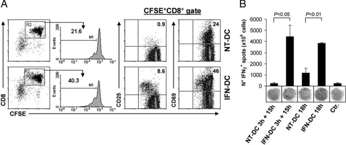
It has recently been reported that type I IFN can greatly enhance presentation and CD8 T cell cross-priming by stimulating CD8α DC that have engulfed tumor cells undergoing immunogenic apoptosis by CTX treatment
[37, 133]. A deep mechanistic study revealed that type I IFN favor cross-priming through multiple actions on CD8α DC, i.e., (i) by promoting intracellular Ag persistence through phagosomal alkalinization and, thus cross-presentation; (ii) by sustaining the survival of Ag bearing DC selectively through the upmodulation of antiapoptotic genes; and (iii) by activating DC. Overall, these data suggest that type I IFN cross-prime CD8 T cells against apoptotic cell-derived Ag both by licensing DC and by enhancing cross-presentation [37, 134]. Recently, Schreiber and collaborators revealed an obligate role for type I IFN in cancer immunoediting thus strengthening the definition of these cytokines as central coordinators at the tumor-host interface [135, 136]. Endogenously produced type I IFN are required, in immunocompetent mice, for rejection of highly immunogenic 3‘-methylcholanthrene-induced (MCA) sarcomas and also prevent the outgrowth of primary carcinogen-induced tumors. Furthermore, several MCA sarcomas derived from IFNAR1-/- mice were rejected in a lymphocyte-dependent manner in wild-type mice, thus suggesting that tumors arising in the absence of IFN-αβ responsiveness are more immunogenic as a group than tumors arising in immunocompetent mice that almost invariably form progressively growing tumors when transplanted into wild-type recipient. A more recent study from same group, elucidated the role of endogenous type I IFN in driving host-protective, antitumor responses. They showed that type I IFN act early during the initiation of the immune response and that innate immune cells represent the essential responsive cells for the generation of protective antitumor immunity. Whereas type I IFN-unresponsive mice showed a defect in the priming of tumor-specific CTL, reconstitution of type I IFN sensitivity in innate immune cells was sufficient to restore this deficit and resulted in tumor rejection. Type I IFN major physiological function is selectively directed toward a single host cell population i.e., DC, and, at least in part, type I IFN function to enhance the capacity of CD8α+ DC to cross-present antigen to CD8 T cells, thus playing an
essential role in tumor-specific T cell priming and tumor elimination.
Type I IFN, and in particular IFNα, became the first biological or immune therapy approved as an anticancer treatment when it received regulatory approval in 1986. There were two recombinant DNA products that were extensively studied and eventually approved for widespread use, IFNα2a (Intron A®) and IFNα2b (Roferon®).
Lessons taken from the history of IFN research have allowed to define and ameliorate the modalities of clinical use of these immune response modifiers. In a recent study published by Kirkwood and colleagues in melanoma patients treated with the high-dose IFNα regimen [137], a striking correlation between clinical response to IFNα and autoimmune events was observed. The results of this study, strongly supporting the concept of IFNα acting as an immune adjuvant, are somehow consistent with the hypothesis of a possible role of DC in the pathogenesis of autoimmune responses [138] and may lead to new perspectives for identifying categories of patients responding to the IFNα therapy [139].Similarly, this study further supports the interest of using IFNα in association with cancer vaccines. In this regard, a pilot Phase I-II trial to determine the effects of IFNα, administered as an adjuvant of Melan-A/MART-1:26–35(27L) and gp100:209–217(210M) peptides in stage IV melanoma patients has recently been carried out [140], providing a first experimental rationale in humans for the use of these cytokines as an adjuvant of cancer vaccines.
On the whole the ―state-of-the-art‖ role of type I IFN in tumor immunity and immunotherapy offers new opportunities for fostering interactions between clinicians and researchers with the common goal of achieving a rapid clinical exploitation of the emerging knowledge in the field.