Molecular and Functional Characterization
of Three Different Postzygotic Mutations in
PIK3CA-Related Overgrowth Spectrum
(PROS) Patients: Effects on PI3K/AKT/mTOR
Signaling and Sensitivity to PIK3 Inhibitors
Daria C. Loconte1☯, Valentina Grossi1,2☯, Cristina Bozzao3☯, Giovanna Forte4,
Rosanna Bagnulo1, Alessandro Stella1, Patrizia Lastella5, Mario Cutrone6,
Francesco Benedicenti7, Francesco C. Susca1, Margherita Patruno1, Dora Varvara1,
Aldo Germani1, Luciana Chessa3, Nicola Laforgia8, Romano Tenconi9, Cristiano Simone1,2, Nicoletta Resta1*
1 Division of Medical Genetics, Department of Biomedical Sciences and Human Oncology (DIMO), University of Bari‘Aldo Moro’, Bari, Italy, 2 National Cancer Institute, IRCCS Oncologico Giovanni Paolo II, Bari, Italy, 3 Department of Clinical and Molecular Medicine, "Sapienza" University of Rome, Rome, Italy, 4 Cancer Genetics Laboratory, IRCCS“S. de Bellis”, Castellana Grotte, Italy, 5 Center for Rare Diseases-Internal Medicine "C. Frugoni", University Hospital of Bari, Bari, Italy, 6 US Dermatologia Pediatrica, Ospedale dell'Angelo Ulss 12 Mestre, Venezia, Italy, 7 Genetic Counseling Service, Department of Pediatrics, Regional Hospital of Bolzano, Bolzano, Italy, 8 Neonatology and NICU Section, Department of Biomedical Sciences and Human Oncology (DIMO), University of Bari‘Aldo Moro’, Bari, Italy, 9 University of Padova, Padova, Italy
☯ These authors contributed equally to this work. *[email protected]
Abstract
Background
PIK3CA-related overgrowth spectrum (PROS) include a group of disorders that affect only the terminal portion of a limb, such as type I macrodactyly, and conditions like fibroadipose overgrowth (FAO), megalencephaly-capillary malformation (MCAP) syndrome, congenital li-pomatous asymmetric overgrowth of the trunk, lymphatic, capillary, venous, and combined-type vascular malformations, epidermal nevi, skeletal and spinal anomalies (CLOVES) syn-drome and Hemihyperplasia Multiple Lipomatosis (HHML). Heterozygous postzygotic PIK3CA mutations are frequently identified in these syndromes, while timing and tissue specificity of the mutational event are likely responsible for the extreme phenotypic variability observed.
Methods
We carried out a combination of Sanger sequencing and targeted deep sequencing of genes involved in the PI3K/AKT/mTOR pathway in three patients (1 MCAP and 2 FAO) to identify causative mutations, and performed immunoblot analyses to assay the phosphory-lation status of AKT and P70S6K in affected dermal fibroblasts. In addition, we evaluated
a11111
OPEN ACCESS
Citation: Loconte DC, Grossi V, Bozzao C, Forte G, Bagnulo R, Stella A, et al. (2015) Molecular and Functional Characterization of Three Different Postzygotic Mutations inPIK3CA-Related Overgrowth Spectrum (PROS) Patients: Effects on PI3K/AKT/mTOR Signaling and Sensitivity to PIK3 Inhibitors. PLoS ONE 10(4): e0123092. doi:10.1371/ journal.pone.0123092
Academic Editor: Wayne A Phillips, Peter MacCallum Cancer Centre, AUSTRALIA Received: November 19, 2014 Accepted: February 27, 2015 Published: April 27, 2015
Copyright: © 2015 Loconte et al. This is an open access article distributed under the terms of the
Creative Commons Attribution License, which permits unrestricted use, distribution, and reproduction in any medium, provided the original author and source are credited.
Data Availability Statement: All relevant data are within the paper and its Supporting Information files. Funding: V.G. is supported by an Italian Association for Cancer Research (AIRC) fellowship. This study was partially supported by FIRB– FUTURO IN RICERCA RBFR12VP3Q_003 (to C.S.) from the Italian MIUR and by Fondi d’Ateneo/conto terzi (to N. R.) from the University of Bari“Aldo Moro”.
their ability to grow in the absence of serum and their response to the PI3K inhibitors wort-mannin and LY294002 in vitro.
Results and Conclusion
Our data indicate that patients’ cells showed constitutive activation of the PI3K/Akt pathway. Of note, PI3K pharmacological blockade resulted in a significant reduction of the prolifera-tion rate in culture, suggesting that inhibiprolifera-tion of PI3K might prove beneficial in future thera-pies for PROS patients.
Introduction
In 1997 Cynthia A. Moore et al. [1] described a new sporadic overgrowth disorder with a com-bination of anomalies including macrocephaly, megalencephaly, cutis marmorata telangiecta-tica congenita and foot abnormalities different from the condition known as cutis marmorata telangiectatica congenita.
In the following years, two megalencephaly (MEG) syndromes were recognized: the mega-lencephaly-capillary malformation (MCAP) syndrome, formerly called macrocephaly-capillary malformation (MCM) syndrome, characterized by involvement of the CNS, growth dysregula-tion with body asymmetry (hemihyperplasia), vascular anomalies and distal limb malforma-tions (polydactyly and syndactyly), and the closely related megalencephaly-polymicrogyria-polydactyly-hydrocephalus (MPPH) syndrome, which lacks the vascular malformations and syndactyly of MCAP. These two easily recognizable and sporadic conditions show similar brain involvement with (hemi)megalencephaly, ventriculomegaly, polymicrogyria, and cere-bellar tonsillar ectopia progressing to Chiari anomaly; this suggests that they are due to de novo mutations of genes of the same pathway [2]. So far, PIK3CA postzygotic mutations have been described in almost all cases of MCAP/MPPH, while mutations in the AKT3 and PIK3R2 genes have been detected in a few cases [2]. Very recently, de novo heterozygous activating mutations in the CCND2 gene (encoding cyclin D2) were identified in MPPH patients lacking upstream PI3K/AKT pathway mutations [3].
Postzygotic mutations in the PIK3CA gene have also been identified in distinct overgrowth syndromes such as CLOVES (congenital lipomatous asymmetric overgrowth of the trunk, lym-phatic, capillary, venous, and combined-type vascular malformations, epidermal nevi, skeletal and spinal anomalies), HHML (Hemihyperplasia Multiple Lipomatosis) and fibroadipose over-growth (FAO) [4–6]. CLOVES syndrome differs from MCAP syndrome for a more marked growth dysregulation, with lipomatous tissues showing complex congenital overgrowth (typical-ly appearing as a truncal lipomatous mass) and a combination of vascular and (typical-lymphatic malfor-mations. FAO shares clinical and molecular features with CLOVES syndrome and may involve the trunk or extremities. It is characterized by progressive segmental overgrowth in various re-gions of the body including visceral, subcutaneous, muscular, fibroadipose, and skeletal tissues.
Recently, for all these clinical entities characterized by the presence of activating somatic mutations in the PIK3CA gene, the new term of PIK3CA-Related Overgrowth Spectrum (PROS) has been proposed so to comprehend the broad range of clinical manifestations in these patients [7].
To identify causative mutations in three patients with clinical symptoms consistent with PROS, we performed Sanger sequencing and targeted deep sequencing of 21 selected genes in-volved in the PI3K/AKT/mTOR pathway in three patients, one affected by MCAP, and two by
Competing Interests: The authors have declared that no competing interests exist.
FAO. In three of them we identified a causative mutation in the PIK3CA gene, which encodes the p110α catalytic subunit of the phosphoinositide-3-kinase heterodimer. Moreover, we eval-uated the phosphorylation status of AKT and P70S6K in primary affected dermal fibroblasts and assessed cell growth upon treatment with PI3K inhibitors.
Materials and Methods
Patient recruitment
All patients signed an informed consent approved by the local ethics committee to participate in this study and to authorize the publication of clinical images. Blood and tissue samples were collected during surgical debulking procedures performed for the treatment of FAO.
DNA Extraction and Sanger Sequencing
Genomic DNA was extracted from peripheral blood cells (PBCs) and tissue samples using the QIAamp Mini Kit (Qiagen, Hilden, Germany), according to the manufacturer’s instructions, and quantified on a BioSpectrometer Plus (Eppendorf, Hamburg, Germany). The entire coding regions of PIK3CA (RefSeq NM_006218.2), including all splice junctions and adjacent intronic sequences were amplified by standard PCR protocols using the AmpliTaq Gold DNA Polymer-ase (Applied Biosystems, London, UK) and the primer pairs listed in Table A inS1 File. Direct sequencing was performed using the BigDye Terminator v1.1 Cycle Sequencing Kit (Applied Biosystems) according to the manufacturer’s instructions on an ABI 310 Genetic Analyzer (Applied Biosystems).
Targeted Deep Sequencing
21 genes involved in the PI3K/AKT/mTOR pathway (PIK3R1, PIK3R2, PIK3CA, PTEN, PDK1, PDK2, KRAS, AKT1, AKT2, AKT3, RICTOR, MAPKAP1, MLST8, MTOR, IRS1, GAB1, GAB2, THEM4, MAPK8I1, PTPN11, RAPTOR) were selected for targeted sequencing, see Table B in S1 File. An Ion AmpliSeq Custom Panel was designed online using Ion AmpliSeq Designer 2.2 (http://www.ampliseq.com/) to analyze the CDSs (+/-25 bp of intronic flanking regions) of these genes. The final custom panel was composed of 554 amplicons divided into 2 primer pools for a total of 66.58 kb of DNA. DNA was quantified using the Qubit dsDNA HS Assay Kit (Life Technologies) on a Qubit2.0 Fluorometer (Life Technologies).
The panel covered 97.45% of the regions of interest (ROI). Libraries were prepared using the Ion AmpliSeq Library Kit v2.0 (Life Technologies), according to the manufacturer's in-structions. One of 16 barcodes of the Ion Xpress Barcode Adapters 1–16 Kit (Life Technolo-gies) was added to each sample. Libraries were quantified with the Qubit dsDNA HS Assay Kit (Life Technologies) on a Qubit2.0 Fluorometer (Life Technologies) and equimolar amounts of each library were used to prepare templates for clonal amplification. Emulsion PCR was per-formed on a OneTouch2 system (Life Technologies) using the Ion PGM Template OT2 200 Kit (Life Technologies). Templates were enriched using Ion OneTouch ES (Life Technologies) and prepared for loading on a 316v2/318v2 chip. Groups of 4 sample libraries were sequenced on each chip. Sequencing runs were performed on an Ion Torrent Personal Genome Machine (Life Technologies) using the Ion PGM Sequencing 200 Kit v2 (Life Technologies), according to the manufacturer’s instructions.
Alignment
Data analysis was performed using Torrent Suite Software v.4.0.2 (Life Technologies). Reads were aligned to the hg19 human reference genome from the UCSC Genome Browser (http://
genome.ucsc.edu/) and to the BED file designed using Ion AmpliSeq Designer. Alignments were visually verified with the Integrative Genomics Viewer (IGV) v.2.3 (www.broadinstitute. org/igv/home).
Coverage Analysis
The mean average read depth and the percentage of reads that mapped on the ROI out of the total number of reads (reads on target) were calculated using the Coverage Analysis plugin (Torrent Suite 4.0 software, Life Technologies). For each sample, the percentage of ROI with a minimum coverage of 200X was calculated using the amplicon coverage matrix file.
Variant Analysis
Variant calling was performed with the Variant Caller plugin configured with somatic high stringency parameters. Variants were annotated using the Ion Reporter 4.0 software (https:// ionreporter.lifetechnologies.com/ir/). Common single nucleotide variants (minor allele fre-quency [MAF]>5%), exonic synonymous variants and intronic variants were removed from the analysis, while exonic non-synonymous, splice site and loss-of-function variants were ana-lyzed. The pathogenicity prediction programs PolyPhen2 and SIFT and splice prediction pro-grams were used to evaluate variants not previously described.
Cell Culture and Reagents
IMR90 human primary fibroblasts (from ATCC) were grown in DMEM supplemented with 10% FBS, 100 IU/ml penicillin and 100μg/ml streptomycin; patients-derived primary fibro-blasts were grown in RPMI supplemented with 10% FBS, 100 IU/ml penicillin, 100μg/ml streptomycin and 1% L-glutamine in a humidified incubator at 37°C and 5% CO2avoiding
confluence at any time. Wortmannin (10μM) and LY294002 (25μM) were purchased from Sigma-Aldrich (Poole, UK) and Selleckchem (Houston, TX), respectively.
Skin biopsies were washed in PBS, then transferred in a 35-mm petri plate containing 2 ml of serum free D-MEM. The tissue was minced into fine fragments using a scissor, and subse-quently transferred in a 15-ml centrifuge tube containing 2 ml of 1 mg/ml Collagenase II dis-solved in serum free D-MEM.
After 2–3 hours at 37°C in a CO2incubator, dissociated clumps of cells and loose cells were
washed in cold serum free D-MEM to remove Collagenase and finally transferred in a 25-cm2 culture flask with 5 ml of complete D-MEM medium (15% Fetal Calf Serum, 1x L-Glutamine, 1x Penicillin/streptomycin) and incubated in CO2-incubator.
After 3–4 days of culture, when cellular growth became evident, medium was replaced twice per week. When several colonies were well expanded with rounded up mitotic cells and 60– 70% confluence was reached, subcultures were set up splitting primary culture by Trypsin-EDTA solution cell detachment.
Molecular studies were performed on cell cultures obtained from the split of primary cul-tures (first subculture, 1stpassage) or on cell cultures obtained from the splitting of confluent first subcultures (2ndpassage).
Quantification of Cell Number
The reported number of primary cells was determined by counting. Supernatants (containing dead/floating cells) were collected, and the remaining adherent cells were detached by Tryp-sin/EDTA (Sigma-Aldrich). Cell pellets were resuspended in 1X PBS and 10μl were mixed with an equal volume of 0.01% trypan blue solution. Viable cells (unstained, trypan blue
negative cells) and dead cells (stained, trypan blue positive cells) were counted with a phase contrast microscope.
Cell Proliferation Assay (WST-1)
Cell proliferation was determined using the Cell Proliferation Reagent WST-1 (Roche, Mann-heim, Germany) as per manufacturer’s instructions. Briefly, cells were seeded into 96-well plates one day before treatment. After 12h, 24h, 36h or 48h of drugs (or DMSO) exposure, 10μl of the Cell Proliferation Reagent WST-1 were added to each well and incubated at 37°C in a humidified incubator for 1h. The absorbance was measured on a microplate reader (Bio-Tek, Seattle, USA) at 450/655 nm. Each assay was performed in 6 replicates and the experiment was repeated six times. The proliferation index was calculated as the ratio of WST-1 absor-bance of treated cells at the indicated time point (12h, 24h, 36h or 48h) to WST-1 absorabsor-bance of the same experimental group at 0h.
Immunoblot Analysis
Immunoblotting analyses were performed according to Cell Signaling Technology instructions (Beverly, USA). Briefly, cells were homogenized in 1X lysis buffer (50 mM Tris-HCl pH 7.4; 5 mM EDTA; 250 mM NaCl; 0.1% Triton X-100) supplemented with protease and phosphatase inhibitors (1 mM PMSF; 1.5μM pepstatin A; 2 μM leupeptin; 10 μg/ml aprotinin, 5 mMNaF; 1 mM Na3VO4). 15 to 20μg of protein extracts from each sample were denatured in 5x Laemmli
sample buffer and loaded into an SDS-polyacrylamide gel for western blot analysis. Western blots were performed using polyclonal anti-β-Actin (Sigma-Aldrich; Cat N° A 2066; dil: 1:10000; rabbit; antigen: C11 [11 aa in C- terminal]), monoclonal anti-phospho-Akt (Thr308) (Cell Signaling Technology; Cat N° #2965, dil: 1:250; rabbit; antigen: synthetic phospho-peptide [KLH-coupled] corresponding to residues around Thr308 of a mouse Akt), polyclonal anti-phospho-Akt (Ser473) (Cell Signaling Technology; Cat N° 9271; dil: 1:250; rabbit; antigen: synthetic phospho-peptide [KLH-coupled] corresponding to residues around Ser473 of a mouse Akt), polyclonal anti-Akt (Cell Signaling Technology; Cat N° #9272; dil: 1:500; rabbit; antigen: synthetic peptide [KLH-coupled] derived from carboxy-terminal sequence of a mouse Akt), polyclonal anti-phospho-p70S6K (Ser371) (Cell Signaling Technology; Cat N° #9208; dil: 1:250; rabbit; antigen: synthetic phospho-peptide [KLH-coupled] corresponding to residues around Ser371 of humane p70S6 kinase). Western blots were developed with the ECL-plus chemiluminescence reagent (GE Healthcare, Uppsala, Sweden) as per manufacturer's instructions.
Statistical Analysis
Statistical significance of the results was analyzed using Student’s t-test. P<0.05 was considered statistically significant.
Results
Patients and clinical findings
Patient 1. Patient 1, a female aged 10 years and 4 months, is the first of two children of a healthy 38-year-old woman and a non-consanguineous 48-year-old man, whose family history was unremarkable. She was conceived naturally. The fetal ultrasound scan at gestational week (gw) 12 was normal, while at gw 21 a choroid plexus cyst and echogenic intracardiac focus were detected, the biparietal diameter was at the 95thcentile at gw 21 and>95thcentile at gw 33 with normal cerebral morphology. She was born at the 34thgw with a weight of 3080 g
(90-97thcentile), length of 49 cm (90thcentile) and head circumference of 35.6 cm (97thcentile). At birth, a diffuse capillary malformation involving the trunk and limbs was observed, associated with cutaneous syndactyly of the 2ndand 3rdtoes (Fig 1a). During the first year of life, a plantar epidermal nevus and a small hemangioma, which subsequently disappeared, were observed, to-gether with left hemihyperplasia (detected at the age of 3 months).
Cardiac and abdominal ultrasound scans were repeatedly normal. Brain MRI scans detected increased peritrigonal signal of white matter at the age of 10 months and focal hemimegalence-phaly with perisylvian polymicrogyria at the age of 7 years. From the age of 7 years, the girl had episodes of generalized tonic-clonic seizures refractory to antiepileptic therapy. She suffers from mild cognitive impairment and attention deficit disorder, and in the last years had temper tantrums and angry outbursts.
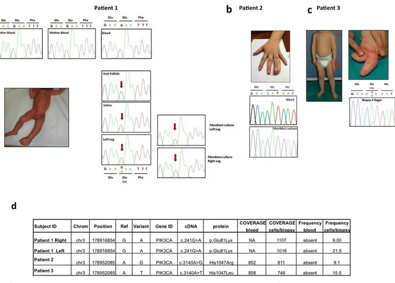
Fig 1. Clinical and mutational spectrum of the three index cases. a Patient 1, clinically diagnosed with MCAP, showing diffuse capillary malformation at the age of 2 months and cutaneous syndactyly between the 2ndand 3rdtoes. The PIK3CA c.241 G>A [p.E81K] mutation detected by Sanger sequencing in affected cells and tissues of patient 1 showed varying levels of the mutant allele depending on the tissue tested. The mutation was absent in the patient's blood and in her parents. b Macrodactyly of the right 4thfinger in patient 2, diagnosed with FAO, at the age of 17 years. Sequence of PIK3CA exon 20 in blood and cultured fibroblasts obtained from patient 2 showing that the mutation is undetectable in these samples. c Patient 3, at the age of 15 months before surgical intervention; note the disproportion of the left 2ndand 3rdfingers and the subcutaneous mass at the left deltoid region. Sanger sequencing validation of the c.3140 A>T [p.H1047L] mutation detected with targeted deep sequencing in the biopsy from the 2ndfinger of patient 3. d List of samples and mutations detected with targeted deep sequencing. Coverage indicates the mean average of reads on target in the regions of interest (ROI) while frequency denotes the percentage of reads with the mutation.
On physical examination at the age of 10 years and 4 months, her weight was 35 kg (25th -50thcentile), height 138 cm (25th-50thcentile) and head circumference 60 cm (+ 5.2 SD). She had left hemihyperplasia, involving face, trunk and limbs (mainly legs) with diffusely soft and thick irregularly marbled skin and prominent capillaries and veins on the trunk, abdomen and limbs. Three small (diameter<0.5 cm) achromic spots were observed on the trunk. Her 3rd right finger was significantly larger than the contralateral one and she had a bilateral proximal 4/5 cutaneous syndactyly of the 2ndand 3rdtoes. Besides macrosomia, she had dysmorphic fea-tures including malar hypoplasia, hypertelorism (inner canthal distance>97thcentile, palpe-bral length + 1 SD), long philtrum (>97thcentile) and high palate, and S-shaped scoliosis.
Patient 2. Patient 2, the only daughter of healthy non-consanguineous parents, presented to our observation at 12 years of age with a history of macrodactyly of the right 4thfinger diag-nosed at birth (Fig 1b). Histological examination carried out after surgical excisions performed at 3 and 5 years of age showed fibrolipomatous tissue. At 6 years of age, disease recurrence was observed, with two nodular areas (14 and 11.2 mm in diameter) revealed by ultrasound scan of the right hand soft tissues. Such areas, located adjacent to the shaft of the middle and distal phalanges, showed uneven distribution and no evidence of bone overgrowth or distortion at X-ray examination. No development of further lesions was observed at follow-up at age 18, except for a small angioma between the 4thand 5thright metacarpal bones, and increased volume of the already existing fibrolipomas of the right 4thfinger.
Patient 3. The proband is the first and only son of healthy, non-consanguineous parents. At birth, macrodactyly of the left 2ndand 3rdfingers was noted. Radiological examination showed that overgrowth involved the soft tissues and all skeletal segments (phalangeal and metacarpal bones) of the affected rays. Thus, a provisional diagnosis of apparently isolated true congenital macrodactyly was done and a periodical clinical follow-up was suggested. Over time, the two enlarged fingers showed disproportionate overgrowth. Moreover, at 6 months of age, a soft swelling appeared at the left deltoid region, which was sonographically compatible with a subcutaneous mass of adipose tissue, and, at 10 months of age, a slight overgrowth of the left arm and forearm was noted. These clinical signs suggested a diagnostic hypothesis of Proteus-like syndrome. Because of the negative functional and postural consequences of exces-sive volume and weight of the abnormal fingers, a surgical intervention was performed when the proband was 1 year and 3 months old to remove the 2ndand 3rdleft fingers by amputation (Fig 1c). Skin biopsies from the affected regions were obtained during this procedure. So far, surveillance measures revealed no abnormalities. Psychomotor development seems to be nor-mal and physical examination revealed no significant craniofacial dysmorphism.
Molecular and biochemical analyses
Mutational analysis of PIK3CA exons and adjacent intronic regions was performed by Sanger se-quencing methods on genomic DNA isolated from blood samples, tissue biopsies, and cultured dermal fibroblasts. No pathogenic variants were found, except for the point mutation c.241 G>A [p.E81K] in PIK3CA exon 1 detected in patient 1. This mutation was identified in hetero-zygosity in hair follicles, saliva, left leg biopsy, and left and right leg cultured dermal fibroblasts of the proband, but was undetectable in the patient's blood and in her parents (Fig 1a).
Considering the limited sensitivity of Sanger sequencing for detecting low-level mosaic muta-tions, we performed targeted deep sequencing of 21 selected genes involved in the PI3K/AKT/ mTOR pathway both in blood and tissue/biopsy/cell culture samples of the three patients.
The mean coverage depth per sample was 1900X with a mean percentage of reads on target of 93.75%. The percentage of ROI with a minimum coverage of 200X was 97.22%, see Table C inS1 File.
This approach confirmed the presence of the c.241 G>A [p.E81K] mutation in the right and left leg biopsies of patient 1, with mutant allele frequencies of 9% and 21.5%, respectively (Fig 1d).
In both patients with a clinical diagnosis of FAO (patients 2 and 3), targeted deep sequenc-ing analysis led to the identification of a PIK3CA mutation in primary fibroblasts samples only. Specifically, a c.3140 A>G [p.H1047R] mutation was identified in cultured dermal fibroblasts of patient 2 with a frequency of 9.1% (74/811 reads) (Fig 1d) and a c.3140 A>T [p.H1047L] mutation was identified in the 2ndfinger tissue biopsy of patient 3 with a frequency of 15.5% (116/746 reads) (Fig 1d). As shown in Fig1band1c, respectively, the c.3140 A>G [p.H1047R] mutation was undetectable by Sanger sequencing, whereas the c.3140 A>T [p.H1047L] muta-tion was visible with this method in DNA derived from fibroblast cultures, but was absent in blood samples (data not shown).
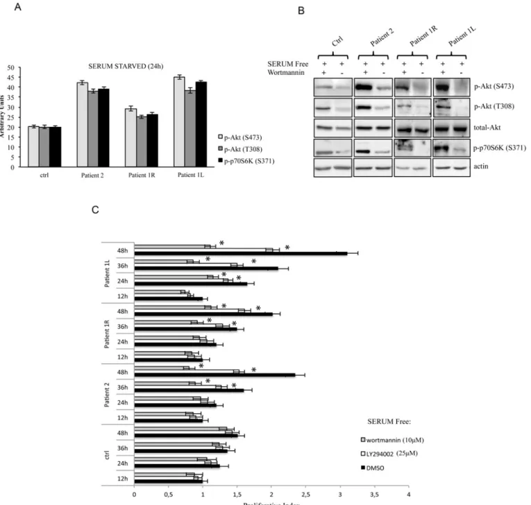
PIK3CA mutations have been previously identified in several types of cancer, where they overactivate the PI3K/AKT/mTOR signaling in the absence of growth factors [8,9]. Thus, to functionally characterize these mutations in our patients, we cultured primary dermal fibroblasts from skin biopsies of overgrowth lesions in the absence of serum. According to our immuno-blotting results (Fig2aand2b), mutant cells (fibroblasts from biopsy of patient 2, and from left and right leg biopsies of patient 1) showed increased levels of phosphorylated AKT at threonine 308, a residue targeted by PI3K in a direct or indirect (through PDK1) manner, when compared to IMR90 primary human normal fibroblasts. Moreover, patients’ cells showed increased phos-phorylation of another AKT residue, serine 473, which is targeted by the TORC2 complex (mTOR/Rictor) in a PI3K-dependent manner [10,11]. In order to fully characterize the PI3K/ AKT/mTOR cascade, we also evaluated the phosphorylation status of p70S6K, a direct substrate of the TORC1 complex (mTOR/Raptor). Our results showed that phosphorylation of serine 371, was significantly increased in overgrowing cells compared to normal healthy cells.
To complete the functional characterization of our patients’ primary fibroblasts, we treated these cells with the PI3K inhibitors wortmannin or LY294002 in the absence of growth factor stimulation. As shown inFig 2b, overactivation of the PI3K/AKT/mTOR pathway was abrogat-ed by pharmacological inhibition of PI3K in all patients testabrogat-ed as shown by the evaluation of the phosphorylation status of p-AKT and p-p70S6K. Importantly, patient-derived fibroblasts showed the ability to grow in culture even in the absence of growth factors, thus mimicking in vivo overgrowth features, while IMR90 primary human normal fibroblasts failed to prolifer-ate in serum starvation conditions (Fig 2c). Moreover, growth factor-independent proliferation in vitro was significantly decreased by the pharmacological blockade of PI3K by both wortman-nin and LY294002 in affected cells (Fig 2c), suggesting that this approach could be beneficial in overgrowth patients.
Discussion
Our data support the hypothesis that PIK3CA mutations can be responsible for a wide clinical spectrum of segmental overgrowth disorders. Identical mutations have been found in pheno-typically distinct disorders; however, to date any correlation with genotype is suggested but not confirmed [12]. The c.241 G>A [p.E81K] mutation identified in a patient with MCAP, patient 1, has been described previously in only one case of MCAP [2]. This mutation involves the resi-due 81 in the Ras-binding domain of PI3K and to our knowledge no functional analysis for this mutation has been ever performed on cells of affected patients.
According to Keppler-Nereuil [7] this patient should be categorized as PROS-A.
The PIK3CA c.3140 A>G [p.H1047R] mosaic mutation identified in patient 2 with over-growth confined only to the right 4thfinger diagnosed at birth, has been observed in patients
Fig 2. Overactivation of the PI3K/Akt pathway is abrogated by pharmacological inhibition of PI3K in all patients tested. a The indicated values are the result of the densitometric analysis of the phosphorylated forms of Akt and p70S6K normalized against total Akt and the loading control, respectively. The presented results are representative of at least three independent sets of experiments (bars represent standard deviation of the mean). b Immunoblot analysis of phospho-Akt (Ser473), phospho-Akt (Thr308), total Akt and phospho-p70S6K (Ser371) in mutant cells (fibroblasts from biopsies of FAO patient 2, and from left [L] and right [R] leg biopsies of MCAP patient 1) compared to IMR90 primary human normal fibroblasts (Ctrl).β-Actin was used as a loading control. Cells were treated with the PI3K inhibitor wortmannin (10μM) for 24 hours in the absence of growth factor stimulation. The presented results are representative of at least three independent sets of experiments. c Patients' affected cells are dependent on PI3K activity for proliferation. Primary fibroblasts obtained from biopsies were cultured in the presence or absence of wortmannin (10μM) and LY294002 (25μM). At the indicated time points, the proliferation index was determined using the WST-1 assay. The results were also confirmed by cell counting with trypan blue staining (data not shown). Each assay was performed in 6 replicates and the experiment was repeated six times. Statistical analysis was performed using Student’s t-tail test; *P<0.05, which was considered statistically significant (bars represent standard deviation of the mean).
with KTS, CLOVES and very severe FAO, but also in cases of isolated macrodactyly [13]. On the basis of the clinical spectrum of this patient (seeresults), after the positive testing for PIK3CA mutation, this patient would be classified as PROS B. It is worth noting that the PIK3CA c.3140 A>G [p.H1047R] change is the most common cancer-associated PIK3CA mu-tation [8,9]. However, in affected tissues of overgrowth disorder patients, it is isolated and asso-ciated to a variable mutation burden, as opposed to what happens in a tumor, where it is one of several somatic mutations.
Similarly, the c.3140 A>T [p.H1047L] mutation identified in patient 3, with clinical presen-tation consistent with FAO, has been already described in FAO and in isolated macrodactyly type I cases, and was also reported in more than 130 human cancers in the COSMIC Database. The initial classification of this patient at referral would have been PROS B, however, after clin-ical follow up, he was more precisely matching the PROS A presentation.
Our functional studies on primary patient-derived fibroblasts showed overactivation of the PI3K pathway and PI3K-dependent proliferation in affected cells for all mutations analyzed. Indeed, patients’derived cells were able to grow in vitro in the absence of growth factors and mitogens, while displaying a significantly reduced proliferation rate upon PI3K pharmacologi-cal inhibition by both wortmannin and LY294002. Very recent reports have demonstrated that oncogenic PIK3CA mutations cause activation of PI3K pathway and proliferative advantage in cells derived from lymphatic malformations [14,15]. Our data, show that the two mutations functionally studied [H1047R, E81K] behave similarly in PROS patient-derived fibroblast.
No malignancies were identified in our patients. Although an increased cancer risk cannot be ruled out in patients with somatic PIK3CA mutations, data are currently very limited. This is not surprising considering that only in recent years researchers began to identify mutations associated with these disorders, which were often misdiagnosed and superficially classified as Proteus or Proteus-like syndromes [16]. Keppler-Noreuil et al. [12] described a patient diag-nosed with FAO harboring a mosaic c.3140 A>T [p.H1047L] mutation that developed prema-lignant features of nephrogenic rests and a second patient, diagnosed with CLOVES, who had an ovarian cystadenoma; In addition, Kurek et al. [4] reported the case of a CLOVES patient with a mosaic c.3140 A>G [p.H1047R] mutation, who was affected by Wilms' tumor. In an-other report, two cases of Wilms' tumor, one case of leukemia, two cases of meningioma and one case of medulloblastoma were described, respectively, in five MCAP and one MPHH pa-tient. However, these patients' diagnoses were merely clinical, with no molecular characteriza-tion of the underlying disorders [2].
Surgical debulking and orthopedic procedures are the only treatments currently available for patients with segmental overgrowth syndromes [16,17]. However, considering that the in-vestigation of small molecule inhibitors of the PI3K signaling network is a promising area of oncology drug development, in the next few years these patients may well benefit from the re-sults of completed and ongoing clinical trials on PI3K inhibitors in cancer therapy. For exam-ple, an improved wortmannin chemical analog, PX-866, displayed antitumor activity in preclinical models and is currently being tested in various clinical trials [18]. For all of these reasons, PROS patients might be regarded as very appropriate candidates for enrollment in tri-als based on PI3K inhibitors. In fact, they display an ideal "clean" cellular setting, as they carry only a single PIK3CA mutation while lacking the host of somatic mutations that usually char-acterize tumors and that can interfere with/modulate the complex mechanisms regulating PI3K/AKT/mTOR signaling. On the other side, they would need long-term therapies and thus should be treated with drugs with very limited and acceptable side effects.
The results presented in this study, strengthen the idea that patients with these rare and often neglected syndromes, may represent the target of future trials using clinically available in-hibitors of the PI3K/AKT/MTOR pathways.
Supporting Information
S1 File. Table 1: Primers and annealing temperature for the PCR amplification of PIK3CA (NM_006218.2), AKT1 (NM_001014432.1), AKT3 (NM_005465.4), and PIK3R2
(NM_005027.2) genes. Table 2: List of PI3K/Akt/mTOR pathway genes selected for targeted deep sequencing. Table 3: Matrix Table
(DOCX)
Acknowledgments
We thank all the patients and their families for their valuable contribution. We thank Dr. Fran-cesco Paolo Jori for his helpful discussion during the preparation of the manuscript and editorial assistance. V.G. is supported by an Italian Association for Cancer Research (AIRC) fellowship. This study was partially supported by FIRB—FUTURO IN RICERCA RBFR12VP3Q_003 (to C.S.) from the Italian MIUR and by Fondi d’Ateneo/conto terzi (to N.R.) from the University of Bari“Aldo Moro”.
Author Contributions
Conceived and designed the experiments: NR CS. Performed the experiments: DCL CB RB VG GF AG LC FCS. Analyzed the data: NR CS AS. Wrote the paper: NR CS RT AS. Performed ge-netic counselling to families of affected individuals: PL DV MP. Provided clinical data and per-formed biopsies from patients: MC FB NL RT.
References
1. Moore CA, Toriello HV, Abuelo DN, Curry CJ, Hall BD, Higgins JV, et al. Macrocephaly-cutis marmora-tatelangiectaticacongenita: a distinct disorder with developmental delay and connective tissue abnor-malities. Am J Med Genet.1997; 70:67–73. PMID:9129744
2. Rivière JB, Mirzaa GM, O'Roak BJ, Beddaoui M, Alcantara D, Conway RL, et al. De novo germline and postzygotic mutations in AKT3, PIK3R2 and PIK3CA cause a spectrum of related megalencephaly syn-dromes. Nat Genet. 2012; 44:934–940. doi:10.1038/ng.2331PMID:22729224
3. Mirzaa GM, Parry DA, Fry AE, Giamanco KA, Schwartzentruber J, Vanstone M, et al. De novo CCND2 mutations leading to stabilization of cyclin D2 cause megalencephaly-polymicrogyria-polydactyly-hydrocephalus syndrome. Nat Genet. 2014; 46:510–515. doi:10.1038/ng.2948PMID:24705253
4. Kurek KC, Luks VL, Ayturk UM, Alomari AI, Fishman SJ, Spencer SA, et al. Somatic mosaic activating mutations in PIK3CA cause CLOVES syndrome. Am J Hum Genet. 2012; 90:1108–1115. doi:10.1016/ j.ajhg.2012.05.006PMID:22658544
5. Biesecker LG, Peters KF, Darling TN, Choyke P, Hill S, Schimke N, et al. Clinical differentiation be-tween Proteus syndrome and hemihyperplasia: description of a distinct form of hemihyperplasia. Am J Med Genet. 1998; 79:311–318. PMID:9781913
6. Lindhurst MJ, Parker VE, Payne F, Sapp JC, Rudge S, Harris J, et al. Mosaic overgrowth with fibroadi-pose hyperplasia is caused by somatic activating mutations in PIK3CA. Nat Genet. 2012; 44:928–933. doi:10.1038/ng.2332PMID:22729222
7. Keppler-Noreuil KM, Rios JJ, Parker VER, Semple RK, Lindhurst MJ, Sapp JC, et al. PIK3CA-Related Overgrowth Spectrum (PROS): Diagnostic and Testing Eligibility Criteria, Differential Diagnosis, and Evaluation. Am J Med Genet Part A 2014; 167A:287–295. doi:10.1002/ajmg.a.36836PMID:
25557259
8. Samuels Y, Wang Z, Bardelli A, Silliman N, Ptak J, Szabo S, et al. High frequency of mutations of the PIK3CA gene in human cancers. Science. 2004 304:554. PMID:15016963
9. Kang S, Bader AG, Vogt PK. Phosphatidylinositol 3-kinase mutations identified in human cancer are oncogenic. Proc Natl Acad Sci U S A. 2005; 102:802–807. PMID:15647370
10. Dalle Pezze P, Sonntag AG, Thien A, Prentzell MT, Gödel M, Fischer S, et al. A dynamic network model of mTOR signaling reveals TSC-independent mTORC2 regulation. Sci Signaling. 2012;27; 5(217):ra25. doi:10.1126/scisignal.2002469PMID:22457331
11. Gan X, Wang J, Su B, Wu D. Evidence for direct activation of mTORC2 kinase activity by phosphatidyli-nositol 3,4,5-trisphosphate. J Biol Chem. 2011; 286:10998–11002. doi:10.1074/jbc.M110.195016
PMID:21310961
12. Keppler-Noreuil KM, Sapp JC, Lindhurst MJ Parker VE, Blumhorst C, Darling T, et al. Clinical delinea-tion and natural history of the PIK3CA-related overgrowth spectrum. Am J Med Genet A. 2014; 164:1713–1733.
13. Rios JJ, Paria N, Burns DK Israel BA, Cornelia R, Wise CA, et al. Somatic gain of function in PIK3CA in patients with macrodactily. Hum Mol Genet. 2013 22:444–451. doi:10.1093/hmg/dds440PMID:
23100325
14. Boscolo E, Coma S, Luks VL Greene AK, Klagsbrun M, Warman ML, et al. AKT hyper-phosphorylation associated with PI3K mutations in lymphatic endothelial cells from a patient with lymphatic malforma-tion. Angiogenesis 2014 Nov 26. [Epub ahead of print].
15. Osborn AJ, Dickie P, Neilson DE, Glaser K, Lynch KA, Gupta A, et al. Activating PIK3CA alleles and lymphangiogenic phenotype of lymphatic endothelial cells isolated from lymphatic malformations. Hum Mol Genet. 2015 24:926–933 doi:10.1093/hmg/ddu505PMID:25292196
16. Biesecker L. The challenges of Proteus syndrome: diagnosis and management. Eur J Hum Genet. 2006; 14:1151–1157. PMID:16883308
17. Tosi LL, Sapp JC, Allen ES, O'Keefe RJ, Biesecker LG. Assessment and management of the orthope-dic and other complications of Proteus syndrome. J Child Orthop. 2011; 5:319–327. PMID:23024722
18. Chen Y, Wang BC, Xiao Y. PI3K: a potential therapeutic target for cancer. J Cell Physiol. 2012; 227:2818–2821. doi:10.1002/jcp.23038PMID:21938729