SHELL MICROSTRUCTURES IN LOPINGIAN BRACHIOPODS: IMPLICATIONS FOR FABRIC EVOLUTION AND CALCIFICATION
CLAUDIO GARBELLI
State Key Laboratory of Palaeobiology and Stratigraphy, Nanjing Institute of Geology and Palaeontology, Chinese Academy of Sciences, 39 East Beijing Road, Nanjing, Jiangsu 210008, P.R. China.
Dipartimento di Scienze della Terra A. Desio, Università di Milano, Via Mangiagalli 34, 20133 Milan, Italy. E-mail: [email protected] To cite this article: Garbelli C. (2017) - Shell microstructures in Lopingian brachiopods: implications for fabric evolution and calcification. Riv. It. Paleontol. Strat., 123(3): 541-560.
Abstract. The study of the shell microstructure of brachiopods is fundamental to understand their
evolu-tionary history and their biomineralization process. Here, species of forty Lopingian brachiopods genera, represen-tative of twenty-seven different families, are investigated using the Scanning Electron Microscope. The investiga-ted specimens come from different paleogeographic localities in the Palaeotethys/Neotethys oceans. The studied brachiopods show a large variability of the shell fabric, which is mainly related to the organization of its structural units: laminae, fibers and columns, possibly crossed by pseudopunctae or punctae. For the Strophomenata, the laminar fabric of Productida is crossed by pseudopunctae with taleolae and the laminae are often organized in packages, with the blades oriented about perpendicular to each other; this feature is less evident in the laminar Or-thotetida, which bear pseudopunctae without taleoae. For the Rhynchonellata, fibrous fabrics are either impuctate in the Spiriferida, most Athyridida and Rhynchonellida, or with punctae, as observed in the Orthida, Terebratulida and in the Neoretziidae (Athyridida). The fibers show a range of sizes and shapes also in the same specimens and the transition to the columnar layer is different than in Strophomenata.
The arrangement of the structural units revealed that the disposition of the organic membranes, on which biomineralization took place, was highly variable among the taxa. On the other hand, two distinctive features are analogous among distantly related groups, i.e. the Strophomenata and the Rhynchonellata: the presence of a co-lumnar tertiary layer underlying the secondary fabric and the alternations between fibers/laminae of the secondary layer and columns of the tertiary layer. This suggests that there are common factors controlling the development and evolution of the shell fabric in all rhynchonelliformean brachiopods that can be linked to their taxonomical position, to their environmental requirements and to constraints imposed by their low-energy life-style. This should be taken into account to understand how these calcifying organisms responded and will respond to environmental and climate change in past and future times.
Received: March 21, 2017; accepted: October 05, 2017
Keywords: Rhynchonelliformea; Strophomenata; biomineralization; taxonomy; columnar layer.
I
ntroductIonThe shell microstructure of calcifying organ-isms plays a critical role since it can affect parameters such as exoskeleton mechanical proprieties, func-tionality and metabolic cost (Palmer 1983; Palmer 1992; Schmahl et al. 2012). Due to the growing concern about the effects of anthropogenic CO2, modern calcifying organisms have been studied to assess how they modify the shell microstructure, in response to global warming and acidification of seawater (e.g. Roger et al. 2012; Cross et al. 2016; Milano et al. 2016). This knowledge is a key step to understand how these organisms can survive in a changing environment. Unfortunately,
compara-tive and experimental studies on modern organisms cannot fully address the evolutionary response on the long-term timescale (Cross et al. 2015). From this perspective, the fossil record of brachiopods provides the best archive to study the evolution of shell microstructure through geological time (Wil-liams & Cusack 2007).
Modern rhynchonelliformean brachiopods display a small number of taxa, which bear mul-tilayered calcite shells with different hierarchical microstructures and mechanical proprieties (Pérez-Huerta et al. 200; Griesshasber et al. 2007; Schmahl et al. 2008; Göetz et al. 2009). However, during the Paleozoic, they have been one of the major calcify-ing phyla. Two classes of rhynchonelliformean bra-chiopods dominated the Paleozoic benthic communi-ties (e.g. Curry & Brunton 2007): the Strophomenata,
which became extinct at the end of the Permian, and the Rhynchonellata, still extant. These two taxa show general differences in their body plan orga-nization and shell microstructure (Williams 1968; Williams 1997). In the two classes, the multilayered shell biocomposite is different, as the secondary layer is laminar in the Strophomenata, while it is fi-brous in the Rhynchonellata (Williams 1997). The tertiary layer is morphologically similar in the two classes, being columnar in both (Göetz et al. 2009; Garbelli et al. 2014a). Cambrian and Ordovician brachiopods had already evolved different types of organocarbonate fabric (Williams 1970; Brun-ton 1972), but only certain fabric types survived the end-Permian extinction event (e.g. Erwin 2006; Shen et al. 2011; Garbelli et al. 2017).
Brachiopods low-Mg calcite shells are highly resistant to diagenesis; thus, they have a high poten-tial to preserve the original shell microstructure and fabric during the fossilization process (i.e. Angio-lini 1993; Garbelli et al. 2012). The study of various shell microstructures of fossils can return informa-tion about physiological changes of biomineraliza-tion in these organisms. The purposes of this paper are: 1) to investigate the microstructure of shell fab-ric in several Lopingian brachiopod taxa, with a
fo-cus on describing its variability, and 2) to disfo-cuss its implication on the evolution of Rhynchonellifor-mea brachiopods fabric. This can help us in a better understanding of macroevolutionary patterns of biomineralization and to assess the possible key fac-tors, which promote fabric change during a period of environmental and climatic perturbations at a global scale, such as during the Lopingian.
MaterIalsandMethods
Materials. The shell microstructure of several species
be-longing to 39 genera of Lopingian brachiopods (Tabs 1, 2) was stud-ied using a Scanning Electron Microscope (SEM).
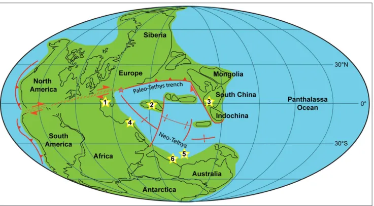
A total of 450 brachiopods shells have been analyzed. The observed specimens were collected from different paleogeographic localities in the Palaeotethys/Neotethys oceans (Fig. 1). The mate-rial comes from the Nesen Formation, Alborz Mountains, northern Iran (Angiolini & Carabelli 2010; MPUM 9907-10050); Julfa Forma-tion, Ali Bashi Formation and Boundary Clay, Ali Bashi Mountains, northwestern Iran (Ghaderi et al. 2014; Garbelli et al. 2014b; MPUM 11616-11657, MPUM 11723-11724); Selong Group, southern Tibet (MPUM 11703-11707); Gyanyima Formation, southwestern Ti-bet (Shen et al. 2006, 2010; MPUM 11682-11702); Bulla Member, Dolomites, Northern Italy (Posenato 2001, 2009; Brand et al. 2012; MPUM 11720-11722); Gomaniibrik Formation, Hazro, Turkey (un-published data collected by A. Baud: MPUM 11708-11719); Chang-hsing Limestone and Dalong Formation, South China (unpublished data collected in 2014 by L. Angiolini, G. Crippa, C. Garbelli, S.Z. Fig. 1 - Lopingian paleogeographic reconstruction showing the setting of the formations from which the brachiopod shell structure was
in-vestigated. The yellow stars refer to the paleogeographic positions of the different studied localities: 1 - Southern Alps (Dolomites), 2 - Northern Iran, 3 - South China, 4 - Turkey, 5 - Southwestern Tibet, 6 - Salt Range (Pakistan) (modified after Muttoni et al. 2009; Angiolini et al. 2015).
Class Order Family Genus Layers observed Structures observed Comments
Strophomenata
Orthotetida Wagen, 1884 Meekellidae Stehli, 1954
Orthotetina Schellwien, 1900 L, C? P, no T, S massive shell composed mainly of laminae, which seem very compact
Alatorthotetina He Xi-Lin & Zhu Mei-Li,
1985 L P, no T, S
Meekella White & St. John, 1867 L P, S the specimen does not show good preservation
Productida Sarytcheva & Sokol-skaya, 1959
Strophalosidae Schuchert,
1913 Marginalosia Waterhouse, 1978 L rather thick laminar layer, >0.5 mm, poor preservation Aulostegidae Muir-Wood &
Cooper, 1960 Edriosteges Muir-Wood & Cooper, 1960 Pr?, L P Richthofeniidae Waagen,
1885 Richthofenia Kayser, 1881 L P, T
shell wall extremely rich in pseudopunctae, some of which are strongly persistent through it.
Permianellidae He & Zu,
1979 Permianella He & Zu, 1979 L P, T Monticuliferidae
Muir-Wood & Cooper, 1960 Costatumulus Waterhouse in Waterhouse & Briggs, 1986 L the specimen does not show good preservation Linoproductidae Stehli,
1954 Linoproductus Chao, 1927b L P, T, E cross bladed lamination evident Productidae Gray, 1840 Tyloplecta Muir-Wood & Cooper, 1960 L, C P, T, S, E
massive shells, both laminar and columnar are thick, frequent alternation of the two
Araxilevis Sarytcheva in Sarytcheva &
Sokoloskaya, 1965 L, C P, T, S
Productellidae Schuchert, 1929
Cathaysia Ching in Wang, Ching & Fang,
1966 L P, T
Paryphella Liao in Zhao & others, 1981 L P, T
Haydenella Reed, 1944 L P, T
Costiferina Muir-Wood & Cooper, 1960 L P, T, E, S
Retimarginifera Waterhouse, 1970 L P, T, E, S thick laminar layer, >2mm
Transennatia Waterhouse, 1975 L, C? P, T
Spinomarginifera Huang, 1932 L, C P, T, E, S alternation of fibrous and columnar layers; high variability for the presence of columnar layer Lyttoniidae Wagen, 1883 Leptodus Kayser, 1883 L P, T, E cross bladed lamination
Table 1
Class Order Family Genus Layers observed Structures observed Comments
Rhynchonellata
Orthida Schuchert & Cooper, 1932
Schizophoriidae Schuchert
& LeVene, 1929 Acosarina Cooper & Grant, 1969 Pr, F Pu
Enteletidae Waagen, 1884 Enteletes Fischer de Waldheim, 1825 Pr, F Pu keel and saddle
Peltichia Jin & Liao in Jin & Sun, 1981 F Pu thick fibrous layer, >1 mm, small fibers
Spiriferida
Martiniidae Waagen, 1883 Martinia M'Coy, 1844 F, C Ambocoeliidae George, 1931 Paracrurithyris Liao, 1981 F Choristitidae Waterhouse, 1968 Alphaneospirifer Gatinaud, 1949 F, C? Spiriferellidae Waterhouse, 1968 Spiriferella Chernyshev, 1902 F, C? Trigonotretidae Schuchert,
1893 Neospirifer Frederiks, 1924 F, C thick columnar layer Reticulariidae Waagen, 1883 Squamularia Gemmellaro, 1899 F
Elythidae Frederiks, 1924 Bullarina Jin & Sun, 1981 F
Elythidae Frederiks, 1924 Permophricodothyris Pavlova, 1965 F, C alternation of fibrous and columnar layers Spiriferinida Ivanova, 1972 Paraspiriferinidae Cooper & Grant, 1976 Paraspiriferina Reed, 1944 Pr, F Pu
Athyridida Boucot, Jhonson &
Staton, 1964 Athyrididae Davidson, 1881
Araxathyris Grunt, 1965 F, C
Transcaucasathyris Shen et al., 2004 F, C
Comelicania Frech, 1901 F, C
Neoretziidae Dagys, 1972 Hustedia Hall & Clarke, 1893 Pr, F Pu very small fibers, ~4 µm in width Rhynchonellida Khun, 1949
Stenoscismatidae Oehlert,
1887 Stenoscisma Conrad, 1839 F, C massive columnar layer Wellerellidae Licharew,
1956 Uncinunellina Grabau, 1932a F Terebratulida Waagen, 1883
Notothyrididae Licharew,
1960 Notothyris Waagen, 1882 F, C Pu Gillediidae Campbell, 1965 Hemiptychina Waagen, 1882 F, C Pu Dielasmatidae Schuchert,
1913 Dielasma King, 1859 F, C not well preserved
Table 2
Tab. 1 - List of studied taxa and synthetic results of characters detected in the shell fabric of Strophomenata; Pr - primary layer; L - laminar layer; C - columnar layer; P - pseudopunctae; S - spines; E - endospines; T- taleolae.
Tab. 2 - List of studied taxa and synthetic results of characters detected in the shell fabric of Rhynchonellata; Pr - primary layer; F - fibrous layer; C - columnar layer; Pu - punctae.
Shen, D.X. Yuan; MPUM 11658-11861). The material is housed at Dipartimento di Scienze della Terra “A. Desio”, Università di Milano (MPUM catalogue numbers).
Methods. The studied brachiopod specimens were cut along
their longitudinal and transverse axes, embedded in resin, polished and then etched with 5% HCl for 15 seconds. In addition, acetate peels of some specimens were prepared with a cellulose acetate film and acetone (CH3)2CO. The exposed surfaces were metal coated with Au by the sputtering process and then inspected with two scanning electron microscopes (SEMs): 1) a Cambridge S-360 featuring a LaB6 source and an acceleration voltage of 20kV, located at the Diparti-mento di Scienze della Terra A. Desio, Milan, Italy (DPT); 2) a LEO-1530VP with an acceleration voltage between 10 and 15kV, located at the Nanjing Institute of Palaeontology and Geology, Nanjing, China.
Thin sections of specimens were also analyzed by cathodo-luminescence with a cold cathode luminoscope (Nuclide ELM2) op-erating at 10 kV with a beam current of 5-7 mA. The instrument is located at DPT. Exposure to the electronic beam (before taking pho-tomicrographs) was on the order of 1530 seconds, not to force shell material to luminescence, and it was consistent for all specimens. In addition, light exposure for photographs was uniform and set to 1.2 seconds for consistency with a Nikon Coolpix 4500, operating at 400 ISO. Cathodoluminescence (CL) microscopy is a powerful technique to study biominerals, and is particularly important in paleontology to assess the preservation of carbonate shells (Machel 2000; Barbin 2013; Angiolini et al. 2012). Results are presented in the supplemen-tary materials.
r
esults:
ultrastructure,
MIcroMorphologyandshellfabrIcof
l
opIngIan brachIopodsThe investigated taxa show the typical bra-chiopod shell successions described by Williams
(1997), composed of an outermost primary layer, a secondary fibrous or laminar layer and, when pre-sent, an innermost columnar tertiary layer. The pri-mary layer is not always present because it is only a few micrometers thick, and it easily undergoes corrasion. This outermost layer has been observed Fig. 2 - A) Shell sequences showing the outermost primary layer (pr) and the inner fibrous layer (f) crossed by punctae (pu), Hustedia sp. MPUM
11659 (CH60-15); B) transition from the outermost primary layer (pr) and the fibers (f), which preserve their original shape and are oriented subparallel to the outer shell surface, Paraspiriferina alpha MPUM 11676 (CH12-3); C/D) details of outermost primary layer
(pr) showing different morphologies, possibly related to the degree of alteration; the first one is coarser, the second one more com-pact, Enteletes lateroplicatus MPUM10000 (IR 332-1) and Transcaucasathyris sp. MPUM 11658 (CH30-4) respectively.
Fig. 3 - A) cross-bladed laminar layer showing packages of laminae orthogonally oriented (arrows), Paryphella sp. MPUM 11671
(CH136-2); B) shell thicker than 4 millimeters, but com-posed entirely of a laminar fabric, Costiferina indica MPUM
11684 (GY79); C) laminar layer crossed by pseudocpunctae (arrows); the laminae are folded to produce the external or-namentation of costellae, Alatorthotetina sp. MPUM 11711
(EBHZ80-16); D/E) details of the laminae composed of aligned blades/laths; in longitudinal section (E) discon-tinuities are evident between the structural units (arrow),
Cathaysia sp. MPUM 11664 (CH71-8); F) pseudopunctae
formed by cone in cone inwardly deflected laminae with an inner core of calcite, the taleola(t), planar view, Spinomar-ginifera sp. MPUM 11616 (JU117-1); G) details of C
show-ing a pseudopuncta, without taleola, crossshow-ing the secondary shell and deflecting the laminae inwardly, Alatorthotetina sp.
MPUM 11709 (EBHZ65-12); H) pseudopunctae formed by inwardly deflected laminae protruding in the inner shell to form an endospine, Haydenella sp. MPUM 11677 (CH4-5); I)
laminar layer in a coral-like Richtofenioid, crossed by nume-rous pseudopunctae (arrows), Richthofenia lawrenciana MPUM
11682 (GY52); J) longitudinal section of the distal part of a spine composed of laminar secondary layer with a channel (ch) filled by diagenetic calcite, Spinomarginifera helica MPUM
11710 (EBHZ71-10); K/L) cross sections of an isolate spi-ne and of the proximal portion arranged sub-parallel to the outer shell surface respectively, Spinomarginifera helica MPUM
11710 (EBHZ71-10); in all the pictures the asterisks (*) in-dicate the outermost part of the shells.
in the Rhynchonellata Enteletes, Acosarina, Transcau-casathyris, Paraspiriferina, Hustedia and traces of the
outermost layer were observed in Paracrurithyris; the
layer usually appears to be recrystallized (Figs 2A, B, C, D). Its thickness is rather constant along the shell and slightly varies between taxa. The thinnest primary layer observed belongs to Paraspiriferina (~
15 µm), and the thickest one belongs to Transcauca-sathyris (~ 50 µm), indicating a range of thickness
similar to the one observed in modern Rhynchonel-lata (Williams & Cusack 2007).
The outermost primary layer was not obser-ved in the analyzed Strophomenata specimens.
The other two inner layers are thicker; thus, they are more easily preserved compared to the ou-termost layer.
Laminar fabric
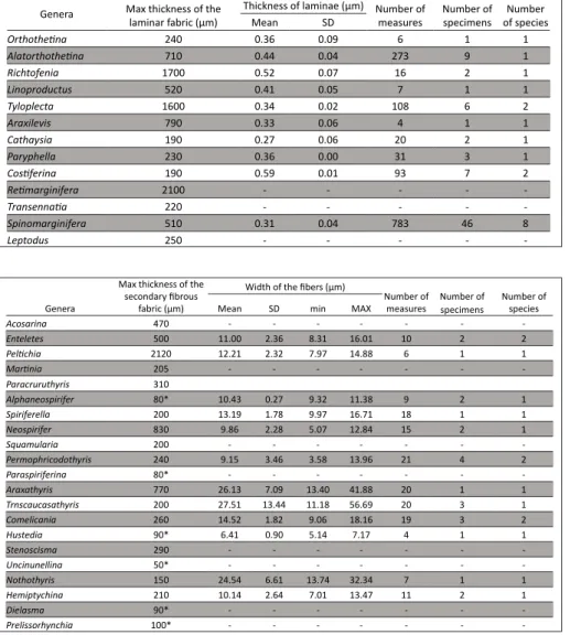
In adult specimens, the laminar secondary la-yer is usually between 50 and 400 μm thick, but it exceeds 1 mm in some genera, such as
Alatorthoteti-na (Orthotetida), and Tyloplecta and Araxilevis (both
Productida) (Figs 3A, B, C). In some genera of Pro-ductida, such as Retimarginifera and Costiferina,
lami-nar fabrics thicker than 2 and 3 mm respectively have been observed. Table 3 summarizes the maximum thickness of the laminar fabric observed for several genera. The laminae are composed of single bla-des/laths, which are 0.6-2 μm wide (Figs 3D, E; Fig. 4). The thickness of laminae is between 0.2 and 0.6 μm, and a summary of the obtained measurements is presented in Tab. 3. In the species represented by a high number of specimens, i.e. the species of
Spinomarginifera, there are significant differences in
the mean thickness between individuals of the same species. The thinnest laminae have been measured in Spinomarginifera iranica, and can be thinner than
0.2 μm. The thickest laminae have been observed in
Costiferina, and can be thicker than 0.7 μm. The
bla-des length is not measurable in transverse or longi-tudinal sections, but observations along the fracture surfaces suggest that they can be longer than 50 μm.
Genera Max thickness of the
laminar fabric (µm) Thickness of laminae (µm) Number of Mean SD measures Number of specimens of speciesNumber
Orthothetina 240 0.36 0.09 6 1 1 Alatorthothetina 710 0.44 0.04 273 9 1 Richtofenia 1700 0.52 0.07 16 2 1 Linoproductus 520 0.41 0.05 7 1 1 Tyloplecta 1600 0.34 0.02 108 6 2 Araxilevis 790 0.33 0.06 4 1 1 Cathaysia 190 0.27 0.06 20 2 1 Paryphella 230 0.36 0.00 31 3 1 Costiferina 190 0.59 0.01 93 7 2 Retimarginifera 2100 - - - - -Transennatia 220 - - - - -Spinomarginifera 510 0.31 0.04 783 46 8 Leptodus 250 - - - - -Table 3
Tab. 3 - Summary of the data mea-sured in the shells with lami-nar secondary fabric.
Genera
Max thickness of the secondary fibrous
fabric (µm)
Width of the fibers (µm)
Number of
measures Number of specimens Number of species Mean SD min MAX
Acosarina 470 - - - -Enteletes 500 11.00 2.36 8.31 16.01 10 2 2 Peltichia 2120 12.21 2.32 7.97 14.88 6 1 1 Martinia 205 - - - -Paracruruthyris 310 Alphaneospirifer 80* 10.43 0.27 9.32 11.38 9 2 1 Spiriferella 200 13.19 1.78 9.97 16.71 18 1 1 Neospirifer 830 9.86 2.28 5.07 12.84 15 2 1 Squamularia 200 - - - -Permophricodothyris 240 9.15 3.46 3.58 13.96 21 4 2 Paraspiriferina 80* - - - -Araxathyris 770 26.13 7.09 13.40 41.88 20 1 1 Trnscaucasathyris 200 27.51 13.44 11.18 56.69 20 3 1 Comelicania 260 14.52 1.82 9.06 18.16 19 3 2 Hustedia 90* 6.41 0.90 5.14 7.17 4 1 1 Stenoscisma 290 - - - -Uncinunellina 50* - - - -Nothothyris 150 24.54 6.61 13.74 32.34 7 1 1 Hemiptychina 210 10.14 2.64 7.01 13.47 11 2 1 Dielasma 90* - - - -Prelissorhynchia 100* - - - -Table 4
Tab. 4 - Summary of the data mea-sured in the shells with fi-brous secondary fabric.
In several Productida, i.e. Spinomarginifera, Haydenel-la, Cathaysia, Tyloplecta, Fusiproductus, Leptodus, Ara-xilevis, Transennatia, Perygeyerella, and Costiferina, the
laminae are organized in packages with the axis of blades oriented about perpendicular to each other (Fig. 3B). This feature is less evident in the inve-stigated Orthotetida. The orthotetid Alatorthotetina
and Orthotetina have pseudopunctae composed only
of a slightly arcuate, anteriorly inclined trail of cone in cone laminae that are inwardly deflected, without any evidence of internal core forming a taleola (Fig. 3G). In the studied Productida, the laminar layers are crossed by pseudopunctae with taleolae (Fig. 3F) and a certain variability in the size and density of pseudopuncation between taxa has been observed (compare Figs 3A, B with 3I). Pseudopunctae can produce endospines when they protrude inwardly in the shell wall (Fig. 3H). Spines are hollow and have and internal tubular structure (Figs 3L, M, N).
Fibrous fabric
The fibrous layer is composed of stacked fi-bers, but a certain amount of variability was obser-ved in the thickness and size of the fibers, their sha-pe, and their reciprocal arrangement. The maximum thickness of this fabric ranges from a few tens of micrometers in Hustedia (Fig. 2A) up to 2 mm in Pel-tichia (Fig. 5A). The mean width of the fibers in cross
section varies from approximately 6 µm to 27 µm (Tab. 4; Figs 5B, G, H, I, L). Even the cross-section outline varies, from a “keel and saddle” profile to a more sub-diamond outline. In some taxa, such as
Transcaucasathyris, Araxathyris, and Notothyris, the size
and shape of the fibers are not consistent in all the secondary layer, but, gradual, significant differences
were observed (Figs 5D, E, G, H, O). In these taxa, the width of fibers show a relative higher standard de-viation, due to the huge differences in the minimum and maximum width of fibers. The maximum width can be three times wider than the minimum one (see Tab. 4). In other genera, such as Peltichia, Paracrurithy-ris, PermophricodothyParacrurithy-ris, and Comelicania, the shape and
size of fibers are more homogeneous in different re-gions of the shell (Figs 5C, L) and standard deviation is smaller. Also in these taxa, differences in fibers size and shape are observed in the umbonal region, where convolute fibers are present. Hustedia has the
smal-lest fibers, with a maximum width of 10 µm, con-firming the observation of MacKinnon (1974) for the Retziidina. About the orientation of the fibers through the shell substance, in Peltichia (Orthida) and Paracrurithyris (Spiriferida) there are abrupt changes
in the orientation of the main longitudinal axis of the fibers (Figs 5A, C). On the other hand, in other groups, the directions of fibers are more consistent, as in Permophricodothyris. In some genera the change in
orientation of the longitudinal axis is gradual, and it is coupled with an increase of fiber size and a change of shape (Figs 5D, M). For example, in the cross sec-tion of Transcaucasathyris, Araxathyris, and Notothyris,
the outermost fibers are smaller and flatter, with a keel and saddle outline, whereas inwardly they beco-me larger, with a diamond shape outline.
In the order Orthida, Spiriferidina and the fa-mily Neoretziidae (Order Athyridida), these fabrics are perforated by punctae (Figs 2A, 5K, 6A-F). The diameter has the same order of magnitude in dif-ferent genera. These perforations deflect the fibers outwardly (Figs 6E, F). In cross section, the infilling of the channels, which perforate the secondary layer, shows regularly disposed hole, which could be traces of internal structures of the mantle extensions (Figs 6A, C). In the Orthida Acosarina, in the Spiriferinida Paraspiriferina and in the the Neoretziidae Hustedia,
the infill of the punctae was detected through the re-crystallized primary layer (Figs 6B, F), but no canopy has been observed.
Columnar fabrics
Different taxa of both classes, Strophomenata and Rhynchonellata, bear a well-developed columnar tertiary layer. Its thickness easily exceeds 1 mm (Figs 7B, F). The genera, which produce a well-developed columnar layer, are Tyloplecta, Araxilevis, and Spinomar-ginifera in the Productida; Permophricodothyris, Martinia, Fig. 4 - Schematic drawing illustrating the structural organization
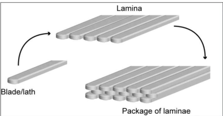
of the laminar fabric. Single long shaped crystals of calcite, the blades, are aligned with the major axis parallel to each others, composing a single lamina; several laminae are pa-cked together to form a layer.
Neospirifer, and Spiriferella in the Spiriferida; Araxathy-ris, TranscaucasathyAraxathy-ris, and Comelicania in the
Athyridi-da; Notothyris and Hemiptychina in the Terebratulida. In
some genera, only traces of columnar layer have been observed because of the poor preservation: Alphane-ospirifer, Stenoscisma, and Dielasma. In a few species, an
intraspecific variability for the development of the columnar layer was observed: some species of Spi-nomarginifera from the Julfa (Iran) and Gomaniibrik
(Turkey) formations show a well-developed colum-nar tertiary layer. On the other hand, none of the numerous conspecific specimens investigated from the Nesen Formation (Iran) has shown any well-de-veloped tertiary layer.
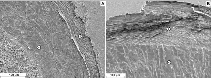
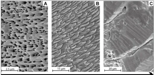
The morphology of the columnar layer is very similar among classes, but the Rhynchonellata show polished surfaces with well-defined accretion of mi-crometric bands (Fig. 7H) more frequently than Stro-phomenata, in which the columns tend to have a co-arser appearance (Fig. 7C). At sub-micrometric scale the structure is different (Figs 7D, I): the columns of Strophomenata appear with smaller and coarser micro-granular units than the columns of Rhyncho-nellata.
In several genera, intercalations of laminar/ fibrous layers inside the columnar layer have been observed (Figs 7A, E). This feature was observed in
Tyloplecta, Araxilevis, and Spinomarginifera in the
Pro-ductida, in Permophricodothyris in the Spiriferida (Fig.
7E). The transition from the laminar/fibrous to the columnar layer shows a certain pattern in variability. In the Strophomenata, the transition is restricted to a few micrometers with a rather abrupt change of fabric (Fig. 7J); also in the Rhynchonellata, some taxa show this feature (Fig. 7E), but others show a gradual shift from fibers to columns (Fig. 7K) in the growth direction.
d
IscussIonVariability in Lopingian brachiopod shell microstructure and its implications
The observed variability in microstructure is in agreement with the already known differences betwe-en the laminar fabric in the Strophombetwe-enata and the fibrous one in the Rhynchonellata (Williams 1997; Williams & Cusack 2007), but it also reveals some important aspects of the ontogenetic development of the tertiary columnar layer.
Laminar fabrics. Despite the homogeneous
basic structure of the laminae, which are compo-sed of aligned structural units (Fig. 4), and even though these fabrics are virtually indistinguisha-ble between the Orthotetida and Productida, some differences are present. For example, lami-nae organized in packages with the axis of blades oriented about perpendicular to each other are more frequently observed in the Productida than in the Orthotetida (compare Figs 3A, 3C). The-se two orders have also differences related to the pseudopunctae, the structures crossing the shell, which are slightly arcuate, anteriorly inclined trail of inwardly deflections of the laminae (Williams 1997). In Orthotetina and Alatorthotetina
(Orthoteti-da), the pseudopunctae are composed of deflected laminae, layered around a core of amalgamated laminae. On the other hand, in all the Producti-da, the pseudopunctae possess an inner rod-core of calcite called taleola. These two types of pseu-dopunctae correspond to those described by Wil-liams (1997).
The reason why Orthotetida and Producti-da show these differences could be explained by the independent origin and evolution of the two groups (Dewing 2004). In fact, Orthotetida pro-bably evolved from a group of the ancestral Bil-lingselloidea, in which the fabric is stratiform la-minar (Williams 1970; Williams & Cusack 2007);
Fig. 5 - A) fibrous layer in which several changes in the direction of fiber growth are evident (arrows), Peltichia sp. MPUM 11660
(CH60-8); B) details of A showing the fibers in cross sec-tion with a keel and saddle outline; C) fibrous layer in which the structural elements are consistently oriented parallel to the outer shell surface, Paracrurithyris pygmaea MPUM MPUM
11661 (CH30-11); D/E) fibrous layer in which the fibers change their orientation, progressively modifying the orien-tation of the growth axis from the outer to the inner shell,
Transcauscasathyris sp. MPUM 11620 (JU136-1); F)
accretio-nary bands on the fibers, Transcaucasathyris sp. MPUM 11621
(JU140-2); G) cross section of the fibers in the outermost layer of the same taxon of D, Transcaucasathyris sp. MPUM
11618 (JU121-1; H) cross section of the fibers in the inner-most fibrous layer of the same taxon of D, Transcaucasathyris
sp. MPUM 11622 (JU89 -1); I) cross section of the fibers in, Terebratulida fam. gen. sp. ind. MPUM 11683 (GY6-12); J) cross section of the fibers in Hustedia sp. MPUM 11662
(CH30-15); K) shell composed of an outermost primary layer (pr) and a fibrous secondary layer (f) crossed by pun-ctae (arrows), Acosarina minuta MPUM 11665 (CH72-11);
L) fibrous secondary layer of Comelicania sp. MPUM 11720
(VB9B-1); M) fibrous secondary layer of Transcaucasathyris
sp. MPUM 11723 (JU1-1); in all the pictures the asterisks (*) indicate the outermost part of the shells.
instead, the primitive Productida evolved from the Plectambonitoidea, which bear a fibrous fabric (Brunton 1972). It is possible to infer that Orthoteti-da and ProductiOrthoteti-da had different abilities to biomine-ralize their shell due to the following reasons;
(1) the two orders have a distinct origin of the laminar fabric;
(2) they bear two different types of pseudo-punctae;
(3) in the Productida, the pseudopunctae with taleola greatly vary in diameter and length in the same shell and they can be differently spaced depending on the shell position and taxon; instead, the pseudopun-ctae of Orthotetida seem more homogeneous in the same shell.
These differences may be related to the mo-bility of the mantle, the amo-bility of membrane secre-tion, and the capacity of changing secretion rate of calcium carbonate (Williams 1968). For example, the accelerated apical growth of pseudopunctae can pro-duce tubercles or endospines in the interior surface of valves in Productida, as observed in genera such as Spinomarginifera. In certain species of
Richthofe-nioids, the endospines of ventral valve are branched and amalgamated to form a net (i.e. the coscinidium), which was covered by the mantle epithelium, ex-posing more surface to the environment (Williams 1997). These features are not present in Orthotetida, revealing differences in the biomineralization process between the two groups, despite the similarity in the fabric and shell structure. In addition, the produc-tion of spines indicates the differences in growing abilities of the two groups. The growth mechanisms of spines are disparate, as illustrated by Alvarez & Brunton (2001) and Pérez-Huerta (2013). Alvarez & Brunton (2001) showed that the Productida grew spines through a peculiar process; they developed hollow spines filled by mantle tissue where the tip is composed of cells, thus the spines can potential-ly grow endlesspotential-ly. These spines are common in all Permian Productida, while the Orthotetida did not develop such spines. This supports the occurrence of significant differences between these groups in terms of shell biomineralization. All these observa-tions suggest that the Productida had a more plastic shell fabric and a greater flexibility to modify the shell growth. This is consistent with the evolutionary history of the two groups, with the Productida rea-ching the highest level of morphological shell varia-tion (Brunton et al. 2000).
Fibrous fabric. Three factors affect the
varia-bility of this fabric: the size, the shape of the fi-bers, and the orientation of their longitudinal axis through the shell substance. Basing on previous observations on Paleozoic brachiopods (e.g. Ma-cKinnon 1974), the range in size of the fibers is a taxonomic feature. For example, the athyridid genus
Hustedia has rather small fibers, reaching up to 10
µm in width, with rounded keel and saddle, as also described by MacKinnon (1974). From the same suborder Athyrididina, but under a different family,
Transcaucasathyris shows fibers with a smoothed keel
and saddle outline in the outer part of the shell (Fig. 5G); however, the fibers become diamond shaped inwardly (Fig. 5H). In species of this genus, the ou-ter fibers have a small size, up to 10 µm in width, but mature inner fibers can be as wide as 40 µm. Diamond shaped fibers with large size have been observed in other taxa of this order (MacKinnon 1974), but they belong to another suborder than the studied one, the Koninckinidina. This reveals that the shape and size of fibers could be a diagnostic feature for lower taxonomic ranks, probably at the generic or specific level. However, in Araxathyris
and Transcaucasathyris, which are close relatives
(An-giolini & Carabelli 2010; Garbelli et al. 2014b), the change of fiber shape and size is very similar, and this indicates that the overall organization of shell fabric is more significant to draw phylogenetic rela-tionships between different genera, than the obser-vation of single structural elements. In the same family Athyrididae, Comelicania has a different shell
fabric organization, with fibers showing a consistent keel and saddle shape through the all secondary la-yer, and the width of the fibers comprised between 10 and 20 µm (Fig. 5L). It is worthy of note that
Comelicania is also assigned to a different subfamily,
the Comelicaninae (Posenato 2001; Alvarez & Rong Jia-yu 2002), whereas both Araxathyris and Trancau-casarthyris are in the subfamily Spirigerellinae (Shen
et al. 2004). This observation suggests that the ove-rall shell fabric organization could be indicative of phylogenetic relationship at low taxonomic rank, such as family, subfamily, and genera.
The taxa possessing a secondary fibrous shell show a different ability of producing accessory structures perforating the shell layers: the punctae. Six different genera, belonging to four different or-ders, have been studied here: Acosarina, Enteletes, and Peltichia in the Orthida; Paraspiriferina in the
Spiriferi-nida; Hustedia in the Athyridida; Notothyris, Hemypty-china, and Dielasma in the Terebratulida. The
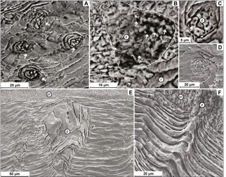
pun-ctae show the same organization when crossing the secondary shell. Here, their diameter is between 10 and 20 µm, but it becomes wider close to the prima-ry layer (Fig. 6E). In the Schizophoridae Acosarina,
the infill of the puncta penetrates the thin layer co-vering the secondary layer, which is assumed to be a recrystallized primary layer, as proposed by Williams & Harper (2000). In addition, this infill retains some voids, both in the primary and secondary layer (Figs 6B, C). These voids are regularly disposed, with a diameter of approximately 1-2 µm, and they could represent the trace of the microvillous branches of the punctae, which also perforate the primary
shell. In analogy to the modern craniformis, which bear branched punctae perforating all the shell lay-ers (Williams 1997), this observation seems to sup-port the hypothesis that the punctae were in direct contact with periostracum without any evidence of a canopy, as in the primitive enteletoid Schizoporia
(Williams & Harper 2000). On the other hand, it is difficult to exclude the possibility of presence/ absence of a canopy in the shell, because this thin structure, located in the uppermost part of the pri-mary layer, is easily lost during the diagenesis. This infill was not observed in the recrystallized primary layer of Enteletes or Peltichia. In the former, the fibers
suture upward the punctae, adjacent to the primary layer (Fig. 6E). In the second one, the primary lay-Fig. 6 - A/C) Cross section of the punctae and detail of the infilling (if) showing numerous voids (arrows), Acosarina sp. MPUM 11665
(CH72-11); B) infilling (if) of a puncta in the primary layer; the infill shows voids (arrows), Acosarina sp. MPUM 11665 (CH72-11); D) cross
section of a puncta in Paraspiriferina alpha MPUM 11676 (CH12-3); E) longitudinal section of a puncta in Orthida with the infilling
(if) which does not cross the primary layer (pr), Enteletes sp. MPUM 10000 (IR332-1); F) puncta crossing the outer secondary layer,
deflecting the fibers outwardly and penetrating into the inner part of the primary layer (pr), infilling (if), Hustedia sp. MPUM 11659
er was absent in the studied specimens. Note that the punctae of Peltichia were smaller, less dense and
scarcely persistent through the shell. These obser-vations suggest that the family Enteletidae, which evolved from the Schizophoriidae (Williams & Har-per 2000), could have evolved different types of punctae that did not perforate the primary layer, but a preservation bias cannot be excluded.
In the Neoretziidae Hustedia, the infill of
punctae permeated the primary recrystallized layer, highlighting that the puncta was connected to the outer periostracum (Fig. 6F), but there are not evi-dences of a canopy.
For the Terebratulida, no specimen showed the primary layer being preserved, hampering com-parison with modern taxa.
Despite the possible differences in punctae among Permian brachiopods, the presence of the-se perforations impothe-ses developmental constrains to the outer epithelium of the mantle. In addition, they bear specific biological functions, such as stora-ge compartments, sensorial activity, and protection against boring organisms (Owen & Williams 1969; Thayer 1977; Curry 1983; Pérez-Huerta et al. 2009). To be noted that several authors found a positive relationship between density of punctae and seawa-ter temperature (Campbell 1965; Peck et al. 1987; Ackerly et al. 1993), suggesting that the variability in the distribution and density of these structures di-scloses disparate thriving abilities among taxa. The major variation is in Peltichia, where the punctae
ap-pear less dense, when compared with those of Aco-sarina, another species of Orthida. These two
spe-cies occur together in the Lopingian-Lower Triassic successions of South China, but only Acosarina has
been recovered in the Extinction Interval (Shen & He 1991). On the other hand, Peltichia disappears
below the extinction horizon (Shen & Shi 2007), suggesting that density of punctae may have played some role in the differential survivorship during the Lopingian.
Columnar fabrics. The columnar layer is
pre-sent in both classes, but not in all the studied taxa, confirming the homoplastic origin of this feature at different taxonomic levels (Smirnova & Popiel-Barcyz 1991). The development of the columnar layer during ontogenesis is similar to the one de-scribed in Williams (1968), but the studied material reveals a few distinctive features in some taxonomic
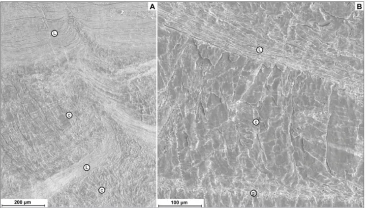
groups. In the Strophomenata, the transition to the columnar layer is principally controlled by a speci-fic activity of the mantle, which ceases secretion of the organic strand that delimits the morphology of single blades and laminae and starts to produce a continuous columnar layer. However, in the Rhyn-chonellata, the transition may vary from gradual to abrupt. For example, when Transcaucasathyris and Co-melicania are compared, it is evident that the
transi-tion is gradual in the former and abrupt in the latter (see Figs 8A, B). This probably corresponds to a gradual demise of organic sheet production in spe-cies of the former genus, and an abrupt demise in the latter.
Another interesting feature is that this shift between the two secretory regimes can be rever-sible in several taxa of both classes. The shell fa-bric of species of Permophricodothyris
(Rhynchonel-lata) shows intercalations of fibrous and columnar layers (Fig. 7E, see fig. 4F in Garbelli et al. 2012). On the other hand, species of Transcaucasathyris –
in the same class – do not show these alternations, highlighting a difference in the ability to modulate the production of the organic matter sheets. Ana-logous examples are also present in the Stropho-menata, such as Tyloplecta, Araxilevis, and Spinomar-ginifera, where there are intercalations of laminae
and columns (Fig. 7A, see fig. 4C in Garbelli et al. 2012). The function and cause of the reversion in the secretory mechanism remain unclear. To figure
Fig. 7 - A) Shell composed of an alternating sequence of laminar (l) and columnar layers (c), Spinomarginifera helica MPUM 11708
(EBHZ15-15); B) massive columnar-like layer, Araxilevis in-termedius MPUM 9959 (IR 311-6b); C) bands of accretion
and discontinuity between two adjacent prisms, Araxilevis intermedius MPUM 9959 (IR 311-4); D) prisms surface
show-ing a microgranular-like texture, Tyloplecta persica MPUM
9949 (IR317-4); E) fibers (f) intercalated between two co-lumnar layers (c), Permophricodothyris iranica MPUM 10044
(IR332-5); F) thick columnar layer, Comelicania sp. MPUM
11721 (VB9B-2); G) shell composed of an outer fibrous (f) and an inner columnar (c) layer, Transcaucasathyris araxensis
MPUM 11723 (JU10-4); H) details of F showing bands of accretion on the prisms surface, Comelicania sp. MPUM
11721 (VB9B-2); I) details of columnar layer showing the boundaries between microgranules, Transcaucasathyris araxen-sis MPUM 11620 (JU136-1); J) transition between laminar
and columnar layer, Spinomarginifera iranica MPUM 11617
(JU1-2); K) transition from fibrous to columnar layer in the Athyridida Transcaucasathyris sp., the arrow shows the change
in orientation of the fiber into a prism, Transcaucasathyris sp.
MPUM 11619 (JU131-4); in all the pictures the asterisks (*) indicate the outermost part of the shells.
out whether this character is controlled by the onto-genetic program that regulates shell production, by environmental stimuli or by the interplay of both these factors, it is important to understand bra-chiopod biomineralization. Angiolini et al. (2012) reported that species of Gigantoproductus show
in-terruptions in the secretion of the columnar layer in correspondence of growth lines represented by irregular laminae or grains. The authors interpreted it as a feature related to spawning or more proba-bly seasonal growth interruptions. Similar features have also been observed in large productids, such as Araxilevis and Tyloplecta. It is important to
un-derstand that in these taxa, such kind of intercala-tions of laminar and columnar fabrics are present throughout the shell. In the taxa here studied, these intercalations tend to be thicker in the outermost shell, where they show a clear laminar fabric. The intercalations become thinner inwardly, where they appear as the growth lines described in Angiolini et al. (2012) (Fig. 9). This could highlight that there is a common physiological mechanism for both inter-calations of laminar layers and growth lines, despite their different pattern. The growth lines observed in species of Gigantoproductus show a frequency of
for-mation caused by regular environmental perturba-tions (Angiolini et al. 2012). On the other hand, the intercalations recorded in the species here studied show a less organized pattern of distribution and the laminar intercalations have a different thickness.
Laminar and columnar fabrics must have different mechanical proprieties and the higher
or-ganic content in the former confers more toleran-ce to deformation, as observed in the fibrous fa-bric (Schmahl et al. 2012). On the other hand, the small size of structural units, in this case the bla-des, confers higher hardness (Goetz et al. 2009). Therefore, we can hypothesize that the intercala-tions are related to interacintercala-tions with the substrate during the growth of the organism, which could be more affected by mechanical stress in juvenile stages. This explains also why outwardly the inter-calations are thicker, when the shell is thinner, if compared to subsequent life stages. In addition, the mechanical stress due to the substrate is ran-dom and this could explain why the intercalations are irregularly disposed, in contrast to the growth lines observed by Angiolini et al. (2012), which are caused by regular seasonal fluctuations.
Permophricodothyris (Spiriferida) and Comelica-nia (Athyridida) bear a columnar prismatic layer
and show similar microstructures as those obser-ved in the Productida. In the former, the columnar layer is interrupted by intercalations of fibrous la-yer, which can vary in thickness; in the latter, there are evident interruptions of columns growth, but without any evidence of fibers. These analogies would disclose that a common genetic mechanism drives the development of this fabric feature in all rhynchonelliformean brachiopods. On the other hand, Productida show a higher frequency of in-tercalations than taxa of the other orders. The di-verse mechanical stress imposed by the semi-in-faunal life style typical of most of the Productida Fig. 8 - Comparison of shell sequence in two taxa of Athyridida; A) shell sequence in Transcausathyris sp., showing a gradual change from fibers
to prisms, with fibers growing in size inwardly; B) shell sequence of Comelicania sp., showing a clear transition from secondary fibrous
to columnar fabric, with fibers rather homogeneous in size and shape through all the shell; in all the pictures the asterisks (*) indicate the outermost part of the shells.
(Strophomenata), compared to the epifaunal free or attached lifestyles in the taxa of Rhynchonel-lata, could be one of the factors which acted on evolutionary time scale to promote the acquisition of a laminar shell fabric in the phyletic lineage of this group.
Paleobiological and evolutionary impli-cations
Different life styles and ecologies, coupled with the small genome size of the Phylum Bra-chiopoda (Adachi et al. 2013), suggest that the ana-logies observed in the Permian brachiopod shell structure are the result of repetitive adaptations, as it has been proposed for the numerous convergent shell morphologies (Brunton et al. 2000).
The main differences in shell structure of calcifying Rhynchonelliformea, besides the pre-sence/absence of punctae and pseudopunctae, are related to the fabrics they can produce: laminar (composed of blades), fibrous, columnar, or seve-ral combinations of these, and to the size and sha-pe of the structural units. However, the observa-tions on the newly studied materials and previous published studies (Williams 1968; Brunton 1972;
Williams 1970; Williams & Brunton 1993; Dewing 2004) indicate that:
(1) even if the size of blades composing the laminae can be larger than those observed in the Permian taxa investigated here (also see Dewing et al. 2004), the laminar fabric appeared to have evolved from a fibrous one (see Brunton 1972) by a reduction of size of the structural elements, and a consequent change in their relative spatial orga-nization;
(2) the fibers seem to have a large range of variation in size (Tab. 4) and the fibers can have a wide range of outlines, from diamond to keel and saddle, or very flat in shape (Fig. 5);
(3) the size of the structural elements can be significantly different among specimens of the same species from different stratigraphic positions (Garbelli et al. 2017);
(4) for some species, it has been observed a gradual and continuous change in size and shape from fibers to columns (Fig. 8A), suggesting that an ontogenetic gradual change from one fabric to another is possible.
These observations reveal that simple varia-tions in size, shape and arrangement of structural Fig. 9 - Alternation between laminar (l) and columnar (c) fabric in the Productida Tyloplecta persica; inwardly the intercalations become thinner,
until they appear similar to the growth lines (gl) of Angiolini et al. (2012); A) outermost shell, Tyloplecta persica MPUM 9942 (IR877-1);
units could lead to the evolution of fabrics, through modifications of the organization of organic mem-branes, delimiting the structural units. In addition, chemico-structural changes in the organic sheets can be relevant, as it happens in modern taxa, where col-umns are enveloped in a different organic substrate than the fibers (Williams & Cusack 2007). If the dis-position of the organic membranes during the growth is defined by an ontogenetic program, the changes in the overall fabric organization provides important in-dications about the phylogenetic relationships among brachiopods (Williams 1956; Williams & Brunton 1993). The observation of analogies reveals that the overall shell fabric organization could be useful only at low taxonomic ranks (e.g. genus, subfamily, family). An important factor that must be taken in ac-count to understand fabric evolution is its impact on the energetic balance of the organisms. The meta-bolic cost of CaCO3 precipitation is ~5% of the
energy that is required to produce proteinaceous or-ganic fractions (per unit of shell; Palmer 1992). This means that the laminar fabric of the Strophomenata Productida and Orthotetida are energetically more expensive than the fibrous fabric of the orders be-longing to Rhynchonellata, since the laminae require a larger fraction of organic membrane to be envel-oped, because they are composed of small structural units, i.e. the blades (Fig. 10). Additionally, the colum-nar layer shows a microstructure more similar to an inorganic precipitate (Goetz et al. 2009; von Allmen et al. 2010); a shell containing a columnar layer can be produced at a lower metabolic cost, because there is less production of organic membranes. Pseudopunc-tae and tubular hollow spines represent an additional complexity and cost in laminar fabrics; also punctae
add a cost to fibrous fabrics, but, from a metabolic perspective, they could provide some advantages, since they operate as storage compartments. On the contrary, tubular hollow spines and pseudopunctae are just structures for the shell functionality.
Therefore, each different type of shell entails a different metabolic investment, which plays a criti-cal role in survivorship, especially during events such as the end Permian global mass extinction (Clapham & Payne 2011; Garbelli et al. 2017), when the ma-rine carbonate system was strongly perturbed and the sweater temperature raised (Kump et al. 2009; Brand et al. 2012; Clarkson et al. 2015; Brand et al. 2016; Garbelli et al. 2016).
Outlining the differences in the cost of pro-duction of the shell could lead to a better under-standing of the paleobiology of extinct groups. For example, productid brachiopods preferred low nutri-ent settings, instead spire bearing brachiopods, like athyridids and spiriferids, proliferated in high nutrient settings, suggesting that they have different physiolo-gies (Pérez-Huerta & Sheldon 2006). In fact, these brachiopod groups show the following differences:
(1) their secondary layer fabrics are different; (2) the concavo-convex (most of the produc-tids) and biconvex (spiriferids and athyridids) bra-chiopods have a different ratio between body vol-ume and shell: this could mean that the repartition of costs between shell biocomposite and the soft-body parts is different between productids and spiriferids/ athyridids, with the former having a lower ratio;
(3) productids show a higher frequency of in-tercalations of laminar secondary layer in the colum-nar layer than the other orders;
(4) productids present pseudopunctae and Fig. 10 - Comparison of three
dif-ferent fabric types in which the size of the structural units increases and the ratio of enveloping organic mem-branes decreases: A) laminar fabric; B) fibrous fabric; C) columnar fabric.
variably developed tubular hollow spines; spiriferids do not have any complication of the shell structure, being impunctated and showing spines composed only of primary layer;
(5) athyridids present a more complex situa-tion, with some clades evolving punctae and others developing spines, which grow in a different way if compared with those of productids (Alvarez & Brunton 2001).
These differences suggest that productids had assigned a larger fraction of their limited ener-gy budget to shell building compared to spiriferids and athyridids. Since re-allocating energy from shell formation to support increase of metabolic cost during ocean warming and acidification affects
survivorship ability (Mackenzie et al. 2014), the
productids could have been more sensitive to this
environmental perturbations than spiriferids and athyridids.
The mechanical characteristics conferred to the shell are another important factor to understand fabric evolution. Studying modern Rhynchonellifor-mea, Goetz et al. (2009) reported that different fa-brics affect the mechanical proprieties of the shell, pointing out that this feature easily undergoes se-lective pressure related to environmental and eco-logical conditions. Noteworthy the most derived brachiopods lineage of the Strophomenata, i.e. the Productida, which proliferated in the semi-infaunal environment, bear a laminar fabric coupled with a shell that is commonly concavo-convex to planocon-vex (Brunton et al. 2000). Excluding older stocks of Strophomenata, i.e. the Plectambonitoidea, which may have a fibrous fabric with a semi-infaunal habi-tus (Brunton 1972; Congreve et al. 2015), the unique condition of the Productida could indicate that the laminar fabric confers mechanical advantages related to the substrate interactions of this life-style.
c
onclusIonThis study has shown that there is variability in the shell structure of Rhynchonelliformea during the Lopingian, and this is related to:
- the type of structures crossing the shell sub-stance, which have their origin in the deep evolutio-nary history of the analyzed taxa;
- the type of fabric succession composing the shell, i.e. the presence/absence of the columnar layer;
- the pattern of change in the secretory me-chanism and the relative alternation between fi-brous/laminar and columnar layers;
-the amount of intercrystalline organic mat-ter, i.e. the organic membranes separating the struc-tural units.
The mechanism producing/evolving the dif-ferences in shell structure remains unclear. It can be argued that there are some ontogenetic constrains in the development of shell fabric which are com-mon to all Rhynchonelliformea brachiopods. On the other hand, there should have been some selec-tive environmental force causing the differences in shell structure and acting on the evolutionary time-scale. The effects of these factors are difficult to be evaluated in short time laboratory experiments (i.e. Cross et al. 2016), which test the response of sin-gle organisms during their lifetime, or even smaller intervals. From this point of view, the analysis of shell fabric evolution in fossil brachiopods provi-des important information to understand how and why their fabric changed in different environmental conditions. In particular, at lower taxonomic levels, shell microstructure may be useful to infer phyloge-netic relationships.
Acknowledgments: I would like to thank Lucia Angiolini for the
continuous support and the precious suggestions. I also acknowledge Aymon Baud, Shuzhong Shen and Renato Posenato for providing part of the material for the ultrastructure study. Thanks to the technicians of University of Milan, Curzio Malinverno, Agostino Rizzi and An-drea Risplendente, and to Yang Fan from NIGPAS. I am also grateful for the suggestion provided by the reviewers Renato Posenato and Al-berto Pérez-Huerta. This work has benefitted of the financial support of SAF/China from MIUR and funding from the China Postdocto-ral Science Foundation (Grant N 2016M591939) to Claudio Garbelli, PRIN to Elisabetta Erba and funding from Lucia Angiolini.
RefeRences
Ackerly S.C., Cisne J.L., Railsback L.B. & Anderson T.F. (1993) - Punctual density in the Ordovician orthide brachiopod Paucicrura rogata: anatomical and paleoenvironmental variation. Lethaia, 26: 17-24.
Adachi K., Kuramochi T., Kimura K.S. & Okumura S. (2013) - First Extensive Examination of Genome Size in Phy-lum Brachiopoda (Lamp Shells) Collected from Japan. J. Shellifish Res., 32(2): 539-541.
Alvarez F. & Brunton C.H.C. (2001) - Fundamental differenc-es in external spine growth in brachiopods. In: Brunton C.H.C., Cocks M.R.L. & Long S.L. (Eds) - Brachiopods Past and Present - The Systematics Association Special Volume Series, 63: 108-118.
Angiolini L. (1993) - Ultrastructure of some Permian and Tri-assic Spiriferida and Arthyridida (Brachiopoda). Riv. It. Paleontol. Strat., 9: 283-306.
Angiolini L. & Carabelli L. (2010) - Upper Permian brachio-pods from the Nesen Formation, North Iran. Spec. Pap. Palaeontol., 84: 41-90.
Angiolini L., Stephenson M., Leng M.J., Jadoul F., Millward D., Aldridge A., Andrews J., Chenery S. & Williams G. (2012) - Heterogeneity, cyclicity and diagenesis in a Mis-sissippian brachiopod shell of palaeoequatorial Britain. Terra Nova, 24(1): 16-26.
Angiolini L., Zanchi A., Zanchetta S., Nicora A, Vuolo I., Berra F., Henderson C., Malaspina N., Rettori R., Vacha-rd D. & Vezzoli G. (2015) - From rift to drift in South Pamir (Tajikistan): Permian evolution of a Cimmerian terrane. J. Asian Earth Sci., 102: 146-69.
Barbin V. (2013) - Application of cathodoluminescence mi-croscopy to recent and past biological materials: a de-cade of progress. Miner. Petrol., 107(3): 353-362.
Brand U., Posenato R., Came R., Affek H., Angiolini L., Azmy K. & Farabegoli E. (2012) - The end-Permian mass ex-tinction: a rapid volcanic CO2 and CH4-climatic catas-trophe. Chem. Geol., 322-323: 121-144.
Brand U., Blamey N., Garbelli C., Griesshaber E., Posenato R., Angiolini L., Azmy K., Farabegoli E. & Came R. (2016) - Methane hydrate: Killer cause of Earth’s greatest mass extinction. Palaeoworld, 25: 496-507.
Brunton C.H.C. (1972) - The shell structure of Chonetacean brachiopods and their ancestors. Bull. British Mus. (Nat. Hist.) Geology, 21: 1-26.
Brunton C.H.C., Lazarev S.S. & Grant R.E. (2000) - Produc-tida. In: Kaesler R.L. (Ed.) - Treatise of Invertebrate Palaeontology (Part H, Brachiopoda Revised): 350-368. Geological Society of America - University of Kansas Press, Boulder, CO.
Campbell K.S.W. (1965) - Australian Permian terebratuloids. Bull. Bureau Min. Res., Geol., Geoph. Australia, 68: 1-113. Chuang S.H. (1994) - Observations on the reproduction and
development of Liothyrella neozelanica (Thomson 1918) (Terebratulacea, Articulata, Brachiopoda). J. Roy. Soc. New Zeal., 24: 209-218.
Clapham M.E. & Payne J.L. (2011) - Acidification, anoxia, and extinction: A multiple logistic regression analysis of ex-tinction selectivity during the Middle and Late Permian. Geology, 39:1059-1062.
Clarkson M.O., Kasemann S.A., Wood R.A., Lenton T.M., Daines S.J., Richoz S., Ohnemueller F., Meixner A., Poulton S.W. & Tipper E.T. (2015) - Ocean acidification and the Permo- Triassic mass extinction. Science, 348: 229-232.
Congreve C.R., Andrew Z.K. & Patzkowsky M.E. (2015) - Phylogenetic revision of the Strophomenida, a diverse and ecologically important Paleozoic brachiopod order. Palaeontology, 58(4): 743-758.
Cross E.L., Peck L.S., Lamare M.D. & Harper E.M. (2016) - No ocean acidification effects on shell growth and re-pair in the New Zealand brachiopod Calloria inconspicua (Sowerby, 1846). ICES J. Mar. Sci., 73: 920-926.
Curry G.B. (1983) - Microborings in Recent brachiopods and the functions of caeca. Lethaia 16: 119-127.
Curry G.B., Peck L.S., Ansell A.D. & James M. (1989) - Physi-ological constraints in fossil and recent brachiopods. T. Roy. Soc. Edin.-Earth, 80: 255-62.
Dewing K. (2004) - Shell structure and its bearing on the phylogeny of Late Ordovician- Early Silurian stropho-menoid brachiopods from Anticosti Island, Quebec. J. Paleontol., 78 (2): 275-286.
Erwin D.H. (2006) - Extinction: how life on earth nearly ended 250 million years ago. Princeton University Press, Princeton, N.J., 296 pp.
Garbelli C., Angiolini L., Jadoul F. & Brand U. (2012) - Mi-cromorphology and differential preservation of Upper Permian brachiopod low-Mg calcite. Chem. Geol., 299: 1-10.
Garbelli, C., Angiolini, L., Brand U. & Jadoul F. (2014a) - Bra-chiopod fabric, classes and biogeochemistry: implica-tions for the reconstruction and interpretation of sea-water carbon- isotope curves and records. Chem. Geol., 371: 60-67.
Garbelli C., Angiolini L., Shen S.Z., Crippa G., Yuan D.X., Bahrammanesh M., Abbasi S. & Birjandi M. (2014b) - Additional brachiopod findings from the Lopingian suc-cession of the Ali Bashi mountains, NW Iran. Riv. It. Paleontol. Strat., 120(1): 119-126.
Garbelli C., Angiolini L., Brand U., Shen S.Z., Jadoul F., Po-senato R., Azmy K., & Cao C.Q. (2016) - Neotethys seawater chemistry and temperature at the dawn of the latest Permian extinction. Gondwana Res., 35: 272-285. Garbelli C., Angiolini L. & Shen S.Z. (2017) -
Biomineraliza-tion and global change: A new perspective for under-standing the end-Permian extinction. Geology, 45: 19-12. Garilli V., Rodolfo-Metalpa R., Scuderi D., Brusca L., Parrinel-lo D., Rastrick S., Foggo A., Twitchett R.J., Hall-Spencer J. & Milazzo M. (2015) - Physiological advantages of dwarfing in surviving extinctions in high-CO2 oceans. Nat. Clim. Change, 5: 678-682.
Ghaderi A., Garbelli C., Angiolini L., Ashouri A.R., Korn D., Rettori R. & Gharaie M.H.H. (2014) - Faunal change near the end-Permian extinction: the brachiopods of the Ali Bashi mountains, NW Iran. Riv. It. Paleontol. Strat., 120(1): 27-59.
Goetz A.J., Griesshaber E., Neuser R.D., Luter C., Huhner M., Harper E. & Schmahl W.W. (2009) - Calcite mor-phology, texture and hardness in the distinct layers of rhynchonelliform brachiopod shells. Eur. J. Mineral., 21: 303-315.
Griesshaber E., Schmahl W.W., Neuser R., Pettke T., Blüm M., Mutterlose J. & Brand U. (2007) - Crystallographic texture and microstructure of terebratulide brachiopod shell calcite: an optimized materials design with hierar-chical architecture. Am. Mineral., 92: 722-734
He W.H., Twitchett R.J., Zhang Y., Shi G.R., Feng Q.L.,Yu J.X., Wu S.B. & Peng X.F. (2010) - Controls on body size dur-ing the Late Permian mass extinction event. Geobiology, 8(5): 391-402.
his-tory. Rev. Mineral. Geochem., 54: 329-356.
Knoll A. H., Bambach R.K., Payne J.L., Pruss S. & Fischer W.W. (2007) - Paleophysiology and end-Permian mass extinction. Earth Planet. Sci. Lett., 256: 295-313.
Kump L.R. Bralower T.J. & Ridgwell A. (2009) - Ocean acidi-fication in deep time. Oceanography, 22: 94-107.
Machel H.G. (2000) - Application of cathodoluminescence to carbonate diagenesis. In: Pagel M., Barbin V., Blank P. & Ohnenstetter D. (Eds) - Cathodoluminescence in Geo-sciences. Springer, New York: 271-301.
Mackenzie C.L., Ormondroyd G.A., Curling S.F., Ball R.J., Whiteley N.M. & Malham S.K. - (2014). Ocean warm-ing, more than acidification, reduces shell strength in a commercial shellfish species during food limitation. PLoS ONE 9(1): e86764.
MacKinnon D.I. (1974) - The shell structure in spiriferide brachiopoda. Bull. British Mus. (Nat. Hist.) Geology, 5(3): 189-258.
Milano S., Schöne B.R. & Witbaard R. (2016) - Changes of shell microstructural characteristics of Cerastoderma edule (Bivalvia) - A novel proxy for water temperature. Palaeo-geogr., Palaeoclimatol., Palaeoecol., 465: 405-416.
Muttoni G., Gaetani M., Kent D.V., Sciunnach D., Angiolini L., Berra F., Garzanti E., Mattei M. & Zanchi A. (2009) - Opening of the Neo-Tethys Ocean and the Pangea B to Pangea A transformation during the Permian. Geo-Arabia, 14: 17-48.
Palmer A.R. (1983) - Relative cost of producing skeletal or-ganic matrix versus calcification – evidence from marine gastropods. Mar. Biol., 75: 287-292
Palmer A.R. (1992) - Calcification in marine molluscs: How costly is it? P. Natl. Acad. Sci USA, 89: 1379-1382. Peck L.S., Clarke A. & Holmes L.J. (1987) - Size, shape and the
distribution of organic matter in the Recent Antarctic brachiopod Liothyrella Uva. Lethaia, 20: 33-40.
Peck S.L., Brockington S. & Brey T. (1997) - Growth and me-tabolism in the Antarctic Brachiopod Liothyrella uva. Phil. T. Roy. Soc. B., 352: 851-858.
Pérez-Huerta A. & Sheldon N.D. (2006) - Pennsylvanian sea level cycles, nutrient availability and brachiopod paleo-ecology. Palaeogeogr., Palaeoclimatol., Palaeoecol., 230: 264-279. Pérez-Huerta A., Cusack M., Zhu W., England J. & Hughes J.
(2007) - Material properties of brachiopod shell ultra-structure by nanoindentation. J. R. Soc. Interface, 4: 33-39. Pérez-Huerta A., Cusack M., McDonald S., Marone F., Stam-panoni M. & MacKay S. (2009) - Brachiopod punctae: a complexity in shell biomineralisation. J. Struct. Bio., 167(1): 62-67.
Pérez-Huerta A. (2013) - Functional morphology and modi-fications on spine growth in the productid brachiopod Heteralosia slocomi. Acta Palaeontol. Pol., 58(2): 383-390. Posenato R. (2001) - The Athyridoids of the transitional beds
between Bellerophon and Werfen Formations (upper-most Permian, southern Alps, Italy). Riv. It. Paleontol. Strat., 107: 197-226.
Posenato R. (2009) - Survival patterns of macrobenthic ma-rine assemblages during the end-Permian mass extinc-tion in the western Tethys (Dolomites, Italy). Palaeogeogr.,
Palaeoclimatol., Palaeoecol., 280: 150-167.
Roger L.M., Richardson A.J., McKinnon A.D., Knott B., Mat-ear R. & Scadding C. (2012) - Comparison of the shell structure of two tropical Thecosomata (Creseis acicula and Diacavolinia longirostris) from 1963 to 2009: potential implications of declining aragonite saturation. ICES J. Mar. Sci., 69: 465-474.
Schmahl W.W., Griesshaber E., Merkel C., Kelm K., Deuschle J., Neuser R.D., Göetz A.J., Sehrbrock A. & Mader W. (2008) - Hierarchical fibre composite structure and mi-cromechanical properties of phosphatic and calcitic bra-chiopod shell biomaterials - an overview. Mineral. Mag., 72: 541-562.
Schmahl W.W., Griesshaber E., Kelm K., Goetz A., Jordan G., Ball A., Xu D., Merkel C. & Brand U. (2012) - Hier-archical structure of marine shell biomaterials: biome-chanical functionalization of calcite by brachiopods. Z. Kristallogr., 227: 793-804.
Smirnova T.N. & Popiel-Barcyz E. (1991) - Characterisics of the shell ultrastrucutre in Terebratellacea. In: D. I. MacKinnon, D. E. Lee & J. D. Camplbell (Eds) - Bra-chiopods Through Time, Proceedings of the 2nd In-ternational Brachiopod Congress, University of Otago, Dunedin, New Zeland, 1990. Balkema. Rotterdam: 159-165.
Shen S.Z. & He X.L. (1991) - Changhsingian brachiopod as-semblage sequence in Zhongliang Hill, Chongqing. Jour-nal of Stratigraphy 15(3):189-196.
Shen S.Z., Cao C.Q., Henderson C.M., Wang X.D., Shi G.R., Wang Y. & Wang W. (2006) - End-Permian mass extinc-tion pattern in the northern peri-Gondwanan region. Palaeoworld, 15(1): 3-30.
Shen S.Z., Cao C.Q., Zhang Y.C., Li W.Z., Shi G.R., Wang Y., Wu Y.S., Ueno K., Henderson C.M., Wang X.D., Zhang H., Wang X.J. & Chen J. (2010) - End-Permian mass ex-tinction and palaeoenvironmental changes in Neotethys: evidence from an oceanic carbonate section in south-western Tibet. Global Planet. Change, 73: 3-14.
Shen, S. Z., Crowley J.L., Wang Y., Bowring S.A., Erwin D.H., Sadler P.M., Cao C.Q., Rothman D.H., Henderson C.M., Ramezani J., Zhang H., Shen Y.A., Wang X.A., Wang W., Mu L., Li W.Z., Tang Y.G., Liu W.L., Liu L.J., Zeng Y., Ji-ang Y.F. & Jin Y.G. (2011) - Calibrating the end-Permian mass extinction. Science ,334: 1367-1372
Shi G.R., Zhang Y.C., Shen S.Z. & He W.H. (2016) - Nearshore-offshore-basin species diversity and body size variation patterns in Late Permian (Changhsingian) brachiopods: Palaeogeogr., Palaeoclimatol., Palaeoecol., 448: 96-107.
Thayer C.W. (1977) - The function of punctae in articulate brachiopods. J. Paleontol. Supp., 51: 2-28.
Von Allmen K., Nägler T.F., Pettke T., Hippler D., Griessha-ber E., Logan A., Eisenhauer A. & Samankassou E. (2010) - Stable isotope profiles (Ca, O, C) through mod-ern brachiopod shells of T. septentrionalis and G. vitreus: implications for calcium isotope paleo-ocean chemistry. Chem. Geol., 269: 210-219.
Watson S., Peck L.S., Tyler P.A., Southgate P.C., Tan K.S., Day R.W. & Morley S.A. (2012) - Marine invertebrate
skel-eton size varies with latitude, temperature and carbon-ate saturation: implications for global change and ocean acidification. Glob. Change Biol., 18(10): 3026-3037. Williams A. (1956) - The calcareous shell of the Brachiopoda
and its importance to their classification. Biol. Rev., 31: 243-287.
Williams A. & Rowell A.J. (1965a) - Brachiopod anatomy. In: Moore R.C. (Ed.) - Treatise on Invertebrate Paleontol-ogy (part H): 6 -57. Geological Society of America and University of Kansas Press, New York and Lawrence. Williams A. & Rowell A.J. (1965b) - Brachiopod morphology.
In: Moore R.C. (Ed.) - Treatise on Invertebrate Paleontol-ogy (part H): 57-115. Geological Society of America and University of Kansas Press, New York and Lawrence. Williams A. (1970) - Origin of laminar-shelled articulate
bra-chiopods. Lethaia, 3: 329-342.
Williams A. & Cusack M. (2007) - Chemicostructural diversity of the brachiopod shell. In: Selden P.A. (Ed.) - Treatise on Invertebrate Paleontology (Part H, Brachiopoda
Re-vised): 2397-2521. Geological Society of America - Uni-versity of Kansas Press, Boulder, CO.
Williams A. (1968) - Evolution of the shell structure of articu-late brachiopods. Spec. Pap. Paleontol., 2: 1-55.
Williams A. (1970) - Origin of laminar-shelled articulate bra-chiopods. Lethaia, 3: 329-342.
Williams A., Brunton C.H.C. & innon D.I. (1997) - Morphol-ogy. In: Kaesler R.L. (Ed.) - Treatise on Invertebrate Palaeontology (Part H, Brachiopoda Revised). Introduc-tion: 321-422. Geological Society of America - Univer-sity of Kansas Press, Boulder, CO.
Williams A. & Brunton C.H.C. (1993) - Role of the shell struc-ture in the classification of the orthotetidine brachio-pods. Palaeontology, 36: 931-966.
Williams A. (1997) - Shell structure. In: Kaesler R.L. (Ed.) - Treatise on Invertebrate Palaeontology (Part H, Bra-chiopoda Revised). Introduction: 267-320. Geological Society of America - University of Kansas Press, Boul-der, CO.