Università degli Studi di Ferrara
DOTTORATO DI RICERCA IN SCIENZE
FARMACEUTICHE
CICLO XXI
Coordinatore: Prof. Manfredini Stefano
Design and Synthesis of
New A
2A
and A
3
Adenosine Receptors
Antagonists
Settore Scientifico Disciplinare CHIM/ 08
Dottoranda Tutore Dott. Saponaro Giulia Prof. Simoni Daniele
Contents
Chapter 1 1. Introduction 3
1.1. A2A Adenosine Receptor 5
1.1.1. Pharmacology 5
1.1.2. A2A Adenosine Receptor Antagonists 9
1.1.3. Medicinal Chemistry 10
1.1.4. Clinical Development and Patents 16 1.2. A3 Adenosine Receptor 21
1.2.1. Pharmacology 21
1.2.2. A3 Adenosine Receptor Antagonists 24
1.2.3. Medicinal Chemistry 25
1.2.4. Clinical Development and Patents 33
Chapter 2 2. Design and Synthesis 35
2.1. First Project 36
2.2. Second Project 43
Chapter 3 3. Results and Conclusions 49
3.1. First Project 50
3.2. Second Project 56
3.3. Conclusions 64
Chapter 4 4. Experimental Section 66
Chapter 1
Chapter 1
Chapter 1
Chapter 1
Introduction
Introduction
Introduction
Introduction
Chapter 1 Introduction
3
1. Introduction
The purine nucleoside adenosine is consensually identified as a ma-jor local regulator of tissue function especially when energy supply fails to meet cellular energy demand. Due to its ability to equalize energy intake to metabolic demand in the 1980s it earned the reputa-tion of a “retaliatory metabolite”.1
Adenosine is omnipresent, released from almost all cells, and gener-ated in the extracellular space by breakdown of ATP through a series of ectoenzymes, including apyrase (CD39) and 5′-nucleotidase (CD73).2 The latter dephosphorylates extracellular AMP to adeno-sine, regulating the limiting step for its formation. Extracellularly, adenosine concentration is kept in equilibrium by reuptake mecha-nisms operated through the action of specific transporters. Then in-side the cell it is phosphorylated to AMP by adenosine kinase or de-graded to inosine by adenosine deaminase (ADA). Intracellularly, adenosine formation is dependent upon the hydrolysis of AMP by an intracellular 5-nucleotidase or hydrolysis of
S-adenosyl-homocysteine. It is estimated that the levels of adenosine in the in-terstitial fluid are in the range 30-300 nM.3
Adenosine concentrations increase under metabolically unfavorable conditions. Tissue hypoxia, for example, leads to an enhanced breakdown of ATP and the increased generation of adenosine. In addition to this route, the release of adenosine might be potentiated by hypoxia-dependent inhibition of the salvage enzyme adenosine kinase which rephosphorylates the nucleoside to AMP.4
As adenosine is unstable and its half-life is limited by deamination or cellular reuptake, hypoxia-induced increase typically affects only lo-cal adenosine receptor signaling. As adenosine is not released in a
Chapter 1 Introduction transmitter or hormone-like fashion, it is likely to belong to the group of autacoids.
Adenosine mediates its effects through activation of a family of four G-protein-coupled adenosine receptors (ARs), named A1, A2A, A2B
and A3. These receptors differ in their affinity for adenosine, in the
type of G proteins that they recruit, and finally in the downstream signaling pathways that are activated in the target cells. A1 and A3
ARs display high and low affinity for adenosine, respectively, and are inhibitory toward regulation of adenylyl cyclase activity. By contrast, activation of high-affinity A2A and low-affinity A2B subtypes stimulates
adenylyl cyclase leading to an increase of cyclic AMP (cAMP) levels. Early pharmacological evidence for the existence of ARs has been provided by specific antagonism by methylxanthines, caffeine, and theophylline of adenosine-induced effects in the heart and brain.5 These receptors are widely distributed through the body, and their presence on basically every cell makes them an interesting target for the pharmacological intervention in many pathophysiological situa-tions linked to an increase of adenosine levels.
The first recorded report describing evidence for an ARs originates from 1976. Now, 30 years later, advances in understanding the role of adenosine and its receptors in physiology and pathophysiology as well as new developments in medicinal chemistry of these receptors have enabled researchers to identify potential therapeutic areas for drug development.
With the combination of pharmacological data, using selective ligands and genetically modified mice, important progress has been made toward an understanding of the role of ARs in a variety of dis-eases, such as inflammatory conditions, sepsis, heart attack,
ische-Chapter 1 Introduction
5 mia-reperfusion injury, vascular injury, spinal cord injury, chronic ob-structive pulmonary disease (COPD), asthma, diabetes, obesity, in-flammatory bowel disease, retinopathy, and Parkinson’s Disease (PD). Nonselective AR antagonists are used to maintain wakefulness (caffeine) and, less commonly at present, treat bronchospasm (theo-phylline, amino(theo-phylline, enprofylline). Currently a number of new se-lective AR agonists and antagonists are in testing for a variety of new indications.
1.1. A2A Adenosine Receptor
1.1.1. Pharmacology
The gene for the A2A AR has been cloned from several species
in-cluding dog,6 rat,7,8 human,9 guinea pig,10 and mouse11 and demon-strated a high degree of homology among human, mouse, and rat.12 The A2A AR stimulates adenylyl cyclase activity through the coupling
with Gs proteins leading to activation of cAMP-dependent protein
kinase A. This in turn phosphorylates and activates various recep-tors, ion channels, phosphodiesterases, and phosphoproteins like CREB and DARPP-32.13-15 Activation of protein kinase C has been also reported in PC12 cells.16 In brain striatum the A2A subtype
stimu-lates Golf, another member of the Gs subfamily of G proteins.17 In
ad-dition A2A AR can interact with different types of Ca 2+
channels to ei-ther increase intracellular Ca2+ or decrease Ca2+ influx18,19 and is in-volved like the other adenosine subtypes in the modulation of ERKs activity.20
Due to a long carboxy terminal domain, the A2A AR shows a greater
molecular weight (45 kDa) in comparison to the other subtypes (36-37 kDa). The A2A AR C terminus has been defined as a crowded
Chapter 1 Introduction place where different accessory proteins may interact such as D2
-dopamine receptors,21 R-actinin,22 ADP-ribosylation factor nucleotide site opener (ARNO),23 ubiquitin-specific protease (USP4),24 and translin-associated protein X (TRAX).25 The lack or presence of such different partners may explain conflicting results deriving by A2A ARs
activation, e.g., neuroprotection versus neurotoxicity.26
Within the brain A2A ARs are richly expressed in the striatum,
nu-cleus accumbens, and olfactory tubercle. A coexpression of A2A with
D2 dopamine receptors has been reported in the GABAergic
striato-pallidal neurons where adenosine and dopamine agonists exert an-tagonistic effects in the regulation of locomotor activity. Activation of A2A ARs in striatopallidal neurons decreases the affinity of D2
recep-tors for dopamine, antagonizing the effects of D2 receptors (Fig.1).
The negative interaction between A2A and D2 receptors is at the basis
of the use of A2A antagonists as a novel therapeutic approach in the
treatment of PD.27 In addition, A2A ARs may have an important role in
the neurodegenerative process. Accordingly, a neuroprotective effect was demonstrated after caffeine intake or A2A AR inactivation against
dopaminergic neurodegeneration in a neurotoxin model of PD.28 Concomitantly, two large prospective epidemiological studies have strongly associated caffeine consumption to a reduced risk of devel-oping PD.29,30 Last, the recent discovery that the A2A can form
func-tional heteromeric receptor complexes with other Gprotein- coupled receptors such as D2 and the mGlu5 receptors has also suggested
new opportunities for the potential of A2A antagonists in PD. 21
In the future development of bivalent ligands, able to activate D2 and block
Chapter 1 Introduction
7 promising strategy for the treatment of this neurodegenerative dis-ease.31-33
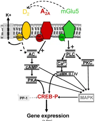
Figure 1. Functional interactions between dopamine D2, adenosine A2A and
me-tabotropic glutamate 5 receptors in striatopallidal neurons.34
In addition to the protection against striatal and nigral neuron loss by A2A antagonists, there are data also supporting their protective role
outside the basal ganglia.35 Local injection of an A2A antagonist
pre-vents glutamate-dependent death of neurons in hippocampal cor-tex36 and also reduced cortical damage in a variety of ischemic stroke models. In A2A knockout (KO) mice transient focal ischemia
caused less neuronal damage in comparison to their wild-type (WT) littermates.37 Therefore, it seems that tonic activation of A2A ARs may
Chapter 1 Introduction neuroprotective effects induced by endogenous A1 activation.
Re-cently, selective inactivation or reconstitution of A2A ARs in
bone-marrow cells revealed their contribution to the development of ischemic brain injury.38
The involvement of A2A ARs in neuroprotection is likely to be
com-plex as stimulation of this subtype also diminishes brain damage af-ter excitotoxic and traumatic injury.39,40
A2A-mediated protection has been reported against ischemia in the
myocardia, kidney, and liver and in ischemia-reperfusion injury in the spinal cord.41-44
High expression of A2A ARs has been found in platelets, leukocytes,
vascular smooth muscle and endothelial cells with important implica-tions in the regulation of inflammatory responses. It is now well es-tablished that stimulation of the A2A AR in immune cells induces
anti-inflammatory effects, mostly due to its ability to increase cAMP lev-els, which has strong immunosuppressive effects.45 Stimulation of A2A ARs inhibits neutrophil adherence to the endothelium,
degranula-tion of activated neutrophils and monocytes, plus superoxide anion generation. A2A ARs have been recently defined as sensors and
ter-minators of proinflammatory activities. The strongest evidence for the key role of A2A in inflammation derived by the elegant study of Ohta
et al.46 using mice deficient in A2A ARs. In this model the lack of A2A
subtype leads to increased tissue inflammation and damage, thus suggesting a negative and nonredundant regulatory role for the A2A
AR. This model permits one to appreciate that adenosinergic regula-tion of immune cells is fundamental in normal physiological control of inflammation in vivo in spite of the fact that other Gs-protein-coupled
catheco-Chapter 1 Introduction
9 lamines, prostaglandins, dopamine, and histamine.45 Interestingly, the A2A AR has been demonstrated to be involved in promotion of
wound healing and angiogenesis in healing wounds.47,48
Moreover, it plays an active role in the pathogenesis of dermal fibro-sis, suggesting a role for antagonists as novel therapeutic approach in the treatment and prevention of dermal fibrosis in diseases such as scleroderma.49
1.1.2. A2A Adenosine Receptor Antagonists
The discovery and development of potent and selective A2A AR
an-tagonists became, in the last 10 years, an attractive field of research to the discovery of new drugs for the treatment of neurodegenerative disorders, such as PD.
Different compounds have been deeply investigated as A2A AR
an-tagonists, which could be classified in two great families: nitrogen polyheterocyclic systems and styrylxanthine derivatives. Table 1 summarizes the examples of A2A AR antagonists reported in this
Chapter 1 Introduction
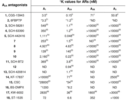
Table 1. Affinity of AR antagonists at the A1, A2A, A2B and A3 ARs.
°Ki values for ARs (nM)
A2A antagonists A1 A2A A2B A3 1, CGS-15943 3.5a 0.15b 71c 50.8d 2, 8FBPTP *3.351 *1.251 *ND *ND 3, SCH-58261 54955 1.155 >1000055 >1000055 4, SCH-63390 35055 1.255 >1000055 >1000055 5, SCH-442416 1,11154 0.04854 >1000054 >1000054 7 25353 1.553 ND53 >1000053 8 4,92755 4.6355 >1000055 >1000055 9 13955 14055 >1000055 >1000055 10 2,16055 0.2255 >1000055 >1000055 11, SCH-BT2 36955 3.855 >1000055 >1000055 12 ND 0.9459 ND ND 13, SCH-420814 ND 1.160 ND ND 14, KF-17837 >1000062 7162 ND 250062 15, CSC *2800063 *5463 ND >1000063 16, BS-DMPX *1200 *8.2 ND ND 17, KW-6002 283066 3666 180066 >300066 18, ST-1535 72 6.6 352 >1000 °
Binding experiments at recombinant hA1, A2A, A2B and A3 ARs, unless noted; *Binding
experiments at rat brain (A1) and striatum (A2A) ARs; ND not determined. a
Ongini, E.; Dionisotti, S.; Gessi, S.; Irenius, E.; Fredholm, B. B. Naunyn Schmiedebergs Arch.
Phamacol. 1999, 359, 7. b
Varani, K.; Gessi, S.; Dionisotti, S.; Ongini, E.; Borea, P. A.
Br. J. Pharmacol. 1998, 123, 1723. c
de Zwart, M.; Vollinga, R.; Beukers, M. W.; Slee-gers, D. F.; von Frijtag Drabbe Kuenzel, J. K.; de Groote, M.; Ijzerman, A. P. Dru. Dev
Res. 1999, 48, 95. d Klotz, K.-N.; Hessling, J.; Hegler, J.; Owaman, C.; Kull, B.; Fred-holm, B. B.; Lohse, M.J. Naunyn Schmiedebergs Arch. Phamacol. 1998, 357, 1.
1.1.3. Medicinal Chemistry
Pyrazolo[4,3-e][1,2,4]triazolo[1,5-c]pyrimidines (PTPs)
9-Chloro-2-furan-2-yl-[1,2,4]triazolo[1,5-c]quinazolin-5-ylamine named CGS-15943 (1, Figure 2) represented the first potent but poorly selective antagonist for the A2A AR subtype.
50
Bioisosteric re-placement of the phenyl ring of CGS-15943 with an N7-substituted pyrazole led to the first example of an adenosine antagonist display-ing the pyrazolo-triazolo-pyrimidine (PTP) core named 8FBPTP (2,
Chapter 1 Introduction
11
8-(4-fluorobenzyl)-2-(2-furyl)-8H-pyrazolo[4,3-e][1,2,4]triazolo[1,5-c]pyrimidin-5-amine, Figure 2).51 Some structural features of this compound highlighted the essential requirements for the A2A affinity,
i.e., the furyl moiety and the free amino group at the 5- position. Starting from these observations Baraldi et al.52,53 focused their in-terest on the pattern of substitution on the pyrazolo preserving the other structural elements.
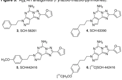
Figure 2. Structural relationships between CGS15943 and 8FBPTP (the first
A2A AR antagonist) 2, 8FBPTP 1, CGS15493 N N N N O NH2 Cl N N N N N N O NH2 F
Several alkyl, aryl, and phenylalkyl substituents have been intro-duced at both the N7 and the N8 positions. The biological data de-rived from the molecules obtained, indicated that the best radicals were phenylalkyl chains and among these it was possible to discern the length of the spacer introduced between the phenyl ring and the pyrazolo nitrogen that was optimized in two or three carbon atoms. Two selected compounds of this family named SCH-58261 (3, Figure 3, 5-amino- 7-(β-phenylethyl)2-(2-furyl)-pyrazolo[4,3-e][1,2,4]triazolo- [1,5-c]pyrimidine) and SCH- 63390 (4, Figure 3, 5-amino-7-(3-
phenylpropyl)2-(2-furyl)-pyrazolo[4,3-e][1,2,4]triazolo[1,5-c]pyrimidine)52,53 proved to be potent and selective A2A AR
antago-nists both in rat and human models. It was also noted that the N7 de-rivatives were more selective for the A2A AR than the corresponding N8 derivatives. 53
Chapter 1 Introduction From the family of SCH compounds, 5-amino-7-[3-(4-
methoxyphenyl)propyl]-2-(2-furyl)pyrazolo[4,3-e]-1,2,4-triazolo[1,5-c]pyrimidine (SCH-442416, 5, Figure 3) was selected for the
devel-opment of a new positron emission tomography (PET) ligand, whose chemical structure allows an easy introduction of a methyl group by direct O-alkylation of the phenolic function with [11C]CH3I under
alka-line conditions.54 The aim of this study was to use [11C]SCH 442416, (6, Figure 3) as a new ligand for the in vivo imaging of A2A ARs using
PET. The in vitro binding in the brain and periphery, the good signal-to-noise ratio observed between 5 and 15 min after injection, and the low occurrence of radioactive metabolites all suggested that [11C]SCH-442416 was applicable as the first non-xanthine ligand suitable for the in vivo imaging of A2A ARs using PET. In addition, the
data obtained from the binding experiments showed a higher affinity of the title compound for hA2A vs rat ARs (0.048 vs 0.5 nM).
54 3, SCH-58261 4, SCH-63390 N N N N N N O NH2 N N N N N N O NH2
Figure 3. A2A AR anatgonists (Pyrazolo-triazolo-pyrimidines).
6, [11C]SCH-442416 N N N N N N O NH2 [11C]H3CO N N N N N N NH2 H3CO 5, SCH442416 O
Chapter 1 Introduction 13 12, Analog related to SCH-58261 N N N N N N N N O O NH2 O 13, SCH-420814
Water-Soluble A2A Adenosine Receptor Antagonists
The major restriction of the tricyclic adenosine antagonists was the low solubility in aqueous media that limited the pharmacological screening. Starting from this limit Baraldi et al.53-55 reported a second generation of pyrazolo-triazolo-pyrimidines bearing oxygenated sub-stituents on the phenylalkyl chains at the position (compounds 7-10). The most interesting compounds are depicted in Figure 4. Com-pound 7 displayed the best value of A2A AR affinity indicating that the
4-hydroxy group positively influenced the receptor interaction but was not enough for reaching a good profile of water solubility.
N N N N N N NH2 N N N N N N NH2 11, SCH-BT2 Figure 4. Water-soluble A2A AR antagonists.
N N N N N N N NH2 N O 7: R = OH 8: R = COOH 9: R = SO3H 10: R = NH2 R S O O N N HCl O O O H3CO
Chapter 1 Introduction A water-soluble analogue of SCH-58261, named SCH-BT2 (11, Fig-ure 4), was prepared by introduction of a 4-methyl-piperazine-1-sulfonyl moiety at the para position of the phenyl ring. SCH-BT2 al-tered neither motor behaviour nor produced postural asymmetry by itself. However, when infused concomitantly with levodopa (L-DOPA, capable of inducing modest controlateral rotational behavior), SCH-BT2 significantly potentiated the number of contraversive rotations.56-58 Very recently, a novel series of 3-substituted 8-furyl-[1,2,4]-triazolo[1,5-i]purin-5-amine analogs related to SCH-58261 was re-ported as A2A AR antagonists.
59
Most of the N3- substituted aryl piperazine and piperidine analogs demonstrated in vivo A2A receptor
binding affinity and A1 receptor selectivity profiles superior to those of
SCH-58261. In these series compound 12, Figure 4, displayed both superior in vitro and promising in vivo profiles.
Neustadt et al.60 recently reported the arylpiperazine derivatives of pyrazolo[4,3-e]triazolo[1,5-c]pyrimidines with antagonist activity on the A2A AR. Among these derivatives, SCH-420814 (13, Figure 4)
demonstrated potent antagonist activity at the A2A AR.
Structure-activity relationship studies revealed additional compounds incorpo-rating an aryl-piperazine side chain that also showed potent oral ac-tivity in the haloperidol-induced catalepsy model in rats.
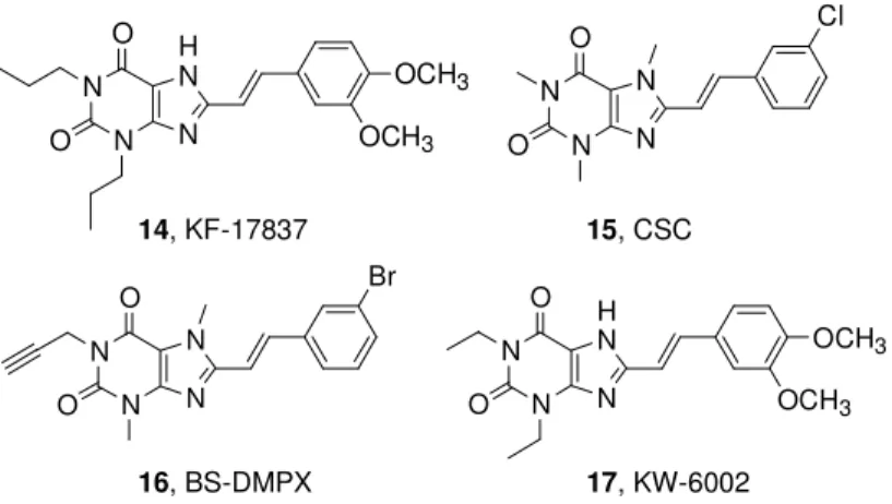
Styrylxanthines
1,3-Dipropyl-7-methyl-8-(3,4-dimethoxystyryl)-xanthine (14, KF-17837, Figure 5) was the first A2A AR antagonist in this chemical
class of compounds.61,62 The 3-chlorostyrylcaffeine 15 (CSC, Figure 5) was identified as being less potent than KF17837 but with an in-creased selectivity vs A1 AR subtype.
Chapter 1 Introduction
15
Figure 5. A2A AR antagonists (styrylxantine).
N N N H N OCH3 OCH3 O O 14, KF-17837 N N N N Cl O O 15, CSC N N N N Br O O N N N H N OCH3 OCH3 O O 16, BS-DMPX 17, KW-6002
Introduction of a propargyl at the 1- position in combination with the 8-styryl group in compound 16 (BS-DMPX, Figure 5) increased affin-ity to the A2A AR with retention of selectivity.
65
1,3-Diethyl-7-methyl-8-(3,4-dimethoxystyryl)- xanthine 17 (KW-6002, also named istradefyl-line, Figure 5) is an 8-styrylxanthine with high affinity for the rat stri-atal A2A AR.
66
Due to its high affinity and selectivity, a radiolabeled derivative, [11C]-KW-6002 labeled at the aromatic O-methyl position, was developed to be used in pharmacological testing to trace the A2A
ARs in vivo.67,68
9H-Purine derivatives
Minetti et al., on the basis of the molecular modeling of a number of potent AR antagonists, designed and synthesized a number of 2-alkyl-substituted purine derivatives as A2A AR antagonists.
69
From them ST-1535 (2-n-butyl-9-methyl- 8-[1,2,3]triazol-2-yl-9H-purin-6-ylamine 18, Figure 6), was the most interesting.
Chapter 1 Introduction N N N N N NH2 N N 18, ST-1535
Figure 6. 9H-Purine derivative
1.1.4. Clinical Development and Patents
PD is a progressive, incurable disorder with no definite preventive treatment, although drugs are available to alleviate the symptoms and/or slow down the progress of the disease. Current therapy is based on dopamine replacement therapy, the most common drug treatments being dopaminomimetic agents, including L-DOPA, a do-pamine precursor, as well as direct or indirect dodo-pamine receptor agonists. L-DOPA is the mainstay in the treatment of PD but, be-cause of tolerance problems and a wide range of adverse reactions, including involuntary movements and vomiting, a strong demand for new therapies exists. Among the various strategies, A2A AR blockers
are considered a potential approach to treatment of the disease.27, 70 KW-6002, an adenosine A2A antagonist, is currently undergoing
phase III clinical trials at Kyowa Hakko for the oral treatment of PD. As monotherapy or combination therapy with L-DOPA or dopamine agonists, it has been shown to improve the symptoms of the disease in a parkinsonian monkey model without increasing the incidence or severity of dopaminergic-related side effects or inducing or worsen-ing dyskinesia. The company had been developworsen-ing the drug for the treatment of depression, but phase II studies were discontinued. In mice and rats, KW-6002, like other A2A AR antagonists,
dose-dependently prevented reserpine and haloperidol-induced catalepsy, suggesting that it modulates dopaminergic neurotransmission.71,72
Chapter 1 Introduction
17 On the other hand, in D2 receptor knockout mice, which are a model
of motor impairment that resembles PD, blockade of A2A ARs with
KW-6002 rescued the behavioral parameters and reestablished al-tered enkephalin and substance P expression, suggesting a non-dopaminergic mechanism for the antiparkinsonian activity of KW-6002.73 KW-6002 improved motor disability in experimental nonhu-man primate parkinsonian models. Coadministration of KW-6002 and L-DOPA/benserazide potentiated the motor effects of levodopa (30%) without increasing the dyskinetic response.74,75 Recently low doses of KW-6002 coadministered with low doses of L-DOPA at-tenuated the development of L-DOPA-induced dyskinesia as well as rotational responses to repeated L-DOPA in hemiparkinsonian mice. These results encourage consideration of future A2A antagonist trials
in PD that are aimed at reducing the development rather than the expression of dyskinesia.76 Kyowa Hakko Kogyo has completed three phase III studies of KW-6002 in development for the treatment of PD (registration number [clinicaltrial.gov] 6002- EU-007, 6002-US-013, or 6002-US-018). KW-6002 has a specific antagonistic effect on the A2A AR in the brain. The studies were conducted in PD patients
with wearing-off phenomenon on treatment with DOPA alone or L-DOPA administered concomitantly with other PD medications. Two studies were conducted in North America and one study was con-ducted in 14 countries of the European Union and other regions. KW-6002 was administered for 12-16 weeks. The primary endpoint was the reduction in the percentage of awake time spent in the “off” state, which served as an indicator of the improvement in the wear-ing-off phenomenon. One of the North American studies revealed a statistically significant reduction in the percentage of awake time
Chapter 1 Introduction spent in the off state. The other North American study and the trial conducted in the European Union/other regions did not demonstrate a significant reduction in percentage of awake time per day spent in the off state compared with placebo patients but showed a significant improvement or a trend toward improvement in one of the secondary endpoints, the motor function score, assessed using the Unified Parkinson’s Disease Rating Scale subscore III. Kyowa Hakko in-tended to submit a new drug application to the Food and Drug Ad-ministration in the latter half of 2006. The long-term safety of KW-6002 in patients who have completed KW-6002-EU-007, KW-6002-US-013, or 6002-US-018 studies has been assessed in an extension phase III study started in October 2004 (registration number [clinicaltrial.gov] 6002-INT-001). Other open-label phase III studies of the continued safety of KW- 6002 for patients who completed the prior double-blind study 6002-INT-001 started in March 2005 (registration number [clinicaltrial.gov] 13711A) and in October 2005 (registration number [clinicaltrial.gov] 6002-US-025) and are currently recruiting patients. Phase II trials are also under way by the company for the treatment of restless legs syndrome (RLS).
KW-6002 has been patented as a therapeutic agent for behavioral disorders,77 anxiety78 and higher brain dysfunction,79 in medicinal composition with dopaminergic agents, monoamine oxidase-B (MAO-B) inhibitors, or catechol-O-methyltransferase (COMT) inhibi-tors for PD, RLS, and attention deficit hyperactivity disorder,80 in me-dicinal composition with antidepressant agent such as the serotonin and/or norepinephrine reuptake inhibitors for depression81 and for disease accompanied by chronic muscle/skeleton pain82 and drug dependence.83
Chapter 1 Introduction
19 SCH-420814 is a selective, orally active A2A AR antagonist
discov-ered by scientists at Schering-Plough and currently under phase II investigation for PD.60 It reversed haloperidol-induced catalepsy in rats and potentiated L-DOPA induced turning behaviour in neuro-toxin 6-hydroxydopamine (6-OHDA)-lesioned rats. Also, it was effec-tive in the 1-methyl-4-phenyl-1,2,3,6-tetrahydropyridine (MPTP) monkey model of PD and several rodent models of depression. Pharmacokinetic profiling revealed oral availability of 57%, 41% and 4% in rats, dogs, and cynomolgus monkeys, respectively. SCH-420814 and SCH-412348 were tested in vivo in rats treated with the A2A agonist CGS-21680, which reduces locomotion. At doses
rang-ing from 0.1 to 1 mg/kg, both compounds dose-dependently reversed the effects of the A2A agonist 2-p-(2-carboxyethyl)phenethylamino-5’-N-ethylcarboxamidoadenosine (CGS 21680). They also potentiated
L-DOPA-induced turning behaviour in 6-OHDA-lesioned rats at the same dose ranges. These results suggest these agents may have potential in PD as well as in other conditions associated with reduced dopaminergic activity.60
Both SCH-420814 and SCH-412348 have been patented for PD84 and other involuntary movement disorders.85 Moreover, SCH 420814 has been patented as a method for treating anxiety disorders includ-ing panic disorder, agoraphobia, obsessive-compulsive disorders, social phobia, and posttraumatic stress disorder.78
ST-1535 is an A2A AR antagonist in preclinical phase at Sigma-Tau.
The compound displayed A2A AR antagonist activity in vivo as it
in-creased spontaneous motor activity in mice and was able to antago-nize haloperidol-induced catalepsy at a dose of 10 mg/kg. It also ex-hibited antidepressant activity in the mouse forced swim test.
Poten-Chapter 1 Introduction tially useful for the treatment of PD and other motor disorders, it was selected for in vivo characterization in animal models.86 ST- 1535 (10, 20 and 40 mg/kg, per os (po)) when administered alone to MPTP-treated common marmosets produced a dose-related in-crease in locomotor activity and tended to reverse motor disability. Treatment with a threshold dose of L-DOPA (2.5 mg/kg, po) pro-duced an increase in locomotor activity and again tended to reverse motor disability.87,88 ST-1535, at oral doses of 5 and 10 mg/kg, an-tagonizes catalepsy induced by intracerebroventricular administra-tion of CGS 21680 in mice. Oral ST-1535, at 1.25 and 2.5 mg/kg, po-tentiates L-DOPA effects in reducing haloperidol-induced cata-lepsy.89 ST-1535 potentiates the effects of a threshold dose of L-DOPA in unilaterally 6-OHDA-lesioned rats.88
Subchronic (18 days, twice a day) ST-1535 (20 mg/kg ip) + L-DOPA (3 mg/kg ip) did not induce sensitization to turning behavior or ab-normal involuntary movements during the course of treatment, indi-cating a low dyskinetic potential of the drug; acute administration of ST-1535 (20 mg/kg ip) proved capable of reducing jaw tremors in a tacrine model of Parkinson’s disease tremor, thus representing a po-tential new compound, with long-lasting activity, for the treatment of PD.90 ST-1535 has been patented for the treatment of PD and other motor disorders, Alzheimer’s disease, Huntington’s disease, Wilson’s disease, and neurodegenerative conditions including cerebral ische-mia.91
Chapter 1 Introduction
21 1.2. A3 Adenosine Receptor
1.2.1. Pharmacology
The A3 AR is the only adenosine subtype cloned before its
pharma-cologic identification.92 It was originally isolated as an orphan recep-tor from rat testis, having 40% sequence homology with canine A1
and A2A subtypes. 93
Homologs of the rat striatal A3 AR have been
cloned from sheep and human. Interspecies differences in A3 AR
structure are large, showing the rat A3 AR only 74% sequence
ho-mology with sheep and human.
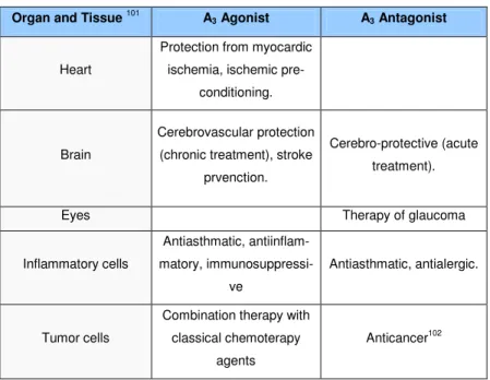
Table 2 . Distribution and therapeutical potential of A3 AR
A3 ARs activation inhibits adenylyl cyclase activity by coupling with Gi
proteins.94 In the rat mast cell line RBL- 2H3 and rat brain, A3 ARs
stimulation activate phospholipase C through Gq proteins.95,96 The A3
Organ and Tissue 101 A3 Agonist A3 Antagonist
Heart
Protection from myocardic ischemia, ischemic
pre-conditioning.
Brain
Cerebrovascular protection (chronic treatment), stroke
prvenction.
Cerebro-protective (acute treatment).
Eyes Therapy of glaucoma
Inflammatory cells Antiasthmatic, antiinflam-matory, immunosuppressi-ve Antiasthmatic, antialergic. Tumor cells
Combination therapy with classical chemoterapy
agents
Chapter 1 Introduction AR is widely distributed with its mRNA expressed in testis, lung, kid-neys, placenta, heart, brain, spleen, liver, uterus, bladder, jejunum, proximal colon and eye of rat, sheep and humans (Table 2).92,97-100 A dual role of A3 ARs has been reported in the brain. In particular, it
seems that chronic preischemic administration of the agonist IB-MECA induces a significant neuronal protection and reduction of the subsequent mortality, while acute administration of the drug results in a pronounced worsening of neuronal damage and postischemic mortality.
Mice with functional deletions of the A3 AR (A3 AR-/-) reveal a
num-ber of CNS functions where the A3 ARs play a role, including
no-ciception, locomotion, behavioral depression and neuroprotection. Consistent with previous reports of the neuroprotective actions of A3
AR agonists, A3 AR-/- mice show an increase in neurodegeneration
in response to repeated episodes of hypoxia suggesting the possible use of A3 agonists in the treatment of ischemic, degenerative
condi-tions of the CNS.103 To date, much evidence supports that activation of A3 ARs is crucial for cardioprotection during and following
ische-miareperfusion and it is likely that a consistent part of the cardiopro-tective effects exerted by adenosine, once largely attributed to the A1
AR, may now in part be ascribed to A3 AR activation.104,105 The
mo-lecular mechanism of A3 AR cardioprotection has been attributed to
regulation of ATPsensitive potassium channels. The cardioprotective effects of A3 ARs were also detected in mice overexpressing low
lev-els of A3 ARs without detectable adverse effects, while higher levels
of A3 expression lead to the development of a dilated
cardiomyopa-thy.106 Similar data were observed in the case of A1 ARs
Chapter 1 Introduction
23 In addition to reducing injury in myocardial and vascular tissues, other beneficial actions at the inflammatory level have been attrib-uted to the A3 subtype. For example, A3 ARs are expressed in
hu-man neutrophils where they are involved together with A2A in the
re-duction of superoxide anion generation108 and have been implicated in suppression of tumor necrosis factor alpha (TNFR) release in-duced by endotoxin from human monocytes.109 Moreover, A3
activa-tion seems to inhibit degranulaactiva-tion and superoxide anion producactiva-tion in human eosinophils.110 Transcript levels for the A3 subtype are
ele-vated in the lungs of asthma and COPD patients, where expression is localized to eosinophilic infiltrates. Similar evidence was observed in the lungs of ADA-deficient mice that exhibited adenosine-mediated lung disease. Treatment of ADA-deficient mice with MRS 1523, a se-lective A3 antagonist, prevented airway eosinophilia and mucus
pro-duction. These results are in contrast to experiments performed in human eosinophils ex vivo, where chemotaxis was reduced by A3 AR
activation, suggesting that significant differences exist between the impact of A3 signaling on eosinophil migration ex vivo and in the
whole animal.111 The functional role of the A3 subtype in the
patho-genesis of asthma remains controversial and differences in the pharmacology of A3 subtype from different species render it difficult
to understand whether an A3 AR agonist or antagonist is better for
use in antiasthmatic therapies. A very interesting area of application of A3 ligands concerns cancer therapies. The possibility that A3 AR
plays a role in the development of cancer has aroused considerable interest in recent years.112 A3 subtype has been described in the
regulation of the cell cycle and both pro- and antiapoptotic effects have been reported depending on the level of receptor activation.113-116
Chapter 1 Introduction A3 activation has been demonstrated to be involved in inhibition of
tumor growth both in vitro and in vivo, leading to the development of A3 agonists in clinical trials for colon carcinoma. The molecular
mechanisms involved in the anticancer effects induced by A3
ago-nists included regulation of the WNT pathway.117 On the other hand, it has been reported that adenosine upregulates HIF-1R protein ex-pression and vascular endothelial growth factor (VEGF) protein ac-cumulation by activating A3 AR subtype in tumoral cells, suggesting a
role for A3 subtype in the regulation of angiogenesis. 118
Overexpres-sion of the A3 subtype has been demonstrated in colon cancer
tis-sues obtained from patients undergoing surgery in comparison to normal mucosa. Overexpression in tissues was also reflected at the level of peripheral blood cells, rendering this adenosine subtype a possible marker for cancer detection.119 Similar data were also found in the case of arthritis, where A3 activation shows beneficial effects
by suppression of TNFR production.120,121 Adenosine receptors have been implicated in many ocular and systemic ischemic diseases (e.g., retinal ischemia). The A3 KO mouse showed lower intracellular
pressure, suggesting a role for A3 antagonists in the therapy of
glau-coma.122,123
1.2.2. A3 Adenosine Receptor Antagonists
A3-selective AR antagonists have been postulated as novel
anti-inflammatory and antiallergic agents; recent studies also indicated a possible employment of these derivatives as antitumor agents. In re-cent years many efforts have been made to search for potent and selective hA3 AR antagonists (Table 3).
Chapter 1 Introduction
25
Table 3
Kia values for ARs (nM)
A3 antagonists A1 A2A A2B A3 19, PSB-10126 1700b 2700b ND 0.43 20, KF-26777127 1800 470 620 0.20 21128 >1000 >1000 >1000 0.80 22, OT-7999129 °°>10000 °°>10000 °°>10000 0.95 23, MRS1097133 5930c 4770c NDe 108 24, MRS1191133 40100c <10%c NDe 31.4 25, MRS1334134 >100c >100c NDe 2.69 26, MRS1523135 15600c 2050c NDe 18.9 27, MRE-3008-F20141 1200 141 2100 0.82 28, MRE-3005-F20143 250 60 200 0.04 31, VUF-5574148 >10000c >10000c NDe 4.0 a
Binding experiments at recombinant hA1, A2A, A2B and A3 ARs, unless noted; b Binding
experi-ments at human cortex (A1), striatum (A2A) ARs.; c Binding experiments at rat cortex (A1), striatum
(A2A) ARs.; d IC50 values; e ND = not determined.
1.2.3. Medicinal Chemistry
Xanthines
Natural antagonists for ARs, such as caffeine and theophylline, show in general low affinity for the A3 AR subtype.
124
Different positions of the xanthine core have been modified with the aim of improving A3
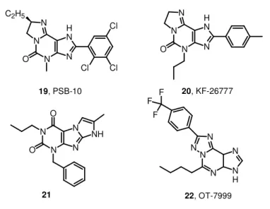
AR affinity. A series of tricyclic imidazo[2,1-i]purinones and ring-enlarged analogues derived from xanthine derivatives has been pre-pared as AR antagonists. In comparison with xanthines, the tricyclic compounds exhibit increased water solubility due to a basic nitrogen atom, which can be protonated under physiological conditions.125 Among this series PSB-10, 8(R)-ethyl-4-methyl-2-(2,3,5-trichlorophenyl)-4,5,7,8-tetrahydro-1H-imidazo[2,1-i]purin-5-one (19, Figure 7), is a high-affinity ligand for A3 ARs (hA3Ki = 0.43 nM) with
high selectivity over hA1 and hA2A ARs (Ki = 1700 and 2700 nM,
Chapter 1 Introduction studies in CHO cells expressing recombinant hA3 ARs (IC50 = 4
nM).126 Another similar compound is 2-(4-bromophenyl)-7,8-dihydro-4-propyl-1H-imidazo[2,1-i]purin-5(4H) one, also named KF-26777 (20, Figure 7), endowed with subnanomolar affinity to hA3 ARs (Ki =
0.20 nM) and high selectivity over A1, A2A, and A2B ARs (9000-,
23500- and 31000-fold, respectively). It concentration-dependently inhibited 2-chloro-N6 -(3-iodobenzyl)-N-methyl-5’-carbamoyl-adenosine (Cl-IB-MECA) -induced [35S]guanosine 5’-O-(3-thiotriphosphate) ([35S]-GTPγS) binding to human embryonic kidney 293 cells (HEK293) (IC50 = 270 nM) and enhanced intracellular Ca
2+
concentration in human promyelocytic cells (KB = 0.42 nM). This
agent was indicated for potential interest for treatment of brain ischemia and inflammatory diseases such as asthma.127
The discovery of 1-benzyl-3-propyl-1H,8H-imidazo[2,1-f]purine-2,4-diones by cyclization between the 7- and 8- positions of the xanthine core lead to 21 (Figure 7), a highly potent and selective A3 adenosine
receptor antagonist.128 This compound shows a subnanomolar affin-ity (hA3Ki = 0.8 nM) toward the desired receptor target with a
note-worthy selectivity versus the other adenosine receptors subtypes. In this field of research the triazolopurine derivatives in which the xanthine structure is extended are also reported. One example is OT-7999 (22, Figure 7), which proved to be a potent and selective hA3 AR ligand. In receptor binding assays, OT-7999 displayed high
affinity for the A3 AR (Ki = 0.95 nM) and >10500-fold selectivity
rela-tive to other AR subtypes. Significant reductions in intraocular pres-sure were obtained in cynomolgus monkeys at 2-4 h following topical application to the eye of OT-7999 (500 mcg).129,130
Chapter 1 Introduction 27 N N O O N N NH N N N N H N O C2H5 Cl Cl Cl 19, PSB-10 N N N N H N O 20, KF-26777
Figure 7. A3 AR antagonists (xanthines).
N N N H N N N F F F 22, OT-7999 21
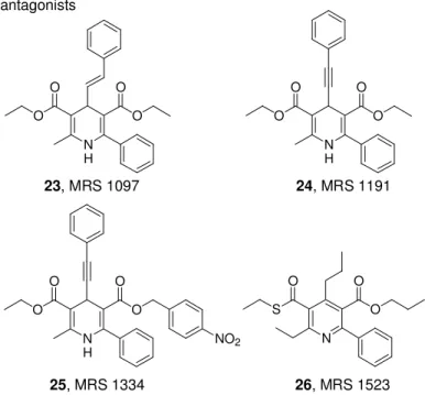
1,4-Dihydropyridine and Pyridines
Starting from the experimental observations that 1,4- dihydropyridi-nes bind A1 adenosine receptors in the rat brain,
131,132
Jacobson et al. used the 1,4-dihydropyridine nucleus as a template for probing the SAR profile at the A3 AR subtype.133 SAR studies of adenosine
receptor antagonists indicated that sterically bulky groups are well tolerated at the 4-, 5-, and 6-positions. The combination of substitu-tions led to the discovery of MRS 1097 (2-methyl-6-phenyl-4-styryl-1,4-dihydro-pyridine-3,5-dicarboxylic acid diethyl ester, 23, Figure 8), MRS 1191, methyl-6-phenyl-4-phenylethynyl-1,4-dihydro-pyridine-3,5-dicarboxylic acid 5-benzyl ester, 24, Figure 8), and MRS 1334 (2-
methyl-6-phenyl-4-phenylethynyl-1,4-dihydro-pyridine-3,5-dicarboxylic acid 3-ethyl ester 5-(4-nitro-benzyl) ester, 25, Figure 8) as the first A3 antagonists related to 1,4-dihyropyridines.
Chapter 1 Introduction
Figure 8. Dihydropyridine and Pyridine derivatives as A3 AR antagonists N H O O O O N H O O O O N H O O O O NO2 N S O O O 23, MRS 1097 24, MRS 1191 25, MRS 1334 26, MRS 1523
In this study, they also synthesized pyridine derivatives133,134 through oxidation of the corresponding 1,4-dihydropyridine. In this class of compounds, small groups at the 4-position were found to be essen-tial such as in MRS 1523 (6-ethyl-5-ethylsulfanylcarbonyl-2-phenyl-4-propyl-nicotinic acid propyl ester, 26, Figure 8), which showed fa-vourable affinity at the hA3 AR subtype. Comparing the structural
re-quirements for the two related classes of compounds indicated that bulky substituents at the 4- position and a 5-benzyl ester, which are affinity enhancing in dihydropyridines, are not well tolerated in the pyridine series for A3 receptor binding. At other positions, structural
parallels occur between corresponding dihydropyridine and pyridine analogues.135
Chapter 1 Introduction
29
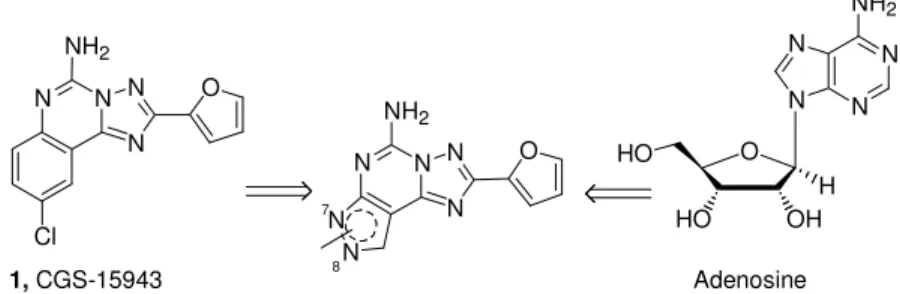
Pyrazolo-triazolo-pyrimidines (PTPs)
The pyrazolo-triazolo-pyrimidine nucleus, due to its strong structural correlation with the nonselective antagonists CGS-15943, 1, and the adenine nucleus present in the endogenous modulator adenosine (Figure 9), has been strongly investigated in the past decade as a prototypical template for adenosine antagonists.
N N N N O Cl NH2 N N N N NH2 O OH HO H HO N N N N N N O 1, CGS-15943 Adenosine
Figure 9. Structural correlation with CGS-15943 and the adenine nucleus
present in the adenosine.
7
8
NH2
The triazolo-quinazoline derivative CGS-15943 represented the start-ing point for searchstart-ing for new potent and selective hA3 adenosine
receptor antagonists.
MRS-1220, a 5-N-phenylacetyl derivative of CGS-15943, in receptor binding studies displayed Ki values of 305 ± 51, 52.0 ± 8.8 and 0.65
± 0.25 nM for rat A1, A2A, and hA3 receptors, respectively, being 470-
and 80-fold selective for hA3 ARs vs rat A1 and A2A ARs,
respec-tively. MRS-1220 also antagonized the effects of an A3 agonist in
functional assays.136,137
An innovative series of tricyclic compounds (MRE series) reported by Baraldi’s group represented new selective A3 AR antagonists. In this
class attention was focused on the N8 patterns of substitution due to the quite complete inactivity of the N7-substituted derivatives at the hA3 subtype (e.g., SCH-58261).
Chapter 1 Introduction N N N N N N NH 27, MRE-3008-F20 N N N N N N NH 28, MRE-3005-F20 Figure 10. A3 AR antagonists (pyrazolo-triazolo-pyrimidines).
N H O N H O H3CO N O O
MRE-3008-F20 (27, Figure 10), one of several high affinity antago-nists, is an A3 AR ligand (Ki = 0.29 nM against
4-aminobenzyl-5’-N-methylcarboxamidoadenosine ([125I]-AB-MECA) binding to human receptors expressed in HEK293 cells) with high selectivity over rat A1
and A2A ARs (Ki > 10000 and 1993 nM, respectively) as well as hA1
and hA2A ARs (Ki = 1197 and 141 nM, respectively). 138
The com-pound showed antagonist activity in a functional assay being capable of blocking the effect of IB-MECA on cAMP production in CHO cells (IC50 = 4.5 nM).
139-141
The tritium-labeled compound was able to bind hA3 ARs expressed in CHO cells with a KD value of 0.82 nM and a Bmax value of 297 fmol/mg protein and represents the first
high-affinity, selective radiolabeled antagonist for this subtype resulting in a useful tool for characterization of A3 ARs in both normal and
patho-logical conditions.142 The isosteric replacement of the phenyl with a 4-pyridyl moiety provided higher hydrosolubility and led to the first water-soluble hA3 antagonist (MRE-3005-F20, 28, Figure 10) which
is an ideal candidate for the pharmacological and clinical investiga-tions of the hA3 AR subtype.
Chapter 1 Introduction
31 In molecular modeling studies reported by Moro et al. on pyrazolo-triazolo-pyrimidines, a combined target-based and ligand-based drug design has been carried out to define a novel pharmacophore model for the hA3R antagonists. A high-throughput docking strategy has
been applied on the pyrazolo-triazolo-pyrimidine series. All low-energy docked conformations have been superimposed and used to characterize the common features crucial to the recognition process. A novel target-based pharmacophore model has been proposed for human A3 AR antagonists. A CoMFA (comparative molecular field
analysis) approach has been used as an alternative scoring function for prediction of ligand receptor binding affinity. The new target-based pharmacophore model was coherent with the structure-activity relationships collected on the pyrazolo-triazolo-pyrimidine ana-logues.144,145
Moreover, very recently Botta, Martinelli and Baraldi et al. performed a pharmacophoric study using the software Catalyst, which yielded three different common feature hypotheses for antagonists of the hA3R. The three pharmacophores referred to a recurring scheme
consisting of three hydrophobic interactions lying at the vertexes of a triangle. They seemed particularly good in handling pyrazolo-triazolo-pyrimidine derivatives.146 These results confirm the importance of this tricycle as the most potent class of A3 AR antagonists.
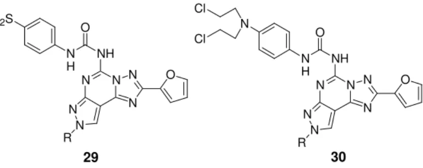
Fluorosulfonyl- and Bis(ββββ -chloroethyl)amino-phenylamino-pyrazolo[4,3-e]1,2,4-triazolo[1,5-c]pyrimidines
Synthesis of irreversible A3 antagonists was realized to provide
use-ful tools for structure-activity studies. Electrophilic groups, specifically sulfonyl fluoride and nitrogen mustard (bis-(β-chloroethyl)amino)
Chapter 1 Introduction moieties, have been incorporated at the 4-position of the aryl urea group (compunds 29 and 30, Figure 11).147
N N N N N N R NH 29 O N N N N N N R NH 30 O
Figure 11. A3 irreversible antagonists.
O N H FO2S O N H N Cl Cl
Compounds containing a fluorosulfonyl moiety proved to be irre-versible antagonists at the hA3 AR (at 100 nM, 79% of inhibition),
while the corresponding nitrogen mustard derivatives were unable to covalently bind this receptor subtype. This difference in the receptor interaction between the 29 and 30 series has been explained on the basis of chemical reactivity of the two different groups: the -SO2F
group is highly reactive versus all nucleophilic functions, while the nitrogen mustard reacts only with amino functions.
Isoquinoline and Quinazoline Urea Analogues as Antagonists for the Human Adenosine A3 Receptor
A structure-affinity analysis reported by IJzerman et al.148 indicated that at the 2- position of the quinazoline ring or the equivalent 3-position of the isoquinoline ring a phenyl or heteroaryl substituent in-creased the A3 AR affinity in comparison to unsubstituted or aliphatic
derivatives. Combination of the optimal substituents in the two series led to the potent hA3 AR antagonist
N-(2-methoxyphenyl)-N’-[2-(3-Chapter 1 Introduction
33 pyridyl)quinazolin-4-yl]urea (VUF5574, 31, Figure 12) with a Ki value
of 4 nM and a selectivity of at least 2500- fold vs A1 and A2A ARs. In
an in vitro functional assay the compound competitively antagonized the inhibition of cAMP production induced by the adenosine agonist NECA in CHO cells expressing hA3 ARs with a pA2 value of 8.1.
148 N N HN O N 31, VUF-5574
Figure 12. Quinazoline urea derivative
N
H OCH
3
1.2.4. Clinical Development and Patents
At the moment there are not A3 antagonists in clinical phases.
How-ever, in light of the plethora of biological effects attributed to A3 ARs,
substantial efforts in medicinal chemistry have been addressed to develop antagonists for the A3 subtype.
149
As a result a number of molecules are in biological testing as therapeutic agents for asthma and COPD, glaucoma, cancer and stroke.
Use of A3 antagonists has been patented for inhibition of tumor
growth.150 The pre- or coadministration of pharmaceutical composi-tions comprising high-affinity adenosine A3 receptor antagonists,
such as MRE-3008-F20, has been patented for synergistically ac-centuating the response to chemotherapy consisting of taxane (e.g., paclitaxel), vinca alkaloid (e.g., vincristine), camptothecin (e.g., iri-notecan), or antibiotic (e.g., doxorubicin) treatment.151 The claim fur-ther embodies the prevention of multidrug resistance (MDR) and
tar-Chapter 1 Introduction geted tumors include those expressing MDRassociated protein (MRP), A3 ARs, or P-glycoprotein, as found in leukemia, melanoma,
and carcinoma of the pancreas, ovary, and lung. Moreover, MRE-3008-F20 has been also patented for the treatment of cardiac hy-poxia, allergic diseases, cerebral ischemia, and cancers with high concentrations of A3 ARs.
152
Other patents of A3 antagonists also concern their use for cognitive
disorders, multiple sclerosis, neurodegeneration, PD, stroke, trau-matic brain injury,153 asthma and COPD,154-157 glaucoma158 and ar-thritis.159
Chapter
Chapter
Chapter
Chapter 2
2
2
2
Design and Synthesis
Design and Synthesis
Design and Synthesis
Design and Synthesis
Chapter 2 Design and Synthesis
2. Design and Synthesis
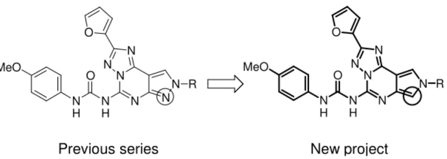
2.1. First Project: Pyrrolo[3,4-e][1,2,4]triazolo[1,5-c]pyrimidines In the last 10 years the pyrazolo-triazolo-pyrimidine (PTP) nucleus has distinguished as an attractive key intermediate for obtaining adenosine receptor antagonists due to its strong structural correla-tion with the non-selective AR antagonist CGS15943 (1, Fig. 2 ). A wide number of compounds originated from the structure-activity op-timization work based on the systematic substitution of the N5, N7,
N8, C2 or C9.160
According to the literature results, a structure-activity relationship (SAR) profile of the pyrazolo-triazolo-pyrimidines could be deline-ated.
The furan ring at the 2- position of the nucleus is fundamental for the affinity toward all four adenosine receptor subtypes.
The presence of the free amino group at the 5- position and an ary-lalkyl chain at the N7 position of the PTPs are essential for both affin-ity and selectivaffin-ity at the A2A AR, whereas the concurrent presence of
the 4-methoxy-phenyl carbamoyl moiety and small alkyl chain (such as methyl or ethyl) at the 5- and 8- position, respectively, play an im-portant role in determining potency and selectivity at human A3 AR.
Figure 13. N N N N N O R N H N H O MeO N N N N N N O R N H N H O MeO
Chapter 2 Design and Synthesis
37 In order to identify a new series of A3 AR antagonists and with the
aim to better investigate the role of the nitrogen at the 7- position on the interaction with ARs, we performed a synthetic strategy for the preparation of the pyrrolo[3,4-e][1,2,4]triazolo[1,5-c]pyrimidine nu-cleus which can be considered the 7-deaza-analogue of the pyra-zolo[4,3-e][1,2,4]triazolo[1,5-c]pyrimidine core (Fig. 13).
As depicted in scheme 1, commercially available uracil (32) has been employed as starting material. The 1,3-dibenzyl-1H-pyrimidine-2,4-dione 33 was obtained via a bis-alkylation with benzylbromide.161 Treatment of 33 with p-toluenesulphonylmethyl isocyanide (TosMIC) in presence of 60% NaH gave the desired 1,3-dibenzyl-1,6-dihydro-pyrrolo[3,4-d]pyrimidine-2,4-dione 34 which was then alkylated with the appropriate alkyl halide to furnish 6-alkyl-derivatives (35a-d). The debenzylation at the 1- and 3- positions with AlCl3 in anhydrous
tolu-ene provided derivatives 36a-d. The 2,4-dichloro-6-alkyl-6H-pyrrolo[3,4-d]pyrimidines 37a-d were obtained by treatment of 36a-d with POCl3 and DBU. Selective substitution of the chlorine atom at
the 4-position with furoic acid hydrazide followed by a Dimroth rear-rangement led to the desired
pyrrolo[3,4-e][1,2,4]triazolo[1,5-c]pyrimidine nucleus (39a-d). Compounds 40a-d were obtained
treating derivatives 39a-d with a solution of ethanol saturated with ammonia. These were converted into the corresponding phenyl urea derivatives 41a-c by reaction with 4-methoxy-phenylisocyanate.
Chapter 2 Design and Synthesis HN N H O O N N O O Bn Bn N N O O Bn Bn NH HN N H O O N R N N Cl Cl N R N N N R Cl NH HN O O N N Cl N R N N O N N H2N N R N N O N N N H N R N N O N H O MeO Scheme 1 a = CH3 b = CH2CH2CH3 c = CH2CH2Ph d = CH2CH2CH2Ph i ii iii N N O O Bn Bn N R iv v vi vii viii ix 32 33 34 35a-d
36a-d 37a-d 38a-d
39a-d 40a-d
41a-c
REAGENTS: i) NaOH 10%, tetrabutylammonium bromide, benzylbromide,
CH2Cl2, 80 °C, 18h; ii) NaH, TosMIC, Et2O, DMSO, rt, 5h; iii) K2CO3, RX, DMF, 40-80 °C, 4h; iv) AlCl3, toluene, 40 °C, 1h; v) POCl3, DBU, 50 °C, 4h; vi) 2-Furoic acid hydrazide, TEA, 1,4-dioxane, rfx, 5h; vii) HMDS, BSA, 120 °C, 18h; viii) EtOH sat. ammonia sol., 60 °C, 18h; ix) 4-OCH3-phenyl isocyanate, THF, 50 °C, 18h.
Chapter 2 Design and Synthesis 39 N N N H N N N O N H O MeO N N N H N N N O N H O N 41a 41e Figure 14.
A relevant problem of the pyrazolo-triazolo-pyrimidines was the typi-cally low water-solubility which could limit their employment as pharmacological and diagnostic tools.
Compound 5-{[(4-methoxy-phenyl)carbamoyl]amino}-(2-furan-2-yl)-8-methyl-8H-pyrrolo[3,4-e][1,2,4]triazolo[1,5-c]pyrimidine (41a, hA1Ki =
800 nm, hA2AKi = 500 nm, hA2BIC50 = 838 nm and hA3Ki = 15 nm) is
characterized by good binding data but, unfortunately, by low water-solubility, so we tried to improve the hydrophicity of this compound by introducing 4-pyridil moiety on the side chain at the 5- position (Fig.14), accordingly to a similar efficient strategy previously re-ported.143
Because of the reactivity and instability of the 4-pyridil isocyanate, this intermediate was prepared as depicted in scheme 2 , starting from the commercially available nicotinic acid hydrazide, which after reaction with sodium nitrite under acid conditions afforded the corre-sponding acyl azide. The latter was heated at reflux for 2 hours in dry toluene to give the isocyanate upon Curtius rearrangement. The crude isocyanate was heated for 5 hours in dry toluene with com-pound 40a to give the desired urea derivative 41e.143
Chapter 2 Design and Synthesis N H N O NH2 N N3 O N N C O N N N N N O N H N H O N Scheme 2
REAGENTS: i) NaNO2, HCl acq., 1h, 0 °C; ii) Toluene, 2h, rfx; iii) Toluene, 5h, 100 °C.
i iii 40a 41e ii Pyrazolo[3,4-e][1,2,4]triazolo[1,5-c]pyrimidines
In order to complete the SAR studies on this class of compounds, we decided to synthesis a novel series of
pyrazolo[3,4-e][1,2,4]triazolo[1,5-c]pyrimidine derivatives which can be considered
the structural isomers of the parent
pyrazolo[4,3-e][1,2,4]triazolo[1,5-c]pyrimidine derivatives (Fig. 15).
Figure 15
Starting from the data obtained from the previous series of pyrazolo-[4,3-e]triazolo-pyrimidine, we introduced at the N8 or N9 positions, small alkyl chain, such as methyl or propyl, and arylalkyl chain, such as phenylethyl or phenylpropyl. These modifications allowed us to explore the interaction of this side of the molecule with the adenosine
N N N N N N O N H N H O MeO N N N N N N O R N H N H O MeO R
Chapter 2 Design and Synthesis
41 receptors. In addition to the substitution at the pyrazole ring, we stud-ied the 5- position of the PTP structure introducing a free amino group, the 4-methoxy-phenyl moiety, a chlorine atom, morpholine and substituted piperazine rings.
For the synthesis of these new compounds we followed the synthetic strategy depicted in scheme 3. The 3-methyl-pyrazole (42) has been oxidized with KMnO4 and then nitrated at the 4 position with HNO3
and H2SO4. The carboxylic function was converted into the
corre-sponding carboxamide via esterification and subsequent treatment with a solution of NH4OH.
162
4-Nitro-1H-pyrazole-3-carboxylic acid amide 45 was alkylated with the appropriate alkyl halide and K2CO3
in DMF to give an approximately 1:1 mixture of the two isomers a and b which were efficiently separated via column chromatography. The nitro group was then reduced with hydrogen in presence of a catalytic amount of C/Pd 10% and intermediates 50-53a,b were con-verted into the corresponding
1/2-methyl-1,4-dihydro-pyrazolo[4,3-d]pyrimidine-5,7-dione 54-57a,b by heating with an excess of urea.
The 5,7-dichloro-1/2-methyl-1H-pyrazolo[4,3-d]pyrimidines 58-61a,b were obtained by treatment of 54-57a,b with POCl3 and DBU.
Selec-tive substitution of the chlorine atom at the 7- position with furoic acid hydrazide followed by a Dimroth rearrangement led to the desired pyrrolo[3,4-e][1,2,4]triazolo[1,5-c]pyrimidine nucleus (66-69a,b). Compounds 74-77a,b were obtained treating derivatives 66-69a,b with a solution of ethanol saturated with ammonia. These were con-verted into the corresponding 4-methoxy-phenyl urea derivatives 78-81a,b by reaction with 4-methoxy-phenylisocyanate. Compound 66a was also reacted with different primary and secondary amines to give final derivatives 70-73.
Chapter 2 Design and Synthesis N N H N N H COOH N N H COOH N N H CONH2 O2N O2N N N CONH2 O2N R N N CONH2 H2N R HN N H N N O O R N N N N Cl Cl R N N N N NH Cl R HN O O N N N N Cl R N N O N N N N N H R N N O N H O MeO N N N N H2N R N N O N N N N R' N N O R = CH3 R = CH2CH2CH3 R = CH2CH2Ph R = CH2CH2CH2Ph N N N N R R 70: R' = cycloexylamine 71: R' = morpholine 72: R' = 4-methyl-piperazine 73: R' = 4-phenyl-piperazine a b Scheme 3 i ii iii iv vi vii viii ix xii xi v x 42 43 44 45 46-49a,b
50-53a,b 54-57a,b 58-61a,b 62-65a,b
66-69a,b
78-81a,b 74-77a,b
REAGENTS: i) KMnO4, rfx, 4hrs; ii) HNO3, H2SO4, 100 °C, 4hrs; iii) a:H2SO4, EtOH, rfx,
10hrs; b:NH4OH 30%, 100 °C, 4hrs; iv) alkyl halide, K2CO3, DMF, rt, 10hrs; v) H2, C/Pd
10%; vi) Urea, 250 °C; vii) POCl3, DBU, 80 °C, 8hrs; viii) 2-Furoic acid hydrazide, TEA,
1,4-dioxane, rfx, 5h; ix) HMDS, BSA, 120 °C, 18h; x) EtOH sat. ammonia sol., 60 °C, 18h; xi) 4-OCH3-phenyl isocyanate, THF, 50 °C, 18h; xii) Amines, 2-methoxyethanol,
Chapter 2 Design and Synthesis
43 2.2. Second Project: Imidazo[2,1-i]purin-5-ones as A3 adenosine
receptor antagonists with improved water solubility
The aim of the second project of this PhD thesis was to obtain A3
adenosine receptor antagonists with high selectivity and affinity along with increased water-solubility.
We focused our attention on the imidazo[2,1-i]purin-5-one scaf-fold,126,127,163-166 obtained by the fusion of a third imidazoline ring on the xanthine bicycle, as an interesting tricyclic structure useful for the development of ARs ligands. A particular attention has been in past played to the substitution at the 2- position of the
imidazo[2,1-i]purinone nucleus, which theoretically corresponds to the 8-position
of the original xanthine core. The crucial role played by the 2-substituent in the subtype selectivity of the AR antagonists so far re-ported has been established. Compound KF20274 (82, Fig. 16),163 substituted at the 2- position with a 3-noradamantyl moiety can be structurally associated to the xanthine labelled as KW3902, (1,3-dipropyl-8-(3-noradamantyl)xanthine). The 3-noradamantyl function showed to be able to induce A1 selective antagonist activity in both
the tricycle KF20274 and the xanthine analogue KW3902. The main advantage claimed for the annelation approach of the xanthine core into the imidazo[2,1-i]purinone scaffold, is the enhancement of water solubility awarded by the imidazoline basic nitrogen which has been reported to be subject to protonation at physiological pH. Compound 83, (Fig. 16) (R)-7,8-dihydro-8-ethyl-2-(4-bicyclo[2.2.2]octan-1-ol)-4-propyl-1H-imidazo[2,1-i]purin-5(4H)-one,164 is a particularly potent A1
AR antagonist with good selectivity over the other three AR subtypes and high water solubility (>100 mg/mL) and showed a good in vivo profile after oral administration in a rat diuresis model. Müller and
co-Chapter 2 Design and Synthesis worker explored the imidazopurinone nucleus introducing at the 2- position substituents previously known to promote A2A or A3 AR
activ-ity in the corresponding 8-substituted xanthine analogues.165 Com-pound 84 (Fig. 16) has been conceived as water soluble A2A AR
an-tagonist as tricyclic congener of 8-styrylxanthines, while, derivative PSB11, (85, Fig. 16), (R)-4-methyl-8-ethyl-2-phenyl-4,5,7,8-tetrahydro-1H-imidazo[2,1-i]purin-5-one, exhibited a Ki value of 2.3
nM for A3 receptor and good selectivity vs all other adenosine
recep-tor subtypes. The radiolabelled derivative of this compound ([3H]PSB-11) exhibited a KD value of 4.9 nM and a Bmax value of
3500 fmol/mg of protein.166 The 2-(2,3,5-trichlorophenyl) substituted analogue, PSB10, showed inverse agonist activity in binding studies in CHO cells expressing recombinant hA3 ARs (IC50= 4 nM).126
N N N H N O N H N N N N O N H 82, KF20274 N N N H N O N H 85, PSB 11 84 N N N H N O N H 83 OH Figure 16
In a recent study performed in Baraldi’s laboratories, a wide series of 8-heterocyclyl-substituted xanthine derivatives has been identified as
Chapter 2 Design and Synthesis
45 very potent and selective human A2B AR antagonists.
167
With this se-ries, whose design based on the structure of the 8-phenyl-xanthine derivative MRS1754 (N-(4-cyanophenyl)-2-[4-(2,3,6,7-tetrahydro-2,6-dioxo-1,3-dipropyl-1H-purin-8-yl)phenoxy]acetamide),168 it was dem-onstrated that the phenyl and the pyrazole rings may occasionally behave as bioisosters.
Given these findings, in the present study we evaluated the effect of the replacement of the 2-phenyl ring of PSB11 and congeners with differently substituted 5-membered heterocycles, in particular 1,3- and 1,5- disubstituted pyrazoles or a 3- substituted isoxazole. At the 4- position an allyl or a benzyl group have been introduced whereas, the 8- position has been functionalized with a methyl or an ethyl, as the efficacy of such substituents was suggested by previous SAR studies on different series of xanthine-related ARs antagonists. Moreover, with the aim to verify a possible enantioselective interac-tion between the newly reported series of imidazo[2,1-i]purinones and ARs, for a selected number of compounds the pharmacological properties of the optically pure enantiomers have been compared to those of the corresponding racemates.
The synthesis of the 2-heterocyclyl tricyclic purinone derivatives has been performed, in analogy to described procedures, as depicted in scheme 7.126,163,165,169
1-Subsituted-5,6-diaminouracils 95a,b170 and the appropriate pyra-zole/isoxazole carboxylic acids were reacted in DMF solution in presence of 1-ethyl-3-[3 (dimethylamino)propyl]carbodiimide hydro-chloride (EDAC) as condensing agent, followed by ring closure with sodium hydroxide at reflux to afford the desired
3-allyl/benzyl-8-Chapter 2 Design and Synthesis Me HN NH2 O MeO N MeO O NH2 N N OMe O HO N N OMe O RO N N OH O RO Scheme 5
REAGENTS: i) Et2O, 1h, 0 °C; ii) benzene/ CH3COOH 1:1, 1h, rfx; iii)
CH3I/ benzyl bromide, K2CO3, dry acetone, 2h, rt; iv) NaOH 10%, MeOH, 1h, rfx. i ii iii iv 91a: R = Me 91b: R = Bn + OMe O MeO O 88 89 90a,b 91a,b Me O O OEt O H N NH2 + N N OMe O N N OMe O + N N OH O i ii REAGENTS: i) EtOH, 3H, rfx;
ii) NaOH 10%, MeOH, 1h, rfx.
86a 86b
87a,b Scheme 4
[(substituted)isoxazol/pyrazol-3/5-yl]-1H-purine-2,6(3H,7H)-dione de-rivatives (96a-j).167
The diamino uracils 95a,b were obtained by reduction of the corre-sponding nitroso uracils using sodium dithionite.167
The substituted pyrazole carboxylic acids (87a,b and 91a,b) were prepared according to procedures reported in literature (Scheme 4 and 5).151