International PhD Program in Neuroscience
XXIX Cycle
Coordinator: Prof. Salvatore Salomone
TGF-β1 PATHWAY AND AGE-RELATED MACULAR DEGENERATION
PhD thesis
Vincenzo Fisichella
Tutor: Prof. Filippo Caraci Co-tutor: Prof. Claudio Bucolo
BIOMETEC
Department of Biomedical and Biotechnological Sciences Section of Pharmacology.
Medical School- University of Catania 2016
TABLE OF CONTENTS
LIST OF ABBREVIATION………4
INTRODUCTION………7
ANATOMICAL LANDMARKS………...7
RETINAL PIGMENT EPITHELIUM………...7
BRUCH MEMBRANE………..8
CLINICAL EVALUATION OF MACULAR DISEASE……….8
INVESTIGATION OF MACULAR DISEASE………...11
MICROPERIMETRY………...11
FUNDUSFLUORESCEINANGIOGRAPHY………11
INDOCYANINEGREENANGIOGRAPHY………..13
OPTICALCOHERENCETOMOGRAPHY………14
FUNDUSAUTOFLUORESCENCE………...15
AGE-RELATED MACULAR DEGENERATION………..16
CLASSIFICATION………...16 EPIDEMIOLOGY……….16 RISK FACTORS………...16 DRUSEN………...17 ANTIOXIDANT SUPPLEMENTATION……….18 NON-EXUDATIVE AMD……….18
RETINAL PIGMENT EPITHELIAL DETACHMENT ………...19
RETINAL PIGMENT EPITHELIAL TEAR……….19
CHOROIDAL NEOVASCULARIZATION (CNV)………..20
HAEMORRAGIC AMD……….21
AGE RELATED MACULAR DEGENERATION AND ALZHEIMER’S DISEASE ………. .21
AMYLOID β (Aβ)………...21
THE DEPOSITION OF Aβ CAUSES AMD AND AD………..23
GENETIC BACKGROUND………...24
THE Aβ DEPOSITION IN THE BRAIN AND MACULAR AREA OF RETINA………... 25
IMAGING STUDIES OF AD AND AMD………. 25
TGF-β1 SIGNALING PATHWAY: SMAD AND NON-SMAD DEPENDENT PATHWAYS……….…...26
NEUROPROTECTIVE EFFECTS OF TGF-β1 AGAINST Aβ-INDUCED NEURODEGENERATION….29 DESIGN OF THE PRESENT RESEARCH……….30
CHAPTER I………...31
CHAPTER II………49
GENERAL DISCUSSION AND CONCLUSIONS………..62
LIST OF ABBREVATIONS
RPE Retinal pigment epithelium VA Visual acuity
BCVA Best-corrected VA PH Pinhole VA
CNV Choroideal neo-vascularization RAPD Relative afferent pupillary defect FA Fluorescein angiography
CCD Charge-coupled device FAZ Foveal avascular zone DR Diabetic retinopathy RVO Retinal vein occlusion
ICGA Indocyanine green angiography PCV Polypoidal choroidal vasculopathy OCT Optical coherence tomography SS Swept-source
CSR Central serous retinopathy FAF Fundus autofluorescence
AMD Age-related macular degeneration GA Geographic atrophy
PED Retinal pigment epithelial detachment RAP Retinal angiomatous proliferation CFH Complement factor H
SNP Single nucleotide polymorphism
ARMS2 Age-related mavulopathy susceptibility 2 AREDS Age-related eye disease study
VEGF Vascular endothelial growth factor PDT Photodynamic therapy
PIGF Placental growth factor
rtPA recombinant tissue plasminogen activator AD Alzheimer’s disease
Aβ Amyloid β
APP Amyloid precursor protein BACE-1 beta-secretase 1
PEDF Pigment epithelium-derived factor SERPINF1 Serpin family F member 1
ARPE-19 Adult retinal pigment epithelial cell line-19 RAGEs Receptor for advanced glycation end products NF-kB Nuclear factor- kappa B
EC Esterified cholesterol PC Phosphatidylcholine
MCP-1 Monocyte chemoattractant protein-1 C3 Complement component 3
C5 Complement component 5 iC3b inactivated C3b
IL-1β Interleukin-1beta
TNF-α Tumor necrosis factor alpha IL-6 Interleukin-6
IL-8 Interleukin-8
MMP2 Matrix metalloproteinase 2 MMP9 Matrix metalloproteinase 9 ROS Reactive oxygen species
RSAD2 Radical S-adenosyl methionine domain containing 2 mRNA messenger RNA
PET Positron emission tomography PiB Pittsburgh compound-B
TGFβ-1 Trasforming growth factor beta-1 TGFβ-2 Trasforming growth factor beta-2 TGFβ-3 Trasforming growth factor beta-3 LAP Latency-associated peptide ALK5 Activin-like kinase 5 TGFβRI TGFβ receptor I
TGFβRII TGFβ receptor II
SMAD Small mother against decapentaplegic
R-SMAD Small mother against decapentaplegic receptor ERK Extracellular-regulated kinase
PI3K/AKT Phosphatidylinositol-3-kinase/protein kinase B CSF Cerebrospinal fluid
MCI Mild cognitive impairment hAPP human beta-amyloid precursor NFT Neurofibrillary tangles
ALK5 Activin-like kinase 5 NGF Nerve growth factor
BDNF Brain-derived neurotrophic factor GDNF Glial-derived neurotrophic factor TRKB Tropomyosin receptor kinase B Bcl-2 B cell lymphoma-2
Bcl-xl B cell lymphoma-extra large
INTRODUCTION Anatomical landmarks
The macula is a round area at the posterior pole, lying inside the temporal vascular arcades. It measures between 5 and 6 mm in diameter, and subserves the central 15-20° of the visual field. Histologically, it shows more than one layer of ganglion cells, in contrast to the single ganglion cell layer of the peripheral retina. The inner layers of the macula contain the yellow xanthophyll carotenoid pigments lutein and zeaxanthin in far higher concentration than the peripheral retina (hence the full name 'macula lutea'- yellow plaque).
The fovea is a depression in the retinal surface at the centre of the macula, with a diameter of 1.5 mm - about the same as the optic disc.
The foveola forms the central floor of the fovea and has a diameter of 0.35 mm. It is the thinnest part of the retina and is devoid of ganglion cells, consisting only of a high density of cone photoreceptors and their nuclei (Fig. 1), together with Mϋller cells.
The umbo is a depression in the very centre of the foveola which corresponds to the foveolar light reflect, loss of which may be an early sign of damage.
The foveal avascular zone, a central area containing no blood vessels but surrounded by a continuous network of capillaries, is located within the fovea but extends beyond the foveola. The exact diameter varies with age and in disease, and its limits can be determined with accuracy only by fluorescein angiography (average .6 mm)
Retinal pigment epithelium
The retinal pigment epithelium (RPE) is composed of a single layer of cells that are hexagonal in cross-section. The cell consist of an outer non-pigmented basal element containing the nucleus, and an inner pigmented apical section containing abundant melanosomes.
The cell base is in contact with Bruch membrane, and at the cell apices multiple thread-like villous processes extend between the outer segments of the photoreceptors.
at the posterior pole, particularly at the fovea, RPE cells are taller and thinner, more regular in shape and contain more numerous and larger melanosomes than in the periphery.
RPE cells and intervening tight junctional complexes (zonula occludentes) constitute the outer blood-retinal barrier, preventing extracellular fluid leaking into the subblood-retinal space from the
choriocapillaris, and actively pumping ions and water out of the subretinal space. Its integrity is important for continued adhesion between the two, thanks to a combination of osmotic and hydrostatic forces, possibly with the aid of hemidesmosomal attachments. Facilitation of
photoreceptor turnover by the phagocytosis and lysosomal degradation of outer segments following shedding.
Maintenance of the outer blood-retinal barrier is a key factor, as are the inward transport of
metabolites (primarily small molecules such as amino acids and glucose) and the outward transport of metabolic waste products. The dense RPE pigment serves to absorb stray light.
Bruch membrane
The Bruch membrane separates the RPE from the choriocapillaris and on electron microscopy consists of five distinct elements:
1) The basal lamina of the RPE 2) An inner collagenous layer 3) A thicker band of elastic fibres 4) An outer collagenous layer
5) The basal lamina of the inner layer of the choriocapillaris
The RPE utilizes Bruch membrane as a route for the transport of metabolic waste products out of the retinal environment. Changes in its structure are thought to be important in the pathogenesis of many macular disorders .
CLINICAL EVALUATION OF MACULAR DISEASE The main characteristic symptoms:
o Blurried vision and difficulty with close work may be an early symptom.
o A positive scotoma , in which patients complain of something obstructing central vision, is a symptom of more severe disease. The optic neuropathy, however, leads to a missing area in the visual field(negative scotoma).
o Micropsia (decrease in image size) is caused by spreading apart of foveal cones, and is less common.
o Macropsia (increase in image size) is due to crowding together of foveal cones, and is uncommon.
o Metamorphopsia (distortion of perceived images) is a common symptom that is virtually never present in optic neuropathy.
o Colour discrimination may be disturbed, but is generally less evident than in even relatively mild optic neurophaty.
o Difficulties related to dark adaptation, such as poor vision in dim light and persistence of after-images, may occur.
VISUAL ACUITY
Distance visual acuity (VA) is directly related to the minimum angle of separation (subtended at the nodal point of the eye) between two objects that allow them to be perceived as distinct. In ptactice, it is most commonly carried out using a Snellen chart, which utilizes black letters or symbols (optotypes) of a range of sizes set on a white chart (Fig.2), with the subject reading the chart from a standard distance.
Normal monocular VA equates to 6/6 (metric notation) on Snellen testing. Normal corrected VA in young adults is often superior to 6/6.
Best-corrected VA (BCVA) denotes the level achieved with optimal refractive correction. Pinhole VA a pinhole (PH) aperture compensates for the effect of refractive errors, and
consists of an opaque occlude perforated by one or more holes of about 1 mm diameter. Howewer, PH acuity in patients with macular disease and posterior lens opacities may be worse than with spectacle correction.
Binocular VA is usually superior to the better monocular VA of each eye, at least where both eyes have roughly equal vision.
CONTRAST SENSITIVITY
Is a measure of the ability of the visual system to distinguish an object against its background. A target must be sufficiently large to be seen, but must also be of high enough contrast with its background; a light grey letter will be less well seen against a white background than a black letter. Contrast sensitivity represents a different aspectof visual function to that tested by the spatial resolution tests described above, which all use high-contrast optotypes. A lot of conditions reduce both contrast sensitivity and visual acuity (e.g. amblyopia, optic
neuropathy, some cataracts). The Pelli-Robson contrast sensitivity letter chart is viewed and 1 metre and consists of rows of letters of equal size but with decreasing contrast of 0.15 log units for groups of three letters.
NEAR VISUAL ACUITY
Near vision testing can be a sensitive indicator of the presence of macular disease. The chart is held at a comfortable reading distanceand this is measured and noted. The patient wears any necessary distance correction together with a presbyopia correction if applicable
AMSLER GRID
Evaluates the 20° of the visual field centred on fixation (Fig.3). It is principally useful in screening for and monitoring macular disease, but will demonstrate central visual field defects originating elsewhere.
PUPILS
The pupillary reactions to light are usually normal in eyes with macular disease, although extensive pathology such as a large area of CNV (choroideal neo-vascularization) can give a relative afferent pupillary defect (RAPD). In contrast, an RAPD occurs in relatively mild cases of asymmetrical optic neuropathy.
COLOUR VISION
Colour vision is commonly affected only in proportion to the decrease in visual acuity in macular disease, again in contrast to optic neuropathy were subtle colour desaturation is an early sign.
INVESTIGATION OF MACULAR DISEASE
MICROPERIMETRYThis is a newer investigative technique that has hitherto been used principally in research but may increasingly be incorporated into clinical practice. It measures sensitivity at finely spaced central retinal loci, including in patients with poor fixation, and uses a tracking system based on image registration to facilitate serial monitoring, allowing detection of subtle change.
FUNDUS FLUORESCEIN ANGIOGRAPHY
Fluorescein angiography (FA) should be performed only if the findings are likely to influence management.
Fluorescence is the property of certain molecules to emit light of a longer wavelength when stimulated by light of a shorter wavelength. The excitation peak for fluorescein is about 490 nm ( in the blue part of the spectrum). Stimulated molecules will emit yellow-green light of about 530 nm (Fig.4)
Fluorescein (sodium fluorescein) is an orange water-soluble dye that, when injected intravenously, remains largely intravascular. It is excreted in the urine over 24-36 hours. Outer blood-retinal barrier. The major choroidal vessels are impermeable to both bound
and free fluorescein. However, the walls of the choriocapillaris contain fenestrations through which unbound molecules escape into the extravascular space, crossing Bruch membrane but on reaching the RPE are blocked by intracellular complexes termed tight junctions or zonula occludentes.
Inner blood-retinal barrier is composed principally of the tight junctions between retinal capillary endothelial cells, across which neither bound nor free fluorescein can pass; the basement membrane and pericytes play only a minor role in this regard. Disruption of the
Fig.4: Absorption and emission curves of sodium fluorescein dye. The peak absorption (excitation) is
inner blood-retinal barrier permits leakage of both bound and free fluorescein into the extravascular space
Filters
- cobalt blue excitation: incident white light from the camera is filtered so that blue light enters the eye, exciting the fluorescein molecules in the retinal and choroidal circulations. - Yellow-green barrier filter blocks any blue light reflected from the eye, allowing only
yellow-green emitted light to pass.
Image capture in modern digital cameras uses a charge-coupled device (CCD). Modern device typically require a lower concentration of injected fluorescein to obtain high-quality images, with a lower incidence of adverse effects.
Technique
Adequate pharmacological mydriasis is important to obtain high-quality images; media opacity such as cataract may reduce picture quality. It is important to mention common and serious adverse effects, particularly the discoloration of skin and urine; the patient should be seated in front of the fundus camera, and colour photographs, red-free (green incident light, to enhance red detail)and
autofluorescence images taken as indicated.
An intravenous cannula is inserted and fluorescein (usually 5 ml of 10% solution) is injected in 5-10 seconds; a 5 ml vial of 10% (100 mg/ml) sodium fluorescein contains 500 mg and pictures should be taken over 20-60 minutes following ingestion.
Angiografic phases
Fluorescein enters the eye through the ophthalmic artery, passing into the choroidal circulation through the short posterior ciliary arteries and into the retinal circulation through the central retinal artery. The angiogram consists of the following overlapping phases: The choroidal (pre-arterial) phase typically occurs 9-15 seconds after dye injection and is characterized by patchy lobular filling of the choroid due to leakage of free fluorescein from the
fenestrated choriocapillaris.
The arterial phase start about a second after the onset of choroidal fluorescence, and shows retinal arteriolar filling and the continuation of choroidal fillin. The arteriovenous (capillary) phase shows complete filling of the arteries and capillaries with early laminar flow in the veins in which the dye appears to line the venous wall leaving an axial hypofluorescent strip. This phenomenon reflects initial drainage from posterior pole capillaries filling the venous margins, as well as the small-vessel velocity profile, with faster plasma flow adjacent to
vessel walls where cellular concentration is lower.
The venous phase. Laminar venous flow pregresses to complete filling, with late venous phase
featuring reducing arterial fluorescence.
The late (recirculation) phase demonstrates the effects of continuous recirculation, dilution and elimination of the dye. Fluorescein is absent from the retinal vasculature after about 10 minutes. The dark appearance of the fovea is caused by three factors:
- absence of blood vessels in the FAZ;
- blockage of background choroidal fluorescence due to the high density of xanthophyll at the fovea
- blockage of background choroidal fluorescence by the RPE cells at the fovea, which are larger and contain more melanin and lipofuscin than elsewhere in the retina.
Causes of hyperfluorescence
o Autofluorescentcompounds absorb blue light and emit yellow-green light in a similar fashion to fluorescein, but much more weakly. Autofluorescent lesions classically include optic nerve head drusen and astrocytic hamartoma.
o Pseudofluorescence (false fluorescence) refers to non-fluorescent reflected light visible prior to fluorescein injection.
o A window defect is caused by atrophy or absence of the RPE as in atrophic age-related macular degeneration, a full-thickness macular hole, RPE tears and some drusen.
o Pooling in an anatomical space occurs due to breakdown of the outer blood-retinal barrier (RPE tight junction). As regards the subretinal space, it is characterized by early hyperfluorescence, which, as the responsible leak tends to be only small, slowly increases in intensity and area. Instead , in the sub-RPE space occurs an early hyperfluorescence that increases in intensity but not in size.
o Leakage of dye is characterized by fairly early hyperfluorescence, increasing with time in both area and intensity. It occurs as a result of breakdown of the inner blood-retinal barrier due to: dysfunction or loss of existing vascular endothelial tight junctions as in background diabetic retinopathy (DR), retinal vein occlusion (RVO) and papilloedema; primary absence of vascular endothelial tight junctions as in CNV, proliferative diabetic retinopathy and tumours.
Causes of hypofluorescence
o Masking of retinal fluorescence. Preretinal lesions such as blood will block all fluorescence. o Masking of background choroidal fluorescence allows persistence of fluorescence from
superficial retinal vessels: deeper retinal lesions, subretinal or sub-RPE lesions, increased density of the RPE, choroidal lesions
o Filling defects may results from: vascular occlusion, which may involve the retinal arteries, veins or capillaries; loss of the vascular bed as in myopic degeneration and choroideremia.
INDOCYANINE GREEN ANGIOGRAPHY
Whilst FA is an excellent method of studying the retinal circulation, it is of limited use in delineating the choroidal vasculature , due principally to masking by the RPE. In contrast, the near-infrared light utilized in indocyanine green angiography (ICGA) penetrates ocular pigments such as melanin and xanthophyll, as well as exudate and thin layers of subretinal blood, making this technique eminently suitable. An additional factor is that about 98% of ICG molecules bind to serum protein (mainly albumin), considerably higher than the binding of fluorescein. ICG fluorescence is only 1/25th that of fluorescein so modern digital ICGA uses high-sensitivity videoangiographic image capture by means of an appropriately adapted camera. The technique is similar to that of FA, but with an increased emphasis on the acquisition of later images than that FA.
Diagnosis
Examples of the pathological images are shown under the discussion of individual conditions where relevant.
Hyperfluorescence
A window defect similar to those seen with FA.
Leakage from retinal or choroidal vessels, the optic nerve head or the RPE; this gives rise to tissue staining or to pooling.
Abnormal retinal or choroidal vessels with an anomalous morphology and/or exhibiting greater fluorescence than normal.
Hypofluorescence: blockage of fluorescence. Pigment and blood are self-evident causes, but fibrosis, infiltrate, exudate and serous fluid also block fluorescence.
Indications
- Polypoidal choroidal vasculopathy (PCV): ICGA is far superior to FA for the imaging of PCV.
- Exudative age-related macular degeneration: conventional FA remains the primary method of assessment, but ICGA can be a useful adjunct, particularly if PCV is suspected.
- Chronic central serous chorioretinopathy: in which it is often difficult to interpret areas of leakage on FA.
- Posterior uveitis: ICGA can provide useful information beyond that available from FA in relation to diagnosis and the extent of disease involvement.
- Choroidal tumours.
- Breaks in Bruch membrane. - If FA is contraindicated.
OPTICAL COHERENCE TOMOGRAPHY
Optical coherence tomography (OCT) is a non-invasive, non-contact imaging system providing high resolution cross-sectional images of the posterior segment. Imaging of the anterior segment has also been increasingly adopted. OCT is analogous to B-scan ultrasonography but uses near-infrared light interferometry rather than sounds waves, with images created by the analysis of interference between reflected reference waves and those reflected by tissue. As regards instruments promising newer modalities include swept-source (SS) OCT that can acquire images at a much higher rate and with extremely high retinal element resolution and better imaging depth; choroidal definition is improving rapidly.
Applications
- Macula: the diagnosis and monitoring of macular pathology has been revolutionized by the advent of OCT imaging, e.g. AMD, diabeti maculopathy, macular hole, epiretinal membrane and vitreomacular traction, CSR and retinal venous occlusion.
- Glaucoma: the widespread availability of OCT in ophthalmology suites for the assessment of medical retinal disease has contributed to its increased adoption as an adjunct to clinical and perimetric assessment in the management of glaucoma.
- Anterior segment OCT: has an expanding range of clinical applications such as suspected angle-closure glaucoma and corneal analysis.
FUNDUS AUTOFLUORESCENCE
Imaging of fundus autofluorescence (FAF) using an enhanced fundus camera or scanning laser ophthalmoscopy permits visualisation of accumulated lipofuscin in the retinal pigment epithelium.the scope of its place in the clinical management of macular degeneration and other conditions has not yet been clearly defined. It can be useful, for instance, to demonstrate more extensive macular disease than is visible clinically, in order either to determine the cause of unexplained poor visual acuity or to establish the reason for substantial visual symptoms despite good measured acuity. There is speculation that it may have greater utility in the future in the management of dry AMD once effective therapies become available.
AGE-RELATED MACULAR DEGENERATION
Age-related macular degeneration (AMD) is a degenerative disorder affecting the macula. It is characterized by the presence of specific clinical findings, including drusen and RPE changes, in the absence of another disorder. Later stages of the disease are associated with impairment of vision. Classification
Conventionally AMD has been divided into two main types:
I. Dry (non-exudative, non-neovascular) AMD is the most common form, comprising around 90% of diagnosed disease. Geographic atrophy (GA) is the advanced stage of dry AMD; it has been authoritatively suggest that the term “dry AMD” be used only to describe GA rather than earlier stages of AMD.
II. Wet (exudative, neovascular) AMD is much less common than dry, but is associated with more rapid progression to advanced sight loss. The main manifestations are CNV and PED, though in recent years at least two additional conditions, retinal angiomatous proliferation (RAP) and polypoidal choroidal vasculopathy (PVC), have been included under the umbrella of neurovascular AMD.
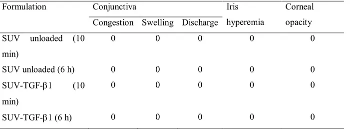
A recent expert consensus committee has provided a clinical classification of AMD (table 1) Epidemiology
AMD is the most common cause of irreversible visual loss in industrialized countries. In the USA, it is responsible for around 54% of severe sight loss (better eye worse than 6/60) in Caucasian, 14% Hispanic and 4% in black individuals. The prevalence increases with age and symptoms are rare in patients under 50 years of age. In the UK, significant visual impairment (binocularly 6/18 or worse) from AMD affects about 4% of the population aged over 75 years and 14% of those over 90, with 1.6% over 75 having binocular acuity of less than 6/60. Patients with late AMD in one eye, or even moderate vision loss due to non-advanced AMD in one eye, have about a 50% chance of developing advanced AMD in the fellow eye within 5 years.
Risk factors
AMD is multifactorial in aetiology, and is thought to involve a complex interaction between polygenic, lifestyle and environmental factors:
Age: is the major risk factor.
Race: late AMD is more common in white individuals than those of other races.
Heredity: family history is important; the risk of AMD is up to three times as high if a first-degree relative has the disease. Variants in many genes have been implicated in AMD risk and protection such as the complement factor H gene CFH, which helps to protect cells from complement-mediated damage, with several times the risk of AMD for homozygotes with a particular single nucleotide polymorphism (SNP), and the ARMS2 gene on chromosome 10. Genes related to lipid metabolism are also thought to be important.
Smoking: doubles the risk of AMD. Hypertension
Dietary factors: high fat intake and obesity may promote AMD, with high antioxidant intake having a protective effect in some groups.
Other factors: such as cataract surgery, blue iris colour, high sunlight exposure and female gender are suspected, but their influence remains less certain.
DRUSEN Histopathology
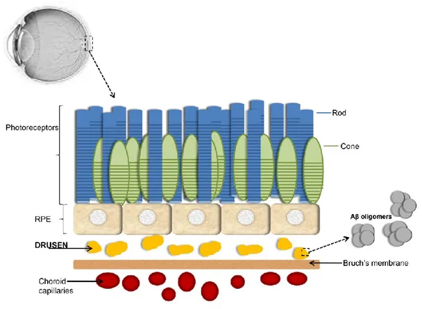
Drusen are extracellular deposits located at the interface between the RPE and Bruch membrane. The material of which are composed has a broad range of constituents, and is thought to be derived from immune-mediated and metabolic processes in the RPE. Their precise role in the pathogenesis of AMD is unclear, but is positively associated with the size of lesions and the presence or absence of associated pigmentary abnormalities. Age-related drusen are rare prior to the age of 40, but are common by the sixth decade. The distributions is highly variable and they may be confined to the fovea, may encircle it or form a band around the macular periphery.
Clinical features
There is a strong association between the size of drusen and the risk of developing late AMD over 5-year period.
a) Small drusen(drupelets), sometimes termed “hard” drusen, are typically well-defined white-yellow and by definition measure <63 µm in diameter.
b) Intermediate drusen are fairly well-defined yellow-white focal deposits at the level of the RPE measuring between 63 µm and 125 µm.
c) Large drusen are less well delineated yellow-white deep retinal lesions measuring over 125 µm in diameter. The presence of large drusen in both eyes is associated with a 13% risk of progression to late AMD over 5 years, but with accompanying bilateral pigmentary abnormalities this rises to about 50%.
OCT
Medium-sized and large drusen are seen as hyper-reflective irregular nodules beneath the RPE, located on or within the Bruch membrane.
Fluorescein angiography
FA findings depend on the state of the overlying RPE and on the affinity of the drusen for fluorescein. Hyperfluorescence can be caused by a window defect due to atrophy of the overlying RPE, or by late
Category Definition (based on presence of lesions within two
disc diameters of the fovea in either eye) No apparent ageing changes No drusen
No AMD pigmentary abnormalities Normal ageing changes Only drupelets
No AMD pigmentary abnormalities Early AMD Medium drusen (>63 µm but <125 µm)
No AMD pigmentary abnormalities
Intermediate AMD Large drusen (>125µm). Any AMD pigmentary abnormalities
Late AMD Neovascular AMD and/or any geographic atrophy
staining.Hypofluorescencedrusen masking background fluorescence are hydrophobic, with a high lipid content, and tend not to stain.
ANTIOXIDANT SUPPLEMENTATION
There is substantial evidence, notably from the Age-Related Eye Disease Study (AREDS, now known as AREDS1) and the follow-up AREDS2, that taking high-dose antioxidant vitamins and minerals on a regular basis can decrease the risk of the development of advanced AMD in individuals with certain
dry AMD features.
The regimen used in AREDS1 consisted of vitamin C, vitamin E, the beta-carotene form of vitamin A, and 80 mg daily of zinc. Because high doses of zinc and beta carotene create problems to other districts, AREDS2 looked at adjusting this components. AREDS2 found that lutein and zeaxanthin are a safe alternative to beta-carotene, and are probably superior ( 18% reduction in risk of advanced AMD). Therefore recommended daily supplementation based on AREDS2, are:
- Vitamin E (400 IU) - Vitamin C (500 mg) - Lutein (10 mg) - Zeaxanthin (2 mg) - Zinc (25-80 mg) - Copper (2 mg)
NON-EXUDATIVE (dry, non-neovascular) AMD Diagnosis
Symptomsconsist of gradual impairment of vision over month or years. Both eyes are usually affected, but often asymmetrically.
Sygnsin approximately chronological order: - numerous intermediate-large soft drusen
- focal hyper-and/or hypopigmentation of the RPE
- sharply circumscribed areas of RPE atrophy associated with variable loss of the retina and choriocapillaris
- drusenoid RPE detachment. OCT
Loss of RPE and morphological alterations of the overlying retina of increasing severity are seen in GA. Outer retina tubulations may be seen; outer retinal corrugations. This recently described phenomenon is an undulating hyper-reflective layer on OCT thought to correspond to the histological finding of basal laminar deposit, a layer that accumulates between the RPE and the RPE basement membrane in AMD. Basal linear deposit is a distinct finding consisting of membranous debris laid down between the RPE basement membrane and the inner collagenous layer of the Bruch membrane that may progress focally to form drusen.
FA
FA of atrophic areas shows a window defect due to unmasking of background choroidal fluorescence, if the choriocapillaris is still intact.
Potential new therapies
An extensive range of therapies shows promise for the treatment of dry AMD. Lampalizumab, for example, is a complement-inhibiting monoclonal antibody injected intravitreally on a monthly basis reduced progression of GA by 44%. More, preliminary evidence suggest a neuroprotective effect of saffron (20 mg/day). Others therapies include subretinal stem cell transplantation and intravitreal injection of a range of drugs including ciliary neurotrophic factor, steroid insert and neuroprotective drugs including brimonidine.
RETINAL PIGMENT EPITHELIAL DETACHMENT
Pigment epithelial detachment (PED) is the condition in which there is fluid beneath the retinal pigment epithelium (RPE). PED has many causes but the most common are AMD and central serous choroidopathy. It is caused by disruption of the physiological forces maintaining adhesion. There are different types of PED:
o Seruos PED: blurred central vision and metamorphosia; an orange dome-shaped elevation with sharply delineated edges, often with a paler margin of subretinal fluid. Are also associated blood and lipid exudation. As regards the therapies, intravitreal injections of vascular endothelial growth factor (VEGF) inhibitor may stabilize or improve vision; combining photodynamic therapy (PDT) with intravitreal anti-VEGF can also be effective. o Fibrovascular PED: by definition fibrovascular PED represents a form of “occult” CNV. It is
much more irregular in outline and elevation than serous PED.
o Drusenoid PED: develops from confluent large soft drusen, and is often bilateral. There are shallow elevated pale areas with irregular edges. The outlook is usually better than other forms of PED, with only gradual visual loss
o Haemorrhagic PED: virtually every haemorrhagic PED has underlying CNV or polypoidal choroidal vasculopathy (PCV). It is characterized by: sudden impairment of central vision, elevated dark red dome-shaped lesion with a well-defined outline and blood may break through into the subretinal space, assuming a more diffuse outline and a lighter red colour.
RETINAL PIGMENT EPITHELIAL TEAR
Tears may occur spontaneously , following laser or after intravitreal injection. Older patients and large irregular PEDs associated with CNV are at higher risk. Occurs a sudden fall in vision with foveal involvement. A crescent-shaped pale area of RPE dehiscence is seen, next to a darker area corresponding to the retracted and folded flap.
CHOROIDAL NEOVASCULARIZATION (CNV)
Choroidal neurovascularization (CNV) consists of a blood vessel complex that extends through Bruch membrane from the choriocapillaris into the sub-RPE (type 1) or subretinal (type 2 ) space. It occurs in many different disorders, usually when Bruch membrane and/or RPE function has been compromised by a degenerative, inflammatory, traumatic or neoplastic process. AMD is the most common causative association, followed by myopic degeneration. Often the CNV is the first lesion in neovascular AMD; understanding of aetiopathogenesis has improved over recent years. The promotion and inhibition of blood vessel growth by cytokines is important, particularly vascular endothelial growth factor (VEGF). It binds to endothelial cell receptors, promoting proliferation and
vascular leakage.
There is an acute or subacute painless blurring of vision, usually with metamorphosia. The CNV itself may be identifiable as a grey-green or pinkish-yellow lesion and medium-large drusenare a typical finding in the same or fellow eye. Also haemorrhage is common with an intra- and subretinal lipid deposition, sometimes extensive.
Treatment with anti-VEGF agents
Inhibitor of VEGF block its interaction with receptors on the endothelial cell surface and so retard or reverse vessel growth. They have become the predominant means of treatment for CNV. Intravitreal injection is the standard method of administration, notable risks including retinal detachment, damage to lens, RPE tears and endophthalmitis. All available anti-VEGF agents seems to have potential for benefit in a range of vascular eye diseases. Every CNV subtypes respond to anti-VEGF therapy, but benefits is only likely in the presence of active disease. Alfibercept (Eylea®) is a recombinant fusion protein that binds to VEGF-A, VEGF-B and placental growth factor (PIGF). It was adopted rapidly into clinical practice, principally because the recommended maintenance regimen consists of one injection every 2 months in contrast to the monthly injections recommended with ranibizumaband bevacizumab. The standard dose is 2 mg in
0.05 ml.
Ranibizumab (Lucentis®) is a humanized monoclonal antibody fragment developed specifically for use in the eye, though is derived from the same parent mouse antibody as bevacizumab. It non-selectively binds and inhibits all isoform of VEGF-A. The usual dose is 0.5 mg in 0.05 ml. Three
main treatment strategies are adopted in AMD.
Bevacizumab (Avastin®) is a complete antibody originally developed to target blood vessel growth in metastatic cancer deposits. Treatment strategies in AMD are similar to those used for ranibizumab.
The dose of bevacizumab is usually 1.25 mg/0.05 ml.
Pegaptanib (Macugen®) was the first anti-VEGF agent approved by regulatory authorities for ocular treatment.
Treatment with photodynamic therapy (PDT)
Verteporfin is a light-activated compound preferentially taken up by dividing cells including neovascular tissue. It is infused intravenously and then activated by diode laser to cause thrombosis. With the avvention of anti-VEGF treatment, PDT is now rarely used for CNV.
HAEMORRAGIC AMD
The visual prognosis for most eyes with extensive subretinal or sub-RPE haemorrhage is relatively poor. Some results superior to the untreated course have been reported for intravitreal anti-VEGF injection alone, and liquefaction of blood by intravitreal recombinant tissue plasminogen activator (rtPA) and pneumatic displacement may be appropriate for large or thick haemorrhage. If the patient takes a coumarin anticoagulant, liaison with the prescribing physician is worthwhile to assess if this could reasonably be stopped- there is an association with massive macular haemorrhage. Antiplatelet drugs do not usually require discontinuation, though aspirin may be associated with a greater risk of CNV than other agents.
AGE-RELATED MACULAR DEGENERATION AND ALZHEIMER’S DISEASE
Both diseases age-relatedmacular degeneration of the eye (AMD) and Alzheimer’s disease of the brain (AD) share a common risk: the age factor. These are two diseases whichcause irreversible damage: AMD is the most common cause of blindness.[Gehrs et al., 2006], AD is the most common
form of dementiain the world[Hirtz et al.,2007].
The brain and the retina are derived from the neural tube and both have blood tissue barriers, for these reasons they have many common features.
Alzheimer’s disease is.mainly characterized by memory loss, with disoriented behaviour and impairments in language, comprehension, and spatial skills also characterizing this disorder. Neuropsychiatric symptoms, such as agitation and psychosis are also frequent in people with AD, and are a common precipitant of institutional care.
There are different clinical and pathological features that pool these two diseases, such as events of oxidative stress and inflammation or the molecular similaritiesof the deposits that are typically accumulated. Also, the condition of oxidative stress and inflammation leads to the activation of the protein aggregation especially in aged post-mitotic cells, such as neurons and RPE [Kaarniranta et al.,2011].
Molecular similarities and molecular links between age-related macular degeneration and AD The most important feature that links AMD and AD is the presence and composition of extracellular deposits, that in AMD are called drusen and in AD are referred to as senile plaques. The presence of large and confluent drusen is a strong risk factor for developing choroidal neovascularisation (CNV), a complication of the wet type of AMD[Bressler et al.,1990]. The typical plaques of AD are deposited in the hippocampus and in the brain cortex and they can be diffuse, primitive, cored or compact amyloid plaques [Atwood et al.,2002].
Amyloid β (Aβ)
The Aβs are 36- to 43-amino acid peptides as the natural products of metabolism. There are two isoforms of secreted Aβ: the Aβ1-40 and Aβ1-42. The proteolysis of amyloid precursor protein (APP) generates Aβ peptides by some sequentially enzymatic hydrolysis at the β-sites by APP-cleaving
enzyme 1 (BACE-1), β- and γ-secretase, and protein complexes that contain presenilin1 at the catalytic core [Haass et al., 2007]. There are two physical forms of aggregates of Aβ: one of them is characterized by 2-6 peptide oligomers that create intermediate assemblies. Another form is represented by fibrils organised into β-pleated sheets forming insoluble fibres of advanced amyloid plaques. In normal retina and in normal brain there are small amounts of Aβ[Anderton et al.,1997] and these levels are subject to increase with age. In the retina these deposits take place primarily among the photoreceptor outer segments and on the interface between the RPE and Bruch’s membrane, but also Aβ is deposited in the vascular network of the inner and the outer retina.The first scientists who identify Aβ drusen in the eye with AMD were Johnson et al.in 2002 [Johnson et al.,2002] and showed that the Aβ was part of a substructural vesicular component within the drusen. Also Dentchev et al. [Dentchev et al.,2003] showed these results demonstrating that Aβ accumulations were exclusive of
drusen in eyes with AMD too.
Anderson et al. [Anderson et al., 2004]demonstrated, through the immunoelectron microscopy, the structural features of the Aβ-containing material in drusen. They showed the ultrastructure of the spherical Aβ-containing elements, which were composed of a central core, with one or more concentric inner rings. The most of the Aβ immunoreactive mature fibrils were associated with the outer layers, which consisted of densely-packed spherical subunits. Luibl et al [Luibl et al.,2006]through the use of an anti-oligomer antibody proved the presence of toxic nonfibrillar oligomers in the drusen, especially in the centre of drusen. They called this structure “amyloid oligomer cores”; usually the size of these cores is the same in both large and small drusen, sometimes
in large drusenoccurs coalescence of smaller drusen.
It is still unclear which form of Aβ is more prevalent in normal human retina and in the drusen of AMD patients between Aβ1-40 or Aβ1-42, in fact Prakasam et al.[Prakasam et al.,2010], through the use of a Aβ ELISA kit measured the levels of this two forms in different eye tissues from bovine and mouse. Also Dutescu et al. [Dutescu et al.,2009]showed that Aβ was not detectable in the retina or the RPE, but significant amounts of Aβ1-40 or Aβ1-42were present in the aqueous and vitreous humors of all the eyes examined. Anyway the Aβ1-40 might be the predominant form in the eye. As regards the brain, the Aβ levels are normally low because usually it is secreted in the cerebrospinal fluid. According to this scenario it would berelevant to understand the role of Aβ in development of AMD. Yoshida et al. [Yoshida et al.,2005]demonstrated that human RPE cells express constitutively all of the genes that are involved in Aβ production: APP, α-, β-, γ-secretase and neprilysin. Also,it is interesting the evidence that the exposure of cultured human RPE cells to Aβ induced a significant increase in the expression of VEGF and a significant decrease in the expression of pigment epithelium-derived factor (PEDF). PEDF is also known as serpin F1 (SERPINF1),it is a multifunctional secreted protein that has anti-angiogenic, anti-tumorigenic, and neurotrophic functions[Filleur et al., 2009]. It was originally identified in the retina[Dawson et al., 1999]and was secreted by RPE cells[Tombran-Tink et al.,1991] .Yoshida et al. have reported that there is an important equilibrium shift between VEGF and PEDF and this balance is crucial for the development of AMD. Neprilysin is a zinc-dependent metalloprotease enzyme that degrades a series of small secreted peptides, in particular the peptide Aβ [Pardossi-Piquard et al.,2006]; it has been demonstrated, through immunohistochemical analyses, that in senescent neprilysin gene-deficient mice (neprilysin -/- mice) there was an up-regulation of VEGF and a down-regulation of PEDF in comparison to wild type mice. Moreover, Ma et al. [Ma et al.,2007]showed that oligomeric forms of Aβ up-regulated VEGF secretion in ARPE-19 cellsthrough the binding to receptors for advanced glycation end products (RAGEs); this mechanism was widely dependent on NF-κBsignalling pathway.
Proteomic analyses revealed an high similarity between the molecular components of senile plaques and drusen, as regards proteoglycans, inflammatory mediators, metal ions (Fe,Cu,Zn), proteases and
clearance-related elements, α2-macroglobulin, cholinesterases, serum amyloid P component, apolipoprotein E, immunoglobulin and basement membrane matrices. Also a lot of complement activators are present in both elements (plaques and drusen)in association with complement components and complement regulatory proteins [Anderson et al.,2010; Hageman et al.,2005; Johnson et al.,2001; Johnson et al.,2006; Eikelenboom et al., 1982; Eikelenboom et al.,1996; Reichwald et al., 2009;Zanjani et al.,2005]. Obviously this suggests the existence of possible similar and common pathways involved in the aetiologiesof both diseases. Recently, Wang et al. [Wang et al.,2010] identified the amount of lipids and proteins present in drusen from 36 human retinas obtained >6h after death; the major components of drusen were esterified cholesterol (EC) and phosphatidylcholine (PC). This suggests that alterations of metabolism of cholesterol and other related molecules could contribute to the genesis of AMD by increasing the production of Aβ.Also in patients at early stage of AD an increased level of total cholesterol has been found [Wolozin et al., 2004]. The deposition of Aβ causes AMD and AD
o Role of Chronic inflammation
Chronic inflammation seems to be a common factor in both diseases[Donoso et al., 2006; Penfold et al., 2001; Cameron et al.,2010; Mandrekar-Colucci et al., 2010]. There are a lot of scientific evidence that demonstratean important role of complement activation in the pathogenesis of AMD [Bonifati et al., 2007; Anderson et al., 2010; Gehrs et al., 2006]and AD, in fact various kind of complement components exists in the brains of AD patients and in the drusen of AMD eyes. All the scientific evidences available, confirm that the complement activation at the level of Bruch’s membrane is a core process in drusen formation, and demolition of the integrity of Bruch’s membrane is linked to wet AMD.
Aβ is known to influence the alternative complement activation pathway; human RPE cells express viz., C3, C5, factor B, factor D, factor H and factor I. Some experiments showed that Aβ binds directly to complement factor I, which blocks its ability to cleave C3b and inactivate iC3b (factor I and factor H are soluble complement-activation inhibitors) [Wang et al., 2008]. This suggests that Aβ triggers the complement system by stopping the activity of factor I, bringing to chronic inflammation in the subretinal tissues. As regard the main activator of the alternative complement pathway (factor B) it has been shown that Aβ did not modulate the expression of factor B in RPE cells, but it directly enhanced the production of monocyte chemoattractant protein-1 (MCP-1)[Wang et al., 2009; Hageman et al., 2005].Aβ also increased the production of IL-1β, TNF-α in macrophages/microglia and the exposure of RPE cells to IL-1β and TNF-α significantly up-regulated factor B. ThereforeAβ stimulates RPE cells to produce MCP-1 , it recruits macrophages/microglia which produce cytokines that acts on factor B in RPE cells, up-regulating its expression. Scholl et al. [Scholl et al., 2008]measured the concentrations of complement activation products in plasma of AMD patients and found that Ba and C3d (markers of chronic complement activation) were significantly elevated, compared to controls.
o The role of microglia and other inflammatory cells
Microglial activation is one of the mechanisms involved in the pathologenesis of AD, in fact Aβs are excellent activators of microglial cells. These cells and reactive astrocytes migrate to fibrillary plaques and the inflammation marker linked to these cells are elevated in the brains of AD patients [Wyss-Coray et al., 2002]. As in all inflammatory processes the phagocytic microglia engulf and degrade Aβwith a subsequentand large production of chemokines (IL-1, IL-6 and TNFα) [Akiyama et al., 2000].Also, microglia express RAGEs which bind Aβ, stimulating in this way the production of
cytokines, glutamate and nitric oxide [Li et al., 2003; Yan et al., 1996].The RPE cells are important for the maintenance of immune balance in the subretinal space with the production of immunosuppressive factors; this normal condition is also maintained by the absence of microglial cells in this site [Chen et al., 2002]. Retinal microglial cells move from their normal position to the inner retina to get close to the RPE in the subretinal space, in AMD eyes and in animal models of AMD[Combadiere et al., 2007]. The activated condition of retinal microglial cells, observed in AMD eyes, have ameboid morphological structures that represent their activated status [Luibl et al., 2006] . Ma et al. [Ma et al., 2009]studied the effects of retinal microglia on RPE cells by co-culturing RPE cells and activated microglial cells. These onescaused changes in the structure and composition of RPE cells, increasing the expression and secretion of pro-inflammatory, chemotactic and proangiogenic molecules, with an increase of VEGF, MMP2, MMP9, too. All these variations of the normal immunological and molecular equilibrium would then enhance the progression of AMD.
o Oxidative stress
It’s a common condition among AMD [Beatty et al., 2000; Brennan et al., 2009] and AD [Darvesh et al., 2010; Querfurth et al., 2010]and it’s linked to age. Photoreceptors are easily exposed to oxidative stress because they contain lipofuscin (a photo-inducible generator of ROS)[Feeney-Burns et al., 1984; Feeney, 1978]and Aβ is a potent mitochondrial toxin that affects the neurosynaptic pool [Priller et al., 2006]in fact induces mitochondrial dysfunction and oxidative stress in RPE cells [Butterfield et al., 2004].
o Alteration of RPE cells gene expression
Microglia and neural cells are the main targets in the brain for the deposition of Aβ. Instead in the retina the major cell type affected by Aβ is RPE cells. Some scientists studied the genome-wide changes in gene expression of RPE cells stimulated with Aβ1-40 by gene microarray and RT-PCR [Kurji et al., 2010]. Essentially the up-regulate genes were member of inflammatory and immune categories, mainly IL-1β, RSAD2 (Radical S-Adenosyl Methionine Domain Containing 2) and IL-8, which are involved also in the angiogenic responses in the RPE/choroidal layers. As regard the Aβ1-42Bruban et al. [Bruban et al., 2009]showed that it induces a disorganisation of cytoskeletal actin filament with also a decrease expression of tight junction proteins (occludin and zonula occludens-1) in RPE cells. The loss of cell adhesion involves in the irreversible damage of the blood-retina barrier. Genetic background
Several research groups have studied the genetic basis of AD and AMD diseases. AMD was the first in which the GWAS (genome-wide association) identified CFH gene as the responsible gene [Edwards et al., 2005;Haines et al., 2005; Henning et al., 2005]. Mainly, variations in regulatory region for complement activation (which contains multiple haplotype) alter the risk of AMD. In fact, for example, modifications in the C3 locus are significantly associated with the development of this disease [Spencer et al., 2008; Yates et al., 2007].This observation further support for the involvement of the alternative pathway of complement activation in the pathogenesis of AMD. The uncontrolled activation of the alternative pathway of complement at the level of Bruch's membrane is thought to be a key element in the process of drusen formation and a major contributing factor to the pathogenesis of AMD [Gehrs et al., 2010].
Patients with AD and AMD
It was not yet perfectly defined the AMD frequency among patients with AD. However, it was reported a common condition of abnormalities and damages of visual pathways in patients with AD, like optic nerve degeneration, ganglion cell degeneration and decreased thickness of the retinal nerve fibre layer [Parisi et al., 2001]. In the retinas of AD patients Aβ is deposited around the major retinal vessels in the inner surface of the retina, as it was showed in amyloid angiophaty of the brain.Aβ depositsare known to damage the ganglion cell layer, cause the death of the ganglion cells with a thinning of nerve fibre layer, mimicking glaucoma.
The Aβ deposition in the brain and macular area of retina
It is clear that the accumulation of Aβ due to loss of balance between its production and clearance, may be the initiating factor bothin AD and AMD. There are two enzymes that regulate the steady-state levels of Aβ: proteases neprilysin and insulin-degrading enzyme. Neprilysin degrades Aβ monomers and oligomers [Shirotani et al., 2001] , in fact its reduction causes a cerebral accumulation of Aβ [Iwata et al., 2001]. Iwata et al. [Iwata et al., 2002]showed that the neprilysin levels were lower in 132-week-old mice compared to 10-week-old. The mRNA level of this enzyme was significantly lower in AD brains than in control human brains [Russo et al., 2005], so the down-regulation of neprilysin is linked to the deposition of Aβ in normal aging brain and in AD disease.
Imaging studies of AD and AMD
The detection of Aβ deposition is essential to diagnose AD and AMD at an early stage. The most successful non-invasive technique is probably positron emission tomography (PET) with 11C-labelled Pittsburgh Compound-B (PiB) [Klunk et al., 2004]. Instead, Higuchi et al. [Higuchi et al.,2005]used19F- and 1H-MRI to resolve the negative aspects of PET. This technique has relatively high resolution and the elimination of radiation exposure. The retina can be directly observed through the pupil of the eye. Today , improvements in ocular imaging, e.g., spectral domain optical coherence tomography make possible the identification of retinal Aβ. It would be extremely beneficial to detect the presence of Aβ, and in particular Aβ oligomers, within the drusen of AMD patients.
Common therapies to AMD and AD
The therapies, with anti-amyloid agents, developed for the treatment of AD may also be used for AMD, considering the similitudes between these two disease.Butovsky et al. [Butovsky et al., 2006]showed some interesting results as regards a treatment with Copaxone (glatiramer acetate). The T cell-based vaccination with Copaxone in AD mouse model led a reduction of the cognitive decline, elimination of plaque formation and induction of neurogenesis. A lot of clinical trials are in progress: γ-secretase inhibitors (LY450139, BMS-708163, GSI-953, E2012, PF-3084014, NIC5-15), α-secretase activators (EHT0202, MK-0952, MEM1414/R1533) and β-secretase inhibitors (CTS-21166, HPP854). In particular, one therapeutic approach (phase 3 of clinical trial) is underway: two monoclonal antibodies against Aβ (AAB-001, LY2062430) and 10% intravenous immune globulin. These antibodies bind Aβ activating the complement system and Fc-receptor-mediated phagocytosis by microglia[Fu et al., 2010]. It would be interesting to consider a drug that enhances the activity of neprilysin,too.
Since it was shown that Alzheimer’s Aβ is also an important factor in AMD[Yoshida et al., 2005] many more studies have focused on the similarities in the pathogenesis and characteristics of AD and AMD, starting from accumulation of the Aβ up to the consequences caused by this deposits.
Growing evidence suggests that a deficit of neurotrophic factors such as Transforming-Growth-Factor-β1 (TGF-β1) can significantly contribute to the pathogenesis of amyloid-related neurodegenerative disorders such as Alzheimeir’s disease. The deficiency of TGF-β1 signaling has been shown to increase both Aβ accumulation and Aβ-induced neurodegeneration in AD models (Caraci et al. 2011).
Presently no studies have been conducted to examine β1 levels in AMD. Nevertheless TGF-β1signaling is required to maintain retinal vascular stability and retinal function in adult mice [Walshe et al., 2009], and TGF-β1 signaling has been implicated in several retinal diseases [Carmeliet and Jain, 2011]. It is therefore important to understand how TGF-β1signaling in the CNS and in particular in the retina is regulated, because rescue of TGF-β1 signaling might represents a new strategy to promote neuroprotection in amyloid-related neurodegenerative disorders such as AD and AMD.
TGF-β1 signaling pathway: smad and non-smad dependent pathways
It has been hypothesized that neurotoxicity of Aß in vivo is limited by the presence of endogenous protective factors that may be lacking in the AD brain such as TGF-β1 [Caraci et al. 2011]. Transgenic mice lacking TGF-β1 show enhanced neuronal susceptibility to different neurotoxic insults [Brionne et al.,2003].
TGF-β1 is a member of TGF-beta superfamily, which consists of several groups of highly conserved multifunctional cell-cell signaling proteins of key importance in the control of tissue homeostasis [Ten Dijke et al., 2004].
The TGF-β subfamily includes three isoforms in mammals, TGF-β1, 2 and 3, which are important modulators of cell survival, inflammation, and apoptosis [Taipale et al, 1998], and also exert a central role in immune suppression, and repair after injury [Li et al., 2006]. The three TGFβs are all synthesized as homodimeric proproteins (proTGFβ) that are around 400 amino acids in size and products of separate genes. The proTGFβs are cleaved intracellularly by furin into a larger C-terminal pro-region also known as latency-associated peptide (LAP), and a shorter N-terminal active peptide, which forms the mature homodimers (25-kDa). LAP remains non-covalently associated with the mature TGFβ 25-kDa dimer before the complex is secreted [Dubois et al., 1995]. The association between the TGF-β1, 2, and 3 prodomains (LAPs) and the corresponding mature growth factors prevents signaling through the β high affinity receptors [Lawrence et al.,1984]. Thus, TGF-bioactivity requires dissociation from LAP, a process termed latent TGF-β activation. ‘
Extracellular activation of TGF-β is a critical but incompletely understood process in vivo. In particular, an important and unresolved issue in TGF-β biology regards the connection between matrix incorporation and activation of the latent TGF-β. A variety of molecules, from protons to different proteases, such as plasmin and trombospondin, have been described as latent TGFβ activators [Annes et al.,2003]. It seems that inactive TGF-β stored in tissues can be activated in response to iniury and subsequent extracellular matrix perturbations. After TGF-β is released from its latency-associated peptide, it becomes able to initiate its diverse cellular responses by binding to, and activating specific cell surface receptors that have intrinsic serine/threonine kinase activity.
All three TGF-β isoforms interact with a high-affinity transmembrane receptor complex consisting of the activin-like kinase 5 (ALK5)/TGF-β type I receptor and the TGF-β type II receptor (TβRII) subunits [Caraci et al. 2011]. Several studies have demonstrated that ligand binding to TβRII induces the assembly of type I and type II receptors into complexes with the subsequent phosphorylation and activation of ALK5, which then propagates the signal inside the cell through the phosphorylation of receptor-regulated Smads (Smads: Smad2, Smad3, Smad5 and Smad8). The interaction between R-Smads and (ALK5)/TGF-β type I receptor is facilitated by the Smad anchor for receptor activation (SARA) [Shi et al., 2003]. Phosphorylated R-Smads form heteromeric complexes with Smad4. These complexes accumulate in the nucleus, where they regulate gene expression in a cell-type-specific and ligand dose-dependent manner through interactions with transcription factors and specific promoter elements of target genes.
Smad6 and Smad7 are inhibitory Smads, which are known to counteract the signalling of R-Smads through different mechanisms [Ten et al., 2004]. Inhibitory Smads bind to activated type I receptors, thus inhibiting the phosphorylation and the following nuclear translocation of R-Smads. Furthermore, they can recruit E3-ubiquitin ligases targeting the receptor complex to the ubiquitin degradation pathway with the following inhibition of TGF-β/Smad signaling cascade.
Recent evidence suggest that TGF- rough the activation of smad-independent pathways such as the extracellular-regulated kinase (ERK) pathways [Caraci et al., 2008], the nuclear factor κB (NF-κB) pathway [König et al., 2005], and the phosphatidylinositol-3-kinase (PI3K)/Akt pathway [Caraci et al., 2008].
Role of TGF-β1 in the brain and in amyloid-related neurodegenerative disorders: the example of AD
In the CNS β2 and 3 isoforms account for almost all the β immunoreactivity, while TGF-β1 expression has been found to be constitutive only in the meninges and choroid plexus and, most importantly, in some specific brain regions such as the hippocampus and the cortex [Vivien et al., 2006]. Interestingly, TGF-β1 expression and release increase significantly in response to CNS lesions. Astrocytes and microglia seem to be the major sources of TGF-β1 in the injured brain [Finch et al., 1993], and several studies have shown that TGF-β1 induction during iniury exerts a central role in preventing neurodegeneration [Caraci et al.2011].
An increased expression of TGF-β1 has been observed with age [Finch et al. 1993], and a protective role has been suggested for this neurotrophic factor in longevity [Salvioli et al., 2009]. Aging is characterized by an increased level of pro-inflammatory markers such as IL-6, TNF-α or IL-1β [Franceschi et al., 2000]. This state of sub-clinical, chronic inflammation has been called “inflamm-ageing”, and seems to be involved in the pathogenesis of several age-related disorders such as cancer, diabetes, cardiovascular pathologies and AD [Franceschi et al., 2000]. The protective role of TGF-β in aging and longevity has been suggested by in vitro and in vivo studies [Carrieri et al.,2004]. Increased plasma levels of bio-active TGF-β1 have been found in both male and female centenarians as compared to younger control subjects [Carrieri et al. 2004]. Similar results have been obtained by Forsey et al. [Forsey et al., 2003] in octogenarian and nonagenarian subjects. Salvioli et al. [Salvioli et al., 2009] have also proposed that this age-related increase of TGF-β1 might counteract the pro-inflammatory status observed during aging, thus preventing the development of age-related disorders such as cancer and AD.
Changes in TGF-β1 serum and cerebrospinal fluid (CSF) levels have also been analyzed in AD. In particular, increased TGF-β1 levels have been found in CSF of AD patients [Tarkowski et al., 2002; Chao et al., 1994], whereas a reduction of both its active (25 kDa) and inactive (50 kDa) forms has been reported in AD plasma [Mocali et al., 2004].
Recently a single nucleotide polymorphisms (SNPs) at codon +10 (T(C) and +25 (G/C) that affects the levels of expression of TGF-β1 has been associated with an increased conversion of Mild Cognitive Impairment (MCI) in AD [Arosio et al., 2007]. Other studies have demonstrated that that both the +10 C allele and the CC genotype are over-represented in AD when compared to HC, and, that CC genotype might act as a risk factor for the development of Late-Onset AD (LOAD), independently of apolipoprotein status [Caraci et al. 2012].
Many reports also describe a significant impairment of TGF-β1 signaling in AD brain [Wyss-Coray et al., 2006; Caraci et al. 2011; Lee et al. 2006; Ueberham et al. 2006; Chalmers et al. 2007; Tesseur et al. 2006]. The study by Tesseur et al.[2006] strongly points to a causal role for of TGF-β signalling dysfunction in age-dependent neurodegeneration and AD pathogenesis . The authors found that the expression of TGF-β type II receptor (TβRII) by neurons is reduced very early in the course of AD, and this alteration seemed to be specific for AD and was not observed in other neurodegenerative conditions such as Parkinson’s disease, frontotemporal dementia, or Lewy body dementia. The authors also found that a deficiency of TGF-β signalling, in a mouse model of AD, promoted both Aβ deposition and neuronal loss [Tesseur et al. 2006]. Moreover, Tesseur et al. [2006] have shown that the impairment of TGF-β signaling in neuroblastoma cells resulted in neuritic dystrophy and increased levels of secreted Aβ and β-secretase-cleaved soluble amyloid precursor protein. These data suggest that a deficiency of TGF-β/TβRII signaling axis might exert a pathogenetic role in AD, depriving cortical neurons of trophic support, and finally promoting Aβ-induced neurodegeneration.
However, the role of TGF-ß1 in AD pathophysiology is not unequivocal, and conflicting results have been reported recently. TGF-ß1 is known to induce the expression of the APP gene in several different cell culture systems [Lesne et al. 2003] and might thus increase Aß production. The co-expression of TGF-ß1 in transgenic AD mice accelerates the deposition of Aß in cerebral blood vessels [Wyss-Coray et al. 1997], and transgenic mice overexpressing TGF-ß1 develop AD-like vascular alterations [Gaertner et al. 2004]. In addition, vessel-derived TGF-ß1 has been suggested to contribute to inflammatory processes in the AD brain [Harris-white et al. 1998; Grammas et al. 2002]. Town et al. [2008] have found that blocking TGF-Smad 2/3 signaling reduces cerebrovascular β-amyloid deposits and Aβ abundance in Tg2576 mice, and these events result in promotion of Smad1/5/8 signaling with increased infiltration of Aβ-containing peripheral macrophages around cerebral vessels and β-amyloid plaques.
Overall data from the literature seem to suggest that TGF-ß1 can promote Aβ deposition in cerebral blood vessels, but reduces Aβ accumulation in the brain parenchyma [Wyss-Coray et al., 2006]. In particular, it has been demonstrated that a modest increase in astroglial TGF-β1 production in aged transgenic mice expressing the human beta-amyloid precursor protein (hAPP) results in a 50% reduction of Aβ load in the hippocampus, and a decrease in the number of dystrophic neurites [Wyss-Coray et al., 2001].
Deficiency of TGF-ß1 signaling is also involved in tau pathology and NFT formation. Luterman et al. [2000] found that low levels of TGF-ß1 mRNA negatively correlated with NFT in the AD brain, thus suggesting that a deficiency of TGF-ß1 might also contribute to the cascade of events that result in the development of NFT-bearing neurons. The relationship between tau hyperphosphorylation and