Ther Adv Musculoskel Dis 2017, Vol. 9(1) 3 –10 DOI: 10.1177/ 1759720X16671928 © The Author(s), 2016. Reprints and permissions: http://www.sagepub.co.uk/ journalsPermissions.nav Therapeutic Advances in Musculoskeletal Disease
Introduction
Systemic sclerosis (scleroderma, SSc) is an auto-immune connective tissue disease characterized by skin and internal organ fibrosis, coupled with
widespread vascular pathology [Korn, 2001]. Skeletal muscle involvement in SSc was first con-sidered in 1876, as a minor component of the dis-ease associated with disuse [Medsger et al. 1968].
Histopathological findings in systemic
sclerosis-related myopathy: fibrosis and
microangiopathy with lack of cellular
inflammation
Claudio Corallo, Maurizio Cutolo, Nila Volpi, Daniela Franci, Margherita Aglianò, Antonio Montella, Chiara Chirico, Stefano Gonnelli, Ranuccio Nuti and Nicola Giordano
Abstract
Objectives: The objective of this study was to identify specific histopathological features of skeletal muscle involvement in systemic sclerosis (SSc) patients.
Methods: A total of 35 out of 112 SSc-patients (32%, including 81% female and 68% diffuse scleroderma) presenting clinical, biological and electromyographic (EMG) features of muscle weakness, were included. Patients underwent vastus lateralis biopsy, assessed for individual pathologic features including fibrosis [type I collagen (Coll-I), transforming growth factor β (TGF-β)], microangiopathy [cluster of differentiation 31 (CD31), pro-angiogenic vascular endothelial growth factor A (VEGF-A), anti-angiogenic VEGF-A165b], immune/ inflammatory response [CD4, CD8, CD20, human leucocyte antigens ABC (HLA-ABC)], and membranolytic attack complex (MAC). SSc biopsies were compared with biopsies of (n = 35) idiopathic inflammatory myopathies (IIMs) and to (n = 35) noninflammatory myopathies (NIMs). Ultrastructural abnormalities of SSc myopathy were also analyzed by transmission electron microscopy (TEM).
Results: Fibrosis in SSc myopathy (81%) is higher compared with IIM (32%, p < 0.05) and with NIM (18%, p < 0.05). Vascular involvement is dominant in SSc muscle (92%), and in IIM (78%) compared with NIM (21%, p < 0.05). In particular, CD31 shows loss of endomysial vessels in SSc myopathy compared with IIM (p < 0.05) and with NIM (p < 0.01). VEGF-A is downregulated in SSc myopathy compared with IIM (p < 0.05) and NIM (p < 0.05). Conversely, VEGF-A165b is upregulated in SSc myopathy. The SSc immune/inflammatory response suggested humoral process with majority (85%) HLA-ABC fibral neoexpression and complement deposits on endomysial capillaries MAC, compared with IIM (p < 0.05), characterized by CD4+/CD8+/B-cell infiltrate, and NIM (p < 0.05). TEM analysis showed SSc vascular alterations consisting of thickening and lamination of basement membrane and endothelial cell ‘swelling’ coupled to endomysial/perimysial fibrosis.
Conclusions: Fibrosis, microangiopathy and humoral immunity are predominant in SSc myopathy, even if it is difficult to identify specific histopathological hallmarks of muscle involvement in SSc, since they could be present also in other (IIM/NIM) myopathies. Keywords: fibrosis, histopathology, microangiopathy, myopathy, systemic sclerosis
Correspondence to: Claudio Corallo, PhD Scleroderma Unit, Department of Medicine, Surgery and Neurosciences, University of Siena, 53100 Siena, Italy [email protected] Nila Volpi, MD, PhD Daniela Franci, MSc Margherita Aglianò, MD Antonio Montella, MD Chiara Chirico, MD Stefano Gonnelli, MD Ranuccio Nuti, MD Nicola Giordano, MD Scleroderma Unit, Department of Medicine, Surgery and Neurosciences, University of Siena, Italy
Maurizio Cutolo, MD, PhD
Research Laboratory and Academic Division of Clinical Rheumatology, Department of Internal Medicine, University of Genova, Genova, Italy Original Research
Nowadays, SSc skeletal muscle involvement turns out to be a common feature, with prevalence from 14% to 79% [Paik et al. 2014]. This variable prevalence results from the heterogeneous criteria used to define muscle involvement in SSc, includ-ing clinical, biological, electromyographic (EMG) and histological features of muscle weakness [Olsen et al. 1996]. In fact, SSc myopathy can occur for different reasons, such as result of non-autoimmune etiologies: malnutrition, disuse, or other neuromuscular disorders [Clements et al. 1978]. Another important issue related to defin-ing specific criteria for recognizdefin-ing ‘SSc myopa-thy’ as such an entity, is the exclusion (or not) of SSc overlap syndromes, since inflammatory myo-pathies such as polymyositis (PM) or dermatomy-ositis (DM) are a common feature in these syndromes [Pope, 2002]. Despite the absence of definite criteria for the diagnosis of ‘SSc myopa-thy’, researchers agree on the fact that SSc-muscle involvement is a negative prognostic feature impacting survival, and has also been associated with cardiopulmonary complications and even sudden cardiac death [Follansbee et al. 1993]. For these reasons, in the present study we tried to identify histopathological hallmarks of ‘SSc myo-pathy’ that could be unique for the disease itself and not common to other idiopathic inflammatory myopathies (IIM) or to other noninflammatory myopathies (NIM).
Methods
Patients and diagnostic procedures
We admitted 112 SSc patients in our institution from 2010 to 2015. Patients who had been diag-nosed according to the 2013 American College of Rheumatology and European League Against Rheumatism (ACR/EULAR) diagnostic criteria for SSc [Van Den Hoogen, 2013], were enrolled. In total, 35 out of the 112 (32%, including 81% female and 68% diffuse scleroderma) who pre-sented clinical, biological and EMG features of muscle involvement, were enrolled. Table 1 shows the main clinical and biological features of enrolled patients. Patients underwent biopsy of the vastus lateralis muscle. Specimens were fro-zen in liquid nitrogen-cooled isopentane and stored at −80°C until use. Cryostat sections were submitted to diagnostic routine histological and histochemical stains. SSc muscle biopsies were compared with (n = 35, site-matched) biopsies of patients with IIM, diagnosed following current
clinicopathological criteria [Dalakas, 2010] [(n = 15 PM; n = 10 DM; n = 10 inclusion body myosi-tis (IBM)], and to (n = 35, site-matched) biopsies of patients with NIM (n = 21 (A) metabolic myo-pathies, n = 14 (B) congenital myopathies): (A)
n = 5 aspecific myopathic changes at biopsy; n =
4 statin associated rhabdomyolysis; n = 4 mito-chondrial myopathy; n = 3 muscle glycogenosis;
n = 5 steroid myopathy. (B) n = 2 calpainopathy; n = 3 mild dystrophinopathy; n = 2 myotonic
dys-trophy type 1; n = 2 myotonic dysdys-trophy type 2;
n = 2 facioscapulohumeral dystrophy; n = 3
oligo-symptomatic familial hyperckemia). All subjects signed an informed consent, with allowance for scientific utilization of muscle samples for research purposes, in accordance to the principles of the 1975 Declaration of Helsinki (revised Hong Kong 1989) and the entire study protocol was approved by the institutional review board of the Ethics Committee of the University of Siena.
Histology and histoenzymatic stains
Cryostat 10 µm thick sections were submitted to routine hematoxylin–eosin and modified Gomori trichrome for morphological evaluation. Histoenzymatic stains for nicotinamide adenine dinucleotide (NADH) tetrazolium reductase, succinic dehydrogenase, cytochrome c oxidase, Periodic Acid Schiff (PAS) were also carried out.
Immunohistochemistry
Immunohistology for diagnostic routine analysis [CD4, CD8, CD20, human leucocyte antigens ABC (HLA-ABC), membranolytic attack com-plex (MAC)] (Dako, Glostrup, Denmark) was carried out on 7 µm thick cryostat sections on silane-coated slides (StarFrost; Knittel Gläser, Braunschweig, Germany). Vascular involvement was assessed by endothelial marker [cluster of differentiation 31 (CD31)] (Dako), pro-angio-genic vascular endothelial growth factor A (VEGF-A) (abcam, Cambridge, UK) and anti-angiogenic VEGF-A165b (abcam). Muscle fibro-sis was assessed by the analyfibro-sis of type I collagen (Coll-I) (abcam) and transforming growth factor β (TGF-β) (abcam). All the reactions were per-formed by immunoperoxidase technique, by horseradish peroxidase (HRP)-labeled polymer (Dako), and 3,3’-diaminobenzidine (Sigma-Aldrich, Milan, Italy) for visualization. Negative controls were performed by the omission of the primary antibody.
Transmission electron microscopy analysis
Ultrastructural analysis of SSc muscle specimens was performed routinely after fixation in 2.5% glutaraldehyde for 3 h at 4°C, post fixation in 1% osmium tetroxide, and embedding in araldite. Ultrathin sections were stained with uranyl ace-tate and lead citrate and were observed at Philips EM-10 electron microscope (Huntsville, Alabama, USA).
Quantitative analysis on immunohistochemical slides
Morphometry was carried out by a Zeiss AxioPlan2 microscope equipped with AxioVision 4.6 soft-ware (Carl Zeiss Vision GmbH, Hallbergmoos, Germany). Microangiopathy (CD31+ endomysial vessels, VEGF-A and VEGF-A165b), inflammation (deposits of CD4, CD8, CD20 reactive cells), major histocompatibility complex (MHC)-I com-plex fibral neoexpression HLA-ABC, complement deposition MAC and fibrosis (Coll-I, TGF-β) were evaluated as histopathological parameters of muscle involvement. The density of CD31+ endomysial vessels was expressed as a capillary to fiber ratio of muscle area, by counting vessels on
the whole sections immunostained for CD31. VEGF analysis consisted of VEGF-A165b/VEGF-A ratio of consecutive sections. Inflammatory (CD20, CD4, CD8), fibrotic (Coll-I, TGF-β) and comple-ment deposition MAC scores were assessed on three randomly selected fields at 100× magnifica-tion by automatized colorimetric pixel evaluamagnifica-tion, detecting the peroxidase reaction product. Each score was expressed as the marker+ percentage of the total area.
Statistical analysis
Data were evaluated by GraphPad Prism 6® soft-ware for Windows. Analysis of variance (ANOVA) was performed by Kruskal–Wallis test for multiple groups. Significance was set at p < 0.05. Data are expressed as means ± standard deviations (SD).
Results Histopathology
General myopathic changes, such as increased variability of fiber diameter, scattered atrophic fibers and occurrence of internalized nuclei were Table 1. Clinical and biological features of enrolled patients.
SSc patients lSSc dSSc
Number of subjects (35) 11 (32%) 24 (68%)
Female sex, N (%) 7 (63%) 21 (87%)
Disease duration years (1st
non-Raynaud’s) 6.4 ± 4.8 7.9 ± 5.6
At the time of muscle biopsy
(years) 4.5 ± 3.7 4.9 ± 3.7 ANA titer ⩾ 1:160 11 (100%) 24 (100%) Anti-CENP-B 11 (100%) 0 (0%) Anti-Scl-70 0 (0%) 24 (100%) Anti-PM-Scl/Anti-Jo-1 0 (0%) 0 (0%) Increased CK (>200 mcg/l) 5 (45%) 15 (62%) Increased LDH (>190 u/l) 4 (36%) 15 (62%)
Increased aldolase (>7.5 u/l) 3 (27%) 9 (37%)
Increased myoglobin (>85 ng/ml)
Weakness (BMC testing <5) 3 (27%)11 (100%) 7 (29%)24 (100%)
EMG myopathic motor units 11 (100%) 24 (100%)
lSSc, limited systemic sclerosis; dSSc, diffuse systemic sclerosis; ANA, Anti-nuclear antibodies; CENP-B, Centromere Protein B; Scl-70, DNA Topoisomerase I; PM-Scl, exosome; Jo-1, histidyl tRNA synthetase; CK, Creatinine kinase; LDH, Lactate dehydrogenase; BMC, British Medical Council; EMG, Electromyography.
Pharmacological treatment: SSc patients enrolled in the study were in treatment with Iloprost (11 lSSc and 24 dSSc) for secondary Raynaud’s phenomenon, with dual endothelin-1 receptor antagonist Macitentan (4 lSSc and 16 dSSc) for pul-monary arterial hypertension (PAH), with dual endothelin-1 receptor antagonist Bosentan (2 lSSc and 4 dSSc) for digital ulcers, with prokinetic and proton pump inhibitor agents (1 lSSc and 2 dSSc). It is important to underline that all the SSc patients enrolled in the study stopped treatments with low dose corticosteroids (<4–8 mg/die) and with immunosuppres-sive drugs at least three months before muscle biopsy.
detected. Multifocal endomysial fibrosis and scat-tered dilated endomysial capillaries were also observed. Necrosis/regeneration was not promi-nent, with occasional single fiber necrosis in a minority of subjects.
Immunohistology and quantitative analysis
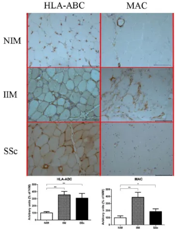
Immunohistology for diagnostic routine analysis (CD4, CD8, CD20, Figure 1; HLA-ABC, MAC, Figure 2) were reported with the relative quanti-tative analysis. The inflammatory cells result was present only in IIM group. Limited or no reactiv-ity was recorded in NIM (p < 0.001) and SSc (p < 0.001) groups, while the sarcolemmal/cyto-plasmic stain of HLA-ABC and MAC deposition on endomysial capillaries is statistically higher in SSc myopathy (p < 0.01; p < 0.05) and IIM (p < 0.01; p < 0.01) groups compared with the NIM group. Moreover, HLA-ABC and MAC stainings in the SSc myopathy group seem to be compara-ble to those of IIM group. In Figure 3, vascular
involvement, represented by A, VEGF-A165b, CD31 and fibrotic component (represented by Coll-I and TGF-β) are reported. The VEGF-A165b:VEGF-A ratio is statistically higher in SSc myopathy (p < 0.01) and IIM (p < 0.05) groups compared with the NIM group. The density of CD31+ endomysial vessels is statistically decreased in the SSc myopathy group compared with the IIM (p < 0.05) and NIM (p < 0.01) groups. In addition, Coll-I and TGF-β reveal strong expression in activated endomysial and perimysial myofibroblasts of the SSc myopathy group, and also in the fibroblasts and in some inflammatory cells surrounding the necrotic fib-ers in the IIM group. Faint Coll-I and TGF-β immunoreactivity is recorded in the resident endomysial and perimysial fibroblasts of the NIM group. Quantitative analysis shows that Coll-I and TGF-β are statistically higher in the SSc-myopathy group compared with the IIM (p < 0.05; p < 0.01) and NIM (p < 0.01; p < 0.01) groups.
Figure 1. Representative images of immunohistological distribution (top) of CD20, CD4, CD8 on consecutive sections. Bars are set at 100 µm. Quantitative analysis (bottom) reveals that inflammatory cells are
predominant in IIM group compared with NIM (***p < 0.001, Kruskal–Wallis) and SSc (***p < 0.001, Kruskal– Wallis) groups.
TEM analysis
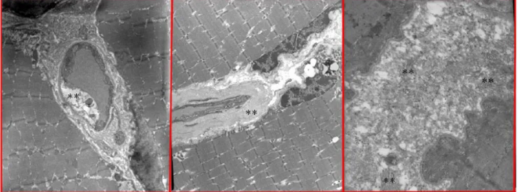
Figure 4 shows a transmission-electron-micros-copy (TEM) analysis of SSc muscle biopsies. Thickening and lamination of the basement membrane (middle), coupled to endothelial cell ‘swelling’ (left) are the main hallmark of SSc microangiopathy. Moreover, considerable endomysial fibrosis (right) with a large deposition of collagen is also observed.
Discussion
The most important problem when analyzing the literature of SSc-related muscle involvement is the total absence of specific criteria for the diag-nosis of such an entity [Ranque et al. 2007]. This is because clinical, biological, EMG and histo-pathological features are very heterogeneous, and
also because SSc-associated myopathy often includes several distinct entities with different pathogenetic mechanisms involved, and with dis-tinct outcomes [Ranque et al. 2007; Morrisroe
et al. 2015]. Even when focusing on muscle
histo-pathological findings only, no clear-cut classifica-tion has emerged [Paik et al. 2015]. Therefore, novel insights on the immunopathological nature of SSc muscle involvement are needed. For this reason, in this study we tried to identify histo-pathological hallmarks that could be specific for SSc myopathy and not common to other IIM and NIM. Whether the association of IIM and SSc constitutes an overlap syndrome or should be considered a SSc manifestation still remains controversial [Troyanov et al. 2005; Bhansing
et al. 2014]. We demonstrated that fibrosis
(81%) and microangiopathy (92%) are the main Figure 2. Immunohistological distribution (top) of ABC and MAC. Sarcolemmal and cytoplasmic HLA-ABC neoexpression is predominant in IIM and SSc groups. Necrotic fibers, identified by deposits of the terminal complex of complement, or MAC are particularly common to IIM group, while MAC deposits on capillary walls are particularly abundant in SSc group. Bars are set at 100 µm. Quantitative analysis (bottom): HLA-ABC is statistically higher in SSc myopathy (**p < 0.01, Kruskal–Wallis) and IIM (**p < 0.01, Kruskal– Wallis) groups compared with NIM group. MAC is highly upregulated in IIM (**p < 0.01, Kruskal–Wallis) and SSc-myopathy (*p < 0.05, Kruskal–Wallis) groups compared with NIM group.
HLA-ABC, human leucocyte antigens ABC; IIM, idiopathic inflammatory myopathies; NIM, noninflammatory myopathies; SSc, systemic sclerosis.
histopathologic hallmarks of SSc-related myopa-thy. We evidenced a reduced vascularization of muscle fibers through the analysis of CD31, and the prevalence of anti-angiogenic isoform VEGF-A165b, as confirmed by other researchers, finding increased VEGF-A165b plasmatic levels in SSc patients [Manetti et al. 2013]. However, these vascular alterations are not specific to SSc; they could be present also in other IIM myopathies, such as PM and DM, as has been well described in the literature [Volpi et al. 2013]. Regarding the inflammatory infiltrate, our study detected an absence of inflammatory cells in the majority of SSc cases (85%). MAC capillary deposits, com-patible with a humoral immune process, suggest
that endothelial injury and intimal proliferation may be mediated by complement-fixing anti-bodies [Evans et al. 1987]. Loss in muscle small endomysial vessels in SSc myopathy indicates a vasculopathy, with proliferative changes of the vessels wall, as evidenced by ultrastructural examination, as an early step in tissue damage, in analogy with skin and internal organs [Asano and Sato, 2015]. Nevertheless, HLA-ABC upregula-tion is considered an immunohistologic hallmark of inflammatory myopathies [Dalakas, 2010]. In view of our consistent finding of MHC-I fibral staining on SSc myopathy, with no relevant infiltrates or myonecrosis, a role of pleiotropic TGF-β might be speculated about as a negative Figure 3. Immunohistology (top) and relative quantitative analysis (bottom) of CD31 (left), VEGF-A,
VEGF-A165b (middle) and Coll-I, TGF-β-A (right) in NIM, IIM and SSc groups, respectively. CD31+ vessels
(arrows) are abundant and surrounding myofibers in NIM group, while they decrease in particular in the SSc group. The density of CD31+ endomysial vessels, expressed as capillary to fiber ratio of muscle area, is
statistically decreased in SSc myopathy group compared with IIM (*p < 0.05, Kruskal–Wallis) and to NIM (**p < 0.01, Kruskal–Wallis) groups. VEGF-A and VEGF-A165b seem equally expressed in small endomysial vessels of NIM group; in SSc group, VEGF-A shows only the stain of endomysial vessels (arrows), while VEGF-A165b also shows a diffuse cytoplasmic stain of myofibers (arrows). The VEGF-A165b:VEGF-A ratio is statistically higher in SSc myopathy (**p < 0.01, Kruskal–Wallis) and IIM (*p < 0.05, Kruskal–Wallis) groups compared with NIM group. Coll-I and TGF-β are expressed only by some resident endomysial fibroblasts (arrows) in NIM group, while they are expressed by both infiltrate cells and activated fibroblasts surrounding necrotic fibers in IIM group (arrows). In SSc group, endomysial activated fibroblasts or myofibroblasts are strongly reactive to Coll-I and TGF-β (arrows). Quantitative analysis demonstrated that endomysial expression of Coll-1 is significantly higher in SSc-myopathy group compared with IIM (*p < 0.05, Kruskal–Wallis) and to NIM (**p < 0.01, Kruskal–Wallis) groups. TGF-β is strongly upregulated in SSc myopathy group compared with IIM (**p < 0.01, Kruskal–Wallis) and NIM (**p < 0.01, Kruskal–Wallis) groups. Bars are set at 100 µm.
IIM, idiopathic inflammatory myopathies; NIM, noninflammatory myopathies; SSc, systemic sclerosis; TGF-β-A,
transforming growth factor β-A; VEGF-A, pro-angiogenic vascular endothelial growth factor A; VEGF-A165b, anti-angiogenic vascular endothelial growth factor A.
modulator of cell immunity [Morikawa et al. 2016], in reducing the extent of muscle inflam-mation, at least in the early phases of muscle involvement, and acting as a pro-fibrotic factor. This hypothesis is based on the documented upregulation of TGF-β in scleroderma lesional skin, where it induces the activation of myofi-broblasts [Nikitorowicz-Buniak et al. 2015]. However, it must be remembered that the litera-ture reported some cases of SSc myopathy with inflammatory cells, predominantly CD8+ T lym-phocytes with lower levels of CD4+ cells, few B cells and no complement capillary deposits [Bhansing et al. 2014], as well as some cases with a majority of CD4+ T cells and B cells and com-plement deposits [Ranque et al. 2007]. Therefore, data related to the inflammatory infiltrate and the nature of immune response seem controver-sial and different possible patterns are envisaged. In conclusion, our suggestion is to study and characterize each case of muscle involvement in SSc from the histological/immunohistological point of view, because of the heterogeneity of its manifestations. This advice is not simply specu-lative, but responds to the need to set up the suit-able therapy for each case. In fact, high-dose steroids represent the first line treatment for all myositides except IBM [Dalakas, 2010]; in SSc myopathic patients, highly susceptible to renal crisis, corticosteroids at the lowest effective dose should be restricted to subjects with histologi-cally proven inflammation. Further studies should follow to determine whether histopatho-logic findings in SSc-related myopathy could influence clinical outcomes of specific targeted therapies.
Funding
This research received no specific grant from any funding agency in the public, commercial, or not-for-profit sectors.
Conflict of interest statement
The authors declare that there is no conflict of interest.
References
Asano, Y. and Sato, S. (2015) Vasculopathy in scleroderma. Semin Immunopathol 37: 489–500. Bhansing, K., Lammens, M., Knaapen, H., van Riel, P., van Engelen, B. and Vonk, M. (2014) Scleroderma-polymyositis overlap syndrome versus idiopathic polymyositis and systemic sclerosis: a descriptive study on clinical features and myopathology. Arthritis Res Ther 16: R111.
Clements, P., Furst, D., Campion, D., Bohan, A., Harris, R., Levy, J. et al. (1978) Muscle disease in progressive systemic sclerosis: diagnostic and therapeutic considerations. Arthritis Rheum 21: 62–71.
Dalakas, M. (2010) Inflammatory muscle diseases: a critical review on pathogenesis and therapies. Curr
Opin Pharmacol 10: 346–352.
Evans, D., Cashman, S. and Walport, M. (1987) Progressive systemic sclerosis: autoimmune arteriopathy. Lancet 1: 480–482.
Follansbee, W., Zerbe, T. and Medsger, T. (1993) Cardiac and skeletal muscle disease in systemic sclerosis (scleroderma): a high risk association. Am
Heart J 125: 194–203.
Figure 4. Transmission electron microscopy analysis of SSc muscle biopsies. (Left) Endothelial cell ‘swelling’ (asterisks) is one of the main hallmarks of microangiopathy (original magnification 6600×). (Middle)
Thickening and lamination of the basement membrane (asterisks) are also recorded (original magnification 5200×). (Right) Extent endomysial fibrosis with large deposition of collagen (asterisks) (original magnification 15,500×).
Korn, J. (2001) Pathogenesis of systemic sclerosis; arthritis and allied conditions. In: Koopman, W. (ed.),
Arthritis and allied conditions. Baltimore, MD: Williams
& Wilkins, pp. 1643–1654.
Manetti, M., Guiducci, S., Romano, E., Bellando-Randone, S., Lepri, G., Bruni, C. et al. (2013) Increased plasma levels of the VEGF165b splice variant are associated with the severity of nailfold capillary loss in systemic sclerosis. Ann Rheum Dis 72: 1425–1427.
Medsger, T., Rodnan, G., Moossy, J. and Vester, J. (1968) Skeletal muscle involvement in progressive systemic sclerosis (scleroderma). Arthritis Rheum 11: 554–568.
Morikawa, M., Derynck, R. and Miyazono, K. (2016) TGF-β and the TGF-β family: context-dependent roles in cell and tissue physiology. Cold Spring Harb
Perspect Biol 8: a021873.
Morrisroe, K., Nikpour, M. and Proudman, S. (2015) Musculoskeletal manifestations of systemic sclerosis. Rheum Dis Clin North Am 41: 507–518.
Nikitorowicz-Buniak, J., Denton, C., Abraham, D. and Stratton, R. (2015) Partially evoked epithelial-mesenchymal transition (EMT) is associated with increased TGF-β signaling within lesional scleroderma skin. PLoS ONE 10: e0134092.
Olsen, N., King, L. and Park, J. (1996) Muscle abnormalities in scleroderma. Rheum Dis Clin North
Am 22:783–796.
Paik, J., Mammen, A., Wigley, F. and Gelber, A. (2014) Myopathy in scleroderma, its identification, prevalence, and treatment: lessons learned from cohort studies. Curr Opin Rheumatol 26: 124–130. Paik, J., Wigley, F., Lloyd, T., Corse, A., Casciola-Rosen, L., Shah, A. et al. (2015) Spectrum of muscle histopathologic findings in forty-two scleroderma patients with weakness. Arthritis Care Res (Hoboken) 67: 1416–1425.
Pope, J. (2002) Scleroderma overlap syndromes. Curr
Opin Rheumatol 14:704–710.
Ranque, B., Authier, F., Berezne, A., Guillevin, L. and Mouthon, L. (2007) Systemic sclerosis-associated myopathy. Ann N Y Acad Sci 1108: 268–282.
Troyanov, Y., Targoff, I., Tremblay, J., Goulet, J., Raymond, Y. and Senécal, J. (2005) Novel classification of idiopathic inflammatory myopathies based on overlap syndrome features and
autoantibodies: analysis of 100 French Canadian patients. Medicine (Baltimore) 84: 231–249. Van Den Hoogen, F., Khanna, D., Fransen, J., Johnson, S., Baron, M., Tyndall, A. et al. (2013) 2013 classification criteria for systemic sclerosis: an American College of Rheumatology/European League against Rheumatism collaborative initiative. Arthritis
Rheum 65: 2737–2747.
Volpi, N., Pecorelli, A., Lorenzoni, P., Di Lazzaro, F., Belmonte, G., Aglianò, M. et al. (2013) Antiangiogenic VEGF isoform in inflammatory myopathies. Mediators Inflamm 2013: 219313.
Visit SAGE journals online http://tab.sagepub.com