UNIVERSITÀ DEGLI STUDI DELLA TUSCIA DI VITERBO
DIPARTIMENTO DI SCIENZE ECOLOGICHE E BIOLOGICHE
Corso di Dottorato di Ricerca in
Genetica e Biologia Cellulare – XXVIII Ciclo
CHARACTERZATION OF SPECIES-SPECIFIC AND
TUMOR-RELATED DIFFERENCES IN p53 CENTROSOMAL
LOCALIZATION AND ACTIVITIES
s.s.d. BIO/11Tesi di dottorato di:
Dott.ssa Claudia Contadini
Coordinatore del corso Tutore
Prof. Giorgio Prantera Dott.ssa Silvia Soddu
06.05.2016
INDEX
INDEX 2
ABSTRACT 4
INTRODUCTION 6
The centrosome in animal cells 6
The centrosome duplication cycle 7
Multipolarity with or without centrosome amplification 9
Role of p53 in centrosome duplication 11
p53 and centrosome duplication in mouse cells 12
p53 and centrosome duplication in human cells 14
p53 mitotic centrosomal localization in human cells 15
p53 centrosomal localization diagnoses ataxia-telangiectasia homozygotes and
heterozygotes 17
AIM 19
RESULTS 20
Human p53-MCL is not cell specific 20
In mouse cells p53 localizes at the centrosome during the whole cell cycle in a
microtubule- and ATM-independent manner 21
p53 centrosomal localization is species-specific 23
Loss of p53 in MEFs induces centrosome amplification 24
p53-MCL is highly stable 26
Loss of p53 in HFs induces PCM fragmentation, centriole disengagement, and loss of
spindle integrity 28
Loss of p53 in HFs induces mitotic catastrophe 29
Loss of p53 in U2OS cells induces centrosome amplification and accumulation of bi- and
DISCUSSION 32
MATHERIAL AND METHODS 35
ABSTRACT
The centrosome is the major microtubule-organizing center in animal cells and consists of two centrioles surrounded by pericentriolar material (PCM). Like DNA, the centrosome duplicates once per cell cycle and in mitosis the two centrosomes form the poles of the mitotic spindle, which segregates the chromosomes into the two daughter cells. Numeral abnormalities of centrosomes (centrosome amplification) occur frequently in cancer, and are considered to be the major cause of chromosome instability (CIN), which accelerates acquisition of malignant phenotypes during tumor progression.
Recent studies have shown that several proteins involved in the DNA damage response localize at the centrosomes and regulate cell cycle and centrosome duplication cycle. Among them, the tumor suppressor p53 was shown to localize at the centrosomes and to prevent numerical centrosome alterations by controlling centrosome duplication and cytokinesis process (Tarapore & Fukasawa Oncogene 2002; Fukasawa Biochim Biophys Acta 2008; Shinmura et al., Oncogene 2007). However, these observations have been made in mouse cells, in which loss of p53 induces centrosome amplification and CIN, but not in normal human cells (NHCs). We have previously demonstrated that p53 localizes at the centrosome in NHCs at each mitosis (i.e., p53 mitotic centrosomal localization, p53-MCL) in a microtubule- and ATM-dependent manner (Ciciarello et al., Journal Cell Bio 2001; Tritarelli et al., Mol Biol Cell 2004; Prodosmo et al., J Clin Invest 2013). Nevertheless, the role of p53 at the centrosome in NHCs is still undeterminated, although we know that loss of p53 alone does not induce centrosome amplification and CIN as in mouse cells, suggesting different p53 centrosomal activities in mice and humans (Kawamura et al., Cancer Sci 2006).
Here we show a different mechanism of regulation and localization of p53 at the centrosomes in human vs. mouse cells. In contrast with human cells, we show that in mouse cells endogenous p53 is constitutively present at the centrosome in a microtubule- and ATM-independent manner. Moreover, we show that different cell cycle-related localization of p53 at the centrosomes in mouse and human cells is also associated with different functional activities of p53. In this sense, we analyzed the effects of p53 depletion in normal human (HF) and murine (MEF) fibroblasts. In MEFs we observed the presence of centrosome amplification due to both overduplication and cytokinesis failure as expected after loss of p53 (Tarapore & Fukasawa Oncogene 2002; Fukasawa Biochim Biophys Acta 2008). At variance, in HFs we observed loss of spindle integrity and mitotic catastrophe, rather than centrosome
amplification, but only after inhibition of p53-MCL gained upon strong p53 depletion (i.e., upon double p53 interference). In addition, we showed that human tumor cells, U2OS, succeed in overcoming these centrosome-associated p53 controls. Indeed, p53-depleted U2OS cells show abnormal centrosome number and accumulation of bi- and multi-nucleated cells.
Overall, our data indicate the existence of species-specific and also tumor-related differences in centrosome-associated p53 activities.
INTRODUCTION
The centrosome in animal cells
The centrosome, the primary microtubule-organizing center (MTOC) in animal cells, is a small non-membranous organelle positioned in proximity to the nucleus (Godinho et al., Cancer Met Rev 2009). In the late 1800s, Theodor Boveri observed a small focus of phase-dense material, surrounded by a larger region of less phase density and named it the centrosome, on the basis of its central position in the cell at that time, as it could be detected at that time. Today, we know that a single centrosome contains two centrioles embedded in an electron-dense protein matrix known as pericentriolar material (PCM) and its diameter is 1-2 µm (Fig.1).
Fig. 1 Schematic view of centrosome structure. The mother and daughter centriole are at right angles to
one-another. In each triplet, the most internal tubule is called the A-tubule; the one following it is the B-tubule; and this is followed by the most external one, the C-tubule. The bases of the two centrioles are actually joined by filamentous proteins, which cross the pericentriolar matrix from one centriole to the other (Image modified by Mónica Bettencourt-Dias & David M. Glover Nat Rev Mol Cell Biol 2007).
Several proteins, including many large (200-450kDa) coiled-coiled scaffold proteins, reside at the centrosome as docking sites for a growing number of regulatory and other activities.
Centrioles are strikingly symmetrical barrel-shaped structures, 0.5 µm long and 0.2 µm in diameter, and their symmetry is derived from the nine sets of triplet microtubules that
organize the barrel together with other elements (Doxsey, Nat Rev Mol Cell Biol 2001). Structurally, one centriole has additional appendages at the end farthest from the other centriole and it is called “maternal centriole”. The other centriole is about 80% the length of the mother centriole and it is named the “daughter centriole” (Chretien et al., J Struct Biol 1997). Centrioles are important for the recruitment of the PCM, as centriole loss leads to PCM dispersal and loss of centrosome integrity.
The PCM usually surrounds both centrioles and contains ring-shaped multiprotein complex containing γ-tubulin that is a protein similar to α e β tubulin but found mainly at the centrosome. These γ-tubulin ring complex (γTuRCs) can nucleate microtubules in vitro in the presence of additional factors that are important to assemble onto centrosome. Indeed, cells treated with drugs that depolymerize microtubules grow back primarily from centrosomes. Centrosome-nucleated microtubules are polarized with their rapidly growing (plus) ends in the cytoplasm and their slow-growing (or minus) ends anchored at the centrosome (Howard J & Hyman AA, Nat Rev 2003).
Thus, the centrosome regulates the nucleation of microtubules (MTs) as well as their spatial organization and it is involved in the formation of bipolar mitotic spindle, which is essential for accurate chromosome segregation and cytokinesis process. Furthermore, by controlling the number, polarity, and distribution of MTs, the centrosome coordinate all MT-related functions that include cell shape, polarity, adhesion and motility.
The centrosome duplication cycle
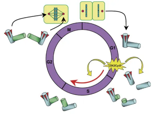
Like DNA, centrioles duplicate semi-conservatively exactly once per cell cycle and the temporal association with DNA replication is controlled by members of the CDK, Aurora, Polo-like, and NIMA families of cell cycle kinases. Initiation of DNA and centrosome duplication is coupled at least partly by G1-specific activation of cyclin-dependent kinase 2 (CDK2)-cyclin E, key factor for both DNA synthesis and initiator of centrosome duplication. Centrosome duplication begins with the physical splitting of the paired centrioles triggered by CDK2-cyclin E, followed by the formation of procentrioles near the proximal end of each pre-existing centriole. Unduplicated centrosomes initiate duplication after activation of CDK2-cyclinE, conversely centrosome just duplicated does not immediately reduplicate in the presence of active CDK2-cycklin E (Fukasawa, Nat Rev 2007). In detail, the centrosome cycle can be divided into several distinct steps: centrosome duplication (S-phase), maturation
and separation (G2-phase), segregation (M-phase) (Fig.2). Centriole duplication occurs during S-phase. The initiation of procentriole assembly takes place before or at the onset of S-phase, when Polo-like kinase 4 (PLK4) initiates centriole assembly and through its autoregulated instability limits centrosome duplication to once per cell cycle (Holland et al., Genes & Dev 2012). After that, procentriole elongation starts during late S-phase and the centriole reaches full-length during the following cell cycle (Azimadeh & Bornens, Cell Sci Rev 2007). After
centriole duplication in S-phase, centrosomes mature throughout G2-phase by assembling PCM around the duplicated centrioles (centrosome maturation); then, the two newly formed centrosomes separate and migrate to the opposite end of the cell (centrosome separation) to assemble the bipolar spindle during mitosis (Fukasawa, Nat Rev 2007). As cells exit mitosis, the two centrioles forming the single inherited centrosome looses their tight orthogonal position and disengage (centriole disengagement) becoming ready for a new duplication cycle.
Fig. 2 Centrosome duplication cycle. Late G1-specific activation of CDK2/cyclin E triggers initiation of both
DNA and centrosome duplication. Centrosome duplication begins with the physical separation of the paired centrioles and with the formation of procentrioles. During S and G2 phases, procentrioles elongate, and two
centrosomes progressively recruit PCM. In late G2, the daughter centriole of the parental pair acquires
appendages (shown as red wedges), and two identical centrosomes are generated. During mitosis, two duplicated centrosomes direct the formation of bipolar mitotic spindles, so that upon cytokinesis, each daughter cell receives one centrosome (Image modified by Fukasawa, Cancer Letters 2004).
Centrosome separation is thought to require the disassembly of a fibrous linker that mediates centrosome cohesion by connecting the centriole pairs. An important factor that acts as a docking site for this linker is C-Nap1, which is found at the proximal end of parental centrioles. C-Nap1 interacts with rootletin, a protein of the ciliary rootlet structure found in many ciliated cells that originates from the basal body and extends towards the nucleus, providing structural support for the cilium (Yang et al., J Cell Biol 2002; Yang et al., Mol Biol Cell, 2006). Rootletin is also found in cells without cilia, forming fibers that proceed from the proximal ends of centrioles. At late G2 to M-phase both proteins dissociate from the centrioles following phopshorylation by NIMA-related kinase (Nek2). Thus, during the cell cycle the phosphorylation of C-Nap1 and rootletin regulates centrosome cohesion that is dependent of the balance between Nek2 and protein phosphatase 1 (PP1) activities (Meraldi & Nigg, Cell Sci 2001).
Multipolarity with or without centrosome amplification
Centrosome duplication is a process heavily regulated by different regulatory proteins that interact with centrosomes, although it is still unclear whether they exert their action locally or globally. Those proteins belong to one of three functional groups: cell cycle regulation, DNA-damage response and/or repair, and nucleocytoplasmic transport. The proteins from each group control centrosome duplication using different mechanisms and pathways and their mutation or absence lead to centrosome defects that interfere with the formation of bipolar spindle. Centrosomes defects can lead to mitotic spindle multipolarity with or without centrosome amplification (Fig.3).
Multipolarity with centrosome amplification may result from centriole overduplication or cytokinesis failure and is characterized by supernumerary centrosome and chromosome instability. Typically, spindles from cells with centrosome amplification exhibit more than two poles, each with at least two centrioles. When cells form mitotic spindles with more than two poles, they fail to undergo cytokinesis, and become binucleated cells in which is triggered the checkpoint response mediated by p53. These cells can eventually undergo cell death probably after massive chromosome missegregation (Ganem et al., Nature 2009). When p53 is absent, these cells continue to cycle and can become very large multinucleated cells that undergo cell cycle arrest or death (Fukasawa et al., Science 1996). Tumor cells can escape these events by clustering amplified centrosomes into functional “pseudo bipolar” spindles, which structurally resemble the “true” bipolar spindles organized by two
centrosomes. In this way, cells with “pseudo bipolar” spindles can undergo normal cytokinesis without chromosomes segregation errors (Quintyne et al., Science 2005).
Fig. 3 Schematic summary of the main causes of spindle multipolarity (Image modified by Maiato and
Logarinho, Nat Rev 2014).
Multipolarity without centrosome amplification is characterized by loss of spindle pole integrity because of premature centriole disengagement, which normally leads to multipolar spindles with single centrioles at individual poles, and PCM fragmentation, which leads to an accumulation of acentriolar poles in addition to two normal poles. Centriole disengagement is important for the centriole division before duplication and for limiting duplication to one event per cell cycle (Brownlee & Rogers, Cell Mol Life Sci 2013). Centriole disengagement is dependent on the activity of separase, a protease that also controls the resolution of sister chromatin cohesion through cleavage of cohesin, and is inhibited by
the spindle assembly checkpoint (SAC), which normally blocks anaphase in the presence of unattached kinetochores (Maiato & Logarinho, Nat Cell Biol 2014). Thus, inefficient inhibition of separase during a mitotic delay or arrest might result in both unscheduled sister chromatid separation and premature centriole disengagement followed by formation of multipolar spindles. How mitotic delay leads to loss of spindle pole integrity remains unclear, but it might be related to the presence of misaligned chromosomes (or chromatids) and inability to satisfy the SAC, which prevents separation of the duplicated chromosomes until each chromosome is properly attached to the spindle apparatus. In addititon, loss of spindle pole integrity might be due to PCM fragmentation. Several centrosomal proteins, including PCM-1, centrin-2, and ninein aggregate at pericentriolar satellites and depletion of these pericentriolar satellite proteins results in PCM fragmentation, generating multipolar spindles (Maiato & Logarinho, Nat Cell Biol 2014). Therefore, a role for pericentriolar satellites during mitosis is emerging as part of the intricate structural and molecular network that ensures spindle pole integrity.
Role of p53 in centrosome duplication
As mentioned, CDK2-cyclin E plays a role in the initiation of centrosome duplication, so proteins that control the CDK2/cyclin E activity may be involved in the regulation of centrosome duplication, such as the p53 tumour suppressor.
p53 plays an integral role in the cell and it is normally present in the nucleus where it functions as a transcription factor involved in cell response to different types of stresses. In particular, p53 is the conductor of a well-orchestrated system of cellular damage detection and control. When damage is sensed, the activity of p53 aids in the decision between repair and induction of cell death (Lakin & Jackson, Oncogene 1999).
Several studies have demonstrated a p53 centrosomal localization during the cell cycle and a function of p53 in regulation of the centrosome duplication cycle independently of DNA damage response (Fukasawa et al., Science 1996; Tarapore and Fukasawa, Oncogene 2002).
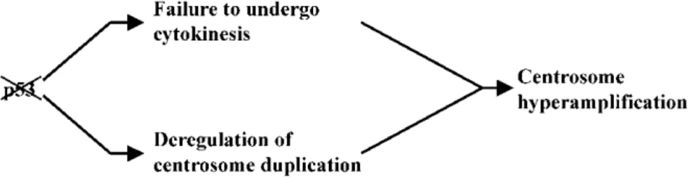
p53 and centrosome duplication in mouse cells
Cells and tissues from p53-deficient mice (p53-/-MEF) show a high frequency of centrosome amplification caused by a failure of cytokinesis process and by a deregulation of centrosome duplication (Fig.4) (Fukasawa et al., Science 1996; Tarapore & Fukasawa, Oncogene 2002).
Fig. 4 Loss of p53 induces centrosome hyperamplification in murine cells. Centrosome hyperamplification
can be induced by two possible mechanisms: cytokinesis failure (doubled genomes as well as centrosomes) and deregulation of centrosome duplication (centrosomes duplicate multiple times) (Image by Tarapore, Oncogene 2002).
In the latter case, p53 controls both initiation of centrosome duplication and suppression of re-duplication. Indeed, there are two major regulatory mechanisms in centrosome re-duplication. The first is to establish the correct timing of initiation of duplication, which ensures the coordination of initiation of centrosome and DNA duplication, and the second is to prevent re-duplication of centrosomes once they have duplicated. In p53+/+ MEFs, initiation of centrosome duplication is tightly coupled with initiation of DNA duplication. In contrast, in p53-/- MEFs, centrosomes initiate duplication early in G1 much before S-phase entry (Tarapore et al., Oncogene 2001a). Furthermore p53-/- MEFs treated with DNA synthesis inhibitors such as aphidicolin (Aph) or hydroxyurea (HU), show multiple copies of centrosomes, in contrast with the Aph-treated p53+/+ MEFs (Hu et al., 1999; Lee et al., 1997; Moro et al., 1995; Tarapore et al., 2001a). Indeed in Aph-treated p53+/+ MEFs no centrosome re-duplication was observed indicating that re-duplication of centrosomes depends on the p53 status (Tarapore & Fukasawa, Oncogene 2002).
This role of p53 in the centrosome regulation in mouse cells is in part due to its transcriptional function on different targets, including p21Waf1/Cip1 (p21), a potent CDK inhibitor (El-Deiry et al., Cell 1993; Harper et al., Cell 1993; Xiong et al., Genes & Dev
1993). p21 protein is expressed constitutively at a basal level throughout the cell cycle in a p53-dependent manner, which restrain untimely activation of CDK2/cyclin E (Tang et al., J Biol Chem 1998, Mussman et al., 2000; Minella et al., Curr Biol 2002), and thus, preventing untimely initiation of centrosome duplication. In the same way, when internal or external stresses temporarily halt DNA synthesis, such as Aph or HU, p53 is stabilized and p21 level increases. p21 upregulation leads to CDK2 inhibition with consequent inhibition of centrosome re-duplication (maintaining the coupling of centrosome duplication and DNA replication) until the stress is relieved (Kawamura et al., Canc Res 2004). In contrast, in cells lacking p53, CDK2 activation in unchecked, leading to centrosome re-duplication and amplification.
In mouse cells, several studies have shown that exogenous p53 localizes to the centrosome (Zajdel and Blair, Oncogene 1988; Brown et al., J Cell Physiol 1994; Moris et al., Exp Cell Res 2000; Tarapore et al., Oncogene 2001b) and that there is a role of the centrosomally localized p53 in the regulation of centrosome duplication. In particular, a centrosome binding-defective mutant of p53 (i.e., p53L13A/L17A mutant characterized by a missense mutation in the N-terminal NES) and a p53 transactivation-defective mutant (i.e., p53D278N) both suppressed centrosome re-duplication in p53-/- MEF, but much less efficiently than wild-type p53 (wt-p53). This observation suggested that both transactivation function and centrosome localization are required for p53 to fully regulate centrosome duplication (Shinmura et al., Oncogene 2007) (Fig. 5).
Fig. 5 p53 controls centrosome duplication in a transactivation-dependent and –independent manners. p53
controls both initiation of centrosome duplication and suppression of re-duplication in a transactivation-dependent manner via up-regulating p21, and in a transactivation-intransactivation-dependent manner potentially via direct physical association with centrosome (Image by Shinmura et al., Oncogene 2007).
p53 and centrosome duplication in human cells
In normal human cells (NHCs), such as human fibroblasts, silencing of endogenous p53 alone does not induce centrosome amplification. Centrosome amplification appears only when the short interfering RNA (siRNA)-mediated silencing of p53 is accompanied by DNA damage with irradiation (Fig.6) suggesting the presence of additional regulatory mechanisms in human cells that ensure the numerical integrity of centrosomes and genomic integrity (Kawamura et al., Cancer Sci 2006). However, examination of human cancer tissues and cultured cells has revealed a significant correlation between loss or mutational inactivation of p53 and the occurrence of centrosome amplification, supporting the idea that p53 mutation alone is not sufficient to induce centrosome amplification in human cells but rather requires additional events (Kawamura et al., Cancer Res 2004).
Fig. 6 Radiation-induced abnormal amplification of centrosomes in p53 short interfering RNA
(siRNA)-treated CCD32SK cells. siRNA-mediated silencing of p53 in NHCs together with DNA damage by irradiation
efficiently induced centrosome amplification. These phenomena were not observed with either siRNA-mediated silencing of p53 or irradiation alone. Centrosome number (= n.) n = 1; n = 2; n ≥ 3. IR, irradiation. (Image by Kawamura et al., Cancer Sci 2006).
p53 mitotic centrosomal localization in human cells
In human cells, we have previously demonstrated that p53 localizes at the centrosome in a specific moment of the cell cycle. In particular, we have shown that p53 moves to the centrosome at the beginning of mitosis (p53-MCL), after nuclear envelope breakdown and that this localization is MT-dependent because is disrupted by MT-depolymerizing agents, such as Nocodazole (Noc) (Ciciarello et al., Journal Cell Bio 2001; Tritarelli et al., Mol Biol Cell 2004) (Fig. 7A-B).
Fig. 7 p53 localizes at the centrosome in human cells during mitosis in a MT - and ATM-dependent manner. (A) p53 localizes at the centrosomes in metaphase (p53-MCL, p53 mitotic centrosomal localization)
but not in interphase in human cells. (B) p53-MCL is inhibited by Nocodazole (NOC) and KU-55933, drugs that affect MT polymerization and ATM activity, respectively.
Noc is a tubulin-depolymerizing agent that inhibits the mitotic spindle assembly and triggers the mitotic spindle checkpoint, which is followed by mitotic arrest (Ciciarello et al., Journal Cell Bio 2001). In addition, we have demonstrated that the protein kinase ATM, one of the major activator of p53, is involved in p53 mitotic centrosomal localization (p53-MCL) (Fig.7B), as shown by the requirement of p53 phosphorylation at Ser15 at the centrosome (Tritarelli et al., Mol Biol Cell 2004).
In general, ATM is activated after DNA damage by autophosphorylation and once activated, it can phosphorylate p53 and its principal negative regulator MDM2, thus preventing p53 cytoplasmatic relocalization and degradation (Shien et al., Cell 1997; Maya et al., Genes & Dev 2001; McGowan, Bioessays 2002). In particular, ATM can phosphorylate
p53-Ser15 facilitating other posttranslational modifications including further phosphorylation at other sides or acetylation.
We have investigated the subcellular localization of p53 in mitosis in the absence of a functional ATM in human lymphoblastoid cells derived from Ataxia-telangiectasia (A-T) patients, who have inactivating mutations in the ATM gene, comparing with human lymphoblastoid cells (AHH1) derived from healthy donor. We have shown that, in A-T cells, p53 fails to localize at the centrosome but it is distributed in discrete “spot”, while in AHH1 cells p53 localizes at centrosomes during mitosis. Furthermore, when ATM is inhibited in the normal AHH1 cells by their exposure to Caffeine, drug that inhibits ATM activity, p53 dissociates from centrosomes during mitosis demonstrating an important role of ATM in p53 MCL in human cells (Fig. 8).
Fig. 8 p53 does not localize at the centrosomes in mitosis in absence of a functional ATM. Immunostaining
of p53 and centrosomes (γ-tubulin) in mitotic lymphoblastoid cells derived from a normal donor (AHH1) or two AT patients (GM02782 and GM03189). In red, γ-tubulin; in green, p53; and in blue, DNA counterstained with DAPI. (A) AHH1 cells. (B) GM02782 (AT). (C) GM03189 (AT). (D) AHH1 cells treated with 5µM Caffeine. (E) GM03189 transfected with wild-type ATM. Bar, 10 µm (Image by Tritarelli et al., Mol Biol Cell 2004).
We also identified phosphorylation at Ser-15 as a functional requirement in this process because the nonphosphorylable p53-S15A mutant is not able to localize at the centrosome when transfected in p53-null K562 cells, in contrast with wt-p53, which is detected at the centrosome of mitotic cells after transfection (Fig.9) (Tritarelli et al., Mol Biol Cell 2004). In addition, we have shown that p53-Ser15 phosphorylation at the centrosome is a transient event because is not possible to detect a positive signal of anti-phospho-p53Ser15 antibody at the centrosome of mitotic cells without pre-treatment with Naf, a general phosphoseryl- and phosphothreonyl-phosphatase inhibitor (Tritarelli et al., Mol Biol Cell 2004).
Fig. 9 Immunostaining of p53 and centrosomes (γ-tubulin) in mitotic K562 cells. In red, γ−tubulin; in green,
p53; and in blue, DNA counterstained with DAPI. (A) K562 cells transfected with wt-p53. (B) K562 cells transfected with p53-S15A. Bar, 10 µm (Image by Tritarelli et al., Mol Biol Cell 2004).
p53-MCL diagnoses ataxia-telangiectasia homozygotes and
heterozygotes
Ataxia-telangiectasia is a rare autosomal recessive multisystemic syndrome caused by mutations in the ATM (ataxia-telangiectasia mutated) gene that result in a lack or inactivation of the ATM protein. A-T syndrome is characterized by several signs : progressive cerebellar degeneration, telangiectasias, immunodeficiency, recurrent infections, insulin-resistant diabetes, premature aging, radiosensitivity, and high risk for malignancy, particularly leukemia and lymphoma in children and epithelial cancers in surviving adults.
Until a short time ago there was no a test sufficiently sensitive and specific for early differential diagnosis, genetic counselling, and carrier prediction, but A-T diagnosis was
based on the combination of clinical features with laboratory tests showing high levels of serum alpha-fetoprotein, cell sensitivity to IR, and reduced or absent levels of ATM protein. We have been drawn a diagnostic test to unambiguously diagnose A-T homozygotes and heterozygotes, using the p53 centrosomal localization in human lymphoblastoid cells (Prodosmo et al., J Clin Invest 2013).
We observed that p53 does not localize at the centrosomes in almost 100% of mitotic lymphoblastoid cell lines (LCLs) or PBMCs derived from A-T patients. Surprisingly, we consistently observed that in A-T heterozygous carriers, p53 localizes at the centrosomes in approximately 50% of the mitotic cells. Based on these findings, we have developed this straightforward, rapid, and inexpensive test to determine mutant ATM zygosity, opening the possibility of performing large-scale screening in the general population for different clinical aspects, such as early diagnosis, genetic counseling, cancer predisposition, susceptibility to IR, and selection for specific targeted therapies. In addition, none of the LCLs derived from patients with genetic diseases causing ataxia (i.e., A-T–like disorder), or showing radiosensitivity (i.e., Nijmegen breakage syndrome, Werner syndrome, or Fanconi anemia group A) have reduced p53-MCL. Similar data were also obtained by analyzing ATR-defective and p53-mutated LCLs (Seckel syndrome and Li-Fraumeni syndrome) or other disorders (Cornelia de Lange syndrome and cylindromatosis). Although the definitive validation of this assay requires larger numbers, our data indicate that the p53-MCL test has a sensitivity and specificity not attained by other existing A-T functional assays.
AIM
Centrosome amplification can occur via deregulated centrosome duplication or cytokinesis failure and is thought to contribute to tumorigenesis by promoting chromosomal instability (CIN) or increasing tumor cells invasiveness (Ganem et al., Nat 2009; Godinho et al., Nat 2014). Indeed, supernumerary centrosomes are usually associated with multipolar spindles responsible of unfaithful segregation of chromosomes during cell division. However, multipolar spindle can be caused not only by centrosome amplification, but also by centriole disengagement and PCM fragmentation (Maiato & Logarinho, Nat Cell Biol 2014).
In normal murine cells, loss of p53 induces centrosome amplification as result of cytokinesis failure and deregulated centrosome duplication or suppression of reduplication. Furthermore, several studies have demonstrated a role of the centrosomally localized p53 in the regulation of centrosome duplication independently of its transactivation function (Tarapore & Fukasawa Oncogene 2002; Shinmura et al., Oncogene 2007; Fukasawa et al., Biochim Biophys Acta 2008). In contrast, in normal human cells, loss of p53 alone does not induce centrosome amplification or CIN (Bunz et al., Cancer Res 2002; Kawamura et al., Cancer Sci 2006) suggesting the existence of species-specific differences in centrosome-associated activity of p53. However, analysis of human cancer tissues and cultured cells has shown a strong correlation between loss or mutational inactivation of p53 and occurrence of centrosome amplification, indicating that p53 mutation alone is insufficient to induce centrosome amplification in human cells, and that other regulatory mechanisms are involved.
We have previously demonstrated that in human cells p53 moves to the centrosome in a particular moment of cell cycle, during mitosis (MCL), although the role of this p53-MCL is still unknown (Ciciarello et al., J Biol Chem 2001; Tritarelli et al., Mol Biol Cell 2004; Oricchio et al., Cell Cycle 2006). Thus, the main aim of this work is to investigate in human cells, compared to murine cells, the role of p53-MCL in the centrosome cycle, first of all, evaluating the differences in p53 centrosomal localization and activity in mice vs. humans. We hypothesize that the different effect of the loss of p53 reported in murine vs. human cells could be correlated with a different p53 centrosomal localization and activity. This will have clear implications for the understanding p53 specific role at the centrosome in human cells independent of DNA damage response.
RESULTS
Human p53-MCL is not cell specific
We have previously demonstrated by IF analysis that p53 localizes at the centrosome during mitosis (p53-MCL) in normal human lymphoblastoid cell lines (AHH1) (Ciciarello et al., 2001; Tritarelli et al., 2003). We asked whether this p53-MCL was cell type specific or not, and performed immunolabeling experiments on another cell type, the adherent normal human fibroblasts (HFs). We observed that in HFs, as well as in AHH1cells, p53 localizes at mitotic centrosomes, indicating that p53-MCL is not cell specific (Fig. 10A). To reinforce and to confirm this observation, we carried out biochemical centrosome isolation from HFs enriched in metaphase and interphase after synchronization by double thymidine block followed by mitotic shake off. To isolate centrosomes from cells, we used a classic protocol of centrosome purification (Blomberg-Wirschell M & Doxsey SJ Methods Enzymol 1998) modified to do not affect p53 centrosomal localization (see Materials and Methods section). Thus, we analysed centrosomes (CS-i) purified from interphase (I) and metaphase (M) enriched HFs by Western Blotting (WB) (Fig. 10B) and we observed p53 signal only in centrosome purified from metaphases, confirming p53-MCL in HFs.
Fig. 10 p53 centrosomal localization in normal human fibroblasts (HFs). (A) Representative IF analyses of
p53 centrosomal localization in human HFs. Cells were immunolabeled with anti-γ-tubulin (red) and anti-p53 (green) Abs; DNA is stained with DAPI (blue). (B) Biochemical analysis of p53 centrosomal localization in isolated centrosomes (CS-i) purified from interphase (I) and metaphase (M) enriched HFs. The anti-p53 Ab specificity was evaluated in total cell extract (TCE) of Cisplatin (CDDP)-treated cells.
In mouse cells p53 localizes at the centrosome during the
whole cell cycle in a microtubule- and ATM-independent
manner
Several studies have demonstrated a role of centrosomal p53 in the regulation of centrosome duplication. These studies were performed in p53-/- MEFs, after transfection of exogenous p53 (Tarapore 2001; Tarapore and Fukasawa 2002; Shinmura et al., 2007). These studies have shown that exogenous p53 localizes at duplicated and unduplicated centrosomes and that both transactivation function and centrosome binding are required to regulate centrosome duplication in p53-/- MEFs (Tarapore at al., 2001a; Shinmura et al., 2007). However, there is no direct evidence on endogenous p53 centrosomal localization in mouse cells. Thus, at the onset of this study, we have begun to characterize the centrosomal localization of the endogenous p53 in p53+/+ MEFs by IF analysis and by biochemical isolation of centrosomes from synchronized cells enriched in metaphase and interphase.
We first evaluated by IF the specificity of a series of anti-p53 antibodies (Abs) by testing their capability to recognize p53 activated by damage (Cispaltin, CDDP) and centrosomal p53 (Fig. 11). We observed that endogenous p53 activated by CDDP treatment is detected in a specific manner by all the anti-p53 Abs tested. In contrast, centrosomal p53 is specifically recognized only by the anti-phospho-p53Ser18 Ab (Cell Signal). Thus, we used this Ab to characterize by IF p53 centrosomal localization in p53 +/+ MEF.
We observed that in murine cells phosphorylated p53-Ser18 localizes at the centrosome only in interphase, also after treatment with Noc and KU55933, indicating that p53 centrosomal localization is microtubule- and ATM-independent (Fig. 12A), differently to human cells. Interestingly, we did not observed signal of phosphorylated p53-Ser18 at the centrosome during mitotis. However, we tested the possibility that non-phopshorylated p53-Ser18 localized also in metaphase, using another Ab anti-p53 (FL393) able to recognize p53 after overexpression in p53 -/- MEF. In this case, we observed p53 centrosomal localization both in interphase and in metaphase (Fig. 12B).
Fig. 12 p53 centrosomal localization in p53 +/+ murine hematopoietic cells (32D) and p53-/- murine embryo fibroblast (p53-/- MEF). (A) Representative IF analyses of p53 centrosomal localization in 32D cell
lines. Cells were immunolabeled with anti-γ-tubulin (green) and anti-p53 ser18P (red) Abs; DNA is stained with DAPI (blue). Mouse cells were treated with Nocodazole (NOC) and KU-55933, microtubules and ATM inhibitors, respectively. (B) Representative IF analyses of centrosomal localization of exogenous p53 wt (mp53wt) transfected, in p53-/- MEF, comparied to the empty vector (pCAG). Cells were immunolabeled with anti-γ-tubulin (red) and anti-p53 (green) Abs.
Thus, these data suggested us the possibility that in murine cells p53 was constitutively present at centrosome during the whole cell cycle. To verify this possibility, we performed experiments of biochemical isolation of centrosome from p53+/+ MEFs. We synchronized cells by double thymidine block and, 6 hrs after the second release, we collected by mitotic shake off the two cell populations, enriched in interphase and in metaphase, from which we isolated centrosomes. By WB analysis, we observed that in MEFs p53 localizes at centrosomes both in interphase and in metaphase, in contrast with the sole mitotic centrosomal localization of human p53 (Fig. 13).
Fig. 13 WB analysis of p53 centrosomal localization in MEFs. Biochemical analysis of p53 centrosomal
localization in isolated centrosomes (CS-i) purified from interphase (I) and metaphase (M) enriched MEFs p53+/+. The anti-p53 Ab specificity was evaluated in total cell extract (TCE) of Cisplatin (CDDP)-treated cells.
p53 centrosomal localization is species-specific
In order to understand if the different p53 centrosomal localization in human vs. murine cells depends on the cellular contest, we transfected human or murine p53 in human (SAOS) and mouse (MEF) p53-/- cells. By IF analysis, we observed that human p53 localizes at centrosomes in metaphase in human SAOS cells, but does not localize at the centrosome in murine MEFs. On the other hand, mouse p53 localizes at centrosome in interphase and metaphase in MEFs, but does not localize at centrosome in human SAOS cells (Fig. 14).
These data indicated that p53 centrosomal localization is species-specific and depends on the cellular context.
Fig. 14 Centrosomal localization of human and mouse p53 transfected in human (SAOS) and murine (MEF) p53-/- cells. Representative IF analyses of species-specific p53 centrosomal localization in human and
murine cells. Cells were immunolabeled with anti-γ-tubulin (red) and anti-p53 (green) Abs; DNA is stained with DAPI (blue).
Loss of p53 in MEFs induces centrosome amplification
Several studies described a role of p53 in the regulation of centrosome number through both its transactivation function and centrosome binding (Tarapore at al., 2001a; Shinmura et al., 2007). These studies have demonstrated that p53 -/- MEFs shown centrosome amplification due to both cytokinesis failure and deregulation in centrosome duplication (Tarapore and Fukasawa 2002). We confirmed these data in murine cells performing experiments of transient p53 interference in p53+/+ MEFs followed by IF analysis of the centrosome number and defects. We used Abs that recognize γ-tubulin (PCM component) and centrin (centriole component), to discriminate the centrosome defects after p53 depletion. We observed multipolarity with centrosome amplification in p53-depleted MEFs. In particular, 48-72 hrs
post transfection we observed an increase in centrosome number due to both cytokinesis defects (i.e., accumulation of bi- and multi-nucleated cells) and overduplicated centrosomes (turquoise insert, several γ-tubulin spots, each of them with two centrioles), compared to the ctrl cells which have a correct number of centrosomes, each of them with two centriole (orange insert) (Fig. 15).
Fig. 15 Centrosome structure and number were evaluated in p53- depleted MEFs. Centrosome and spindle
number and integrity were detected by double IF with the following Abs: γ-tubulin (green) with anti-centrin2 (red); DNA was stained with DAPI (blue). p53-depleted MEFs (n=300) show centrosome amplification (40±5%) (turquoise inserts) compared to their ctrl (10±4%). Scale bar is 1.
Human p53-MCL is highly stable and its depletion
increases γ-tubulin spots in metaphase
To understand if the different p53 centrosomal localization in murine vs. human cells is linked to different p53 biological functions, we decided to perform in HFs the same experiments of p53 interference and IF analysis described for MEFs. The role of p53 at the centrosome in human cells is not clear yet. Kawamura and his group observed that the only loss of p53 in normal human cells (NHCs) does not induce centrosome amplification as in mouse cells, but centrosome amplification occurs if loss of p53 is associated with DNA damage by irradiation (Kawamura et al., 2006). However, the specific role of the centrosomal p53 in centrosome duplication in human cells is still unknown.
Thus, we performed experiments of transient p53 interference in order to deplete p53 from centrosomes. We needed to apply a double pulse of p53 specific siRNAs 48h after the first interference to deplete enough p53, impair p53-MCL and to see centrosomal defects, which occur only in metaphase. Indeed we observed that one pulse of p53 specific siRNAs was sufficient to decrease p53 mRNA level (Fig. 16A), but not enough to deplete p53-MCL and to induce centrosome defects in metaphase (Fig. 16B-C), suggesting a high stability of the centrosomal p53 in NHCs.
Fig. 16 p53-MCL stability in HFs. (A) PCR analysis of p53 mRNA level after 48h from the first p53
interference. (B) Histogram of the percentage of metaphases without p53-MCL after 48hr from the first p53 interference with p53 specific siRNAs. One pulse of p53 siRNAs is not sufficient to deplete p53 from mitotic centrosomes. (C) Histogram of the percentage of metaphases with centrosome defects after p53 interference.
B"
When we succeeded in p53-MCL depletion after the second pulse of siRNAs (fig. 17A), we observed in metaphase an increase of γ-tubulin spots (28±7%), respect to the ctrl cells (4±2%), but no sign of cytokinesis failure or centrosome overduplication (Fig. 17B-C). Indeed, we did not find variation in the percentage of bi- or multi-nucleated cells (sign of cytokinesis failure) and in the percentage of interphases with amplified centrosomes (sign of overduplication) in depleted cells (Fig. 17D). These data suggested that in human cells inhibition of p53-MCL induces a metaphase specific effect characterized by an increment in γ-tubulin spots.
Fig. 17 Loss of p53-MCL in p53-depleted HFs induces a specific effect in metaphase. (A) Histogram of the
percentage of metaphase without p53-MCL after a second pulse of p53 specific siRNAs. (B-C-D) Representative IF (B) and histograms (C-D) of the effect of loss of p53-MCL in HFs. p53-depleted HFs after a second pulse of p53 specific siRNAs show an increase in γ-tubulin sposts in metaphase (C), but not in interphase (D). Centrosome number was detected by IF with the Ab anti-γ-tubulin (red) and DNA was stained with DAPI (blue). (*P-value < 0.05)
A"
C"
Loss of p53 in HFs induces PCM fragmentation, centriole
disengagement, and loss of spindle integrity
Multipolarity can be due to centrosome amplification (i.e., cytokinesis failure and centrosome overduplication) or not (i.e., PCM fragmentation and centrioles disengagment). We observed multipolarity with centrosome amplification in MEFs after loss of p53, as previously reported (REF). Thus, we decided to investigate in detail the centrosome defects seen in p53-depleted HFs by IF analysis using anti-γ-tubulin Ab, to detect PCM, anti-centrin-2 Ab, to detect centrioles, and anti-β-tubulin, to detect spindle poles. IF analysis showed the presence of multipolar spindles whose poles contain centrosomes in different conditions: normal centrosomes (orange insert, two centrin spots per single γ-tubulin spot), disengaged centrosomes (yellow insert, one centrin spot per single γ-tubulin), and fragments of PCM (fuchsia insert, γ-tubulin spots without centrin spots) (Fig. 18). Altogether, these results indicated that p53 depletion in HFs induces loss of spindle integrity rather than centrosome amplification and that a species-specific difference exists in the p53-centrosomal activity in mice and humans.
Fig. 18 Centrosome structure and spindles were evaluated in p53-depleted HFs. Centrosome and spindle
number and integrity were detected by double IF with the following Abs: γ-tubulin (green) with anti-centrin2 (red), and anti-γ-tubulin (green) with anti-β-tubulin (red); DNA was stained with DAPI (blue). p53-depleated HFs (n=500) show multipolar spindle (β-tubulin), disengaged centrosomes (yellow insert) and PCM fragmentation (fuchsia insert) (28±7%) compared to their ctrl (4±2%). Scale bar is 10 µm.
Loss of p53 in HFs induces mitotic catastrophe not mainly
due to mitotic delay
A further characterization of the HF fate was obtained by performing time-lapse experiments (Fig. 19A). In particular, relative to control siRNA transfected cells, p53-depleted HFs showed the presence of mitotic catastrophe in 30% of the analyzed mitosis (n=90) (Fig. 19B). Evaluation of the timing from round up to anaphase or the beginning of mitotic catastrophe (Fig. 19C) showed that approximately 50% of the p53-depleted cells underwent mitotic catastrophe without delay in the prometaphase to anaphase transition. These results suggest that mitotic catastrophe is not mainly due to mitotic delay, an event that induces loss of mitotic spindle integrity by cohesion fatigue.
Fig. 19 p53 depletion is associated with mitotic catastrophe in HFs. Ctrl and p53i HFs are employed as total
population. (A) Stills from time-lapse recording of ctrl and p53i HFs are shown. Ctrl cells divide and return mononucleated in about 3hrs while p53i cells undergo mitotic catastrophe. (B) The histogram show the percentages of cells undergoing mitotic catastrophe; ctrl cells (n=110), p53i cells (n=90). (C) Box-plot graph of the timing from round up to anaphase or the beginning of mitotic catastrophe. The median of timing is 42 minutes in the ctrl and 57 minutes in the p53i cells. Scale bar is 10 µm. (***P-value < 0.001)
C"
B"
A"
Loss of p53 in U2OS cells induces centrosome
amplification and accumulation of bi- and multi-nucleated
cells
We continued our evaluation of p53-MCL activity in human cells. Of relevance, when we preformed the same experiments described above in wt-p53-carrying U2OS tumor cells, we found the depletion of p53-MCL after only one pulse of p53 specific siRNAs (Fig. 20A-B) and the presence of centrosome overduplication and centriole disengagement defects (42±3%) but no PMC fragmentation (Fig. 20C).
Fig. 20 Loss of p53 in p53-depleted U2OS induces centrosome overduplication and centriole disengagement. (A) PCR analysis of p53 mRNA level after 48h from the firts p53 interference. (B) Histogram
of the percentage of metaphase without p53-MCL after one pulse of siRNAs in p53-depleted U2OS. (C) Histogram (left panel) and representative IF (right panel) of the effect of p53 loss in p53-depleted U2OS in centrosome number and integrity. Centrosome defects were detected by double IF with the following Abs: anti-γ-tubulin (green) with anti-centrin2 (red); DNA was stained with DAPI (blue). p53-depleated U2OS cells (n=100) show centrosome amplification (turquoise inserts) and disengaged centrosomes (yellow insert) (42±3%), compared to their ctrl (5±1.2%). (*P-value <0.05; ***P-value < 0.001)
B"
A"
In addition, while normal HFs underwent mitotic catastrophe, U2OS cells failed cytokinesis and accumulated bi-nucleated cells (24±1.4%) and multi-nucleated cells (43±4%) in p53-depleted cells vs. <1% in control cells, 48hrs and 72hrs after transfection respectively, without significant cell death (Fig. 21A-B).
Fig. 21 p53 depletion is associated with accumulation of bi- and multi-nucleated cells in U2OS cells. (A)
Histogram and IF of the percentage of bi-nucleated cells 48hrs after transfection. (B) Histogram and IF of the percentage of multi-nucleated cells 72hrs after transfection. Centrosome number was detected by IF with the anti-γ-tubulin (green) and DNA was stained with DAPI (blue). p53-depleted U2OS cells (n=100) show accumulation of bi- (24±1.4%) and multinucleated cells (43±4%) compared to their ctrl (<1 %). (***P-value <
0.001)
These observations indicate that tumor human cells react in a different manner respect to normal human cells to the loss of the p53 centrosomal control, suggesting the existence of a stringent centrosome-associated p53 functions in human cells that might be lose in tumorigenesis.
DISCUSSION
Different p53 centrosomal behaviour in normal murine vs.
human cells vs. cancer cells
In the first part of our study, we reveal a different p53 centrosomal localization and function in murine vs. human cells, underlying the impossibility to use mouse model to understand the role of centrosomal p53 in the prevention of structural and numerical centrosome alteration involved in human tumorigenesis. Indeed, we know that dysfunction of the centrosome cycle or cytokinesis failure can generate supernumerary centrosomes and abnormal mitotic spindles, which are thought to contribute to tumorigenesis by promoting chromosomal instability (CIN) (Ganem et al., Nat 2009)or increasing tumor cells invasiveness (Godinho et al., Nat 2014).
The p53 tumor suppressor has been extensively implicated in the prevention of structural and numerical centrosome alterations. In stressing conditions, such as inhibition of centrosome protein assembly, loss of centrosome integrity, or failure of centriole duplication, p53 is activated by the p38 stress kinase and induces a p21-dependent G1-arrest (Mikule et al., Nat Cell Biol 2007). During the cell cycle, p53 controls centrosome number ensuring the coordinated initiation of centrosome and DNA replication through p21-mediated regulation of CDK2/cyclin E/A (Meraldi et al., Nat cell Biol 1999) or the inhibition of cell cycle progression upon cytokinesis failure (Nigg, Nat Rev Cancer 2002).
More recently, p53 was shown to act antagonistically with Cyclin B2 on the Aurora-A-mediated regulation of centrosome separation (Nam & van Deursen, Nat Cell Biol 2014). Most of these functions are independent of p53 centrosomal localization and require p53 transcriptional activity. However, centrosome localization-dependent and transcription-independent activities of p53 have also been described in the control of centrosome duplication and suppression of reduplication. However, these observations have been extensively made in murine cells (Fukasawa, Biochm Biophys Acta 2008; Tarapore et al., Oncogene 2002) but they could not be reproduced in NHCs, where silencing of endogenous p53 alone does not induce centrosome amplification or CIN, suggesting the existence of species-specific differences in centrosome-associated activity of p53 (Bunz et al., Cancer Res 2002; Kawamura et al., Cancer Sci 2006).
Consistently with these observations, we show a large body of evidence showing a significantly divergent behavior between human and murine p53-centrosomal localization and activities. We have previously demonstrated that in unperturbed proliferating human cells, p53 does not permanently reside at the centrosome but, at each mitosis, it transiently moves to centrosomes (p53-MCL). We showed that p53-MCL is independent of DNA damage but requires microtubule-dynamics and ATM-mediated p53Ser15 phosphorylation on discrete cytoplasmic p53 foci that move to centrosomes during mitotic spindle formation, where p53 is suddenly dephosphorylated (Ciciarello et al., J Biol Chem 2001; Tritarelli et al., Mol Biol Cell 2004; Oricchio et al., Cell Cycle 2006). This p53 behavior is so consistent as to allow us to develop a functional test for mutant ATM zygosity based on the presence or absence of p53-MCL in cell cycle-reactivated peripheral blood mononuclear cells (PBMCs) (Prodosmo et al., J Clin Inv 2013).
Here, we try to elucidate the functional role of p53-MCL, carrying out a comparative study of the p53 centrosomal localization and function between mouse and humans.
First, we observed a different p53 centrosomal localization between mouse and human cells. At variance with human p53, we show that mouse p53 is constitutively present at the centrosome throughout the cell cycle and its centrosomal localization is independent of both microtubules and ATM. Furthermore, we found as other difference respect to human p53-MCL, that in mouse cells p53 is phophorylated at Ser18 only when it localizes at centrosome during interphase, while it is not phosphorylated at centrosome in metaphase.
Then, we observed a different p53 centrosomal function in human cells comparing to mouse cells. We show that in agreement with its different centrosomal localization, human p53 and mouse p53 have different centrosomal functions. Mouse p53, which localizes at centrosome during the whole cell cycle, is involved in regulation of centrosome duplication and reduplication, and its loss results in centrosome amplification, as demonstrated from other several studies. In contrast, human p53 does not regulate centrosome duplication, reduplication, or separation through its centrosomal localization, and its loss does not result in centrosome amplification. Indeed, we show that suppression of human p53-MCL causes centrosome fragmentation in metaphase, formation of multipolar spindles without centrosome amplification, and mitotic catastrophe, supporting a human-specific role for p53 in the control of spindle pole integrity during mitosis.
In the final part of our work, we show that we could observe these last events only in NHCs and not in cancer cells. In human tumor cells, loss of p53 does not result in centrosome fragmentation and mitotic catastrophe, as in NHCs. In contrast, suppression of human p53-MCL in tumor cells causes centrosome overduplication and accumulation of cytokinesis defects, highlighting the existence of tumor-related differences in centrosome-associated p53 activities in human cells.
An additional piece of evidence supporting the existence of such centrosome/p53-associated differences is the recent discovery of a new NHC-specific “centrosome-loss sensor” (Wong et al., Science 2015). The authors developed a potent and reversible inhibitor of PLK4, the centrinone (LCR-263) that suppresses centriole assembly and causes centrosome depletion in human and other vertebrate cells. As suggested by previous studies, centrosome depletion induces growth arrest in neither human cancer nor normal mouse cells, showing that centrosomes are dispensable for the proliferation of these cells, as demonstrated in
Drosophila cells (Basto el al., Cell 2006). In contrast, in NHCs, centrinone-induced
centrosome depletion results in an irreversible, senescence-like G1-arrest, indicating that centrosomes are critical for the proliferation of NHCs. Of relevance, a molecular characterization of this cell cycle arrest showed that it is dependent on p53, although through a still unknown mechanism. Indeed, none of the well-characterized activating conditions of p53, i.e., DNA damage, p38-mediated stress signaling, Hippo signaling, extended mitotic duration, or segregation errors, has been found to contribute to this irreversible G1-arrest.
Altogether, these results indicate that NHC are different from both normal mouse cells and tumor cells from either species in their p53-mediated controls of centrosome number and activity. Mouse models of Trp53 inactivation/mutation recapitulate many aspects of the oncosuppressing activities of the human TP53 gene. However, homozygous deletion of the
Trp53 gene is compatible with mouse development and viability into adulthood, while in
humans only a single TP53 allele can be mutated/inactivated in the germline (i.e., Li-Fraumeni syndrome). In contrast, the complete absence of p53 often characterizes human tumor cells. The human higher sensitivity to p53 loss indicates the existence of a human-specific, still unknown p53-related characteristic, which is lost during tumor transformation. We believe that the two human-specific, centrosome-associated p53 functions, the p53-MCL-mediated control of spindle pole integrity and the p53-dependent sensor of centrosome loss might fulfill these characteristics. We hypothesize that in NHCs, p53 senses centrosome number and integrity at each mitosis through its direct relocalization at centrosomes in
metaphase. Loss of this sensor should specifically contribute to human tumorigenic transformation, while mouse cells do not possess this type of control.
MATERIALS AND METHODS
The experiments have been repeated from three to five times and the results obtained are presented as means ± SD.
Cells, Culture Conditions
HF (Human fibroblast immortalized with hTERT); U2OS (Human osteosarcoma cell line) p53+/+ MEF (Murine Embryonic fibroblasts p53 wild-type); p53-/- MEF (Murine Embryonic fibroblasts p53 null) were cultured in DMEM with 10% heat-inactivated foetal bovine serum (FBS, Invitrogen) and maintained in a humid incubator at 37°C in a 5% CO2 environment.
For live cell imaging, HF cells were cultured in DMEM medium without phenol red, supplemented with 10% heat-inactivated FBS (Invitrogen).
Cell synchronization
Cells (e.g., HF; MEF) at ∼30% confluency were washed with buffer (PBS) growth medium, and cells were grown in the presence of 2 mM thymidine for 18 h. After the first block thymidine was removed, cells were washed and grown in fresh medium for 9 h to release cells from the block. The second block follows the release by the addition of 2 mM thymidine and cultivation for 17 h. As a result of this synchronization cells progressed synchronously through G2 and mitotic phase and arrested at the beginning of S phase. After release from the thymidine block, >95% of the cells entered S phase (in 0-4 h), progressed into G2 phase (5-6 h) and underwent a synchronous mitosis at 7–8 h.
Centrosome Isolation
Exponentially growing cells were subjected to Cytochalasin B (1µg/ml) and Nocodazole (3µg/ml) treatment for 10 min in ice to disrupt microtubules and actin cytoskeleton and then lysed in a solution of 1mM Tris-HCl (pH 8.0), 0.1% 2-mercaptoethanol, 0.5% Triton X-100. Lysates were poured through a funnel lined with 70-µm nytex membrane (Sigma) and underlined with 1,5 ml 20% Ficoll MW 400,000 or Lympholyte (Cederlane) in PE solution [10mM PIPES, 1mM EDTA, 0,1% 2-mercapoethanol, pH 7.2 with KOH] with 0,1% Triton-X100. Tubes were spun at 25,000g for 20 min at 4 °C and the lysate was aspirated to within 2 ml above the Ficoll cushion. Centrosomes were layered on top of the cushion, collected using a Pasteur pipette and placed in ice until their analysis by Western Blotting.
Western Blotting
For Western Blotting, TCEs and centrosomes purified were prepared in RIPA buffer [50mM Tris-HCl (pH 8), 600mM NaCl, 0.5% sodium deoxycholate, 0.1% SDS, 1% NP40 and 1mM EDTA] supplemented with protease-inhibitor mix (Roche) and Hal Phosphatase Inhibitor Cocktail (Thermo Scientific). Proteins were resolved by SDS-PAGE using NuPAGE® Novex Bis-Tris Gels (Invitrogen), transferred onto nitrocellulose membranes (Bio-Rad), and analyzed with the indicated antibodies (Abs). The following Abs were employed: anti-γ-tubulin mouse monoclonal and rabbit polyclonal Abs (Sigma), anti-p53 rabbit polyclonal Ab (FL393 Santa Cruz Biotech), anti-actin mouse monoclonal Ab (Sigma), anti-GAPDH mouse monoclonal Ab (Santa Cruz Biotech), HRP-conjugated goat anti-mouse and anti-rabbit Abs (Cappel). Immunoreactivity was determined using the ECL-chemioluminescence reaction (Amersham 49 Corp).
Transfection, RNA Interference and PCR
Transfections were carried out by Lipofectamine LTX and PLUS reagent (Life Technologies) following the manufacturer’s instructions using plasmids containing mouse and human p53 wild type or the mutant p53. The amount of plasmid DNA in each sample was equalized by supplementing with empty plasmid.
RNA interference was obtained by using commercially available p53-specific stealths, mix of 3 RNAi sequences (Invitrogene), and by universal negative control stealth RNAi Negative Medium GC Duplexes (Invitrogen). Cells were transduced with 30 nM of siRNA
using RNAiMAX reagent (Invitrogen) according to the manufacturer’s instructions. RNA was isolated 24-48-72 after siRNA transfection by using the RNeasy mini kit (Qiagen S.P.A., Milano, Italy). Total RNA (2 µg) was reverse transcribed using MuLV reverse transcriptase and the reverse transcribed material was used in PCR reactions with the GoTaq® DNA polymerase (Promega).
The following primers were used to amplify:
Human p53 FW 5’-TGACACGCTTCCCTGGATTG-3’ REV 5’-GCTGCCCTGGTAGGTTTTCT-3’ Human GAPDH FW 5’-TCCCTGAGCTGAACGGGAG -3’ REV 5’-GGAGGAGTGGGTGTCGCTGT -3’ Immunofluorescence Microscopy
For immunofluorescence experiments, cells were seeded onto poly-L-lysine coated coverslips. Cells were pre-permeabilized in 0,25% Triton X-100 in PBS for 2 min., fixed 2% formaldehyde, washed in phosphate buffered saline (PBS), permeabilized in 0.25% Triton X- 100 in PBS for 5 min., fixed and permeabilized in ice-cold Methanol at -20°C for 10 min and then blocked in 0.2% Triton X-100, 3% bovine serum albumin (BSA) in PBS for 30 min before the required primary Abs were applied. The following Abs were employed: anti-p53 Abs (DO.7, Thermofisher; 1801, DO.1 Santa Cruz Biotech; Ab1-4-5-7 Millipore), anti-phospho-p53 (Ser18) Ab (Cell Signal), anti-γ-tubulin Ab (Sigma), anti-centrin2 Ab (Santa Cruz Biotech), TRICT-conjugated anti-β-tubulin Ab (AlexaFluor).
Appropriate secondary FITC- or TRITC-conjugated Abs (Alexa-fluor, Invitrogene) were used. DNA was marked with HOECHST 33342 (Sigma). Cells were examined under an Olympus BX53 microscope equipped with epifluorescence and photographs were taken (100 objective) using a cooled camera device (ProgRes MF).
Live Cell Imaging
Cells seeded in 35mm dishes (81153, ibiTreat, Ibidi) and transfected as described above were observed under an Eclipse Ti inverted microscope (Nikon) using a Plan Apo 40X objective (Nikon). During the whole observation, cells were kept in a microscope stage incubator (Basic WJ, Okolab) at 37°C and 5% CO2. DIC images were acquired every 6 minutes over a 48hrs period by using a DS-QiMc camera and the NIS-Elements AR 3.22 software (Nikon). Image and video processing were performed with NIS-elements AR 3.22.
BIBLIOGRAFIA
Azimzadeh J & Bornens M. Structure and duplication of the centrosome. Journal of Cell Science 2007 120: 2139-2142.
Basto R, Lau J, Vinogradova T, Gardiol A, et al. Flies without centrioles. Cell 2006; 125:1375-86.
Blomberg-Wirschell M. & Doxsey SJ. Rapid isolation of centrosomes. Methods Enzymol.1998
Brinkley, B. & Goepfert, T. Supernumerary centrosomes and cancer: Boveri’s hypothesis
resurrected. Cell Motil. Cytoskeleton 1998; 41: 281-288.
Brown CR, Doxsey SJ, White E, Welch WJ. Both viral (adenovirus E1B) and cellular (hsp70,
p53) components interact with centrosomes. J Cell Physiol 1994; 160: 47-60.
Brownlee CW & Rogers GC. Show me your license, please: deregulation of centriole
duplication mechanisms that promote amplification. Cell. Mol. Life Sci. 2013; 70, 1021–
1034.
Bunz F, Fauth C, Speicher MR, Dutriaux A, et al. Targeted inactivation of p53 in human cells
does not result in aneuploidy. Cancer Res 2002; 62:1129-33.
Chrétien D, Buendia B, Fuller SD, Karsenti E. Reconstruction of the centrosome cycle from
cryoelectron micrographs. J Struct Biol. 1997; 120(2): 117-33.
Ciciarello M, Mangiacasale R, Casenghi M, Limongi MZ, et al. p53 displacement from
centrosomes and p53-mediated G1 arrest following transient inhibition of the mitotic spindle. J Biol Chem. 2001; 276:19205-13.
Conduit PT, Wainman A, Raff JW. Centrosome function and assembly in animal cells. Nat Rev Mol Cell Biol 2015; 16: 611-24.
Doxsey S. Re-evaluating centrosome function. Nat Rev Mol Cell Biol. 2001; 2(9): 688-98.
El-Deiry WS, Tokino T, Velculescu VE, Levy DB, Parsons R, Trent JM et al. WAF-1, a
Fukasawa K, Choi T, Kuriyama R, Rulong S, Vande Woude GF. Abnormal centrosome
amplification in the absence of p53. Science 1996; 271(5256): 1744-7.
Fukasawa K. Oncogenes and tumour suppressors take on centrosomes. Nat Rev Cancer. 2007; 7(12): 911-24.
Fukasawa K. p53, cyclin-dependent kinase and abnormal amplification of centrosomes. Biochim Biophys Acta 2008; 1786:15-23.
Ganem NJ, Godinho SA, Pellman D. A mechanism linking extra centrosomes to chromosomal
instability. Nature 2009; 460: 278-82.
Godinho SA, Picone R, Burute M, Dagher R, et al. Oncogene-like induction of cellular
invasion from centrosome amplification. Nature 2014; 510:1667-71.
Gonczy P. Towards a molecular architecture of centriole assembly. Nat Rev Mol Biol Cell 2012; 13:425-35.
Harper JW, Adami GR, Wei N, Keyomarsi K, Elledge SJ. The p21 Cdk-interacting protein
Cip1 is a potent inhibitor of G1 cyclin-dependent kinases. Cell 1993; 75: 805–816.
Holland AJ, Fachinetti D, Zhu Q, Bauer M, et al. The autoregulated instability of polo-like
kinase 4 limits centrosome duplication to once per cell cycle. Genes & Dev 2012; 26:
2684-9.
Howard J & Hyman AA. Dynamics and mechanics of the microtubule plus end. Nature 2003; 422: 753-758.
Kawamura K, Izumi H, Ma Z, Ikeda R, Moriyama M, Tanaka T et al. Induction of centrosome
amplification and chromosome instability in human bladder cancer cells by p53 mutation and cyclin E overexpression. Cancer Res 2004; 64: 4800-4809.
Kawamura K, Morita N, Domiki C, Fujikawa-Yamamoto K et al. Induction of centrosome
amplification in p53 siRNA-treated human fibroblast cells by radiation exposure. Cancer
Sci 2006; 97: 252-8.
Lakin ND, Jackson SP. Regulation of p53 in response to DNA damage. Oncogene. 1999 Dec 13;18(53):7644-55.
Lambrus BG, Uetake Y, Clutario KM, Daggubati V, et al. p53 protects against genome
instability following centriole duplication failure. J Cell Biol 2015; 210: 63-77.
Lingle WL, Barrett SL, Negron VC, D'Assoro AB, et al. Centrosome amplification drives
chromosomal instability in breast tumor development. Proc Natl Acad Sci U S A 2002;
99:1978-83.
Ma Z, Izumi H, Kanai M, Kabuyama Y, et al. Mortalin controls centrosome duplication via
modulating centrosomal localization of p53. Oncogene. 2006; 25:5377-90.
Maiato H, Logarinho E. Mitotic spindle multipolarity without centrosome amplification. Nat Cell Biol. 2014; 16:386-94.
Maya, R., Balass M, Kim ST, Shkedy D, Leal JF, Shifman O, Moas M, Buschmann T, Ronai Z, Shiloh Y, et al. ATM-dependent phosphorylation of Mdm2 on serine 395, role in p53
activation by DNA damage. Genes & Dev. 2001; 15, 1067-1077.
McGowan, C.H. Checking in on Cds1 (Chk2): a checkpoint kinase and tumor suppressor. Bioessays 2002; 24: 502-51.
Meraldi P, Lukas J, Fry AM, Bartek J & Nigg EA. Centrosome duplication in mammalian
somatic cells requires E2F and Cdk2-CyclinA. Nat Cell Biol 1999; 88-93.
Meraldi, P., and Nigg, E. A. Centrosome cohesion is regulated by a balance of kinase and
phosphatase activities. J. Cell Sci. 2001; 114: 3749-3757.
Mikule, Delaval B, Kaldis P, Jurcyzk A, et al. Loss of centrosome integrity induces
p38-p53-p21-dependent G1-S arrest. Nat Cell Biol 2007; 9:160-70.
Minella AC, Swanger J, Bryant E, Welcker M, Hwang H, Clurman BE. p53 and p21 form an
inducible barrier that protects cells against cyclin E–cdk2 deregulation. Curr Biol 2002;
12: 1817-1827.
Morris VB, Brammall J, Noble J, Reddel R. p53 localizes to the centrosomes and spindles of
mitotic cells in the embryonic chick epiblast, human cell lines, and human primary culture: an immunofluorescence study. Exp Cell Res 2000; 256: 122-130.