DOTTORATO DI RICERCA IN SCIENZE CHIMICHE
Ciclo XXVI
Settore Concorsuale di Afferenza: 03/A1 Settore Scientifico Disciplinare: CHIM/01
DEVELOPMENT AND APPLICATIONS OF
HOLLOW FIBER FLOW FIELD-FLOW
FRACTIONATION
IN THE BIOANALYTICAL FIELD.
STUDIES OF AGGREGATION PHENOMENA IN
COMPLEX PROTEIN SAMPLES
Presentata da: Dott.ssa Otilia Maya Tănase
Coordinatore dottorato:
Chia.mo Prof. Aldo Roda
Relatore:
Chia.mo Prof. Pierluigi Reschiglian
Dottoranda: Otilia Maya Tănase Tutor: prof. Pierluigi Reschiglian
Curriculum, indirizzo: Scienze Chimiche, CHIM/01(03/A1)
Titolo tesi: Development and applications of hollow fiber flow field-flow fractionation in the bioanalytical field. Studies of aggregation phenomena in complex protein samples.
Il lavoro della Dott.ssa Otilia Maya Tănase nel triennio di frequenza del Dottorato in Scienze Chimiche si è rivolto allo sviluppo di metodi di frazionamento in campo-flusso (FFF) in particolare basati sull’impiego e sviluppo della versione miniaturizzata del canale separativo FFF chiamata hollow fiber FFF (HF5), in accoppiamento a tecniche di caratterizzazione spettroscopica e ottica, come metodi analitici innovativi per lo studio di campioni proteici complessi e fenomeni di aggregazione. Questo studio trova applicazioni in diversi campi che spaziano dal campo delle proteine terapeutiche, allo studio dei processi di aging alla base dello sviluppo di malattie neurodegenative.
Nel corso dei primi due anni di dottorato, il lavoro di ricerca è stato rivolto allo sviluppo di metodi HF5 accoppiati a tecniche di rivelazione molto sensibili (spettroscopia UV/Vis, fluorescenza e multi-angle light scattering – MALS) per la caratterizzazione dimensionale e morfologica di aggregati proteici spesso presenti nei biofarmaci (anticorpi e/o proteine utilizzate come veicolanti per biofarmaci) e formati durante il processo di sviluppo della loro formulazione. Tali metodi sono risultati caratterizzati da elevata sensibilità, selettività e riproducibilità dimostrandosi particolarmente adatti ad un controllo di qualità (QC) di campioni proteici ad elevato perso molecolare. I risultati ottenuti hanno dimostrato anche la superiorità dell’HF5 rispetto alla cromatografia ad esclusione dimensionale (SEC) – il punto di riferimento nelle pratiche di controllo qualità per proteine terapeutiche. La HF5 accoppiata a tecniche di rivelazione sensibili offre una notevole selettività rispetto alla SEC potendo separare analiti in un ampio intervallo di pesi molecolari, pertanto permette un’accurata valutazione della composizione delle formulazioni proteiche; spesso in SEC lo spazio cromatografico disponibile per la separazione di specie ad alto peso molecolare è molto limitato, causando la co-eluizione di specie con pesi molecolari simili.
Nel terzo anno del Dottorato, la Dott.ssa Otilia Maya Tănase ha trascorso un periodio di ricerca all’estero presso il Laboratorio di Patologia, Immunologia e Microbiologia (Albert Einstein College of Medicine, Yeshiva University, NY, Stati Uniti) sotto la supervisione della prof. Laura Santambrogio. Durante tale periodo si è occupata dell’inserimento della tecnologia HF5 nei laboratori e procedure biochimiche utilizzate per lo studio dei fenomeni di aggregazione proteica attinenti al processo di aging (immunosenescenza). Ha quindi sviluppato un metodo Hf5-MALS per la caratterizzazione dimensionale e conformazionale di questi sistemi proteici complessi. L’HF5 accoppiata al MALS è stata applicata come prima dimensione separativa per lisati cellulari interi, di origine diversa e a diversi stadi dell’aging, pertanto contenenti diversi livelli e tipologie (covalenti e non-covalenti) di aggregati proteici. Inoltre, ha avuto l'opportunità di apprendere conoscenze su tecniche complementari per la caratterizzazione di tali aggregati (SDS-PAGE, Native PAGE e FPLC). I risultati hanno confermato la capacità dell’HF5 accoppiata al MALS per colmare il “gap” di tecniche adatte per la caratterizzazzione di aggregegati proteici sub micrometrici.
Il lavoro di ricerca all’estero è stato concluso con un manuscritto in fase avanzata di preparazione, che verrà inviato alla rivista Nature Methods.
Il progetto di dottorato ha permesso Dott.ssa Otilia Maya Tănase di approfondire le ricerche nel campo della chimica analitica/tecniche separative, iniziate con il lavoro di tesi e proseguite nel periodo successivo come borsista.. Durante il periodo di tesi la dottoranda ha dimostrato serietà nel lavoro di laboratorio e capacità di lavoro autonomo. La dottoranda ha trascorso un periodo di formazione presso Wyatt Technology Europe (Germania), azienda con la quale il gruppo di ricerca in Chimica Analitica ha una collaborazione stretta. La collaborazione ha avuto come frutto l’implementazione del prototipo miniaturizzato HF5 nel sistema separativo Eclipse® DUALTECTM prodotto dall’azienda tedesca, il primo sistema FFF commerciale del suo genere – sistema che ha
costituito la strumentazione fondamentale nel lavoro di ricerca della dottoranda.
Nell’ambito del suo progetto di ricerca, la Dott.ssa Otilia Maya Tănase ha mostrato un’elevata capacità di adattamento a diversi ambienti culturali e diverse strutture organizzative, ha contribuito a mettere le basi e mantenere collaborazioni internazionali e ha messo alla prova le sue capacità di problem-solving mediante la proposta e sviluppo di soluzioni innovative. Le sue doti naturali verso l’apprendimento delle lingue straniere, l’orientamento verso l’organizzazione, l’attenzione ai dettagli, la pazienza e la precisione, le hanno permesso di lavorare con costanza ed in modo efficace sul raggiungimento degli obiettivi a lungo termine, mostrandosi responsabile per il proprio lavoro e per le persone di cui ha coordinato il lavoro in laboratorio.
Ha inoltre svolto attività di tutorato nel laboratorio di Chimica Analitica presso il CdL in Scienze Farmaceutiche Applicate (Imola) dimostrando buone capacità organizzative e didattiche. Inoltre, la Dott. ssa Otilia Maya Tănase si è mostrata disponibile e responsabile ad organizzare, coordinare e seguire il lavoro sperimentale di laureandi nel laboratorio di Chimica Analitica (Scienze della separazione) del Dipartimento "G. Ciamician".
L’attività svolta dalla dottoranda si è finora concretizzata con due articoli pubblicati su riviste internazionali con referee; altri manoscritti sono in fase di avanzata preparazione e saranno presto inviati a riviste internazionali. La dottoranda è stata anche co-autore di sei comunicazioni a congressi nazionali e internazionali, mostrando spiccate capacità di raccolta, elaborazione e preparazione del materiale scientifico da divulgare. Inoltre, la dottoranda ha regolarmente illustrato il suo lavoro mediante relazioni scritte e presentazioni orali in riunioni con gli altri collaboratori del progetto durante tutto il triennio.
Recent advances in the fast growing area of therapeutic/diagnostic proteins and antibodies - novel and highly specific drugs - as well as the progress in the field of functional proteomics regarding the correlation between the aggregation of damaged proteins and (immuno) senescence or aging-related pathologies, underline the need for adequate analytical methods for the detection, separation, characterization and quantification of protein aggregates, regardless of the their origin or formation mechanism.
Hollow fiber flow field-flow fractionation (HF5), the miniaturized version of FlowFFF and integral part of the Eclipse DUALTEC FFF separation system, was the focus of this research; this flow-based separation technique proved to be uniquely suited for the hydrodynamic size-based separation of proteins and protein aggregates in a very broad size and molecular weight (MW) range, often present at trace levels. HF5 has shown to be (a) highly selective in terms of protein diffusion coefficients, (b) versatile in terms of bio-compatible carrier solution choice, (c) able to preserve the biophysical properties/molecular conformation of the proteins/protein aggregates and (d) able to discriminate between different types of protein aggregates. Thanks to the miniaturization advantages and the online coupling with highly sensitive detection techniques (UV/Vis, intrinsic fluorescence and multi-angle light scattering), HF5 had very low detection/quantification limits for protein aggregates. Compared to size-exclusion chromatography (SEC), HF5 demonstrated superior selectivity and potential as orthogonal analytical method in the extended characterization assays, often required by therapeutic protein formulations. In addition, the developed HF5 methods have proven to be rapid, highly selective, sensitive and repeatable. HF5 was ideally suitable as first dimension of separation of aging-related protein aggregates from whole cell lysates (proteome pre-fractionation method) and, by HF5-(UV)-MALS online coupling, important biophysical information on the fractionated proteins and protein aggregates was gathered: size (rms radius and hydrodynamic radius), absolute MW and conformation (shape and mass distribution about the gravity center).
Hollow fiber flow field-flow fractionation, HF5 Multi-angle light scattering, MALS
Intrinsic fluorescence detection, FLD
Analytical techniques hyphenation/coupling Antibody instability
Therapeutic protein formulation
Protein aggregation, aggregation mechanisms Antibody self-association
HF5 performance, specificity, repeatability, FFF selectivity, robustness, resolution, efficiency, sample recovery
Limit of Detection, LoD and Limit of Quantification, LoQ Reactive oxygen species, ROS
Senescence, immunosenescence Oxidative stress
Protein carbonylation, age-related post-translational modifications Irreversible protein damage and protein aggregation
a Page
Table of contents a - d
Preamble I - X
CHAPTER 1:PROTEIN AGGREGATION PHENOMENA 1
1.1. Introduction to the analytical problem……….. 1
1.1.1. The protein aggregates have a wide size range………. 2
1.1.2. Most protein aggregates are reversible………. 3
1.1.3. The life span of protein aggregates is variable………... 3
1.1.4. The separation is intrinsically distressing for protein aggregates……… 4
1.1.5. Is it possible to replace SEC?... 4
1.2. Protein aggregation phenomena related to biological aging (senescence)……. 6
1.3. Protein aggregation phenomena in therapeutic protein formulations………... 8
1.4. The need for appropriate analytical methodologies………... 10
1.5. References………... 11
CHAPTER 2:FIELD-FLOW FRACTIONATION (FFF)………... 14
2.1. The proposed analytical solution: field-flow fractionation………... 14
2.1.1. Principles of FFF………... 16
2.1.2. Mechanism of FFF……… 17
2.1.3. FFF modes of operation………. 18
2.1.3.1. Normal (Brownian) mode………... 19
2.1.4. FFF techniques: Flow FFF……… 23
2.2. Flow FFF sub-techniques: Hollow fiber flow field-flow fractionation (HF5)… 24 2.2.1. Separation principles in HF5 applied to proteins and protein aggregates……... 26
2.2.2. HF5 retention theory in normal mode……….. 29
2.3. HF5 for particle size characterization………... 31
2.4. HF5 performance evaluation: critical parameters………... 33
b
3.1. HF5 instrumental setup………... 42
3.2. Detection systems………. 43
3.2.1. Spectroscopic detection systems……… 47
3.2.1.1. UV/Vis spectrometry……… 47
3.2.1.2. Fluorescence spectroscopy………. 50
3.2.2. Optical detection systems: light scattering………... 53
3.2.2.1. Multi-angle light scattering (MALS)………... 55
3.3. References………... 61
CHAPTER 4:EXPERIMENTAL SECTION……….. 66
4.1. Part 1: Study of protein aggregation phenomena in therapeutic formulations…... 66
Introduction……….. 68
Synopsis: antibody instability……….. 70
A. Physical instability of antibodies……….. 72
Denaturation……….. 72
Aggregation……….... 72
B. Chemical instability of antibodies………. 74
Disulfide formation/exchange……….... 74
Deamidation………... 74
Isomerization………... 74
Oxidation……… 74
Fragmentation………. 75
Synopsis: therapeutic drugs formulation………. 75
A. Liquid antibody formulations……… 76
Effect of protein concentration……… 76
Effect of formulation pH………. 77
Effect of buffering agents………. 78
Effect of formulation excipients………... 78
B. Lyophilized antibody formulations………. 79
Effect of formulation pH………. 79
Effect of protein concentration ………... 79
Product quality assurance: purity. Analytical methods employed for antibody formulation characterization: separation and protein aggregates levels quantification………... 80
Product quality assurance: stability. Analytical methods employed for therapeutic protein stability tests……… 84
c
Mechanism 2: Aggregation of conformationally-altered monomer……… 88
Mechanism 3: Aggregation of chemically-altered product………. 88
Mechanism 4: Nucleation-controlled aggregation………. 89
References……….. 91
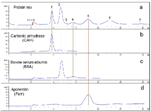
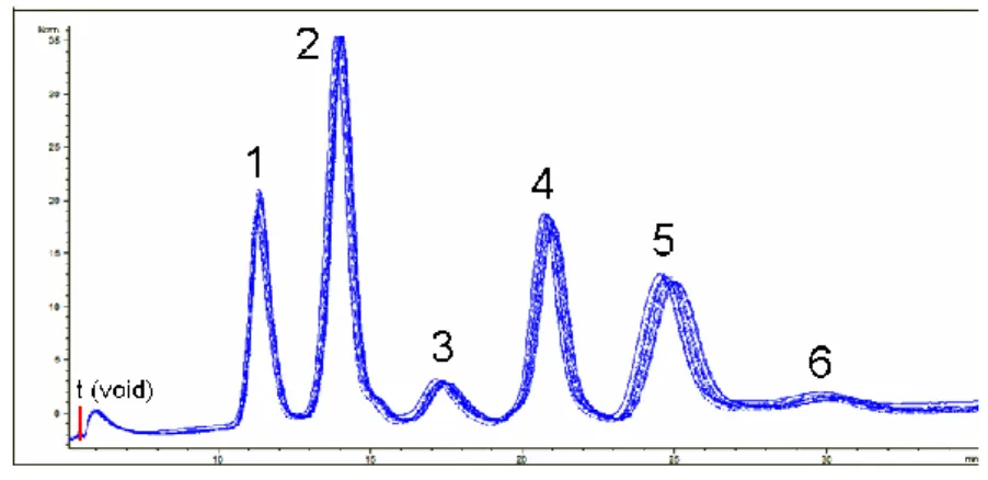
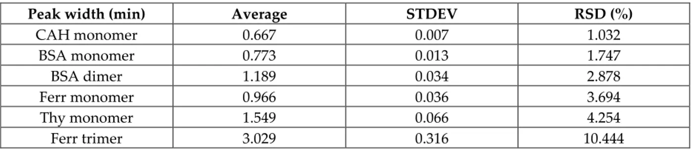
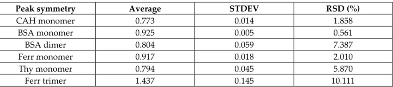
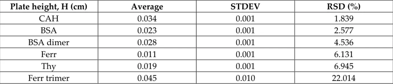
4.1.1. HF5-UV performance evaluation for the separation of a standard protein mixture. HF5-UV method validation tentative: specificity, repeatability (first level of precision), selectivity and robustness……….. 94
4.1.2. HF5-MALS and SEC-MALS performance comparison for the separation of a standard protein mixture. FFF selectivity feature………..…………... 114
4.1.3. Freeze/thaw induced antibody self-association monitoring by HF5-UV and MALS characterization……….. 136
4.1.4. Carrier solutions screening exploratory study by HF5-UV: influence of pH, salt type and concentration, ionic strength and buffering agent on the immunoglobulins (IgGs) stability during separation………. 148
4.1.5. HF5-FLD method optimization for the quantification of aggregate levels in protein formulations. Intrinsic fluorescence-based detection of antibody aggregates: LoD and LoQ estimation. Advantages of miniaturization: UV detection sensitivity boost employing a smaller inner diameter hollow fiber………... 170
4.1.6. HF5-MALS and SEC-MALS performance comparison for the separation of immunoglobulins (IgGs). FFF selectivity feature……….. 192
4.1.7. High reproducibility and low detection limit HF5-MALS method for the characterization of aggregates in protein formulations: AvidinOX®………... 216
4.2. Part 2: Study of oxidative stress-related protein aggregation phenomena during biological aging……….. 240
Synopsis: senescence……… 242
Synopsis: oxidative stress……….. 243
A. Reactive oxygen species (ROS), oxidative stress and senescence………... 244
B. Oxidative stress, senescence and protein damage………...……….. 245
Protein post-translational modifications……… 245
Clearance of oxidized proteins and formation mechanism of protein aggregates: why damaged proteins and their aggregates accumulate………... 247
C. Immunosenescence, oxidative stress, protein damage and accumulation of protein aggregates………... 249 Oxidative stress damage: protein carbonylation……… 250
In vitro and in vivo experimental models of the simulation of aging effects: Paraquat-induced oxidative stress……….. 252
D. Current proteomic methodologies and their limitations………. 253
E. Identification, isolation and proteomic analysis of oxidized proteins and protein aggregates………... 255
d
References……….. 260
4.2.1. Novel methodology based on HF5-MALS for the size-separation, characterization and quantification of oxidative stress-related protein aggregates levels in whole cell lysates 264
This dissertation addresses two major aspects regarding the aggregation phenomena occurring in complex protein samples: the first regards the emerging field of protein therapeutics and the current need for adequate characterization methods for protein formulations; while the second one regards the study of aggregation phenomena occurring in living organisms as a consequence of the natural process of aging, as well as the cause of notorious neurodegenerative disorders.
Therapeutic proteins and, in particular, antibodies, are the compounds most used as biopharmaceuticals. However, the biggest problem is their instability which often leads to aggregation. While aggregation is necessary in particular cases for the proteins to perform their biological activity, the presence of aggregated proteins for therapeutic use is undesirable. Even at very low levels, the aggregates of any kind may not preserve or, even worse, it may impede the desired biological functionality and above all, may represent a potential immunogenic risk. Hence the need to carefully evaluate their clinical relevance [Cromwell et al. 2006].
In order to ensure the efficacy and safety of biopharmaceuticals it is essential to employ suitable analytical methods to monitor/evaluate efficiently and accurately the phenomenon of aggregation during development, production and storage of products. Usually, the various aggregation mechanisms are acting simultaneously and have not been fully elucidated [Mahler et al. 2009, Philo and Arakawa 2009,
II Engelsman et al. 2011]. Moreover, the formed aggregates have sizes in a very wide dimensional range (few nm-mm). The limitations of currently employed analytical methods in terms of sensitivity, speed of analysis, robustness, range of application (Figure 1) or sample complexity and recovery, together with the fact that the term “aggregate” is constantly redefined as a consequence of continuous new findings in the clinical field, have created a “gap”[Philo 2006].
Biological aging, also known as senescence, is a fundamental process which is the main risk factor regarding the development of cancer, cardiovascular and neurodegenerative diseases (Alzheimer, Parkinson and Huntington disease) in vertebrates. In particular, immunosenescence represents the progressive deterioration of the immune system caused by the natural aging process. Inside any living organism, damaged cells are constantly being replaced and proteins are degraded to their constituting amino-acids (proteasome degradation or chaperone-mediated autophagy by lysosome) and re-synthesized. Following senescence, the body gradually loses its ability to repair itself. This is due to loss of equilibrium between the oxidizing species (in particular, the reactive oxygen species) and antioxidants, leading to a pathological condition called oxidative stress [Bandyopadhyay et al. 1999, Squier 2001]. Upon oxidation (most frequently resulting in the carbonylation of particular side chains), the proteins lose their native conformation, becoming thermodynamically unstable, and have a high propensity to form larger assemblies. Moreover, since the degradation mechanisms no longer work efficiently, the aggregates accumulate in the endosomal compartments, increasing in size with advancing age [Cannizzo et al. 2011, Cannizzo et al. 2012]. An appropriate proteomics technology platform, based on a separation technique capable of separating protein aggregates based on their biophysical properties, would allow further analysis on how protein sequence/structure determine their tendency to aggregate, how different post-translational modifications affect unfolding and aggregation and the proteomic machinery associated with their degradation.
III Moreover, it would open a world of possibilities towards the discovery of novel drug targets and early disease markers [Garbis et al. 2005].
Figure 1 [Zölls et al. 2012] depicts the size range of application of the currently employed analytical methods for the characterization of protein particles. As therapeutic proteins, as well as aging proteins can form different types and sizes of aggregates and particles, a case-by-case selection of the appropriate analytical characterization methods is required, since there is no “general solution” to fit all.
Figure 1 – Approximate size range of current analytical methods for the size characterization of sub-visible and visible (protein) particles [Zölls et al. 2012]
The figure shows that no single method is capable of providing a complete characterization, which would cover the whole nm-mm aggregates size range, therefore making the combination of several methods (usually based on different measurement/separation principles) mandatory for an extensive characterization [Philo 2006, Zölls et al. 2012]. The second challenge lies within the data interpretation; since different measurement/separation principle are applied, results may not always be directly comparable and need to be evaluated case-by-case
IV considering the underlying theory. The possibility of applying several orthogonal characterization methods has been proposed as means to bridge this “gap”, by selecting one or more methods for sample-to-sample comparison and searching for trends rather than concentrate on actual numbers. [Carpenter et al. 2010, Carpenter et al. 2012, Zölls et al. 2012].
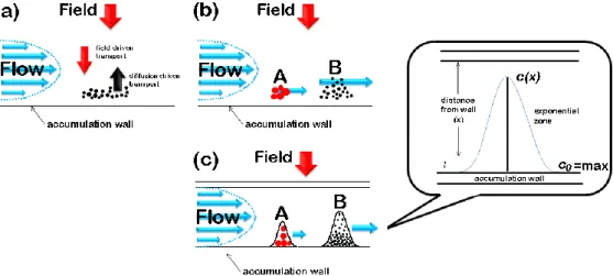
Both protein aggregation topics were faced with the help of field-flow fractionation (FFF), a chromatographic – like flow-based separation technique [Giddings 1966], which is particularly suited for the separation of macromolecules (proteins and protein complexes/aggregates) in a very broad MW range [Zattoni et al. 2007]. FFF and liquid chromatography (LC) employ almost the same instrumental setup, but, since the separation in based on a different principle, the separation device in FFF is an empty channel instead of a chromatographic column. In FFF, the retention does not rely on interaction of the analytes with a stationary phase, but with an external field that is applied perpendicularly to direction of the mobile phase flow. Hence the field-flow dualism required for the separation [Giddings 1973, Giddings 2000].
In flow FFF (Fl FFF or F4), the applied field consists in a second stream of mobile phase applied through the channel section, therefore called cross-flow [Reschiglian and Moon 2008]. This is the FFF variant of choice for the studies reported in this Thesis because of its ability to separate macromolecules and particles in a very wide size (few nm - µm) and MW range. Moreover, the flow field offers intrinsic advantages for the separation of proteins and protein aggregates: (i) the gentle separation mechanism and the lack of a stationary phase; (ii) bio-friendly, and virtually any type of mobile phase can be used; (iii) offers high selectivity in terms of diffusion coefficient differences. Features (i) and (ii) imply that interactions between the proteins and the separation device are negligible. This makes it possible to preserve the native molecular conformation, a fundamental bio-physical property. Feature (iii) is correlated to the separation principle in F4, based on differences in diffusion coefficients.
V The diffusion coefficient (D) is a key parameter which relates to the protein size, shape and surface properties. [Reschiglian and Moon 2008].
In particular, this dissertation is centered on the applications in the bioanalytical field of the miniaturized version of F4, called hollow fiber flow field-flow fractionation (hollow fiber F4 or HF5), which is characterized by a symmetrical channel geometry represented by a cylindrical channel with porous walls. [Johann et al. 2010].
Many efforts have been employed over the years by the Analytical Chemistry research team of the “G. Ciamician” Chemistry Department into developing the HF5 separation device prototype. Thanks to the successful collaboration between the Analytical Chemistry team and Wyatt Technology Europe (Dernbach, Germany), once the HF5 separation device achieved its ready-to-market phase, it was implemented in the Eclipse® DUALTECTM FFF separation system. This novel
implementation made “the first commercial FFF system using both HF5 and AF4 technique integrated into one instrument”, which was launched on the market in 2012. The commercial HF5 channel is equipped with an Enhanced Sealing Technology (EST, patented) facilitating the channel assembly and is also leakage-proof.
In addition to the key advantages offered by F4, HF5 distinguishes itself through unique features, such as: (i) low channel volume, which reduces sample dilution with fractionation, (ii) low flow rates, which makes it possible to be successfully coupled on-line with MS; (iii) disposable usage, which eliminates sample carry-over and possible sample contamination. Features (i) and (ii) also lead to a higher sensitivity by increasing the limit of detection/quantification for protein aggregates. Thanks to all these key advantages, which have led to the promising results described in the literature [Reschiglian et al. 2002, Reschiglian et al. 2004, Reschiglian et al. 2005, Silveira et al. 2005, Roda et al. 2006, Rambaldi et al. 2007, Kang et al. 2008, Kim et al. 2008, Lee et al. 2009, Kang et al. 2010, Lee et al. 2010], HF5 offers unparalleled
VI performances that are fundamental for the characterization of proteins and protein aggregates.
The analytical information was enhanced by HF5 on-line coupled with highly sensitive spectroscopic detection methods, such as UV absorbance and fluorescence
emission detection, or with optical detection methods, like multi-angle light scattering (MALS), which measures the light scattered at multiple angles by the
proteins and protein aggregates fractionated through HF5.
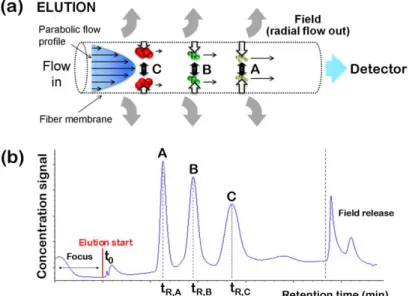
Combined with a concentration detector (like a UV detector), MALS is able to provide directly the size (root mean square radius or rms radius, the mass-average distance between each point in the macromolecule and the center of mass), and the absolute molar weight (MW) of the fractionated proteins and protein aggregates as a function of their elution time. Comparing the rms radius values obtained from the MALS measurements with the hydrodynamic radius (rH) values, which are readily
obtained from the experimental retention time, HF5-MALS allows obtaining information regarding the shape and mass distribution inside the protein aggregates [Reschiglian and Moon 2008].
In Part 1 of this dissertation (Chapter 4, sub-chapter 4.1), HF5 coupled online with MALS, UV-Vis and/or fluorescence detection was proposed to bridge the "gap" of suitable techniques for the characterization of the sub-µm particles. A fast method is proposed for the characterization of a standard protein mixture, with particular regard to the repeatability, resolution, sensitivity, selectivity and robustness. The superior performance of HF5 is demonstrated, especially from the selectivity point of view, when compared to size-exclusion chromatography (SEC) – the benchmark in quality control (QC) of therapeutic proteins. Exploratory methods are proposed for the separation and characterization of self-associated immunoglobulins (IgGs) under different experimental conditions, showing the versatility and the sensitivity of HF5 when coupled with fluorescence detection (FLD) and MALS. The advantages of miniaturization are explored further, sensitivity is improved even more when
VII employing a hollow fiber with a smaller inner diameter for the separation of an IgG with low aggregates levels. A fast method is proposed to compare HF5 and SEC performance, employing an IgG1 monoclonal antibody, once again demonstrating the HF5 superiority in terms of selectivity. Finally, a highly repeatable and sensitive HF5-MALS method, providing insight on the biophysical nature of aggregates, was developed employing an avidin derivate as sample model (AvidinOX®, provided by Sigma Tau).
The aging-related protein aggregation topic, occurring under the effects of oxidative stress, was addressed in collaboration with the Laboratory of Pathology, Microbiology and Immunology, Albert Einstein College of Medicine of Yeshiva University (New York, USA), and the analytical information was enhanced or corroborated by complementary assays performed by the above-mentioned laboratory. In Part 2 of the dissertation (Chapter 4, sub-chapter 4.2), it is demonstrated that HF5 can be used as a first dimension of separation of aging-related protein aggregates from whole cell lysates and, by coupling HF5 with MALS, important biophysical information on the separated aggregates can be gathered (size, molecular weight and conformation). The effectiveness of the separation method was confirmed by both native-PAGE and proteomic analysis of the high molecular weight aggregates. It is shown how, under the effect of the oxidative stress (either induced or naturally occurring in aging organism), protein aggregates are modified and have a smaller size when compared to native aggregates of same molecular weight. Moreover it is shown that oxidative stress-related proteins aggregate and have a different, more compact, molecular conformation. The ability to separate cell lysates under both physiological and denaturing conditions confirmed what previously reported: most of aging-related aggregates are urea-insoluble. Moreover, offline coupling of HF5 with Mass Spectrometry confirmed the presence of several proteins previously reported to be insoluble or with increased tendency to aggregate during aging.
VIII
R
EFERENCES[Bandyopadhyay et al. 1999] Bandyopadhyay U.; Das D. and Banerjee R. K. (1999). "Reactive oxygen species: Oxidative damage and pathogenesis." CURRENT SCIENCE 77 (5): 658-666.
[Cannizzo et al. 2012] Cannizzo E.; Clement C. C.; Morozova K.; Valdor R.; Kaushik S.; Almeida L. N.; Follo C.; Sahu R.; Cuervo A. M.; Macian F. and Santambrogio L. (2012). "Age-related oxidative stress compromises endosomal proteostasis." Cell Press 2 (1): 136-149.
[Cannizzo et al. 2011] Cannizzo E.; Clement C. C.; Sahu R.; Follo C. and Santambrogio L. (2011). "Oxidative stress, inflamm-aging and immunosenescence." Journal of Proteomics 74 (11): 2313–2323. [Carpenter et al. 2012] Carpenter J. F.; Cherney B. and Rosenberg A. S. (2012). The Critical Need for Robust Assays for Quantitation and Characterization of Aggregates of Therapeutic Proteins. Analysis of Aggregates and Particles in Protein Pharmaceuticals. Mahler H.-C. and Jiskoot W. Hoboken, NJ, USA, John Wiley & Sons, Inc.
[Carpenter et al. 2010] Carpenter J. F.; Randolph T. W.; Jiskoot W.; Crommelin D. J. A.; Middaugh C. R. and Winter G. (2010). "Potential Inaccurate Quantitation and Sizing of Protein Aggregates by Size Exclusion Chromatography: Essential Need to Use Orthogonal Methods to Assure the Quality of Therapeutic Protein Products." Journal of Pharmaceutical Sciences 99 (5): 2200–2208.
[Cromwell et al. 2006] Cromwell M. E. M.; Hilario E. and Jacobson F. (2006). "Protein aggregation and bioprocessing." AAPS Journal 8 (3): E572–E579.
[Engelsman et al. 2011] Engelsman J. d.; Garidel P.; Smulders R.; Koll H.; Smith B.; Bassarab S.; Seidl A.; Hainzl O. and Jiskoot W. (2011). "Strategies for the Assessment of Protein Aggregates in Pharmaceutical Biotech Product Development." Pharmaceutical Research 28 (4): 920-933.
[Garbis et al. 2005] Garbis S.; Lubec G. and Fountoulakis M. (2005). "Limitations of current proteomics technologies." Journal of Chromatography A 1077 (1): 1-18.
[Giddings 1966] Giddings J. C. (1966). "A New Separation Concept Based on a Coupling of Concentration and Flow Nonuniformities." Separation Science 1 (1): 123-125.
[Giddings 1973] Giddings J. C. (1973). "The conceptual basis of field-flow fractionation." Journal of Chemical Education 50 (10): 667-669.
[Giddings 2000] Giddings J. C. (2000). Chapter 1: The Field-Flow Fractionation Family: Underlying principles. Field-Flow Fractionation Handbook. Schimpf M. E.; Caldwell K. and Giddings J. C. New York, Wiley: 3-30.
[Johann et al. 2010] Johann C.; Elsenberg S.; Roesch U.; Rambaldi D. C.; Zattoni A. and Reschiglian P. (2010). "A novel approach to improve operation and performance in flow field-flow fractionation." Journal of Chromatography A 1218 (27): 4126–4131.
IX [Kang et al. 2010] Kang D.; Ji E. S.; Moon M. H. and Yoo J. S. (2010). "Lectin-Based Enrichment Method for Glycoproteomics Using Hollow Fiber Flow Field-Flow Fractionation: Application to Streptococcus pyogenes." Journal of proteome research 9 (6): 2855–2862.
[Kang et al. 2008] Kang D.; Oh S.; Reschiglian P. and Moon M. H. (2008). "Separation of mitochondria by flow field-flow fractionation for proteomic analysis." Analyst (133): 505-515.
[Kim et al. 2008] Kim K. H.; Kang D.; Koo H. M. and Moon M. H. (2008). "Molecular mass sorting of proteome using hollow fiber flow field-flow fractionation for proteomics." Journal of Proteomics 71 (1): 123– 131.
[Lee et al. 2009] Lee J. Y.; Kim K. H. and Moon M. H. (2009). "Evaluation of multiplexed hollow fiber flow field-flow fractionation for semi-preparative purposes." Journal of Chromatography A 1216 (37): 6539–6542. [Lee et al. 2010] Lee J. Y.; Min H. K.; Choi D. and Moon M. H. (2010). "Profiling of phospholipids in lipoproteins by multiplexed hollow fiber flow field-flow fractionation and nanoflow liquid chromatography– tandem mass spectrometry." Journal of Chromatography A 1217 (10): 1660–1666.
[Mahler et al. 2009] Mahler H.-C.; Friess W.; Grauschopf U. and Kiese S. (2009). "Protein aggregation: Pathways, induction factors and analysis." Journal of Pharmaceutical Sciences 98 (9): 2909–2934.
[Philo 2006] Philo J. S. (2006). "Is Any Measurement Method Optimal for All Aggregate Sizes and Types? ." The AAPS Journal 8 (3).
[Philo and Arakawa 2009] Philo J. S. and Arakawa T. (2009). "Mechanisms of Protein Aggregation." Current Pharmaceutical Biotechnology (10): 348-351.
[Rambaldi et al. 2007] Rambaldi D. C.; Zattoni A.; Casolari S.; Reschiglian P.; Roessner D. and Johann C. (2007). "An Analytical Method for Size and Shape Characterization of Blood Lipoproteins." Clinical Chemistry 53 (11): 2026-2029.
[Reschiglian and Moon 2008] Reschiglian P. and Moon M. H. (2008). "Flow field-flow fractionation: A pre-analytical method for proteomics." Journal of Proteomics 71 (3): 265–276.
[Reschiglian et al. 2002] Reschiglian P.; Roda B.; Zattoni A.; Min B. R. and Moon M. H. (2002). "High performance, disposable hollow fiber flow field-flow fractionation for bacteria and cells. First application to deactivated Vibrio cholerae*." Journal of Separation Science (25): 490-498.
[Reschiglian et al. 2004] Reschiglian P.; Zattoni A.; Cinque L. and Roda B. (2004). "Hollow-Fiber Flow Field-Flow Fractionation for Whole Bacteria Analysis by Matrix-Assisted Laser Desorption/Ionization Time-of-Flight Mass Spectrometry." Analytical Chemistry 76 (7): 2103–2111.
[Reschiglian et al. 2005] Reschiglian P.; Zattoni A.; Roda B. and Cinque L. (2005). "On-Line Hollow-Fiber Flow Field-Flow Fractionation-Electrospray Ionization/Time-of-Flight Mass Spectrometry of Intact Proteins." Analytical Chemistry 77 (1): 47-56.
X [Roda et al. 2006] Roda A.; Parisi D.; Guardigli M.; Zattoni A. and Reschiglian P. (2006). "Combined Approach to the Analysis of Recombinant Protein Drugs Using Hollow-Fiber Flow Field-Flow Fractionation, Mass Spectrometry, and Chemiluminescence Detection." Analytical Chemistry 78 (4): 1085–1092.
[Silveira et al. 2005] Silveira J. R.; Raymond G. J.; Hughson A. G.; Race R. E.; Sim V. L.; Hayes S. F. and Caughey B. (2005). "The most infectious prion protein particles." Nature (437): 257-261.
[Squier 2001] Squier T. C. (2001). "Oxidative stress and protein aggregation during biological aging." Experimental Gerontology 36 (9): 1539–1550.
[Zattoni et al. 2007] Zattoni A.; Casolari S.; Rambaldi D. C. and Reschiglian P. (2007). "Hollow-fiber flow field-flow fractionation." Current Analytical Chemistry 3 (4): 310-323.
[Zölls et al. 2012] Zölls S.; Tantipolphan R.; Wiggenhorn M.; Winter G.; Jiskoot W.; Friess W. and Hawe A. (2012). "Particles in therapeutic protein formulations, Part 1: Overview of analytical methods." Journal of Pharmaceutical Sciences 101 (3): 914–935.
1.1.
I
NTRODUCTION TO THE ANALYTICAL PROBLEM:
P
ROTEINA
GGREGATIONAggregation is a general term that surrounds several types of protein-protein interactions or characteristics; protein aggregates may emerge from several mechanisms [Philo and Arakawa 2009, Wang et al. 2010] and may be classified in numerous ways, including soluble/insoluble, covalent/non-covalent, reversible/irreversible, and native/denatured. [Cromwell et al. 2006]. In order to address protein aggregation as an issue, an accurate definition of “protein aggregate” is required, even though not often available and definitely, not general.
Protein aggregates sizes cover a very wide size spectrum, from small oligomers to visible “snow” or “flakes” and precipitates, and, as a rule of thumb, only the smaller species have a reversible character. Furthermore, even though less acknowledged, aggregates also display an ample range of life spans, and this aspect impacts greatly on the choice of appropriate detection methods. In addition, the measurement itself may destroy or create aggregates, hence impact on aggregates levels; this aspect represents a major analytical challenge and is decisive as well for appropriate method selection [Philo 2006].
P
ROTEIN
A
GGREGATION
2 Consequently, there is no “general solution to fit all”, since no single analytical method or approach is able to provide a complete answer and, at the same time, able to work in all situations and on all different samples. Despite all the efforts to define the analytical challenge at stake and all the information currently available on the protein aggregation topic, many pharmaceutical scientists fail to acknowledge protein aggregation as an encompassing phenomenon and its implications on the measurement approach and on the data interpretation. In fact, the case-by-case use of complementary (orthogonal) characterization methods is recommended, as well as identifying trends in the obtained results instead on concentrating on raw numbers [Philo 2006, Carpenter et al. 2010, Carpenter et al. 2012, Zölls et al. 2012].
1.1.1.
T
HEP
ROTEINA
GGREGATES HAVE A WIDE SIZE RANGEDue to the complexity of the protein aggregation phenomenon, there is no universal terminology to describe the numerous aggregate sizes or types; however, some of the commonly occurring aggregates are classified by [Philo 2006] as follows: (1) rapidly reversible, non-covalent small oligomers (dimer, trimer, tetramer, and so forth); (2) irreversible, non-covalent oligomers; (3) covalent oligomers (for instance, linked through Schiff bases or disulfide bonds); (4) “large” aggregates ( ≥ decamer); (5) “very large” aggregates (diameter ~50 nm - 3 µm); and (6) visible particulates ( “snow”, “flakes” or “floaters”), which are, most likely, irreversible. Except for the visible particulates, there is a chance that the large and very large protein aggregates have a reversible character, if associated through non-covalent bonds.
In addition, protein aggregates are characterized by the tendency to “evolve” over time, typically into larger and less reversible/irreversible species; moreover, it is highly likely for a protein sample to contain more than one of these types or classes [Philo 2006, Philo and Arakawa 2009].
3
1.1.2.MOST PROTEIN AGGREGATES ARE REVERSIBLE
Despite the common tendency to make a black vs. white distinction, which usually is interpreted as a permanent and irrevocable classification of aggregates into “reversible” and “irreversible”, in reality, though, an aggregate that is irreversible in one context can become reversible in another, indicating the existence of a continuum of aggregation states between reversible and irreversible [Philo 2006].
1.1.3.THE LIFE SPAN OF PROTEIN AGGREGATES IS VARIABLE
The ample range of life spans feature of reversible protein aggregates (their permanence in time) is probably their most neglected; the rates of association/dissociation phenomena between oligomers vary greatly, ranging from ms to several days. Many analytical methods, more in particular, separation techniques, allow only the detection the longer life span species; proteins displaying dynamic self-association (rapid and reversible protein self-assembly), under the effects of the separation method, find themselves under a constant struggle between separation and re-equilibration, because of the law of mass action. Consequently, the separation results often depend on the rates of the association/dissociation process, as well as the equilibrium constants [Philo 2006].
Although either SEC and SV-AUC or Flow FFF methods may successfully resolve multiple peaks, in fact, neither of those peaks represents a pure, individual oligomer, but more likely a dynamic mixture of multiple oligomers; however, if the association/dissociation reactions occur very slowly compared to the duration of the separation, the self-associated protein system behaves like a true mixture, therefore individual oligomers can be resolved. Such reversible, but extremely slow association/dissociation reactions, responsible for the existence of so-called “metastable oligomers”, are rather common [Philo 2006, Philo and Arakawa 2009].
4
1.1.3.THE SEPARATION IS INTRINSICALLY DISTRESSING FOR PROTEIN AGGREGATES
When analyzing protein aggregates, one of the fundamental problems is represented by the simple fact that most characterization techniques and, in particular, separation techniques, have a chance of perturbing the distribution of protein species in the sample to be analyzed; not only the measurement itself may destroy or cause the loss of some aggregates, but new aggregates can also be created by or during the measurement[Philo 2006, Engelsman et al. 2011].
For instance, both SEC and FFF (AF4 or HF5) produce a high sample dilution of the sample during the separation, which potentially induces the dissociation of the reversible aggregates. Moreover, SEC is also infamous for filtration effects and poor sample recovery, which makes the use of an elution buffer containing high levels of salts and/or organic modifiers mandatory; these carrier solution additives may modify the distribution of non-covalent aggregates. Generally less problematic that SEC, the protein recovery in FFF (AF4 and HF5) may also suffer from non-specific adsorption of proteins to the channel wall, therefore some adjustments of the carrier solution are in order. As for the creation of new aggregates, the major impacting factor is the composition of the carrier solution (and changes thereof); however, in both SEC and FFF, protein aggregates may also arise from pre-dilution of the sample with the separation buffer, whose composition is usually different than the protein formulation buffer [Arakawa et al. 2010, Carpenter et al. 2012, Zölls et al. 2012].
1.1.5.IS IT POSSIBLE TO REPLACE SEC?
Despite its notoriety, it is very likely that SEC (and its macro-column version, fast protein chromatography, FPLC) will continue to be the workhorse tool for protein aggregates characterization in the near future. Alternative analytical platforms, such as SV-AUC and FFF (AF4 and HF5) with or without multi-angle scattering (MALS) detection, still have one or more of the following drawbacks: (1) they are not robust
5 enough (do not provide repeatable results) or/and easy to use as routine QC procedures, therefore difficult to validate for lot release, (2) they have low throughput, (3) they require expensive equipment (SV-AUC) or extensive method development (FFF), therefore highly trained personnel, and (4) the software may be very far from being 21 CFR part 11 compliant [FDA 1997].
Despite these shortcomings, the above mentioned analytical methodologies proved themselves to be invaluable as orthogonal methods for SEC, whose purpose is to help determining the reliability of the results provided by SEC. Such cross-validation practices are usually time and resources-efficient, and they can be implemented fairly easily at all stages of drug development, as well as technological platforms in functional and structural proteomics. In addition, these complementary methods can help guiding the development and the improvement of SEC methods [Philo 2006, Arakawa et al. 2010, Carpenter et al. 2010, Engelsman et al. 2011, Carpenter et al. 2012, Zölls et al. 2012].
For all the reasons described in the previous paragraphs, the protein aggregation phenomenon needs a targeted approach; two main fields of research and their applications are explored in this dissertation, and various aspects are described in order to obtain a wholesome understanding of the protein aggregation issue. Moreover, a critical evaluation of the most frequently employed analytical methodologies, which are currently dedicated to the study of protein aggregation, is due in order to find the best course of action in each described case.
In this dissertation, the miniaturized variant of Flow FFF (hollow fiber FFF or HF5) online coupled with spectroscopic and/or optical detection methods, is employed for the study of protein aggregation phenomena. Various aspects, ranging from instrumental and method robustness (and validation), to detection sensitivity and instrumental/method versatility, in terms of aggregates size-range and MW range, as well as versatility in terms of carrier solution choice (enabling the discrimination of different types of protein aggregates), are explored.
6
1.2.
P
ROTEIN AGGREGATION PHENOMENA RELATED TO BIOLOGICAL AGING(
SENESCENCE)
From the pathological and immunological point of view, protein aggregation is a general terminology which describes the association of proteins into larger assembly, following loss of the secondary, tertiary or quaternary protein structure and often leading to loss of biological activity. Protein aggregation is a common biological phenomenon associated to the cellular inability to maintain the homeostasis of their proteome (proteostasis). In physiological conditions, the tendency to aggregate of de novo synthesized unfolded proteins is balanced by the several chaperones that aid their folding [Kopito 2000]. Soluble aggregation is also commonly observed in ubiquitinated unfolded proteins before proteasome degradation or in oxidatively damaged proteins before translocation into the lysosomes by chaperone-mediated autophagy [Kiffin et al. 2004]. Additionally, temporal changes to the cellular homeostasis (temperature, pH, water content and salt/ions concentration) can induce transitory protein unfolding and soluble aggregation. Lately, it has been reported that, during physiological aging, proteostasis becomes gradually compromised and several hundred proteins tend to become more insoluble and aggregate. These proteins have been shown to have common biochemical and biological properties, such as a primary structure with amino acids stretches often found in proteins associated with neurodegenerative diseases and secondary structure with increased beta-sheets [David et al. 2010] or present extensive oxidative post-translational modifications [Cannizzo et al. 2012].
During pathological conditions, protein aggregation is also a very common occurrence giving rise to the group of diseases collectively known as protein conformational diseases. In many degenerative diseases of the CNS, such as Alzheimer’s, Parkinson’s and Huntington’s disease, protein aggregation is a common pathological hallmark [Rubinsztein et al. 2005]. Protein aggregates can be classified according to their biochemical and biophysical characteristics. Biochemically, aggregates can be formed
7 by covalent bonds, through Schiff-base formation, or non covalent bonds, mainly mediated by hydrogen bonds, hydrophobic and electrostatic interactions. The former are irreversible aggregates whereas the latter can be, at least partially, reversed by cellular molecular chaperones. Size-wise aggregates can range considerably, from protein oligomers up to visible cytosolic inclusions, known as the aggresome [Kopito 2000]. The sub cellular location of these aggregates can also vary, from perinuclear to peri-ER or intra-endosomal.
Recently, there has been a strong interest in analyzing protein aggregates for different reasons: (i) to determine how protein primary and secondary structure influence their tendency to aggregate during physiological or pathological conditions; (ii) to map the post-translational modifications observed in chronic inflammatory, metabolic and degenerative diseases that induce protein aggregation, and (iii) to analyze the machinery recruited to the aggregate to aid its disaggregation or to dispose them through proteasomal or autophagy-mediated endosomal degradation.
Consequently, several methods have been employed to separate protein aggregates, including filter assay, analytical ultracentrifugation (AUC), gel electrophoresis and size exclusion chromatography (SEC) [Stegemann et al. 2005, Ishii et al. 2007, Linetsky et al. 2008, Scharf et al. 2013]. However, the first two methods separate the aggregates as a total, thus, making it impossible to analyze the biochemical composition of the aggregates with different MW, shape and biophysical properties. SEC is limited by the pores size of the column used for fractionation (therefore allow for a specific size range of aggregates to be separated), and by the amount of detergent that can be used during the run (which can, in turn, affect the size and composition of the aggregates). Gel electrophoresis does not allow for an accurate analysis of their MW, nor does provide any additional information on the biophysical properties of the aggregates.
Therefore, an analytical methodology with the ability to separate soluble aggregates from terminal aggregates or aggregates with different biophysical properties would
8 allow for a more functional analysis. Indeed, in prion disease, a correlation between the size and the infectivity of the protein’s aggregate was reported [Silveira et al. 2005].
1.3.
P
ROTEIN AGGREGATION PHENOMENA IN THERAPEUTIC PROTEIN FORMULATIONSAntibodies, the most developed therapeutic proteins, are large multi-domain proteins. In particular, human monoclonal antibodies (mAbs) display poor biophysical properties characterized by low stability and unfavorable tendency towards aggregation. Among the factors that lead to these shortcomings, protein formulation plays an important role, even though the core of problem is the primary sequence of the protein itself; moreover, since mAb therapeutic action is so specific, there are significant differences among them (related to Fab differences and the particular
antigen specificity of the monoclonal), which explains the differences in stability and aggregation propensity between antibodies [Lowe et al. 2011].
The final formulation of a therapeutic mAb therefore must be carefully chosen; not only to ensure drug quality (stability and purity), but also to allow drug manufacture at an appropriate scale. Moreover, an effective therapeutic drug requires high-concentration liquid formulations, e.g higher than 100 mg/mL. The mAbs can lose stability and aggregate also, as a result of conformational changes [Schwegman et al. 2009] which can occur upon storage (usually freezing in high-volume vessels [Singh et al. 2009]. Moreover, the constituting amino acids may undergo post-translational modifications during expression, purification, or storage, leading to loss of stability and eventually aggregation [Jenkins et al. 2008].
Known effects of aggregation among therapeutic proteins are: lower in vivo efficacy, increased variability among batches, and perhaps most importantly, immunogenicity in patients [Cordoba-Rodriguez 2008]. Therefore, it is vitally important that for each of the steps of expression, purification, concentration, formulation, storage, and final
9 filling of the mAb, protein degradation, such s aggregation, must be minimized [Lowe et al. 2011].
In the biopharmaceutical field, the aggregates are defined as high molecular weight (MW) protein assemblies; formed either spontaneously, through electrostatic/hydrophobic/hydrogen bonds interactions between native-state monomeric units (also known as protein self-association), or by association between denatured (damaged) monomers. Both types represent a concern from the immunological point a view, and their capacity to enhance immune responses to the monomeric form has been known for over a half a century [Rosenberg 2006]. While large aggregates are more easily identified and separated (eliminated through filtration; for example), as their presence is rigorously regulated by FDA [Zölls et al. 2012], little is known about the immunological effects of protein self-association phenomenon, which may impede the effectiveness of the therapeutic protein by neutralizing the monomer action.
Although it was initially believed that protein-protein favorable interactions can only occur if hydrophobic surfaces are exposed upon denaturation (intended as irreversible damage), leading to aggregation and often causing severe precipitation, an increasing concern regarding the native protein interactions has recently emerged [Cromwell et al. 2006]. In fact, recent literature indicates that even small perturbations in the protein structure may cause the exposure of hydrophobic surface: it is belied that the formation of self-associated antibody species is mediated by electrostatic interactions [Liu et al. 2005], while dipole-dipole interactions are believed to be the cause of fibrillogenic association of b–sheets [Fernández 2005]. Comprehension on how to control the aggregation phenomenon, through thermodynamic and kinetic studies, helps assessing the danger correlated to the presence of associated species during the formulation development of therapeutic proteins. The main rising concerns of the biopharmaceutical industry, which impacts on the safety and efficacy of therapeutic proteins, is represented by protein aggregation and particulate formation in protein formulations, with their very wide
10 size range (nm-µm) being the most pressing issue since no single analytical method is able to resolve it. In fact, many efforts have been employed to fill the “gap” of techniques able to identify, separate and characterize the presence of undesired protein aggregates in the sub-µm size range [Cao et al. 2009, Zölls et al. 2012].
Moreover, many efforts have been developing in order to optimize the time and resources required for protein formulations by means of high-throughput techniques for formulation screening [Capelle et al. 2007, Goldberg et al. 2011], as well as of simplification and customization of the protein formulation process through FDA regulations based on Design of Experiments [Hinz 2006, Feng et al. 2012].
1.4.
T
HE NEED FOR APPROPRIATE ANALYTICAL METHODOLOGIESThe need for adequate analytical methods for the detection, separation and characterization of protein aggregates, regardless of their origin or formation mechanism, justifies the sudden growth of new analysis methods. Since therapeutic proteins, as well as aging proteins, can form different types and sizes of aggregates and particles, a case-by-case selection of the appropriate analytical characterization methods is required: there is no “general solution” to fit all.
No single method is indeed capable to cover the nm-mm aggregates size range, therefore making the combination of several methods (usually based on different measurement/separation principles, therefore orthogonal) mandatory for an extensive characterization, as means to bridge this “gap”[Philo 2006, Zölls et al. 2012].
11
1.5.
R
EFERENCES[Arakawa et al. 2010] Arakawa T.; Ejima D.; Li T. and Philo J. S. (2010). "The Critical Role of Mobile Phase Composition in Size Exclusion Chromatography of Protein Pharmaceuticals." Journal of Pharmaceutical Sciences 99 (4): 1674-1692.
[Cannizzo et al. 2012] Cannizzo E.; Clement C. C.; Morozova K.; Valdor R.; Kaushik S.; Almeida L. N.; Follo C.; Sahu R.; Cuervo A. M.; Macian F. and Santambrogio L. (2012). "Age-related oxidative stress compromises endosomal proteostasis." Cell 2 (1): 136-149.
[Cao et al. 2009] Cao S.; Pollastrini J. and Jiang Y. (2009). "Separation and characterization of protein aggregates and particles by field flow fractionation." Current Pharmaceutical Biotechnology 10 (4): 382-390. [Capelle et al. 2007] Capelle M. A. H.; Gurny R. and Arvinte T. (2007). "High throughput screening of protein formulation stability: Practical considerations." European Journal of Pharmaceutics and Biopharmaceutics 65 (2): 131-148.
[Carpenter et al. 2012] Carpenter J. F.; Cherney B. and Rosenberg A. S. (2012). The Critical Need for Robust Assays for Quantitation and Characterization of Aggregates of Therapeutic Proteins. Analysis of Aggregates and Particles in Protein Pharmaceuticals. Mahler H.-C. and Jiskoot W. Hoboken, NJ, USA, John Wiley & Sons, Inc.
[Carpenter et al. 2010] Carpenter J. F.; Randolph T. W.; Jiskoot W.; Crommelin D. J. A.; Middaugh C. R. and Winter G. (2010). "Potential Inaccurate Quantitation and Sizing of Protein Aggregates by Size Exclusion Chromatography: Essential Need to Use Orthogonal Methods to Assure the Quality of Therapeutic Protein Products." Journal of Pharmaceutical Sciences 99 (5): 2200–2208.
[Cordoba-Rodriguez 2008] Cordoba-Rodriguez R. V. (2008). "Aggregates in MAbs and Recombinant Therapeutic Proteins: A Regulatory Perspective." BioPharm International 21 (11): 44–53.
[Cromwell et al. 2006] Cromwell M. E. M.; Hilario E. and Jacobson F. (2006). "Protein aggregation and bioprocessing." AAPS Journal 8 (3): E572–E579.
[David et al. 2010] David D. C.; Ollikainen N.; Trinidad J. C.; Cary M. P.; Burlingame A. L. and Kenyon C. (2010). "Widespread Protein Aggregation as an Inherent Part of Aging in C. elegans." PLoS Biology 8 (8): e1000450.
[Engelsman et al. 2011] Engelsman J. d.; Garidel P.; Smulders R.; Koll H.; Smith B.; Bassarab S.; Seidl A.; Hainzl O. and Jiskoot W. (2011). "Strategies for the Assessment of Protein Aggregates in Pharmaceutical Biotech Product Development." Pharmaceutical Research 28 (4): 920-933.
[Fda, 06 January 2013]. "CFR - Code of Federal Regulations Title 21." Retrieved 22 February, 2014, from http://www.accessdata.fda.gov/scripts/cdrh/cfdocs/cfcfr/cfrsearch.cfm?cfrpart=11.
12 [Feng et al. 2012] Feng Y. W.; Ooishi A. and Honda S. (2012). "Aggregation factor analysis for protein formulation by a systematic approach using FTIR, SEC and design of experiments techniques." Journal of Pharmaceutical and Biomedical Analysis 57: 143-152.
[Fernández 2005] Fernández A. (2005). "What factor drives the fibrillogenic association of beta-sheets?" FEBS Lett.(579): 6635 - 6640.
[Goldberg et al. 2011] Goldberg D. S.; Bishop S. M.; Shah A. U. and Sathish H. A. (2011). "Formulation development of therapeutic monoclonal antibodies using high-throughput fluorescence and static light scattering techniques: Role of conformational and colloidal stability." Journal of Pharmaceutical Sciences 100 (4): 1306– 1315.
[Hinz 2006] Hinz D. C. (2006). "Process analytical technologies in the pharmaceutical industry: the FDA’s PAT initiative." Analytical and Bioanalytical Chemistry 384 (5): 1036-1042.
[Ishii et al. 2007] Ishii T.; Yamada T.; Mori T.; Kumazawa S.; Uchida K. and Nakayama T. (2007). "Characterization of acrolein-induced protein cross-links." informa healthcare 41 (11): 1253-1260.
[Jenkins et al. 2008] Jenkins N.; Murphy L. and Tyther R. (2008). "Post-translational Modifications of Recombinant Proteins: Significance for Biopharmaceuticals." Molecular Biotechnology 39 (2): 113-118. [Kiffin et al. 2004] Kiffin R.; Christian C.; Knecht E. and Cuervo A. M. (2004). "Activation of Chaperone-mediated Autophagy during Oxidative Stress." Molecular Biology of the Cell 15 (11): 4829–4840.
[Kopito 2000] Kopito R. R. (2000). "Aggresomes, inclusion bodies and protein aggregation." Trends in Cell Biology 10 (12): 524-530.
[Linetsky et al. 2008] Linetsky M.; Shipova E.; Cheng R. and Ortwerth B. J. (2008). "Glycation by ascorbic acid oxidation products leads to the aggregation of lens proteins." Biochimica et Biophysica Acta (BBA) - Molecular Basis of Disease 1782 (1): 22-34.
[Liu et al. 2005] Liu J.; Nguyen M. D. H.; Andya J. D. and Shire S. J. (2005). "Reversible self-association increases the viscosity of a concentrated monoclonal antibody in aqueous solution." Journal of Pharmaceutical Sciences 94 (9): 1928–1940.
[Lowe et al. 2011] Lowe D.; Dudgeon K.; Rouet R.; Schofield P.; Jermutus L. and Christ D. (2011). "Aggregation, stability, and formulation of human antibody therapeutics." Advances in Protein Chemistry and Structural Biology 84: 41-61.
[Philo 2006] Philo J. S. (2006). "Is Any Measurement Method Optimal for All Aggregate Sizes and Types? ." The AAPS Journal 8 (3).
[Philo and Arakawa 2009] Philo J. S. and Arakawa T. (2009). "Mechanisms of Protein Aggregation." Current Pharmaceutical Biotechnology(10): 348-351.
[Rosenberg 2006] Rosenberg A. (2006). "Effects of protein aggregates: An immunologic perspective." The AAPS Journal 8 (3): E501-E507.
13 [Rubinsztein et al. 2005] Rubinsztein D. C.; Difiglia M.; Heintz N.; Nixon R. A.; Qin Z. H.; Ravikumar B.; Stefanis L. and Tolkovsky A. (2005). "Autophagy and its possible roles in nervous system diseases, damage and repair." Autophagy 1 (1): 11-22.
[Scharf et al. 2013] Scharf B.; Clement C. C.; Yodmuang S.; Urbanska A. M.; Suadicani S. O.; Aphkhazava D.; Thi M. M.; Perino G.; Hardin J. A.; Cobelli N.; Vunjak-Novakovic G. and Santambrogio L. (2013). "Age-Related Carbonylation of Fibrocartilage Structural Proteins Drives Tissue Degenerative Modification." Chemistry & Biology 20 (7): 922-934.
[Schwegman et al. 2009] Schwegman J. J.; Carpenter J. F. and Nail S. L. (2009). "Evidence of partial unfolding of proteins at the ice/freeze-concentrate interface by infrared microscopy." Journal of Pharmaceutical Sciences 98 (9): 3239–3246.
[Silveira et al. 2005] Silveira J. R.; Raymond G. J.; Hughson A. G.; Race R. E.; Sim V. L.; Hayes S. F. and Caughey B. (2005). "The most infectious prion protein particles." Nature(437): 257-261.
[Singh et al. 2009] Singh S. K.; Rathore N.; Mcauley A. and Rathore A. S. (2009). "Best Practices for Formulation and Manufacturing of Biotech Drug Products." BioPharm International 22 (6): 32-48.
[Stegemann et al. 2005] Stegemann J.; Ventzki R.; Schrödel A. and Marco A. d. (2005). "Comparative analysis of protein aggregates by blue native electrophoresis and subsequent sodium dodecyl sulfate-polyacrylamide gel electrophoresis in a three-dimensional geometry gel." Proteomics 5 (8): 2002-2009.
[Wang et al. 2010] Wang W.; Nema S. and Teagarden D. (2010). "Protein aggregation—Pathways and influencing factors." International Journal of Pharmaceutics 390 (2): 89–99.
[Zölls et al. 2012] Zölls S.; Tantipolphan R.; Wiggenhorn M.; Winter G.; Jiskoot W.; Friess W. and Hawe A. (2012). "Particles in therapeutic protein formulations, Part 1: Overview of analytical methods." Journal of Pharmaceutical Sciences 101 (3): 914–935.
F
IELD
-F
LOW
F
RACTIONATION
(FFF)
2.1.
T
HE PROPOSED ANALYTICAL SOLUTION:
FIELD
-
FLOW FRACTIONATIONAlbeit a little slower and less visible, but in parallel with traditional techniques such as electrophoresis (gel, GE or capillary, CZE), liquid chromatography (LC) and flow cytometry (FC) [Reschiglian et al. 2005], the development and improvement of
field-flow fractionation (FFF) were motivated by the potential of FFF to fulfill many of the
needs described in the previous Chapters. Since early stages of FFF “the great application range, resolution and versatility for macromolecular and supra-molecular samples, by providing high selectivity and speed, simultaneous measurement, simplified coupling to other measurement devices (…) and applicability to diverse samples over a broad mass‐size range, gentleness in separating delicate species, and flexibility in targeting specific problem areas” make it the ideal candidate for the study of protein aggregation phenomena [Giddings 1993].
Although LC and electrophoresis are still the most employed (traditional) methods for the separation and characterization of biological samples, field-flow fractionation arises as a highly selective, chromatographic-like flow-based separation technique