UNIVERSITÀ DEGLI STUDI DI PISA
Facoltà di Scienze Matematiche, Fisiche e Naturali
Corso di laurea in Chimica
Tesi di Laurea
Production and Characterization of
TusA
Relatori:
Prof. Lorenzo DI BARI
Prof.ssa Annalisa PASTORE
Controrelatore:
Prof.ssa Gloria BARRETTA UCCELLO
Candidato:
Salvatore CINQUERRUI
1
INDEX
Abbreviations 4
1. Abstract 5
2. Introduction
2.1 Sulfur clusters and evolution of life 6
2.2 The role of sulfur in living systems 7
2.2.1 The sulfur containing-amino acids and their biosynthesis 7
2.2.2 Cofactors 10
2.2.2.1 Thiamine 11
2.2.2.2 Molybdenum cofactor (MOCO) 12
2.2.2.3 Lipoic acid and biotin 12
2.2.2.4 Cluster Fe-S 13 [Fe-S] Biogenesis 15 NIF system 16 ISC system 17 IscR 17 IscS 19 IscU 19 IscA 19 HscA and HscB 20 Ferrodoxin 20 IscX 20 SUF system 21
2.2.3 t-RNA posttranscriptional thiomodifications 21
2.3 Friedreich’s ataxia 23
2.4 TusA: State of the art 24
2.5 Aim of the Thesis 26
3. Results and Discussion
2
3.2 TusA Expression 29
3.3 TusA characterization 31
3.3.1 CD analysis 32
3.3.2 Nuclear Magnetic Resonance 40
4. Conclusions 42
5. Materials and Methods
Solutions and buffers 43
5.1 TusA gene Synthesis
5.1.1 Primers Design 45
5.1.2 TusA Cloning 46
5.1.3 pETM11 Transformation and Amplification 46
5.1.4 Plasmid Digestion 47
5.1.5 Ligation 47
5.1.6 TusA Transformation 48
5.1.7 Glycerol Stock 48
5.2 TusA Protein Expression 48
5.2.1Transformation in E. Coli BL21 (DE3) pLysS 48
5.2.2 Transformation in E. Coli BL21 (DE3) 48
5.2.3 Plating 48
5.2.4 Pre-inoculum 49
5.2.5 Inoculum 49
5.3 Protein Purification 49
5.3.1 Purification of a His-tagged protein having a TEV cleavage site 49
5.3.2 His-tag removal 50
5.3.3 Affinity Chromatography 50
5.3.4 Gel Filtration 50
5.3.5 Protein Concentration 50
5.4 Electrophoresis
5.4.1 Agarose gel electrophoresis 51
5.4.2 Polyacrylamide gel electrophoresis 51
3
5.6 NMR Spectroscopy 52
5.7 Circular Dichroism 52
5.8 Mass Spectroscopy 53
4
ABBREVIATIONS
NIF Nitrogen fixation system
PLP Pyridoxal phosphate
SUF Sulfur utilization factor system
Fdx Ferredoxins
FDXR Fdx reductase
[Fe-S] Iron–sulfur clusters
ISC Iron–sulfur cluster system
PCR Polymerase Chain Reaction
NMR Nuclear Magnetic Resonance
SDS-PAGE Sodium Dodecyl Sulphate-PolyAcrylamide Gel Electrophoresis IPTG Isopropyl β‐D‐1‐thiogalactopyranoside
Ni-NTA Ni-Nitrilotriacetic
PBS Phosphate-Buffered Saline
Tris Tris(hydroxymethyl)aminomethane
LB Luria Broth
DMSO Dimethyl sulfoxide
TCEP Tris(2‐carboxyethyl)phosphine
TEV Tobacco Etch Virus
dNTP Deoxynucleotide
CD Circular Dichroism
OD Optical Density
5
1. ABSTRACT
TusA is a small sulfurtransferase protein of 81 amino acid residues encoded by yhhP gene in E. Coli. A great number of organisms have similar proteins.
Yamashino et al. showed that a deletion of yhhP gene in E. Coli cells, grown in standard laboratory rich medium (i.e. Luria Broth), leads to physiological general problems which come out with the formation of filamentous cells. TusA also plays a critical role in 2-thio modification of tRNA at the wobble position of U34. It mediates activated-sulfur transfer from desulfurase IscS to the ternary complex TusBCD, allowing sulfur flow towards TusE/MnmA-tRNA complex.
More recently it has been shown that TusA operates within the Moco-dependent pathway. In more details, TusA is not indispensable for Molybdenum cofactor synthesis, but because it and IscU bind to IscS, sulfur is transferred to a particular metabolic pathway by the availability of IscS binding partners. So in the absence of TusA more IscS is available for iron-sulfur cluster biosynthesis. Since [Fe-S] clusters regulate expression for many genes an overproduction of iron sulfur clusters leads to either inactivity for almost all molybdo-enzymes or higher amount of hydrogenase enzyme.
I cloned tusA (yhhP) gene, expressed and characterized the protein through NMR, Mass Spectroscopy and Circular Dichroism. Results from spectra analysis suggest that TusA after expression and purification is a folded protein with a high thermal stability.
6
2. INTRODUCTION
2.1 Sulfur cluster and evolution of life
Iron-sulfur clusters have important roles in living systems biochemistry. Various metalloproteins such as nitrogenase, NADH dehydrogenase, hydrogenases, Coenzyme Q, cytochrome c reductase and ferrodoxins bear iron-sulfur clusters[1].
They are mainly involved in oxidation-reduction reactions. It is noteworthy that these reactions involves CO, H2, N2, which were probably present in a primordial Earth’s
atmosphere. Moreover the ubiquity of these proteins in most organisms led scientists to theorize that iron-sulfur compounds had an important role in an hypothetical “Fe-S world”: they could be a window between the Biological and the Inorganic world[2].
Many evolutionary theories postulate about a “pioneer inorganic organism” originated in a volcanic hydrothermal flow at high temperature and pressure. It was proposed to be an ancestral precursor involved in a sort of reductive citric acid cycle[3]. It might have been capable to catalyze autotrophic carbon fixation through a set of simple reactions, yielding small organic molecules. These molecules were retained on the mineral surface and acted as ligands, to accelerated their own production. From this autocatalytic system more complex replication systems could have evolved later.
These reactions would have occurred on the surface of minerals which geologists believe the primeval Earth was rich of.From its starting materials (carbon dioxide or an equivalent C1-unit plus a reducing agent) the reaction can be plausible only in presence of a strong
energy source able to drive the reaction. These conditions are fully satisfied by the formation of pyrite from iron and hydrogen sulphide (Eq. 1.1).
FeS + H
2S
FeS
2+ 2H
++ 2e
-(1.1)
Before this theory was published, it was believed that pyrite can be formed only in one way[4] (Eq. 1.2).
FeS + S FeS
2(1.2)
Further studies showed that iron sulphide reacts with hydrogen sulphide in water under anaerobic conditions to yield pyrite and molecular hydrogen[5] (Eq. 1.3).
7
Presence of hydrogen is meaningful because an electron acceptor is needed for pyrite formation. A biological example of this reaction is what is accomplished by hydrogenase, which catalyses the reversible oxidation of H2 in the presence of iron-sulfur cluster.
FeS + H
2S
FeS
2+ H
2(1.3)
The obtained results seemed highly promising, nonetheless some authors showed their criticism towards the chemoautotrophic theory after their inability to reproduce previous results[6]. After all, despite some disputes, it cannot be denied that from an inorganic catalytic system, like a simple Fe-S cluster with a broad nonspecific properties, a more specific and efficient system was originated. In fact from a less elegant and less efficient structure, in selective conditions like the ones dominant in a primeval atmosphere, the association between small clusters and simple proteins could have led to an advantage for the host organisms.
2.2 The role of sulfur in living systems
A very important element in all living systems is sulfur. It is highly incorporated into proteins as amino acids, but also as sulfur-containing cofactors and vitamins, as iron-sulfur clusters, and into RNA molecules after posttranscriptional modifications.
2.2.1 The sulfur-containing amino acids and their biosynthesis
Among the twenty amino acids commonly present in proteins, two of them bear a sulfur atom. Cysteine and Methionine are the main sulfur-containing amino acids, but also homocysteine and taurine can be found, as they play an important role in living systems. The nature employs sulfur other than the canonical oxygen, hydrogen, carbon and nitrogen, because it is less electronegative than oxygen and its replacement with sulfur results in a less hydrophobic amino acid. Furthermore, the thiol side chain in cysteine participate quite often in enzymatic reactions as nucleophile, but also it is readily oxidized to form disulphide which has an important role in proteins.
During every protein translation, methionine and N-formyl methionine are the starting amino acids respectively in Eukaryotes and Prokaryotes. But because of their following removal, it is believed that they do not play any role in protein structure. In eukaryotes,
8
translation starts with association among initiator tRNA (met-tRNAimet), eIF-2 and the 40S
ribosomal subunit at the same time with a molecule of mRNA. Scientists suggested[10] that the hydrophobic nature of methionine lets initiator tRNA and eIF-2 bind together.
Cysteine has a critical role in protein structure: if it is bonded with the sulfur atom of another cysteine, a covalent disulphide bond is formed. The new bond is stronger than the usually weak interactions (hydrogen bond, salt bridges, hydrophobic and Van der Waals interactions) but weaker than a peptide bond. Proteins use disulphide linkage to drive folding, stabilize tertiary structure, increase rigidity and connect each other leading to a quaternary structure. Due to its features, cysteine does stabilize secondary structure as well, unless a disulphide bond is formed, because it would dominate on other weak interactions, breaking the helical regularity that would not be allowed anymore.
S-Adenosylmethionine discovered by G. L. Cantoni in 1952[7] is a molecule involved in methylation and acts as a remarkable coenzyme. It can donate[8,9] its methyl group to vary acceptors, DNA, RNA, amino acid residues, etc.
Figure 1 Structure of methionine, cysteine and S-adenosylmethionine
Human beings and animals are unable to synthetize de novo methionine, so they need to ingest it. Other organisms like plants and microorganisms synthetize methionine from aspartic acid which is converted to β-aspartyl-semialdehyde and then to homoserine by two reduction steps. After the activation of the hydroxyl group conducted by Succinyl-CoA,
9
cysteine react as nucleophile and Succinate is replaced to give Cystathionine, which is cleaved to yield homocysteine. Homocysteine is methylated and methionine is obtained[11]. Cysteine is synthetized in animals starting from Serine which react with Homocysteine to yield Cistathionine, then the enzyme cystathionine gamma-lyase converts it into Cysteine and alpha-ketobutyrate[12].
Figure 2 Methionine biosynthesis. 1. Aspartokinase 2. Aspartate-semialdehyde dehydrogenase 3.
Homoserine dehydrogenase 4. Homeserine O-transsuccinylase 5. Cystathionine-γ-synthase 6. Cystathionine βlyase 7. Methionine synthase
2.2.2 Cofactors
Cofactors are small organic molecules or ions used by enzymes in their catalytic reactions.They can be divided into two main groups: organic cofactors, sometimes further divided into coenzyme and prosthetic groups, and inorganic cofactors that are typically metal ions Cu+, Mn2+, Mg2+, Fe-S clusters.
10
Despite functional groups in proteins are able to catalyse acid-base reactions, nucleophilic-electrophilic reactions and in few cases radical reactions, proteins alone lack on the ability to catalyse redox reactions. Metals provide electrophilic centres and in many cases their availability in multiple oxidation states help electron transfer and redox reactions. Almost all the first row transition metals plus molybdenum, tungsten and magnesium are known to take part in enzymatic reactionS as cofactors. Frequently, amino acid side-chains coordinate metal ion cofactors, either through tightly bound as in metalloenzymes or metal-activated enzymes. The former ones can be isolated with their enzymatic activity still intact since the metal ion is still bounded to the enzyme. The latter ones require an appropriate amount of metal ions in the buffer solution to show enzymatic activity. Furthermore, other than redox reactions, cofactors take part in rearrangements, group transfer and other types of reactions.
Some coenzymes derived from vitamins, for instance thiamine pyrophosphate (TPP) which is a phosphorylated derivative of vitamin B1 (thiamine) and coenzyme B12 which derived
from vitamin B12. A different classification is made taking into account bond strength
between cofactors and enzymes. If the cofactors are loosely or even tightly bounded, but non covalently, they are still able to co-catalyse the reaction and are called coenzymes. It is unnecessary for coenzymes to stay attached to a single enzyme molecule for all the catalytic cycle. If the cofactors, are covalently bounded, whether they are a small molecules or a metal ions, they are called prosthetic group.
The exclusive proprieties of sulfur, together with the vast amount of biomolecules that bears it, give an extraordinary variety of significant functionality.
Another type of sulfur-containing functional group was suggested in the 1980s. It is an activated form of sulfur, named “persulfidic sulfur” (R-S-SH), already characterised in many sulfurtransferase enzymes such as ThiI, NifS, Azotobacter, Rhodanese and
Mercaptopyruvate, involved in biosynthesis of sulfur-containing vitamins. The highly reactivity of the persulfidic group is kept under control thanks to the protected environment of the active site.
2.2.2.1 Thiamine
Thiamine is a small, water soluble vitamin belonging to the B group, which plays an important role in cell metabolism. It is involved in carbohydrates metabolism and biosynthesis of branched chain amino acids.
11
Thiamine is only synthetized by bacteria, fungi and plants[13,14]. The two constituent parts, thiazole and pyrimidine, are separately synthetized and then joined by the action of thiamine phosphate synthase to give ThMP. Its translation is regulated by a negative feedback control. If there is enough thiamine it binds to the mRNAs which translates for the enzymes required for its synthesis, blocking the entire pathway. If thiamine is present in low concentration, no inhibition is carried out. TPP riboswitch is the only one observed in both eukaryotes and prokaryotes[15].
The phosphorylated form of thiamine, thiamine pyrophosphate (TPP), is implicated in carbon-carbon bonds cleavage; among these, the critical α-Ketoacid decarboxylation is carried out by pyruvate decarboxylase. During the decarboxylation step, an electron acceptor is required so as to stabilize the incipient negative charge that is built up on the α-carbon; TPP carries out this role. The sulfur atom on the thiazole ring bears a formally positive charge, that stabilizes the negative charge previously formed on TPP[16].
Figure 3 Chemical structure of Thiamine
2.2.2.2 Molybdenum cofactor (MOCO)
Many enzymes such as sulphite oxidase, xanthine oxidoreductase and aldehyde oxidase[17,18] showed a particular cofactor, essential for their activity, which bears a molybdenum atom coordinated by two sulfur atoms. All the three kingdoms of life maintain its biosynthesis and conserve the genes encoding for molybdenum enzymes[19].
12
N-hydroxylated analogs, such as 6-N-hydrozylaminopurine (HAP), are modified nucleobases that can be exchanged for natural bases in cell metabolism. Lack of molybdenum cofactor (MOCO) in E. Coli results in an hypersensitivity to mutagenic and toxic effects[20].
Figure 4 Chemical structure of molybdenum cofactor required for the activity of many enzymes.
2.2.2.3 Lipoic Acid and Biotin
Lipoic Acid and Biotin are important cofactors of many enzymes involved in central metabolism pathway. Biotin is mainly involved in carboxylation reactions, while Lipoic Acid supports transfer of acyl groups.
Biotin (figure 5) is covalently bounded to the enzyme through an amide group between its carboxyl group and the amino group of a lysine on the enzyme. It accepts an activated carbonyl group formed from bicarbonate and ATP and then transfer it to a suitable substrate.
Lipoic acid (figure 5) is an organosulfur vitamin essential for aerobic metabolism and it derives from octanoic acid. Its catalytic role is mainly carried out by a disulphide group which is able to go through reduction reactions quite easily. Accepted electrons and protons turn it into a dithiol group which is now ready to bind an acyl group to be transfer successively.
13
Figure 5 Chemical structure of Biotin and Lipoic Acid
2.2.2.4 Clusters Fe-S
Clusters Fe-S are among the most structurally and functionally versatile cofactors in
biology. They are widely used by many enzymes to carry out different processes in cell, like DNA repair and replication and RNA modification[27].
In the early 1960s Beinert and Sands through a new electron paramagnetic resonance (EPR) technique, observed in beef heart mitochondria a new signal associated to non-heam iron cofactor[28,29]. In 1962 a plant-type [2Fe-2S] ferrodoxin was isolated from spinach chloroplasts[30]. Immediately afterwards knowledge on iron sulfur proteins grew exponentially.
They are inorganic cofactors bearing till eight iron atoms, quite unstable in air, assembled inside the cells, sometimes present in more than one in the same molecule.
The main ones are the rhombic [2Fe-2S] and the cubane [4Fe-4S], but there are also [3Fe-4S] in enzyme like ferrodoxin I and more complex [8Fe-7S] found on MoFe nitrogenase, able to act as a double electron carrier[31]. In each of them iron is bounded as a cation with sulphide anion as a bridge ligand in a rhombic, or cubic structure. While the oxidation state can change from Fe2+ to Fe3+ for iron, for sulphide it cannot.
14
Figure 6 The four main iron-sulfur cluster configurations and their chemical formulae. Atoms coloring: iron
in orange, sulfur in yellow.
Clusters Fe-S are held up inside the proteins by amino acid side chains that provide various functional groups able to bind them.
Furthermore, since proteins can bind more than one cluster in a defined space, electrons are allowed to move in a long distance inside the polypeptide chain. A longer distance can be swept in a multiprotein system such as Complex I that bears nine different Fe-S clusters[32]. These clusters fit perfectly their role in redox reactions, as they can have many redox states, because the potentials associated with every redox couples can be finely tuned by the environment, hydrogen bonding and the electronic characteristics of the site to which it is bounded. Redox potential can range from 500mV to -500mV, which is a big range for any kind of biological redox reactions.
15
Many studies demonstrated that [Fe-S] clusters can condition proteins structure in their proximity, indeed, they are able to response to solvent effect[33] or to reorganize tertiary structure after cysteine substitution[34].
Endonuclease III is an enzyme involved in DNA repair: it has a [4Fe-4S]cluster that plays a purely structural role, as it controls the structure of a loop crucial to bind and to repair damage DNA[35,36].
Several examples have shown how [Fe-S] clusters take part in transcriptional and translational regulation of gene expression in bacteria[37]. The recognition of particular environmental stimuli involve cluster assembly, conversion or redox reactions[38,39].
The FNR (Fumarate and Nitrate Reduction) protein is able to regulate genes involved in the aerobic and anaerobic respiratory pathways of E. Coli through an oxygen sensing system that convert a dimeric [4Fe-4S]2+ cluster to a monomeric [2Fe-2S]2+ one[40].
Moreover, [Fe-S] clusters are involved in disulphide reduction[34-36] and sulfur donation. The biotin synthase contains a [2Fe-2S] cluster that is degraded during every catalytic cycle to donate the sulfur atom necessary to convert the dethiobiotin to biotin and reassembly soon later in order to restart its catalytic activity[41-42].
[Fe-S] Biogenesis
The [2Fe(µ2-S)2] rhomb is thought to be the basic building block necessary for the construction of more complex structures like the cubane-type [4Fe-4S]. From the latter, [3Fe-4S] and [8Fe-7S] clusters can be assembled.
An additional evidence that Fe-S clusters are probably the most ancient type of prosthetic groups is that their biosynthesis is highly conserved in all three kingdoms of life[43-46]. Three different types of biosynthesis machinery have been shown to be responsible for Fe-S clusters assembly: NIF, IFe-SC and Fe-SUF have been found in bacteria, archea and eukaryotes[47,48].
The ISC system is responsible for Fe-S clusters biosynthesis in bacteria such as E. Coli[49], but with additional proteins ISC is the mitochondrial machinery for Fe-S clusters assembly in Eukarya as well[44,45].
The SUF has the same role of ISC in bacteria but its proteins are expressed during oxidative stress or limited iron concentration.
The NIF system manages iron sulfur clusters biosynthesis in organisms that are involved in nitrogen fixation[49-51].
16
Three are the main actors that a [Fe-S] cluster biosynthetic system requires: scaffold protein(s), sulfur donor and iron donor. Cysteine donates its sulfur atom to a cysteine desulfurase (NifS, IscS and SufS) and an alanine is released. A scaffold protein, NifU and IscU respectively for NIF and ISC systems and SufBCD for SUF system, assembles the clusters. When an already assembled cluster is transferred to an apo-protein (protein lacking in cluster), some other proteins can participate as chaperone, i.e. HscA and HscB for ISC system. The iron donator is still not fully understood. Frataxin is thought to be the iron donor in ISC systems, while its bacterial homolog CyaY might regulate cysteine desulfurase activity[55].
NIF system
The first iron sulfur cluster biosynthetic system identified was NIF that acts for the assembly of nitrogenase. MoFe protein and Fe protein form nitrogenase: the first one carries a [Mo-7Fe-9S] Molybdenum-Iron cofactor and a P [8Fe-7S] cluster, the second one contains only a [4Fe-4S] cluster.
Trying to assemble nitrogenase MoFe protein is a hard challenge, because of its many components expressed by nifS, nifU, nifB, nifE, nifN, nifV, nifQ, nifZ, nifH, nifD, and nifK genes. But just NifS, NifU nd IscAnif are required to NIF system to work[46]. NifS is a cysteine desulfurase, that, as all desulfurase proteins bears a PLP molecule[56,57]. PLP together with a highly conserved Cys325 regulates the function of any NifS-like proteins. The first step is the formation of an adduct between NifS and L-cysteine. Then the thiolate anion of the cysteine of the active site attacks as nucleophile and an L-alanine is released. The last step leads to the transfer of the sulfur from the persulfide to a [Fe-S] scaffold[46,58], NifU in this case. This sulfur transfer might be possible because it has been found that NifS and NifU form a transient complex[59]. Some in vitro experiments show that NifS is needed for cluster loading in NifU but it is not necessary for clusters transfer[60]; similar in
vivo results have not been obtained.
The IscANif and the other A-type proteins have a not clearly understood role. What current evidences suggest is that A-type proteins are able to regulate cluster homeostasis inside the cells, like [Fe-S] clusters storage proteins. They are able to transfer iron-sulfur cluster to apo-proteins but at a lower efficiency than IscU[61]. They can accept from IscU iron-sulfur clusters but are not able to give them back [62]. Lastly a deletion of A-type proteins is neither a cause of death for cells nor leads to any other phenotypic consequence[63].
17
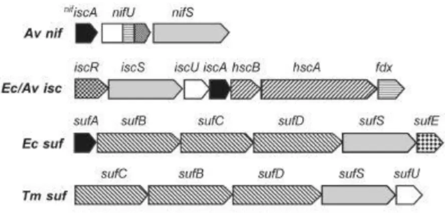
Figure 7 Organization of genes in selected bacterial nif, isc and suf operons.
Av,Azotobacter vinelandii; Ec, Escherichia coli; Tm, Thermotoga maritima.
ISC system
After expression of a A. vinelandii strain lacking of the gene nifS and nifU encoding respectively for NifS and NifU, it was observed nitrogenase activity[64,65] even if it was very low. This result suggested the presence of another system capable to cover for NIF system lack. In the 1996 another L-cysteine desulfurase was found, so the ISC system was discovered[66].
Scientists found that ISC system represents the general system for [Fe-S] cluster biosynthesis in prokaryotes, counting E. Coli and A. vinelandii too[67,68], but also it was found in Eukaryotes[46,69,70].
At least seven genes encode for ISC system. These genes are
iscR-iscS-iscU-iscA-hscB-hscA-fdx-iscX that form a gene cluster and in particular iscRSUA forms an operon.
IscR
The first role associated to IscR was its capability to regulate isc operon expression through a negative feedback control[71]. Following studies proved that IscR is implicated in the regulation of no less than 40 genes[72]. These genes encode for proteins that are directly or indirectly correlated with [Fe-S] clusters functions (periplasmic nitratereductase, hydrogenases-1 and −2,ErpA, NfuA,sufABCDSE (suf) operon[72,73]). However, its main role is to regulate [Fe-S] cluster homeostasis inside the cells. IscR selectively recognizes two different DNA binding sites (named type I and II), if the cluster
18
that it bears is present or not[74,75]. In more details [2Fe-2S]-IscR binds to type I while [2Fe-2S]-IscR and apo-IscR both bind to type II indistinguishably. But in order to accomplish this role another peculiarity is requested: IscR has an atypically ligation scheme for Fe-S clusters, indeed it is constituted of three cysteines and one histidine (Cys)3(His)1. This arrangement makes IscR a poor acceptor for iron sulfur cluster, meaning
that it is able to catch [Fe-S] clusters only at high concentrations.
Finally, it was reported that under anaerobic conditions isc is less expressed than under aerobics ones[75] since oxygen damages iron-sulfur clusters.
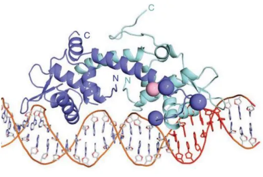
Figure 8 Overall structure of IscR-3CA bound to the hya promoter. The IscR-3CA dimer is shown as a
ribbon representation. Monomeric subunits are shown in purple and cyan; DNA is rendered as a stick model. Natural Structure and Biology (2013)
IscS
IscS like NifS is a cysteine desulfurase that is classified as a group I desulfurase[76]. After purification in E. Coli has been found that IscS is a homodimeric protein of 90kDa that bears a molecule of PLP as cofactor[77].
19
A visual examination of the crystal structure, obtained at a resolution of 2.1Å, has suggested that a conformational change is needed to allow Cys328 to take part into the catalytic cycle[78].
Depending on bacteria, IscS deletions can be lethal (A. vinelandii[51]) or alternatively (E.
Coli[52,79-81]) can lead to growth deformities. After a sulfur atom has been transferred from cysteine to IscS, another transfer occurs towards IscU with the concomitant uptake of iron to form Fe-S clusters.
IscU
IscU has a primary sequence quite similar to the N-terminal domain of NifU, plus three highly conserved cysteines. These evidences led scientists to consider IscU as a scaffold protein like NifU[46,82,83].
In vivo experiments showed that the uptake of iron-sulfur cluster in IscU leads to a
conformational change that requires also IscU and IscS association-dissociation[84].
IscU aggregation states have been largely studied: monomer, dimer and oligomers was found after extraction from E. Coli, but also a dimeric covalently bounded aggregate was observed[85] involving Cys63.
More recently studies have observed two states which quickly interconvert within milliseconds. IscU and IscS form an α2β2 complex with IscSCys328 and IscUCys63 involved in
a disulphide bond[85].
IscA
The role that IscA plays in iron-sulfur cluster biosynthesis is still unclear. During the last twenty years many roles were proposed: scaffold protein for Fe-S clusters biosynthesis[86], iron donor for clusters assembly on IscU[87], it can assemble an air sensitive [2Fe-2S] cluster[88], its metal form can bind ferrodoxin so as to form [2Fe-2S]-ferrodoxin, it can receive iron sulfur cluster from IscU but not the reverse[89].
Anyway, even if it is not as important as IscU, its presence improves ferrodoxin overexpression in E. Coli[90].
HscA and HscB
Other two proteins play an important role in iron-sulfur cluster biosynthesis, HscA that is an Hsp70 (heat shock protein) chaperone and HscB which is a cochaperone.
20
HscA like every Hsp70 protein bears three highly conserved domains: N-terminal ATPase domain (that binds ATP), substrate binding domain (which contains a binding site with a remarkably affinity for hydrophobic residues) and C-terminal domain (which acts like a lid to cover the bounded substrate to the substrate binding domain when ATP is also bounded to the N-terminal domain)[91].
For biotin synthase (BioB) assembly, BioB-HscA complex is observed, but also IscU binds to form a three-members complex, BioB-HscA-IscU[92], which helps the transfer of [Fe-S] cluster to apo-proteins.
HscB mainly stabilizes IscU in its ordered state[93].
Ferrodoxin
Ferrodoxin is a [2Fe-2S] cluster protein that accepts electrons from Fdx reductase (FDXR) that in turn accepts electrons from NADH or NADPH. Those electrons are thought to be used for reduction of sulfur (S0) to sulphide (S2-) in iron sulfur cluster biosynthesis[82,94].
A. vinelandii lacking in fdxD gene that encodes for ferrodoxin undergos to death[95], while
E. Coli its lack comports growth retards[96]. More interestingly depletion of Yah1p, the homologues of fdxD in yeast, leads to iron accumulation in mitochondria and iron-sulfur cluster enzymes inefficiency[97]. These results suggest that Fdx is essential for iron-sulfur cluster biosynthesis since it certainly takes part to an crucial step for electronic transfer.
IscX
IscX is a small protein of 7.7kDa encoded by iscX gene, placed at the end of the isc operon. Previous studies determined its structure and suggested that IscX can play a role in iron-sulfur cluster biosynthesis[98,99]. Its helical structure that exposes many acid residues can bind iron ions[98,100].
Interestingly, organisms which lack CyaY (frataxin in E. Coli) show orthologous of IscX. This can suggest that CyaY and IscX play a similar role[98].
A more recent study[101] confirmed that IscX acts as a regulator in iron-sulfur cluster assembly, indeed it binds to IscS and to IscU separately. Also a ternary complex IscU-IscS-IscX was observed. During the study of the this complex, a low activity of IscS desulfurase was observed. These results led scientists to suggest two main roles for IscX: (i) it reduces IscS desulfurase activity so as to reduce unproductive cysteine conversion (ii) it provides iron to IscU-IscS complex.
21
SUF system
After the discovery of ISC system, it would expected that cells lacking all isc genes would die. This supposition was denied when E. Coli strains with isc delection were still able to grow. The SUF system was soon after discovered[52].
Its expression was shown to be improved under iron-deficent conditions[102] and also activated by H2O2-sensors[103]. Given the above studies and considered that, in order to kill
E. Coli cells, it is strictly required to inactivate both ISC and SUF system, it can be
concluded that SUF system is essential for iron-sulfur cluster biosynthesis under oxidative stress conditions and iron starvation.
2.2.3 tRNA posttranscriptional modifications
RNA molecules can go through post-transcriptional modifications and in particular more than 100 different sulfur containing-nucleosides were identified [104-107]. The roles that modified tRNA plays are critical, indeed it is involved in biogenesis, codon recognition, maintenance of riding frame, structural stability and identification of elements for the translation machinery[108-109].
In E. Coli its synthesis was cleared up and five modifications were identified: 4-thiouridine (s4U) at position eight, 2-thiocytidine (s2C) at position thirty-two, 5-methylaminomethyl-2-thiouridine (mnm5s2U) or 5-cerboxymethylaminomethyl-2-thiouridine (cmnm5s2U) at position thirty-four, 2-methylthio-N6-isopentenyladenosine (ms2i6A) at position thirty-seven.
E. Coli uses two different ways for sulfur containing-nucleosides biosynthesis. The first
one which leads to s4U8 and (c)mnm5s2U34 is independent in Fe-S cluster biosynthesis, the second one which instead leads to s2C32 and ms2i6A37 depends on iron sulfur-cluster biosynthesis[110-111].
Both pathways start with mobilization of sulfur by IscS, that then transfers it in form of persulfide (IscS-SSH) to an acceptor. At this level the pathways diverge, indeed the sulfur atom can be transferred to a specific sulfur-carrier proteins [112-113] or to IscU, a scaffold protein, that together with IscS, is involved in iron-sulfur cluster biosynthesis. Fe-S cluster is then incorporated in modification enzymes which catalyzes tRNA modifications[114-117]. Forouhar et al. reported in 2013 that the sulfur atom is not the one of Fe-S cluster[118], so it has to be determined.
22
Modification at position thirty-four (the wobble position) of tRNA for Glu, Gln and Lys for 5-methyl-2-thiouridine (xm5s2U) is widely observed: 5-methylaminomethyl-2-thiouridine (mnm5s2U) and 5-cerboxymethylaminomethyl-2-thiouridine (cmnm5s2U) in bacterial tRNAs, 5-methoxycarbonylmethyl-2-thiouridine (mcm5s2U) in eukaryotic cytosolic tRNAs, cmnm5s2U in yeast mitochondrial tRNA and 5-taurinomethyl-2-thiouridine (τm5
s2U) in mammalian mitochondrial tRNAs[119].
The wobble position modification allows wobble base pair, which is a matching between two nucleotides in RNA that follows non-standard base pairing.
Figure 9 Sulfur-containing tRNA modification (A)Secondary structure of tRNA and positions of thiolated nucleosides in tRNA. (B) Chemical structure of thiolated nucleosides in E. coli: s4U,4-thiouridine; s2C, 2-thiocytidine;xm5s2U,5-methyl-2-thiouridinederivatives; ms2i6A, 2-methylthio-N6-isopentenyladenosine. (C) Conformation of the xm5s2U: C3’-endo form is preferred because of the steric hindrance of the 2-thio and
2’-OH groups.
Since every codon translating for an amino acid is constituted of three bases, 43=64 possible tRNA molecules should be present inside a cell if every mRNA codon exactly
23
matched with a tRNA anticodon. But three of them are stop codons which bind release factors, so 61 tRNA molecules should follow the canonical Watson and Crick base pairing. However, since most organisms have less than 45 tRNA species, tRNA must match with more than one codon.
One important consequence of the 2-thiouridine modification is the steric effect of the bulky 2-thiocarbonyl group (figure 9c) which is bigger than that of the 2-hydroxyl group and leads to a preferentially C3’-endo conformation for xm5
s2U bases[120,121].
2.3 Friedreich’s ataxia
Spinocerebellar ataxia is a progressive degenerative disease that can be divided into three principal groups: spinal ataxia, cerebellar ataxia and multiple system ataxia[122].
Friedreich’s ataxia (FRDA) is the most common among recessive ataxias. It involves spinal cord’s nerve tissue degeneration characterized by dysarthria, lower limbs areflexia, decreased vibration sense, muscles weakness of legs and positive extensor plantar response[123,124]. Non-neurological signs are also observed: hypertrophic cardiomyopathy[125,126] and diabetes mellitus[127] are the most common. It typically shows its symptoms between the ages of five and fifteen years.
Friedreich’s ataxia is observed when frataxin levels are lower than 70% of the physiological value[128]. From a biological point of view Friedreich’s ataxia is characterized by low iron-sulfur proteins activity such as complex I-III [Fe-S] enzymes, aconitase and succinate dehydrogenase. It seems that ataxia diseases are strictly related to iron-sulfur cluster biogenesis[129,130]. The role that frataxin is thought to play is iron binding chaperone during iron-sulfur cluster are assembly[131]. Frataxin bounded with IscU and ferrochelatase donates iron to [Fe-S][132]. Moreover, under frataxin depletion, iron accumulation in mitochondria can occur leading to oxidative damages catalysed by iron[133].
When the FXN gene located on chromosome nine that encodes for frataxin contains highly repeated GAA intronic sequences, Friedreich’s ataxia occurs. GAA triplet is repeated in the first introne in a way that exceed the normal threshold[134]. Since the mutation involves introne, no abnormal frataxin is synthetized, but instead gene silencing takes place.
24
2.4 TusA: State of arts
TusA is a small protein of 81 amino acids, of about 9094 kDa, with a theoretical pI of 5.18.
The gene encoding for TusA named yhhP was identified in 1998 by Yamashino et al.[135]. Several hypothetical proteins homologous are present in a variety of organisms such as
Haemophilus influenza, Bacillus subtilis, Synechocystis sp., Methanoccus jinnashii (figure
11). The similarity among these genes occurs not only in conserved amino acids which can be aligned but also in protein size, since relatively small sequences (73-84) are observed. Moreover a CPxP motif which can drive protein folding or protein function is common among the organisms cited above.
In 2000 Ishii et al. establish that TusA has a critical role in cells division, indeed cell with
yhhP deletion do not form normal colonies if grown in a rich media (Luria-Bertani
medium) but filamentous cells longer than 10µm.
Its three-dimensional structure was resolved by NMR analysis[136]; it folds in a two-layered α/β-sandwich structure. The first layer is made of four β-sheet strands (β1
residues 10-13, β2 37-43, β3 62-67, β4 73-80), the second layer consists of two α-helices (α1 residues 20-31, α2 48-59); they are wound in a βαβαββ structure with β1 and β2 connected by α1 in a classical right-hand parallel β/α/β motif, while the other β sheets form an antiparallel pattern.A hydrophobic core is shielded between the two layers. It was suggested that the highly conserved Pro53, that probably causes the slight distortion in α2 helix, contributes to this preferable folding.
During 2006 Ikeuchi et al. found that TusA is involved in 2-thiolation of mnm5s2U together with four other proteins encoded by yheL, yheM, yheN, yccK genes and named respectively TusB, TusC, TusD and TusE[137]. TusA, TusD and TusE contain conserved cysteines that may participate in sulfur transfer.
TusA function was studied following [35S] radioactivity in the substrate tRNAGlu(U8C) and data obtained showed that TusA has a critical role in sulfur transfer from the sulfurtransferases IscS to the wobble position of the substrate Uridine. The pathway starts with IscS which transfers an activated sulfur atom from the persulfide group to TusA. They interact to form a dimer[138]. Moreover, TusA bears two cysteines, C19 and C56 highly conserved in bacterial TusA homologous. C19 was identified to be the one implicated in sulfur acceptance from IscS since its substitution with serine does not allow sulfur transfer.
25
Then TusA transfers its sulfur activated atom to TusD, that together with TusB and TusC forms a ternary complex TusBCD. In the last step TusBCD interacts through TusE with MnmA, a 2-thiouridylase of mnm5s2U ATP dependent, that catalyzes the final 2-uridine modification.
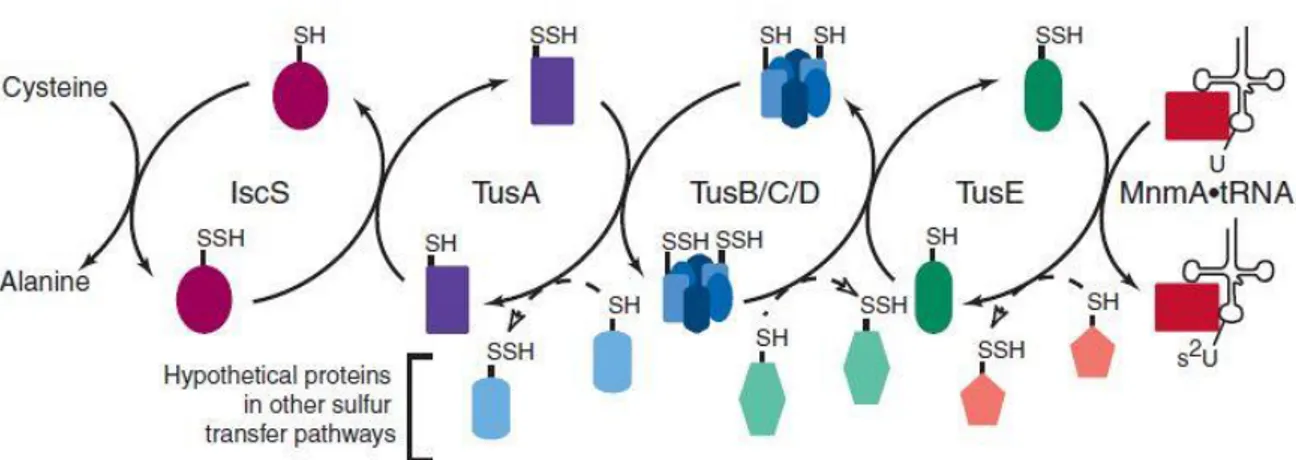
Figure 10 Sulfur transfer mediated by the Tus proteins. Sulfur transfer in s2U biosynthesis is shown with solid arrows. The potential transfer of sulfur from Tus proteins to other proteins and then to a final acceptor is
shown with dashed arrows. Each Tus protein can potentially donate sulfur to multiple proteins, each of which might participate in the thiolation of multiple acceptors.
Dahl et al. have found that TusA and IscU have a comparable dissociation constant with IscS[139]. Since they have to compete for binding on IscS, the equilibrium involving IscS and its partners is shifted to one direction or another when the concentration of one of the two partners is changed. This fact can have important effects in genes regulation. Instead, if TusA is present at low concentration, more IscS is available for IscU, leading to iron sulfur-cluster overproduction inside the cells.
Kozmin et al. demostrated that TusA with IscS are involved in MoCo biosynthesis[140], most likely in the introduction of the two sulfur atoms. It shows another possible role carried out by TusA, since E. Coli strains lacking in MoCo are hypersensitive to the mutagenic and toxic effects of (HAP) N-hdroxylated base analogs.
26
Figure 11 Sequence alignment of YhhP with putative YhhP paralogs and orthologs that are predicted to
occur in microorganisms. Among these aligned sequences, conserved and conservatively substituted residues are highlighted in magenta and dark orange, respectively, as well as the highly conserved CPxP motifs which
are in blue.
2.5 Aim of the thesis
Sulfur is present in many forms in all the organisms in the three kingdoms of life. Since it plays many important roles in living systems a very important challenge to be overcome is to understand the entire sulfur pathway inside the cells.
The main actor in bacteria which directs sulfur sorting is the desulfurase IscS. TusA, a small protein of 81 amino acids, is involved in one of the main pathways for sulfur relay that is initiated by IscS. The transulfurase TusA and the scaffold protein IscU bind to IscS with more or less the same affinity constant. The fact that one pathway can be preferred rather than another depends on many factors.
The aim of this thesis was to clone TusA in E. Coli and fully characterize it with NMR, Mass Spectroscopy and Circular Dichroism experiments. This can help to reach a deep knowledge for the proteins involved in sulfur trafficking and to better understand sulfur pathways. The subject of this thesis reveals to be deeply crucial for further studies on the crucial step in sulfur transfers (i.e. following labelled [35S] in the presence of IscU, IscS, CyaY and TusA).
27
3. RESULTS AND DISCUSSION 3.1 TusA Cloning
The cloning step is critical for the overall PCR experiment. In order to avoid errors and tedious manual selection and reduce costs and time involved in experimentation by lowering the chances of failed experimentations, the usually requirements for primers design were checked by many web-tools: length between 18-30 bases, the difference in Tm
(ΔTm) between forward and reverse primers should be lower than 5°C, the ΔG of the
secondary structure should be minimized till a value of 5Kcal/mol, presence of repeats and runs and secondary priming sites must be avoided, low specificity at the 3’ end to avoid mismatching , dimerization capability and significant hairpin should be absent too.
Table 1 a)shows the regions involved in primers annealing b)recognition sites c)the chosen rimers
a)Regions to cover with primers
TusA gene: the 5’ region
TusA gene: the 3’ region
b)Restriction sites:
Forward restriction enzyme:
NcoI: recognition site after cleavage
Reverse restriction enzyme:
NotI: recognition site after cleavage
c)Primers’ annealing
Forward:
28
The chosen vector was pETM11. It derives from pET (Novagen) backbones and carries, in addition to an antibiotic resistance encoding for kanamycin and a T7 lac promoter, a 6xHis-tag, a protease recognition site and a NcoI recognition site before protein gene. This recognition site has an ATG codon that results very useful since the number of non-native amino acids at the N-terminus can be minimized.
The gene that codifies TusA protein was amplified by PCR using genomic DNA from E.
Coli F11 as a template. PCR colony protocol was used in order to readily obtain TusA gene
from E. Coli.
The expected PCR product carried an extra amino acid, a glycine. This non-native amino acid is essential for adding NcoI restriction site just before TusA gene without having a
mistrascription.
After performing the above mentioned PCR a single band (figure 12) was obtained: this band clearly identifies TusA.
Figure 12 Agarose gel of TusA after PCR
Digestion was optimized in order to avoid partial digest or non-specific DNA cut.
TusA was treated carefully, since it appears to be easily hydrolysed during this step if scrupulous attention was not paid. The best time obtained for a satisfactory digested products, both for TusA and pETM1, was 1.5h.
Restriction enzyme were deactivated rising the temperature till 80°C and ligation was carried out without further purification.
29
In order to check TusA gene sequence after the entire cloning process the whole plasmid was expressed in DH5α E. Coli cells, purified and sent to sequencing. The figure 13 shows a perfect match between TusA E. Coli F11 strain sequence and TusA PCR product..
Figure 13 Alignment of PCR product and yhhP gene
3.2 TusA Expression
The plasmid encoding for TusA was transformed in E. Coli BL21(pLys), and expression was improved after an exhaustive expression test so as to obtain an highest yield-production time ratio.
E.Coli F11 1 NCCCNNGGGAAATTCCNTNAAAAANTTTTG--TTAACTTTAAGAAGGAGA 48 | ||||| |||||||||||||||||| PCR PRODUCT 1 ---AATTTTGATTTAACTTTAAGAAGGAGA 27 E.Coli F11 49 TATACCATGAAACATCACCATCACCATCACCCCATGAGCGATTACGACAT 98 |||||||||||||||||||||||||||||||||||||||||||||||||| PCR PRODUCT 28 TATACCATGAAACATCACCATCACCATCACCCCATGAGCGATTACGACAT 77 E.Coli F11 99 CCCCACTACTGAGAATCTTTATTTTCAGGGCGCCATGGCGACCGATCTCT 148 |||||||||||||||||||||||||||||||||||||||||||||||||| PCR PRODUCT 78 CCCCACTACTGAGAATCTTTATTTTCAGGGCGCCATGGCGACCGATCTCT 127 E.Coli F11 149 TTTCCAGCCCTGACCACACACTCGACGCGCTTGGCCTGCGCTGCCCGGAA 198 |||||||||||||||||||||||||||||||||||||||||||||||||| PCR PRODUCT 128 TTTCCAGCCCTGACCACACACTCGACGCGCTTGGCCTGCGCTGCCCGGAA 177 E.Coli F11 199 CCGGTGATGATGGTGCGCAAAACCGTGCGCAATATGCAGCCTGGCGAAAC 248 |||||||||||||||||||||||||||||||||||||||||||||||||| PCR PRODUCT 178 CCGGTGATGATGGTGCGCAAAACCGTGCGCAATATGCAGCCTGGCGAAAC 227 E.Coli F11 249 GTTGCTGATTATCGCCGACGATCCGGCCACTACCCGCGATATTCCTGGGT 298 |||||||||||||||||||||||||||||||||||||||||||||||||| PCR PRODUCT 228 GTTGCTGATTATCGCCGACGATCCGGCCACTACCCGCGATATTCCTGGGT 277 E.Coli F11 299 TTTGTACCTTTATGGAACACGAACTGGTTGCTAAAGAGACGGATGGACTG 348 |||||||||||||||||||||||||||||||||||||||||||||||||| PCR PRODUCT 278 TTTGTACCTTTATGGAACACGAACTGGTTGCTAAAGAGACGGATGGACTG 327 E.Coli F11 349 CCTTATCGTTATTTGATTCGTAAAGGCGGTTGATAGGCGGCCGCACTCGA 398 |||||||||||||||||||||||||||||||||||||||||||||||||| PCR PRODUCT 328 CCTTATCGTTATTTGATTCGTAAAGGCGGTTGATAGGCGGCCGCACTCGA 377 E.Coli F11 399 GCACCACCACCACCACCACTGAGATCCGGCTGCTAACAAAGCCCGAAAGG 448 | PCR PRODUCT 378 G--- 378
30 Expression 0.1mM IPTG 0.25mM Kan 2h 3h 6h 0.5mM Kan 2h 3h 6h 0.5 mM IPTG 0.25mM Kan 2h 3h 6h 0.5mM Kan 2h 3h 6h
Different parameters were studied: growth time, IPTG and kanamycin concentration, incubation time after induction.
A visual examination of the gel shown in figure 15 tells us that, over a period of four hours, the amount of recombinant protein (shown as a single band on SDS-PAGE) decreases from a background level to a minimum level. From this information it can be concluded that it is substantially useless to push expression over a period of three hours. Longer incubation time may not result in any significant increase in yield, rather it may involve degradation mechanisms which decrease protein concentration.
The optimal concentration of IPTG may vary from protein to protein. In order to test how IPTG concentration may modify TusA expression in BL21, its value was varied between 0.1 and 0.5 Mm, and those samples were left grow for two, three and four hours. Even though we expected to reach the maximum protein concentration after three hours, no differences were found among ours samples. Indeed, although a low IPTG value should induce more slowly, it is enough to obtain an excellent expression even after two hours. Antibiotics were used since the early years of modern biotechnology to select transformed cells from culture medium. pETM11 contains a gene that encode for kanamycin. To study how antibiotics can affect cells growth, kanamycin concentration was varied between 0.25 and 0.5 Mm. Its concentration was not found to have any appreciable effect within the range under study.
31
In conclusion, expression test showed that only induction time has a significant effect on protein yield. Degradation may occur after 3h and such a long time seems not to increase protein amount. Instead, neither IPTG concentration nor kanamycin concentration was found to have any significant effect on protein expression. So standard conditions were used for every expression.
Figure 15 SDS-PAGE of TusA after expression
3.3 TusA Characterization
TusA was overexpressed in E. Coli with an hexa histidine-tag and purified till homogeneity as indicated by ESI-MS spectra (figure 16).
Gel filtration showed a sharp peak with a retention volume between 87 and 100 ml (data not shown). Even though no standard curve was carried out, it seems that TusA does not form dimers or aggregates. So it probably exists as a monomer under working conditions.
32
3.3.1 CD analysis
Proteins can show till four distinct structural levels. Secondary structure is a local regularly repeated structure stabilized by many hydrogen bonds. The most common are alpha helix, beta sheet and turns.
Since Circular Dichroism (CD) shows high sensitivity for proteins secondary structure, it has been largely used to predict various conformations or conformational change of proteins in solution. CD working conditions are closer to the biological environment in which proteins work than the solid one of crystals used in X-rays.
Proteins can be classified on the base of secondary structure: prevalent α-helix constitutes α-rich proteins, prevalent β-sheet constitutes β-rich protein, both α-helix and β-sheet can instead be assembled in two different arrangement α+β with separate regions and α/β with intermixed region.
Each of this arrangement have a characteristic CD spectrum, shown in figure 17. Only α/β and α+β proteins cannot be easily distinguished between each other. Even unordered local secondary structure has a characteristic CD spectrum similar to that of poly(Pro)II. Protein analysis by CD spectroscopy is carried out in the far-UV region (190-250 nm) since at these wavelengths peptide bond is a chromophore.
There are a weak but broad n π* transition at more or less 210 nm and a strong
π π*
transition about at 190 nm.
The far-UV CD spectra of TusA in phosphate buffer at pH 7.2 is shown in figure 18. Each sample was prepared replacing NaCl with NaF so as to avoid NaCl absorbance below 200 nm.The far-UV CD spectra was registered for a solution containing NaF 20 mM and TusA 1.85x10⁻⁵ mM.
After Tscan analysis, TusA far-UV CD spectrum was registered again in order to check possible damages occurred after heating. How it can be observed from TusA spectrum shown in figure 18, TusA has not suffered conformational changes or damages after its
33
heating till a temperature of 90°C, since the two spectra registered before and after Tscan analysis show a perfect match in almost every region between 190 and 250 nm.
Figure 18 CD spectrum of TusA, NaF 20mM
From a visual examination of the spectrum, TusA appears as a β-rich protein since its spectrum is similar to that shown above for β-rich proteins.
Previous studies have determined its structure with different techniques such as NMR and X-rays[141,142]. Even though the latter is carried out with TusA as a crystal in a solid state, it clearly confirmed that TusA has a α+β sandwich structure with a slight difference in favour of the β-sheet.
This spectrum confirms that TusA is constituted of β-sheets more than it is constituted of α-helices. Though for a mixed α+β protein the spectrum should be predominant in α-helix, as the α-helix CD spectrum is quite more intense than that of a β-sheet, it was found that such a critical situation can lead to spectra which present only a broad minimum between 210-220 nm because of the overlapping of many β-sheets and α-helices[143].
There are two sorts of stability for proteins such as enzymes, the first one is the chemical stability that involves chemical bonds, the second one is the conformational stability for the folded state. The latter allows to determine the usefulness of a protein because it shows the practical limits for its uses.
34
To measure the conformational stability of a protein it is required to determine the equilibrium constant and the free energy change (ΔG) for the reaction:
Folded Unfolded
(3.1)
In order to calculate specific thermodynamic parameters, the entire process of unfolding and refolding must be reversible. This reversibility must be checked before denaturation and after renaturation by CD ellipticity curves that should be superimposable.
If the change in ellipticity reveals a monophasic transition between the two states, folded and unfolded, the thermodynamic parameters can be calculate.
The main procedure to calculate these parameters is to follow the ellipticity change at a specific wavelength by increasing of the temperature as a function of many factors. It could be interesting to study protein stability in different conditions such as pH, salt concentrations or ligands concentration.
Considering a two state model, the parameter can be calculate from the denaturation curve by linear extrapolation of the ΔG values[144]
. The sum of the fractions of the folded fF and
the unfolded protein fU is one (fF + fU = 1). Thus, the observed value y at any time will be
y = yFfF + yUfU where yF and yU represent the distinctive values of y for the folded and
the unfolded conformation. Combining these equations yields:
f
U= (
y
F -y)/(y
F -y
U)
(3.2)
and the equilibrium constant, K, and the free energy change, ΔG, can be calculated using:
K =
f
U/(1 -
f
U) =
f
U/
f
F=
(
y
F -y)/(y
-y
U)
(3.3)
and
ΔG = - RT ln K = - RT ln [(
y
F -y)/(y
-y
U)]
(3.4)
where T is the absolute temperature and R is the gas constant (1.987 calories/deg/mol). In the case of CD analysis equations 3.2, 3.3 and 3.4 become:
35
f
U= (θ
F-
θ)/(θ
F- θ
U)
(3.5)
K =
f
U/(1 -
f
U) =
f
U/
f
F=
(θ
F-
θ)/(θ
- θ
U)
(3.6)
ΔG = - RT ln K = - RT ln [(θ
F-
θ)/(θ - θ
U)]
(3.7)
The values of θF and θU are obtained from the pre- and post-transition region by linear
fitting based on least-square analysis.
The Tm value can be calculate from the derivative of the plot θ versus T or graphically
from the transition region of the plot ΔGD versus the absolute temperature when ΔG = 0.
Moreover from this plot other information can be determined, such as ΔSm which is the
slope of the plot and the Van’t Hoff enthalpy ΔHm that can be calculate from the equation
3.8 when ΔG = 0:
ΔG
D= ΔH
m- TΔS
m(3.8)
Furthermore, to estimate the heat capacity change for unfolding (ΔCp) it can be used the
Kirchoff equation (3.9) and the data obtained from the equation are shown above:
d(ΔH
m) / d ΔT
m= ΔC
p(3.9)
ΔCp describes the amount of the curvature of the plot ΔGD versus T. If ΔCpis higher so ΔG
will depend more strongly from the temperature. The best choice to determine it is to plot ΔHm as a function of Tm by carrying out CD experiments at different values of some
parameters. This because the value of ΔCp obtained from the difference between θF and θU
is affected by the error of the arbitral choice for the pre- and post-transition region.
ΔCp is useful for the calculation of other parameters such as the enthalpy of unfolding at
any temperature ΔH(T), the entropy of unfolding at any temperature ΔS(T) and ΔGD(T) at
36
ΔH(T) = ΔH
m+ ΔC
p(T - T
m)
(3.10)
ΔS(T) = ΔH
m+ ΔC
pln (T / T
m)
(3.11)
and the Gibbs-Helmoltz equation that represents the stability curve of the protein[145]:
ΔG
D(T) = ΔH
m(1
– T / T
m) - ΔC
p[(T
m– T) + T ln (T / T
m)]
(3.12)
The temperature-inducing unfolding process of TusA was followed by registering CD spectra at 220 nm at different NaF concentration (Figure 19). After the unfolding process, the CD signal was registered also during cooling in order to check the reversibility of the process (Figure 20).
Figure 19 Difference between thermal-inducing unfolding of TusA at two different salt concentration
To calculate Tm, a best fit of the data was made with a sigmoidal fitting using the
Boltzmann function. The derivative of the obtained curve yields the value of Tm.
The plot ΔGD versus the absolute temperature for the data inside the transition region gave
ΔSm as the slope of the curve.
The Van’t Hoff enthalpy (ΔHm) was calculated considering the equation 3.8 when ΔGD = 0
37
The temperature-independent heat capacity change at constant pressure (ΔCp) was obtained
as the slope of the plot ΔHm as a function of Tm.
38
The temperature of maximum stability (TS) where ΔS = 0 was calculated from the
equation 3.13.
T
S= T
me
[-ΔHm/(TmΔCp )](3.13)
20mM 150mM Tm(°C) 63.5 67.5 ΔSm(cal mol-1 K-1) -115.1 -115.6 ΔHm(Kcal mol-1) 38.7 39.3 TS(°C) 56.9 60.7 yF -11.45 + 5.96x10⁻⁴ (T) -7.40 + 3.06x10⁻⁴ (T) yU -6.00 -3.99 ΔCp(Kcal mol-1 ) 5.73Table 2 Thermodynamics parameters for thermal unfolding of TusA
39
Figure 22 Conformational stability curve of TusA (NaF 150 mM)
TusA is a small protein of 82 amino acids. It acts as a sulfurtransferases to mediate sulfur transfer from IscS to TusBCD complex. It is also known that TusA plays other functions inside the cells, since its presence can skew the sulfur pathway towards iron-sulfur cluster biosynthesis.
From a biophysical point of view TusA shows a typical conduct of small globular proteins[146,147]. It has a thermally reversible denaturation behaviour as described from CD spectra. Moreover TusA is highly stable as demonstrated by the Tm values and salt
concentration leads to a shift towards higher temperature.
This phenomenum can be explained as the stabilization of the negative charges that TusA bears in the side chains. Indeed the presence of negative charges leads to a diffuse repulsion that can be stabilized by the presence of the cation Na+ in solution.
40
3.3.2 Nuclear Magnetic Resonance
Deep knowledge about protein conformation is a prerequisite to study its folding proprieties and stability. The use of NMR spectroscopy in the study of protein biophysical parameters was proved to be highly valuable.
The individual amino acids in a protein structure are affected for the particular chemical environmental which is different from the random coil situation. This means that for an unfolded protein the spectrum resembles the sum of random amino acids. Instead for a folded protein the signals are more often than not shifted from the random coil values. The 1D 1H spectrum of TusA shown in figure 24 reports the protein in its folding state as there is a significant degree of chemical shift dispersion.
Figure 24 1H-NMR spectrum of TusA at 298 K
TusA was also expressed 15N-labelled in order to register a 2D-NMR spectrum, by growing E.Coli cells in M9 minimal medium with (15NH4)2SO4.
A 1H-15N HSQC spectrum with TusA uniformly labelled with 15N was obtained at 25°C and 35°C and it shows (figure 25) dispersed signals indicating a well-defined protein structure.
No differences were found from the spectrum registered at 25°C and 35°C how it was already stated by CD analysis in which TusA showed high stability.
41
Figure 25 1H 15N-HSQC spectrum of TusA at 298 K (red spots) and 308 K (blue spots)
42
4. CONCLUSION
TusA is a small protein involved in sulfur trafficking in prokaryotes. Its roles inside the cells is still under investigation; even though many functions were already confirmed, nothing is known about its stability and its biophysical parameters.
In this study TusA was expressed and many parameters were tested in order to obtain the best profile for its expression in E. Coli. Its stability in cells was proved by the easiness of its bio-production, though some degradation processes occur if the expression is maintained for long time.
TusA was also expressed in 15N-medium in order to register 2D-NMR spectra for an additional prove of its folded state that was deeply studied by CD analysis.
CD spectra confirmed that TusA, as previous found out, present a well-defined secondary structure with an abundance for the β-sheet configuration.
Moreover, thermal-induced unfolding experiments were carried out so as to calculate many thermodynamic parameters. To extract these parameters two assumption were made: (i)TusA is present during the entire range of temperature under consideration only in two conformations, (ii) ΔCp is temperature-independent under the experimental condition. The
process of folding and refolding is reversible and this feature allowed to calculate the thermodynamic parameters Tm, ΔHm, ΔCp and ΔSm, and the results suggested that TusA
has a high melting temperature and it is quite stable till the Tm value.
This work presents the first study about TusA stability. However a more broad study in which more parameters are under study is critical for a complete comprehension of TusA behaviour in physiological condition since it plays a very important role in sulfur sorting.
43
5. MATERIALS AND METHODS Solutions and buffers
- Luria Broth (LB): for 1l of LB 10g of triptone (Sigma), 5g of yeast extract (Sigma), 5g NaCl (Sigma) and 1ml NaOH (Sigma) were solubilised in H2O. The solution was then sterilised using an autoclave.
- LB for plates: to prepare 200ml, 2g of triptone, 1g of yeast extract, 1g NaCl, 0,2ml NaOH and 3g of agar (Sigma) were solubilised in H2O. The solution was then sterilised using an autoclave. When necessary, ampicillin or kanamycin (Euroclone) 1000X was added.
- Ampicillin: the powder (Euroclone) was solubilised in H2O. The solution was sterilised with a filter syringe (Millipore).
- Kanamycin: the powder (Euroclone) was solubilised in H2O in order to reach the final
concentration of 30μl/ml. The solution was filtered with a filter syringe (Millipore). - Tris 1M pH 8: the powder (Sigma) was solubilised in H2O. The pH was lowered to 8
with HCl 1M.
- NaCl 5M: the powder (Sigma) was solubilised in H2O. - IPTG: the powder (Sigma) was solubilised in H2O. - DTT: the powder (Sigma) was solubilised in H2O.
- EDTA 0,5M pH 8: the powder (Sigma) was solubilised in H2O and around 5g of solid NaOH (Sigma) were added to reach the proper pH. Finally the solution was sterilised using an autoclave.
- TBE: in order to prepare a 5X stock solution 54g of Tris (Sigma), 27.5g of boric acid (Sigma) and 20ml of 0.5M EDTA pH 8 were mixed together and H2O was added in order to reach a final volume of 1l.
- PBS (Phosphate-Buffered Saline): 8g NaCl, 0.2g KCl (Sigma), 1.44g Na2HPO4 (Sigma), 0.24g KH2PO4 (Sigma). To prepare 1l of solution, the reagents were dissolved in 800ml of H2O. The pH was adjusted to 7.4 with HCl and then H2O was added to reach the final volume of 1l.
- Equilibrium buffer: 20mM Tris pH 8, 150mM NaCl, 10mM Imidazole, 0.2% v/v Igepal (Sigma), 1mM reducing agent (β-mercaptoethanol or TCEP, Sigma).