A
A
l
l
m
m
a
a
M
M
a
a
t
t
e
e
r
r
S
S
t
t
u
u
d
d
i
i
o
o
r
r
u
u
m
m
–
–
U
U
n
n
i
i
v
v
e
e
r
r
s
s
i
i
t
t
à
à
d
d
i
i
B
B
o
o
l
l
o
o
g
g
n
n
a
a
DOTTORATO DI RICERCA IN
EPIDEMIOLOGIA E CONTROLLO DELLE ZOONOSI
Ciclo XXII
Settore/i scientifico disciplinari di afferenza: VET/05
TITOLO TESI
Survey on the spirilar flora of Lagomorphs
Presentata da: Dott.ssa Joana Marreiros Cabrita Revez
Coordinatore Dottorato
Relatore
Prof. Giovanni Poglayen
Prof. Renato Giulio Zanoni
To Mirko: “As armas e os barões assinalados, Que da ocidental praia Lusitana, Por mares nunca de antes navegados, Passaram ainda além da Taprobana, Em perigos e guerras esforçados, Mais do que prometia a força humana, E entre gente remota edificaram Novo Reino, que tanto sublimaram;” Luís Vaz de Camões (“Os Lusíadas” – Canto I)
“Não me queiram converter a convicção: sou lúcido!” Álvaro de Campos (Fernando Pessoa, “Sou lúcido”)
“A scientist in his laboratory is not a mere technician: he is also a child confronting natural phenomena that impress him as though they were fairy tales.” Marie Currie
Table of contents
Background and objectives 1
Overview of the thesis 5
Part 1: General Introduction – literature review 9
I. Bacterial taxonomy 12
II. Taxonomy of Epsiloproteobacteria 17
Taxonomic history 19
The genus Campylobacter 20
The genus Helicobacter 24
III. Lagomorphs 27
Lagomorpha 27
Production 28
Rabbit intestinal microbiota 30
Epsilonproteobacteria in leporids 30
Zoonoses 31
References 32
Part 2: Experimental work 49
Chapter 1 Campyobacter cuniculorum sp. nov. 51
Isolates 52
DNA extraction and PCR identification 52
16S ribosomal RNA 52
Housekeeping genes rpoB and groEL 54
Whole-cell protein electrophoresis 55
DNA-DNA hybridization 56
Phenotypic characteristics 56
Transmission Electron Microscopy 58
G+C content 58
Conclusion 58
Description of Campylobacter cuniculorum sp. nov. 59
References 59
Chapter 2 Occurrence of Campylobacter and Helicobacter species in leporids 63
Introduction 64
Rabbit Sampling 66
Hare Sampling 67
Cultural examination 68
DNA extraction 69
Direct PCR analysis of the caecal contents 69
Identification of bacterial isolates 70
Results 71
Direct PCR analysis of the caecal contents 71 Cultural examination and bacterial identification 71
Discussion 73
Conclusion 75
References 75
Chapter 3 Virulence determinants of Campylobacter cuniculorum 79
Introduction 80
Materials and Methods 81
Bacterial strains and grow conditions 81
Detection of cdtB gene from Campylobacter isolates by PCR 82 Preparation of mammalian cell cultures 82 Preparation of the bacteria-free supernatants for toxin assays 82 Detection of the CDT activity by cell culture assays 83
Adhesion and Invasion 84
Statistical analysis 84
Results 85
Detection of cdtB gene and CDT activity 85
Adhesion and Invasion 88
Discussion 90
Conclusion 92
References 93
Chapter 4 Antimicrobial susceptibility 97
Introduction 98
Materials and Methods 100
Campylobacter species isolates 100
Escherichia coli isolates 101
MIC determination of Campylobacter species 101
Disk diffusion 102
Results 102
MIC determination of Campylobacter species 102
Escherichia coli isolation 105
Disk diffusion test of E. coli isolates 105
Discussion 109
Conclusion 112
Part 3: General discussion and conclusions 117
Part 4 Summaries 123
Background and objectives
Members of the genera Campylobacter and Helicobacter have been in the spotlight in recent decades because of their status as animals and/or humans pathogens, both confirmed and emerging, and because of their association with food-borne and zoonotic diseases.
In 1991, the taxonomy of Campylobacter and related organisms was thoroughly revised, since this revision several new Campylobacter and Helicobacter species have been described and well-founded crediting a polyphasic taxonomic approach.
There is a paucity of data regarding the occurrence of spiral shaped enteric flora in leporids.
The main research aims of the present study were (i) to address to the characterization of Campylobacter-like organisms isolated from rabbits epidemiologically not correlated, by using a polyphasic approach; (ii) the definition of the prevalence of Campylobacter and Helicobacter in leporids; (iii) the study of some virulence determinants and finally (iv) to monitor of antibiotic susceptibility both in Campylobacter isolates and indicator bacteria.
Overview of the thesis
The objectives laying this thesis focused on the survey of spirilar enteric flora of Lagomorphs, in particular Campylobacter and Helicobacter species in rabbits and hares. The following chapters can be read individually or together. In order to study and understand the significance of the survey, the animals sampled were not correlated epidemiologically.
The first part of the thesis presents an overview of the literature relating to the content of this work. It includes a general introduction to bacterial taxonomy (I) and the bacterial species concept and the taxonomy of members of the Epsilon Proteobacteria class (II), giving an overview of the known species and respective methods currently used for classification and identification. Furthermore, a general introduction to the lagomorphs (III) biology and diseases as well as known zoonotic agents is presented.
The second part presents an overview of the experimental work performed in the framework of this thesis. Chapter 1 describes a new species, belonging to the group of thermotolerant Campylobacter, i.e. Campylobacter cuniculorum isolated from rabbits.
Chapter 2 resolves the prevalence of this new species as well as other genus members
and Helicobacter species in leporids, i.e. rabbits and hares, epidemiologically not correlated.
Chapter 3 focuses on the study of some virulence determinants of C. cuniculorum, including the effects of Cytolethal Distending Toxin (CDT) on mammalian cells, the presence of the gene ecoding CDT (cdtB), and adhesion and invasion properties. In
Chapter 4, results on monitoring antibiotic susceptibilities both in Campylobacter species and indicator bacteria (Escherichia coli) are presented.
The third part of the thesis comprises a general discussion and conclusion of the presented work.
Part 1: General Introduction – literature review
This part will whine (I) bacterial taxonomy followed by a short description of the (II) Taxonomy of the ε-Proteobacteria. Within this taxonomic overview, a short history introduction will be presented and members of the Campylobaceriacea and Helicobactereaceae will be described in detail. Subjects regarding the lagomorphs (III), in particular rabbits and hares, will be presented including a brief introduction, production, normal gut microbiota and respective disorders. Moreover, a short synopsis will illustrate the current knowledge on ε-Proteobacteria in leporids and some known zoonoses associated with lagomorphs.
I. Bacterial taxonomy
Attempts to bring order through categorization to the bewildering variety of organisms have been an ongoing human endeavor (Gevers et al., 2005). Microorganisms are fantastically diverse, and the amount and depth of genetic diversity represented among the named microbial species is greater than that represented among the animals and plants combined (Buckley and Roberts, 2007). In microbiology, bacterial taxonomy (or biosystematics) is an essential discipline that may be defined as the scientific study of the diversity of organisms with the objective of characterizing and arranging them in an orderly scheme. Within biosystematics, three sub disciplines arise: classification, nomenclature and identification (Owen, 2004; Vandamme et al., 1996). Regarding classification, it relates to the orderly arrangements of taxonomic units or taxa into groups on the basis of similarities or defined relationships. Nomenclature instead is referred to the assignment of taxonomic names to the taxa. Finally, identification is naming a distinct taxonomic unit based on the common characteristics or properties that distinguish them from other organisms. Indeed, classification precedes identification, given that it is necessary to describe and characterize the basic taxonomic unit before an isolate can be identified as a member of that unit (Buckley and Roberts, 2007; Gevers et al., 2005; Owen, 2004; Vandamme et al., 1996). In order to completely define modern biosystematics, two additional disciplines are needed: phylogeny (history, origin and evolution of a set of organisms) and population genetics (the variability of populations of bacteria and the formulation of theories that account for its variability) (Buckley and Roberts, 2007; Gevers et al., 2005; Vandamme et al., 1996). Moreover, it is currently a common practice to apply a polyphasic approach in taxonomic studies, incorporating genetic (e.g., DNA-DNA hybridization), phenotypic (e.g., the results of biochemical tests) and phylogenetic data (e.g. rRNA gene sequences), with the aim to create a consensus classification by integrating different kinds of data and information into a classification of the biological entities that contains a minimum of contradictions (Buckley and Roberts, 2007; Gevers et al., 2005; Vandamme et al., 1996).
In the twentieth century technical breakthrough changed the field of bacterial taxonomy in a groundbreaking way by defining a species as the basic taxonomic unit (Staley, 2006). For decades, classification and identification of microorganisms was based on morphological and biochemical characteristics, and a species was defined as “the type culture together with such other cultures or strains of bacteria that are accepted by bacteriologists as sufficiently closely related” (Buchanan, 1955). Nevertheless, there was no effective way to
determine “sufficiently closely related” and bacteriologists are differently competent to pass judgments on all kinds of bacteria (Buckley and Roberts, 2007; Cowan, 1965). However, on the late twentieth century with the introduction of DNA-DNA hybridization method, the species concept was defined as a group of organisms exhibiting 70% or greater DNA relatedness and less than 5% difference in their melting temperature (ΔTm) (Wayne et al., 1987). The DNA-DNA hybridization was believed to allow investigating evolution relationships through a pair wise comparison of the whole-genome sequence content between microorganisms. There are conversely some major drawbacks associated with using of DNA-DNA hybridization for species demarcation (Vandamme et al., 1996). Major disadvantages are: the laborious nature and time-consuming of the technique; different methods are used to determine the level of DNA-DNA hybridization and these methods do not always give the same (quantitative) results; finally, there is the impossibility of establishing a central database (Cho and Tiedje, 2001; Vandamme et al., 1996). Also, the 70% cut-off limit is an empirical value, created to correspond largely with the species designations in use at that time, and is not supported by a theoretical framework of what a prokaryotic species should entail (Gevers et al., 2005).
With the establishment of rapid sequence analysis of either 16S ribosomal RNA or 23S ribosomal RNA (rRNA), gene sequence analysis has become the most common molecular marker used in phylogenetic studies. The reasons for that are their universal presence, constant functionality and the fact that have mosaic structures of highly conserved and more variable domains (Gevers and Coenye, 2007; Vandamme et al., 1996). The analysis of the 16S rRNA gene sequence provides a phylogenetic framework that is particularly useful above species level. However, it has been stipulated that strains exhibiting 97% sequence similarity or less represent separate species. In addition, strains exhibiting >97% may or may not represent a single species, which should then be evaluated by determining DNA-DNA hybridization values (Stackebrandt and Goebel, 1994). Recently, it was proposed that the limit of the 16S rRNA sequence similarity should be increased to 98.7%-99%, since DNA-DNA hybridization is considered as mandatory for testing the genomic uniqueness of a novel species and lower 16S rRNA similarity values frequently do not corroborate a new species delineation (Stackebrandt and Ebers, 2006). However, overreliance on a single character, like 16S rRNA gene sequences, may lead to erroneous conclusions as verified in some members of the ε-Proteobacteria; more than 3% sequence divergence for strains belonging to the same species (as confirmed by DNA-DNA hybridization) has been
Helicobacter species (Hänninen et al., 2005; Hänninen et al., 2003; Vandamme et al., 2000).
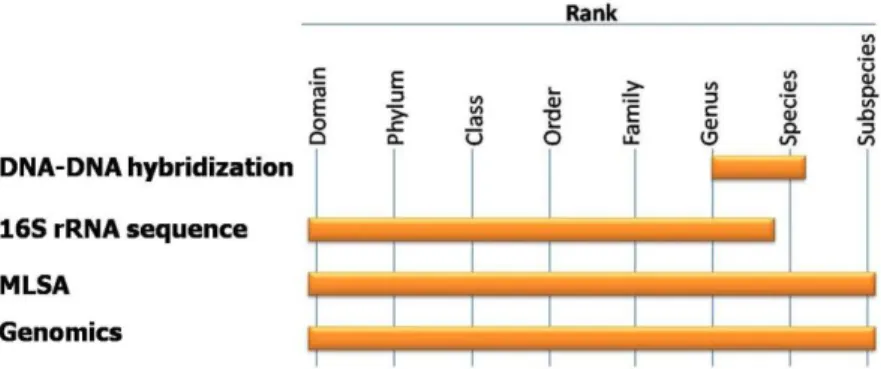
The introduction of several innovative methods, including MultiLocus gene Sequence Analysis (MLSA), whole genome sequencing and rapid DNA typing methods, prompted a re-evaluation of the bacteriological species definition (Stackebrandt et al., 2002). Figure 1 presents the taxonomic resolution of some techniques used for the classification of bacteria.
At present, the innovations mentioned above, MLSA and whole genome sequencing in particular, have been employed on a wider scale. A plethora of new papers has been evaluating diversity within and among species by exploiting whole genome information content and the bacterial species concept emerged from obscurity into the spotlight of high impact factors journals (Doolittle and Papke, 2006; Gevers et al., 2005; Konstantinidis and Tiedje, 2007).
Figure 1: Taxonomic resolution of some of the techniques used for the classification of bacteria. Adapted from Vandamme et al. (1996) and Staley (2006).
Although 16S rRNA gene sequencing is important in identifying a strain to the family or genus level, it is of very little utility for differentiation of species (Staley, 2006), MLSA can be used for analysis below the genus level and for species identification, and such may be a suitable candidate to replace DNA-DNA hybridization in the future (Gevers et al., 2005). In order to fulfill the requirements of suitability of phylogenetic marker, alternative loci have been proposed, such as housekeeping genes. In MLSA, several housekeeping genes are combined to provide a buffer against the distorting effects of recombination at a single locus (Gevers et al., 2005). Although there is no MLSA scheme, various MLSA protocols have been published for numerous genera, with varying combinations of these alternative markers. Some particular examples of the use of housekeeping genes reported for their usefulness as phylogenetic markers in different genera are: rpoB (Korczak et al., 2006) and groEL (Kärenlampi et al., 2004) for Campylobacter genus, gyrB (Hännula and Hänninen,
2007) and groEL (Mikkonen et al., 2004) for the Helicobacter spp., ureAB for the gastric Helicobacter species (Hänninen et al., 2005; O'Rourke et al., 2004). However, these genes are informative within a given genus or family and may not be useful or even present in other taxa. As an example, gyrB is a good phylogenetic marker for Helicobacter species (Hännula and Hänninen, 2007) but not for Vibrio species (Thompson et al., 2007). Nevertheless some genes may be informative in more than one group, and these more widely distributed genes could provide tools for broader comparisons (Gevers and Coenye, 2007). Even though there is quite some sepsis about the existence of a universal alternative marker in addition to the 16S and 23S rRNA genes, the rpoB gene, coding for the RNA polymerase β subunit, has been suggested as a possible candidate (Adekambi et al., 2008; Case et al., 2007). It was demonstrated that it has similar or superior resolution compared to the 16S rRNA gene and showed the highest correlation, next to 16S and 23S, to the whole-genoma based parameter average amino-acid identity (AAI), for inferring phylogenetic relationships above the species rank (Adekambi et al., 2008; Case et al., 2007; Konstantinidis and Tiedje, 2005b).
Of all the technical advances thus far, the emergence of whole–genome sequences may prove to have the biggest impact on bacterial systematics. New derived parameters have been introduced, which compare conserved genes among two organisms as measure to establish evolutionary relatedness (Konstantinidis and Tiedje, 2005a, b). Nevertheless, to evaluate genetic relatedness among ranks above species the use of core genomes is near to impossible, since finding conserved genes among more distantly related genomes is problematic, and even if orthologs are found, they are difficult to compare. Despite the fact that some methods have been developed in order to avoid this problem, and the great utility of those in aiding to create a classification system that is more predictive of genetic and biochemical relatedness (Konstantinidis and Tiedje, 2005b), it would be unrealistic to expect that in the near future bacterial species will be described based on the whole-genome sequences only. Reasons to support the non viability of the whole-whole-genome sequence are mainly the costs as well as the real significance of the method. It has been demonstrated (Konstantinidis et al., 2006) that phylogenetic trees based on MLSA data can bear the same phylogenetic signal as trees based on sequence analysis of the entire common gene pool. This implies that it is not needed the entire genome sequences for all strains under comparison of designed taxonomic frameworks. Nevertheless, in order to understand how to use MLSA data to define species and respective subdivisions from poorly
The current knowledge is very limited in terms of understanding the genetic mechanisms that drive the diversification of prokaryotes, and efforts should be directed at creating a better insight in to these phenomena. However, at the same time it could be concluded that the existing classification system is congruent with the current genomic information and that the existing, primarily 16S rRNA gene sequence and DNA-DNA hybridization based system remains functional and pragmatic (Buckley and Roberts, 2007; Konstantinidis and Tiedje, 2007).
II. Taxonomy of the Epsilonproteobacteria
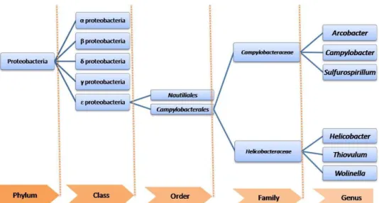
The prokaryotic domain “Bacteria” (or "Eubacteria") are subgrouped into 28 phyla. The epsilon (ε-) Proteobacteria comprise one of the five Classes within the phylum Proteobacteria (Gupta, 2006). This class is a complex phylogenetic lineage within the group of Gram-negative bacteria which includes either mucosal/animal associated or free-living species. Nowadays the order Campylobacteriales consists of two families Campylobacteraceae and Helicobacteraceae (Garrity et al., 2005b) (Figure 2). Moreover, several genera, composed by single free-living species, needs further studies in order to understand which allocation to bacterial family belong. Although the free-living Sulfuricurvum, Sulfurimonas and Sulfurovum have been proposed to be included in the family Helicobacteraceae (On and Owen, 2009), the members of Helicobacteraceae considered here are Helicobacter, Wolinella and Thiovulum as indicated in Bergey‟s Manual of Systematic Bacteriology (Garrity et al., 2005a).
Figure 2: Taxonomic organization of Proteobacteria, with particular attention for Epsilon proteobacteria. Taxonomic ranks are presented on the bottom (orange).
In Figure 3, the phylogeny of several members of epsilonproteobacteria is presented. A great deal of interest in some members of Epsilonproteobacteria stems from the fact that many of these species are host-associated (Helicobacter spp., Campylobacter spp., Wolinella sp.) and comprise important human and animal pathogens (Gupta, 2006).
Figure 3: Phylogeny of Epsilon-proteobacteria based on 16S rRNA gene. The sequences were collected aligned from the Ribosomal Database Project (Cole et al., 2009). Tree was built using iTOL – Interactive Tree Of Life (Letunic and Bork, 2007). Branches for free-living environmental ε proteobacteria sequences are coloured in grey (), Arcobacter in purple (), Campylobacter in orange (), Wolinella in yellow () and Helicobacter in green ().
The family Campylobacteraceae currently comprises the mucosal-associated species belonging to Campylobacter genus, the environmental species from the Arcobacter genus and the free-living environmental bacteria that belong to the genera Sulfurospirillum.
The family Helicobacteraceae includes the type genus Helicobacter, and consists of animal-associated bacteria occurring as commensals or parasites. Thiovulum majus (the only member of the Thiovulum genus) are free-living environmental bacteria species. Finally, the Wolinella genus, have also only one species, Wolinella succinogenes, which are ruminal-associated bacteria.
Taxonomic history. The early history of this class of Gram-negative bacteria started with
the first isolation of a Vibrio-like organism from aborted ovine fetuses by McFadyean and Stockman in 1913, later described as Vibrio fetus (Smith and Taylor, 1919). Subsequently, due to their low DNA base composition, their microaerophilic growth requirements and their nonfermentative metabolism, Vibrio fetus and Vibrio bubulus were transferred into the new genus Campylobacter as Campylobacter fetus and Campylobacter bubulus (Sebald and Véron, 1963). Ten years later, a more comprehensive study of the taxonomy of the microaerophilic Vibrio-like organisms was published, considering four distinct species in the genus Campylobacter: C. fetus (type species), Campylobacter jejuni (isolated from faeces of cattle with diarrhoea, blood of human with gastroenteritis and aborted sheep fetuses), Campylobacter coli (isolated from faeces of pigs with diarrhoea) and two subspecies of Campylobacter sputorum (subspecies sputorum, isolated from human sputum; subspecies bubulus, isolated from bovine vagina and semen) (Véron and Chatelain, 1973).
In the early 1980s, due to the availability of adequate isolation procedures [i.e. filtration technique (Butzler et al., 1973) and selective media (Skirrow, 1977)] manifold of Campylobacter-like organisms were isolated from a variety of human, animal and environmental sources and new species were described (Fox et al., 1989; Lawson et al., 1981; McClung et al., 1983; Neill et al., 1985). Nevertheless, the poor biochemical reactivity and the lack of clear-cut differential characters did not allow a clear classification of several CLOs and their taxonomic positions remained unsolved for many years (Vandamme et al., 2000; Vandamme and On, 2001).
Thanks to the introduction of bacterial phylogeny based on the degree of rRNA cistron similarity in the late 1980s, the taxonomy of Campylobacter began to be more comprehensible. The genus Campylobacter was found to be extremely heterogeneous comprising three distinct phylogenetic lineages which included members of the genera Wolinella and Bacteroides (Vandamme, 2000). The rRNA phylogeny together with phenotypic and other genotypic arguments led Goodwin and colleagues (1989) to move the
Campylobacter to a novel genus, Helicobacter. Two years later, a complete revision of the taxonomy and nomenclature of Campylobacter and related species (CLO, Helicobacter, “Flexispira”, Wolinella and Bacteroides) based on 16S rRNA phylogeny and extensive DNA-rRNA hybridization study, allocated all these microorganisms in the DNA-rRNA superfamily VI (sensu De Ley), later renamed as ε-Proteobacteria (Vandamme et al., 1991). The genus Campylobacter was restricted to those species belonging to the rRNA homology cluster I containing C. fetus. The name Arcobacter was proposed for the aereotolerant Campylobacter species belonging to the rRNA homology cluster II containing Campylobacter nitrofigilis (reclassified as Arcobacter nitrofigilis – type species of the genus). Finally, the rRNA cluster III had included the novel Helicobacter genus, Campylobacter cinaedi and Campylobacter fennelliae (reclassified as Helicobacter cinaedi and Helicobacter fennelliae respectively) and Wolinella succinogenes. The genera Campylobacter, Arcobacter and the free-living CLOs, reclassified as Sulfurospirillum, were included in the same bacterial family Campylobacteraceae (Stolz et al., 2005; Vandamme and De Ley, 1991), sharing similar phenotypic and genotypic features. Only recently the family Helicobacteraceae was described and include Helicobacter, Wolinella and Thiovulum genera (Garrity et al., 2005a), as mentioned above.
The genus Campylobacter (Sebald and Véron, 1963). [adapted from Debruyne et al.,
2008 and Euzéby, 2010a].
Members of the genus Campylobacter have following general characteristics. Cells are slender, spiral curved rods, 0.2-0.8 x 0.5-5 µm. They are Gram negative and do not form spores. Cells in old cultures may form spherical or coccoid bodies, considered to be degenerative forms difficult to detect using PCR methods.
They are typically motile, with a characteristic corkscrew-like motion performed by means of a single polar unsheathed flagellum at one or both ends of the cell. However, cells of some species are nonmotile (Campylobacter gracilis) or have multiple flagella (Campylobacter showae).
Campylobacter species grow under microaerobic atmosphere and have respiratory and chemoorganotrophic type of metabolism. Some species require anaerobiosis for optimal growth or grow only microaerobically in presence of fumarate or hydrogen as an electron donor (Campylobacter concisus, Campylobacter curvus, Campylobacter rectus,
Campylobacter mucosalis, C. gracilis and C. showae). Menaquinone-6 and methyl-substituted menaquinone-6 have been reported as major respiratory quinones in Campylobacter species.
Energy is obtained from amino acids or tricarboxylic acid cycle intermediates, not from carbohydrates which are neither fermented nor oxidized. Campylobacter spp. grow at 35-37ºC, not at 4ºC. Several species are able to grow at 42ºC (thermophilic or, more accurately, thermotolerant Campylobacter species). Gelatin, casein, starch and tyrosine are not hydrolyzed. Oxidase activity is present in all species except C. gracilis. Only Campylobacter jejuni and Campylobacter avium, as well as some strains of Campylobacter curvus, are able to hydrolize hippurate.
The G+C of the DNA ranges from 29 to 47 mol%. Plasmids have been described in a variety of species including C. jejuni, Campylobacter coli, Campylobacter upsaliensis, C. mucosalis, Campylobacter hyointestinalis and C. fetus. Tetracycline and kanamycin resistance were shown to be plasmid-mediated and transferable.
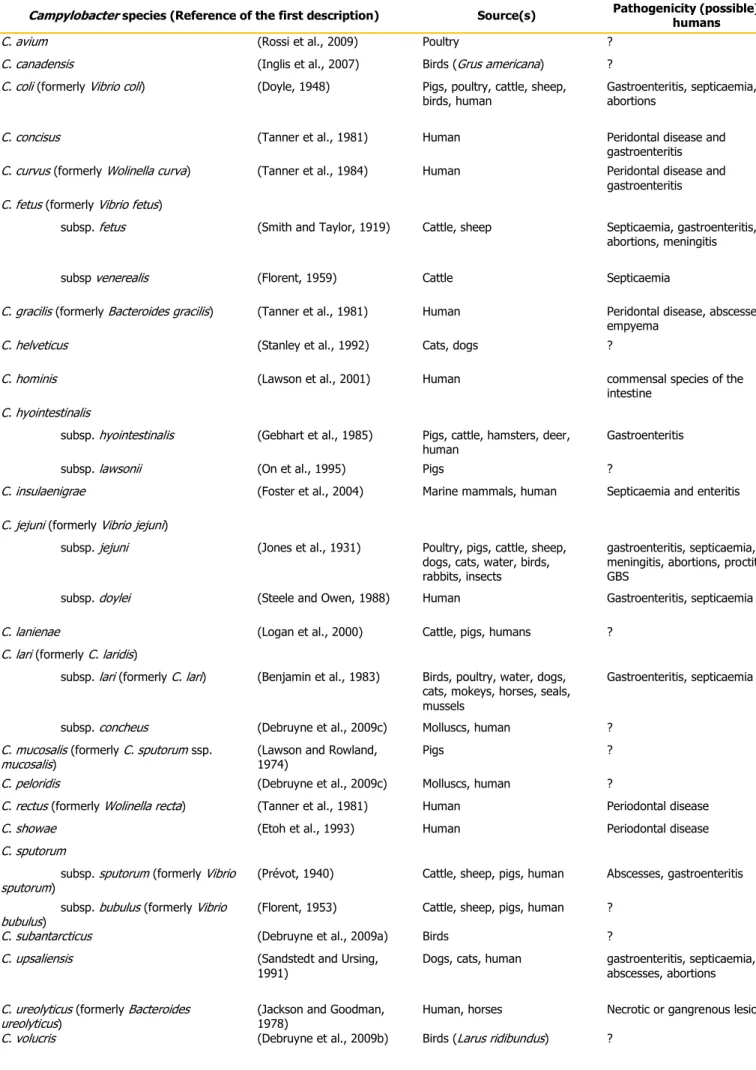
The type species of the Campylobacter genus is Campylobacter fetus (formerly Vibrio fetus, Smith and Taylor, 1919), and at present, there are 23 validly named Campylobacter species (Table 1). Within the genus Campylobacter, the group of thermophilic species, includes at present C. jejuni, C. coli, Campylobacter helveticus, C. upsaliensis, Campylobacter lari, Campylobacter insulaenigreae, C. avium, Campylobacter peloridis, Campylobacter volucris and Campylobacter subantarcticus, form a distinct 16S rRNA phylogenetic subcluster. C. fetus and C. hyointestinalis are also close relatives, while the remaining species form a loose assemblage of predominantly hydrogen-requiring organisms.
Table 1: List of the 23 Campylobacter species, isolation source(s) and human disease association.
Campylobacter species (Reference of the first description) Source(s) Pathogenicity (possible) for
humans
C. avium (Rossi et al., 2009) Poultry ? C. canadensis (Inglis et al., 2007) Birds (Grus americana) ? C. coli (formerly Vibrio coli) (Doyle, 1948) Pigs, poultry, cattle, sheep,
birds, human Gastroenteritis, septicaemia, abortions
C. concisus (Tanner et al., 1981) Human Peridontal disease and gastroenteritis C. curvus (formerly Wolinella curva) (Tanner et al., 1984) Human Peridontal disease and
gastroenteritis C. fetus (formerly Vibrio fetus)
subsp. fetus (Smith and Taylor, 1919) Cattle, sheep Septicaemia, gastroenteritis, abortions, meningitis
subsp venerealis (Florent, 1959) Cattle Septicaemia
C. gracilis (formerly Bacteroides gracilis) (Tanner et al., 1981) Human Peridontal disease, abscesses, empyema
C. helveticus (Stanley et al., 1992) Cats, dogs ?
C. hominis (Lawson et al., 2001) Human commensal species of the intestine
C. hyointestinalis
subsp. hyointestinalis (Gebhart et al., 1985) Pigs, cattle, hamsters, deer,
human Gastroenteritis subsp. lawsonii (On et al., 1995) Pigs ?
C. insulaenigrae (Foster et al., 2004) Marine mammals, human Septicaemia and enteritis
C. jejuni (formerly Vibrio jejuni)
subsp. jejuni (Jones et al., 1931) Poultry, pigs, cattle, sheep, dogs, cats, water, birds, rabbits, insects
gastroenteritis, septicaemia, meningitis, abortions, proctitis, GBS
subsp. doylei (Steele and Owen, 1988) Human Gastroenteritis, septicaemia
C. lanienae (Logan et al., 2000) Cattle, pigs, humans ? C. lari (formerly C. laridis)
subsp. lari (formerly C. lari) (Benjamin et al., 1983) Birds, poultry, water, dogs, cats, mokeys, horses, seals, mussels
Gastroenteritis, septicaemia
subsp. concheus (Debruyne et al., 2009c) Molluscs, human ? C. mucosalis (formerly C. sputorum ssp.
mucosalis) (Lawson and Rowland, 1974) Pigs ? C. peloridis (Debruyne et al., 2009c) Molluscs, human ?
C. rectus (formerly Wolinella recta) (Tanner et al., 1981) Human Periodontal disease C. showae (Etoh et al., 1993) Human Periodontal disease C. sputorum
subsp. sputorum (formerly Vibrio
sputorum) (Prévot, 1940) Cattle, sheep, pigs, human Abscesses, gastroenteritis subsp. bubulus (formerly Vibrio
bubulus) (Florent, 1953) Cattle, sheep, pigs, human ? C. subantarcticus (Debruyne et al., 2009a) Birds ? C. upsaliensis (Sandstedt and Ursing,
1991)
Dogs, cats, human gastroenteritis, septicaemia, abscesses, abortions
C. ureolyticus (formerly Bacteroides
ureolyticus) (Jackson and Goodman, 1978)
Human, horses Necrotic or gangrenous lesions C. volucris (Debruyne et al., 2009b) Birds (Larus ridibundus) ?
There is no simple gold standard for the routine isolation of all Campylobacter species. Simultaneous application of a microareobic atmosphere containing hydrogen with a filtration method and a selective base medium is methodologically the optimal solution. The first isolation method involves filtration of the cells through membrane filters with a pore size of 0.45, 0.65 or 0.8 µm using a nonselective agar medium or broth medium. For selective isolation, different selective media has been described, some using blood agar, others a blood-free agar base basal medium. However, none of these selective supplements supports growth of all of the Campylobacter species.
Incubation at 42ºC will increase selectivity by the elimination or inhibition of many, but not all, other intestinal organisms and is particularly useful for the isolation of the thermotolerant campylobacters. It will, however, inhibit growth of some other Campylobacter species.
Campylobacter species have been isolated from different sources (Table 1). They have been found in the reproductive organs, intestinal tract, and oral cavity of human and different animals.
Some species are pathogenic for humans and animals. In particular, C. jejuni is known worldwide as a major foodborne enteropathogen, with meat, milk and water as most important vehicles, causing as much enteric disease in man as Salmonella and Shigella. It infects people of all ages, more frequently diagnosed in children than adults and with higher incidence in summer season. The organism is found in the intestinal tract of a wide variety of animals and enteric C. jejuni associated disease has been reported also in animals. C. jejuni infection can trigger Guillain-Barré syndrome (an autoimmune disorder affecting the peripheral nervous system) due to similarity between its lipooligosaccharide and human gangliosides.
Other members of the thermophilic species group are C. coli, C. lari, C. insulaenigrae and C. upsaliensis, all of which are known to cause enteritis in human and to be carried in the intestinal tract of a variety of animals.
C. rectus and other oral species are associated with periodontal disease in humans and may cause infections in other part of the body.
C. fetus and C. hyointestinalis are primarily important in veterinary medicine, causing sporadic abortion and reproductive problems in cattle (C. fetus ssp. fetus, C. fetus ssp. venerealis) and sheep (C. fetus ssp. fetus) and enteric diseases in pigs (C. hyointestinalis). C. fetus ssp. fetus also can cause septicemia, meningitis, abortions and enteritis in humans,
The genus Helicobacter (Goodwin et al., 1989) [adapted from Euzéby, 2010b, On et
al., 2005 and Garrity et al., 2005a)]
The Helicobacter genus consists of a group of microorganisms that colonize the mucus layer covering the epithelial surface of the gastrointestinal tract, i.e. oral cavity, stomach, cecum and colon, and internal organs, i.e. liver, of humans and a variety of animal species (Table 2).
Members of the genus Helicobacter are Gram-negative and cells may be curved, spiral, helical or fusiform shaped with 0.2-1.2 x 1.5-10 µm, although variant forms include short or tapered rods, and they are motile by means of flagella. The fact that flagella can be sheathed or unsheathed is one characteristic that helps to differentiate different species of Helicobacter. Spiral cells may be tightly or loosely wound depending on the species, and on the age and condition of the culture. Cells in old cultures or those exposed to air form coccoid bodies.
Helicobacter species grow under microaerobic atmosphere (with or without hydrogen) and have respiratory and chemoorganotrophic type of metabolism. Microaerobic conditions with hydrogen appear to enhance growth on culture media but it is not essential. In opposition to Campylobacter species, Helicobacter spp. do not have a complete Tricarboxylic Acid Cycle. Moreover, these species are assaccharolytic when the sugar catabolism is tested by standard methods, although it has been seen that Helicobacter pylori is able to perform glucose oxidation. They do not hydrolyze gelatin, starch, casein and tyrosine, and are negative for Methyl red and Voges-Proskauer. All species have oxidase activity, and most strains produce catalase.
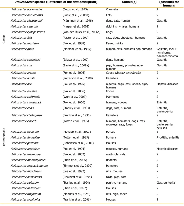
The type species of the Helicobacter genus is Helicobacter pylori (formerly Campylobacter pylori, Marshall et al., 1984), and there are currently 10 validated Helicobacter species isolated from gastric tissue and 22 validated enterohepatic species (Tables 2). Moreover, some Helicobacter species may be commonly (Helicobacter aurati) or occasionally (Helicobacter bilis and Helicobacter muridarum) isolated from both gastric and enterohepatic sites. There is, in addition, a growing list of candidate and unvalidated species (data not shown).
Table 2: List of the Helicobacter species: 10 gastric and 22 enterohepatic species, isolation source(s) and human disease association.
Helicobacter species (Reference of the first description) Source(s) Pathogenicity (possible) for
humans
G
as
tr
ic
Helicobacter acinonychis (Eaton et al., 1993) Cheetahs ? Helicobacter baculiformis (Baele et al., 2008b) Cats ? Helicobacter bizzozeronii (Hänninen et al., 1996) dogs, cats, human Gastritis Helicobacter cetorum (Harper et al., 2002) dolphins, whales, humans ? Helicobacter cynogastricus (Van den Bulck et al., 2006b) Dogs ? Helicobacter felis (Paster et al., 1991) cats, dogs, cheetahs, humans Gastritis Helicobacter mustelae (Fox et al., 1988) Ferret, minks ?
Helicobacter pylori (Marshall et al., 1985) human, cats, primates non-humans Gastritis, MALT lymphoma, adenocarcinoma Helicobacter salomonis (Jalava et al., 1997) dogs, humans Gastritis Helicobacter suis (Baele et al., 2008a) pigs, humans, primates
non-humans Gastritis En te ro hep at ic
Helicobacter anseris (Fox et al., 2006) Goose (Branta canadensis) ? Helicobacter aurati (Patterson et al., 2000) Hamsters ? Helicobacter bilis (Fox et al., 1995) rodents, dogs, cats, sheep, pigs,
humans
Hepatic diseases Helicobacter brantae (Fox et al., 2006) Goose ?
Helicobacter callitrichis (Won et al., 2007) Marmoset ? Helicobacter canadensis (Fox et al., 2000) humans, gooses Enteritis Helicobacter canis (Stanley et al., 1993) dogs, cats, humans Enteritis,
bacteraemia Helicobacter cholecystus (Franklin et al., 1996) Hamsters ?
Helicobacter cinaedi (Totten et al., 1985) humans, hamsters, dogs, cats,
monkeys, rats, foxes Enteritis, bacteraemia, cellulitis Helicobacter equorum (Moyaert et al., 2007) Horses ?
Helicobacter fennelliae (Totten et al., 1985) Humans Proctitis, enteritis Helicobacter ganmani (Robertson et al., 2001) Mouses ?
Helicobacter hepaticus (Fox et al., 1994) mouses, humans Hepatic diseases Helicobacter marmotae (Fox et al., 2002) martmots, cats ?
Helicobacter mastomyrinus (Shen et al., 2005) Rodents ? Helicobacter mesocricetorum (Simmons et al., 2000) Hamsters ? Helicobacter muridarum (Lee et al., 1992) rats, mouses ? Helicobacter pametensis (Dewhirst et al., 1994) birds, pigs, cats ?
Helicobacter pullorum (Stanley et al., 1994) poultry, humans Gastroenteritis Helicobacter rodentium (Shen et al., 1997) Mouses ?
Helicobacter trogontum (Mendes et al., 1996) rats, pigs, sheep ? Helicobacter typhlonius (Franklin et al., 2001) Mouses ?
Within gastric helicobacters, Helicobacter pylori is the most well-known species. H. pylori is established as the primary cause of gastritis and peptic ulceration in humans and has been recognized as a major risk factor for mucosa-associated lymphoid tissue (MALT) lymphoma and adenocarcinoma. The prevalence of this species infection shows large geographical variations and in general it is around 80% in developing countries, whereas in industrialized countries, the prevalence is generally under 40% (Kusters et al., 2006). H. pylori is transmitted from human-to-human and it is mostly acquired in early childhood, most probably from another family member. Although this Helicobacter leads a quiet life in the gastric mucosa for the rest of the host‟s life, about 10–20% of people carrying H. pylori may develop gastrointestinal symptoms of infection (Kusters et al., 2006).
In gastric biopsies of a minority of patients with upper gastrointestinal symptoms (0.17-2.3%) long tightly coiled spiral rods, provisionally named as “H. heilmannii”, can be observed (Baele et al., 2009). In contrast of the human-human transmission described for H. pylori, it has been suggested that “H. heilmannii" group [composed by “H. heilmannii” type one (Helicobacter suis) and H. heilmannii” type two (Helicobacter felis, Helicobacter bizzozeronii, and Helicobacter salomonis)] is transmitted by animals (Meining et al., 1998; Haesebrouck et al., 2009). Nevertheless, several groups have unsuccessfully attempted to recover “H. heilmannii” from human gastric biopsies in vitro. Up to now, H. bizzozeronii is the only “H. heilmannii” species successful cultivated from human gastric samples (Haesebrouck et al., 2009) on two different occasions: from a Danish patient in 1999 (Jalava et al., 2001) and from a Finnish patient with severe dyspeptic symptoms and chronic active gastritis in 2008 (Kivistö et al., 2010).
Transmission of gastric Helicobacter species between individuals is likely to occur via oral– oral contact, whereas enterohepatic Helicobacter species, that are shed in the feces of infected subjects, it is generally assumed that fecal–oral spread is the main route of natural acquisition of these Helicobacter infections (Moyaert et al., 2008).
Several enterohepatic Helicobacter species have been detected in humans, comprising H. canadensis (Fox et al., 2000; Laharie et al., 2009), H. pullorum (Laharie et al., 2009; Steinbrueckner et al., 1997), H. fennelliae (Hsueh et al., 1999), H. cinaedi (Matsumoto et al., 2007) and H. canis (Prag et al., 2007).
III. Lagomorphs
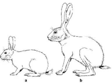
Lagomorpha. The order Lagomorpha is composed by two families: Leporidae (hares and rabbits) and Ochotonidae (pikas) (Matthee et al., 2004). These animals are distributed worldwide either as native or introduced species, showing their flexibility in adapting in different habitats (Chapman and Flux, 1990; Lombardi et al., 2007). Main external morphological differences within leporids, rabbits and hares, can be observed in Figure 4. Although both rabbits and hares share the general characteristic of caecal fermentation of digesta, hares have a smaller stomach and caecum as a proportion of body weight than the rabbit (Stott, 2008).
Figure 4: Comparative outlines of the rabbit (a) and hare (b). Main differences between genera are in leg and ear length. Adapted from Myers et al. (1989).
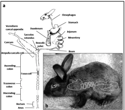
Leporids are herbivores and practive caecotrophy (Hirakawa, 2001). In fact, the gastrointestinal physiology is a very complex system that separates the digestible from the indigestible components of the diet in the proximal colon (Davies and Davies, 2003; Harcourt-Brown, 2002a). The caecum size of rabbit, is proportionally the largest of any mammal. It composes 40-60% of the total volume of the gastrointestinal tract and it is twice the length of the abdominal cavity (Davies and Davies, 2003; Harcourt-Brown, 2002a). A simplified diagram is presented in Figure 5, where it is possible to notice as well the respective topography of this tract.
Figure 5: Simplified schematic diagram of the anatomy of gastrointestinal of the rabbit a (adapted from Harcourt-Brown, 2002a) and respective topography b.
Production. The leporids economic importance, especially in Europe, is the meat, fur and
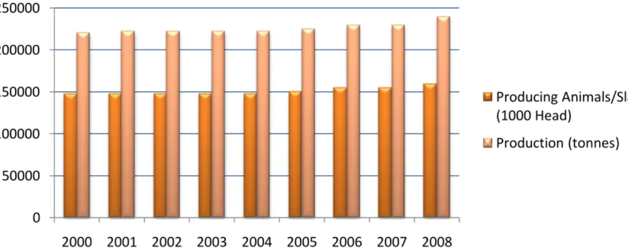
game. In addition, leporids play also a role in an ecological point of view, since they can serve either as prey for some predator species or by contributing to seed dispersal (Delibes-Mateos et al., 2007; Izhaki and Néeman, 1997). Nevertheless, rabbits are becoming important as pet animals (Wagner and Fehr, 2007) and are widely used in research as laboratory animals - in Europe over 260 000 rabbits each year are used in research (Seaman et al., 2008). In what regards meat consumption, rabbits have a bigger role than hares, for their worldwide production as food animal (Chapman and Flux, 1990; Myers et al., 1989). Nevertheless, leporid hunting date back thousands of years in Europe and even today it is important in several countries worldwide (Chapman and Flux, 1990; Cobos et al., 1995; Flux and Aneermann, 1990). Rabbit rearing in the world is mainly directed towards meat production. There are two types of production: industrial (intensive system) and rural (extensive system). Although intensive rearing systems are the most widespread, the rural production is still a common practice in different part of the world (Mendoza et al., 2004). According to FAO statistics, in 2008 China was the world leading production country of rabbit meat with 36% share, followed by Bolivarian Republic of Venezuela with 26% share, and Italy with 13% share of the world production. In Europe, Italy represents the 49% of the European production, followed by Spain with the 15% and France with the 11% (Food and Agriculture Organization of the United Nations, 2008). The Italian production can be seen in Figure 6. According to data from Italian National Union
“Associazioni di Produttori Avi-Cunicoli” (Coniglionline, 2010), in 2005 Italy produced 100.000.000 rabbits, with 3.700.000 total breeders, in which 68% breeders were from rural farm system production, whose production is addressed mainly to the domestic consumption.
Figure 6: Rates of producing rabbits and slaughtered, and production in tones of Italy from 2000 until 2008. Data retrieved from the FAO Statistics database (Food and Agriculture Organization of the United Nations, 2008).
In what concerns consumption per person, Italy leads with around 4.6kg/year, followed by France with 3kg/person and Spain with more than 2Kg/person. In the other countries of the EU, the interest for the rabbit-rearing is rather limited and the consumption per person does not exceed 1 kg (Coniglionline, 2010).
In the intensive meat rabbit industry, control of bacterial disease is a major production challenge (Eady et al., 2007). In Europe, where rabbit farming is well established, approaches to disease control have relied on environmental hygiene, requiring large capital investment in housing and routine inclusion of antibiotics in rabbit feed (Eady et al., 2007; Rodriguez-Calleja et al., 2006; Rodriguez-Calleja et al., 2004). Although microbial contamination of raw meat has always been an important issue for food safety, available information for rabbit meat microbiology is very scarce (Kohler et al., 2008; Rodriguez-Calleja et al., 2006) thus rabbit meat is considered a safe product, since has not been incriminated in outbreaks of foodborne disease (Rodriguez-Calleja et al., 2004). Nevertheless, rabbit has the potential to carry food-poisoning organisms derived from different sources, like gut content or skin (Kohler et al., 2008; Rodriguez-Calleja et al., 2004). Management systems with a reliance on the use of antibiotics are under increasing
0 50000 100000 150000 200000 250000 2000 2001 2002 2003 2004 2005 2006 2007 2008 Producing Animals/Slaughtered (1000 Head) Production (tonnes)
inspection due to the risk of microbial antibiotic resistance spreading to human pathogens (Eady et al., 2007).
Rabbit intestinal microbiota. The caecum is a finely balanced ecosystem composed of a
variety of microorganisms (Harcourt-Brown, 2002b) and the digestive physiology of rabbits is influenced by the bacterial population in the caecum (Abecia et al., 2007). The type of bacteria which are necessary in this environment, with the specific physiological conditions such as those of the rabbit caecum (e.g., rate of passage, characteristics of the input substrate, content density) may differ significantly from those necessary in other herbivorous digestive tracts (Abecia et al., 2005). In a recent metagenomic study (Abecia et al., 2005), it was reported that the rabbit caecal microbiota is highly diverse compared to other herbivorous gut microbiota. Indeed, the majority of taxa detected in the Abecia and colleagues (2005) study represented not just novel species, but also new taxonomic lineages. Various bacterial species are normally found in rabbit cecum such as Bacteroides spp., streptococci, Endophorus spp., Acuformis spp., Eubacterium spp., Fibrobacter spp., Ruminococcus spp., Clostridium, Peptococcus, Peptostreptococcus, and Fusobacterium spp., and many unidentified anaerobic species (Crociani et al., 1984; Fann and O'Rourke, 2001; Harcourt-Brown, 2002b; Monteils et al., 2008). Moreover, coliform bacteria are rarely isolated from normal rabbit cecal contents (Blanco et al., 1994; Camguilhem and Milon, 1989).
The microbiota of the rabbit is affected by several factors such as age, diet and antibiotic usage, thus leading to gastrointestinal disorders (Fann and O'Rourke, 2001; Harcourt-Brown, 2002b). More frequently happens in intensive production systems where animals are under stress and eat artificial as well as medicated diets that may lead to the proliferation of pathogens (Abecia et al., 2005, 2007; Harcourt-Brown, 2002b; Lennox and Kelleher, 2009). The main bacterial pathogens responsible for these diseases are: Salmonella enterica, Escherichia coli, Clostridum spp. and Lawsonia intracellularis (Harcourt-Brown, 2002b; Lennox and Kelleher, 2009).
Epsilon-proteobacteria in Leporids. Although some members of ε-Proteobacteria, in
particular Campylobacter and Helicobacter species, are frequently found in the gastrointestinal tracts of animals, so far there is a paucity of data concerning the presence of these bacteria in leporids. Nevertheless, Campylobacter jejuni have been isolated at low rates in hares (de Boer et al., 1983; Rosef et al., 1983) and in rabbits (Kohler et al., 2008;
Little et al., 2008; Meanger and Marshall, 1989; Prescott and Bruin-Mosch, 1981; Weber et al., 1982). In addition, only one study refers the presence of C. coli in 1% of the hares sampled (Wahlström et al., 2003). No other studies refer to the presence of other Campylobacter species in leporids, with the exception of one French study (Reynaud et al., 1993), that reports the occurence and isolation of a Campylobacter-like organism in high quantities from rabbits. In addition, few authors observed a high amount of spirilar forms in the caecum of rabbits probably referable to Campylobacter-like organisms, however without isolation (Hill, 1985; Ross et al., 1987, 1989).
Regarding Helicobacter species, so far there are no reports about the occurrence of these species in hares; however, there are two reports of occurrence in rabbits (Van den Bulck et al., 2005; Van den Bulck et al., 2006a), that presented results of the low presence of “Helicobacter heilmannii” and H. canadensis or H. pullorum.
In a recent study (Chamorro et al., 2010), that studied the effect of dietary supplementation of glutamine and glutamine and arginine in the intestinal microbiota profile of rabbits, authors report a frequency of detection of both Campylobacter and Helicobacter species by PCR-RFLP, although the authors were not performing isolation. It is important to understand and study the occurrence of Campylobacter and Helicobacter in leporids, since some species of these genera have a zoonotic character. Leporids are important not only as food animals but also as pet animals, thus it is a priority to understand if they can be associated with zoonoses caused by these microorganisms.
Zoonoses. Leporids have been associated with a discrete number of zoonoses. Concerning
wild leporids, the main bacterial diseases transmitted to humans are caused by Yersinia pseudotuberculosis (pseudotubercullosis), Pasteurella multocida (pasteurellosis), Brucella species (brucellosis), Francisella tularensis (tularemia), Yersinia pestis (plague) and Borrelia burgdorferi (Lyme disease) (Chomel, 1992; Harcourt-Brown, 2002c; Orloski and Lathrop, 2003; Telford and Spielman, 1989; Wibbelt and Frölich, 2005).
Regarding rabbits as pet animals, the diseases of major public health importance are rarely encountered and the risk is negligible. However, rabbits can bite, even though it is uncommon, and can inflict painful scratches that can become infected; moreover, owners can develop allergy to rabbit dander (Chomel, 1992; Harcourt-Brown, 2002c). Nevertheless, direct zoonotic bacterial transmission from domesticated rabbits to humans have been reported with the bacterial agents such as Pasteurella multocida, Yersinia
(Chomel, 1992; Harcourt-Brown, 2002c; Raoult et al., 2006). Other diseases to which pet rabbits are susceptible, such as tularemia, are extremely rare and are more commonly transmitted to humans by wild animals (Chomel, 1992).
No viral zoonotic diseases are known that can be transmitted from leporids to humans; other zoonotic diseases caused by parasites and fungus have been reported, such as Cheyletiella parasitovorax (by handling infested animals), Toxoplasma gondii (by eating undercooked meat), and Encephalitozoon cuniculi (ingestion of spores that are shed in the urine of the infected animals) and dermatophytosis by Trichophyton mentagrophytes (Almeria et al., 2004; Chomel, 1992; Frolich et al., 2003; Harcourt-Brown, 2002c).
In what concerns food-borne diseases, the potential bacteria isolated from leporids meat and/ or carcasses are related to Escherichia coli EPEC, Listeria spp., Campylobacter spp., Salmonella enterica and Sthaphylococcus aureus (Kohler et al., 2008).
Nevertheless, it is also considered a zoonoses subject the worldwide emerging problem of antimicrobial resistance, since the spread of resistance microorganisms can occur, thus it is important to monitor the antibiotic resistance.
References
Abecia, L., Fondevila, M., Balcells, J., Edwards, J.E., Newbold, C.J., McEwan, N.R., 2005, Molecular profiling of bacterial species in the rabbit caecum. FEMS Microbiol Lett 244, 111-115.
Abecia, L., Fondevila, M., Balcells, J., Edwards, J.E., Newbold, C.J., McEwan, N.R., 2007, Effect of antibiotics on the bacterial population of the rabbit caecum. FEMS Microbiol Lett 272, 144-153.
Adekambi, T., Shinnick, T.M., Raoult, D., Drancourt, M., 2008, Complete rpoB gene sequencing as a suitable supplement to DNA-DNA hybridization for bacterial species and genus delineation. Int J Syst Evol Microbiol 58, 1807-1814.
Almeria, S., Calvete, C., Pages, A., Gauss, C., Dubey, J.P., 2004, Factors affecting the seroprevalence of Toxoplasma gondii infection in wild rabbits (Oryctolagus cuniculus) from Spain. Vet Parasitol 123, 265-270.
Baele, M., Decostere, A., Vandamme, P., Ceelen, L., Hellemans, A., Mast, J., Chiers, K., Ducatelle, R., Haesebrouck, F., 2008a, Isolation and characterization of Helicobacter suis sp. nov. from pig stomachs. Int J Syst Evol Microbiol 58, 1350-1358.
Baele, M., Decostere, A., Vandamme, P., Van den Bulck, K., Gruntar, I., Mehle, J., Mast, J., Ducatelle, R., Haesebrouck, F., 2008b, Helicobacter baculiformis sp. nov., isolated from feline stomach mucosa. Int J Syst Evol Microbiol 58, 357-364.
Baele, M., Pasmans, F., Flahou, B., Chiers, K., Ducatelle, R., Haesebrouck, F., 2009, Non-Helicobacter pylori helicobacters detected in the stomach of humans comprise several naturally occurring Helicobacter species in animals. FEMS immunology and medical microbiology 55, 306-313.
Benjamin, J., Leaper, S., Owen, R.J., Skirrow, M.B., 1983, Description of Campylobacter laridis, a new species comprising the nalidixic acid resistant thermophilic Campylobacter (NARTC) group. . Curr Microbiol 8, 231-238.
Blanco, J.E., Blanco, M., Blanco, J., Rioja, L., Ducha, J., 1994, Serotypes, toxins and antibiotic resistance of Escherichia coli strains isolated from diarrhoeic and healthy rabbits in Spain. Vet Microbiol 38, 193-201.
Buchanan, R.E., 1955, Taxonomy. Annual review of microbiology 9, 1-20.
Buckley, M., Roberts, R.J., 2007, Reconciling Microbial Systematics and Genomics. Colloquia Reports: Colloquium report September 27-28, 2006. American Society of Microbiology.
Butzler, J.P., Dekeyser, P., Detrain, M., Dehaen, F., 1973, Related vibrio in stools. The Journal of pediatrics 82, 493-495.
Camguilhem, R., Milon, A., 1989, Biotypes and O serogroups of Escherichia coli involved in intestinal infections of weaned rabbits: clues to diagnosis of pathogenic strains. J Clin Microbiol 27, 743-747.
Case, R.J., Boucher, Y., Dahllof, I., Holmstrom, C., Doolittle, W.F., Kjelleberg, S., 2007, Use of 16S rRNA and rpoB genes as molecular markers for microbial ecology studies. Appl Environ Microbiol 73, 278-288.
Chamorro, S., de Blas, C., Grant, G., Badiola, I., Menoyo, D., Carabano, R., 2010, Effect of dietary supplementation with glutamine and a combination of glutamine-arginine on intestinal health in twenty-five-day-old weaned rabbits. Journal of animal science 88, 170-180.
Chapman, J.A., Flux, J.E.C., 1990, Introduction and Overview of the Lagomorphs, In: Chapman, J.A., Flux, J.E.C. (Eds.) Rabbits, hares and pikas: Status conservation action plan. International Union for Conservation of Nature and Natural Resources, Gland, Switzerland, pp. 1-6.
Cho, J.C., Tiedje, J.M., 2001, Bacterial species determination from DNA-DNA hybridization by using genome fragments and DNA microarrays. Appl Environ Microbiol 67, 3677-3682.
Chomel, B.B., 1992, Zoonoses of house pets other than dogs, cats and birds. The Pediatric Infectious Disease Journal 11, 479-487.
Cobos, A., de la Hoz, L., Cambero, M.I., Ordoñez, J.A., 1995, Chemical and fatty acid composition of meat from Spanish wild rabbits and hares. Zeitschrift für Lebensmitteluntersuchung und -Forschung A 200, 182-185.
Cole, J.R., Wang, Q., Cardenas, E., Fish, J., Chai, B., Farris, R.J., Kulam-Syed-Mohideen, A.S., McGarrell, D.M., Marsh, T., Garrity, G.M., Tiedje, J.M., 2009, The Ribosomal Database Project: improved alignments and new tools for rRNA analysis. Nucleic Acids Res. 37, D141-145.
Coniglionline 2010. AVITALIA e le Associazioni Regionali (http://www.coniglionline.com/ass_reg.html, date accessed: 28/02/2010).
Cowan, S.T., 1965, Principles and Practice of Bacterial Taxonomy-a Forward Look. Journal of General Microbiology 39, 143-153.
Crociani, F., Biavati, B., Castagnoli, P., Matteuzzi, D. 1984. Anaerobic ureolytic bacteria from caecal content and soft faeces of rabbit, pp. 83-88.
Davies, R.R., Davies, J.A.E.R., 2003, Rabbit gastrointestinal physiology. Veterinary Clinics of North America: Exotic Animal Practice 6, 139-153.
de Boer, E., Seldam, W.M., Stigter, H.H., 1983, Campylobacter jejuni, Yersinia enterocolitica and Salmonella in game and poultry. Tijdschrift voor diergeneeskunde 108, 831-836.
Debruyne, L., Broman, T., Bergstrom, S., Olsen, B., On, S.L.W., Vandamme, P., 2009a, Campylobacter subantarcticus sp. nov., isolated from birds in the sub-Antarctic region. Int J Syst Evol Microbiol.
Debruyne, L., Broman, T., Bergstrom, S., Olsen, B., On, S.L.W., Vandamme, P., 2009b, Campylobacter volucris sp. nov., isolated from black-headed gulls (Larus ridibundus). Int J Syst Evol Microbiol, ijs.0.013748-013740.
Debruyne, L., Gevers, D., Vandamme, P., 2008, Taxonomy of the family Campylobacteraceae, In: Nachamkin, I., Szymanski, C., Blaser, M. (Eds.) Campylobacter. ASM Press, Washington, pp. 3-25.
Debruyne, L., On, S.L.W., De Brandt, E., Vandamme, P., 2009c, Novel Campylobacter lari-like bacteria from humans and molluscs: description of Campylobacter peloridis sp.
nov., Campylobacter lari subsp. concheus subsp. nov. and Campylobacter lari subsp. lari subsp. nov. Int J Syst Evol Microbiol 59, 1126-1132.
Delibes-Mateos, M., Redpath, S.M., Angulo, E., Ferreras, P., Villafuerte, R., 2007, Rabbits as a keystone species in southern Europe. Biological Conservation 137, 149-156.
Dewhirst, F.E., Seymour, C., Fraser, G.J., Paster, B.J., Fox, J.G., 1994, Phylogeny of Helicobacter Isolates from Bird and Swine Feces and Description of Helicobacter pametensis sp. nov. Int J Syst Bacteriol 44, 553-560.
Doolittle, W.F., Papke, R.T., 2006, Genomics and the bacterial species problem. Genome biology 7, 116.
Doyle, L.P., 1948, The etiology of swine dysentery. American journal of veterinary research 9, 50-51.
Eady, S.J., Garreau, H., Gilmour, A.R., 2007, Heritability of resistance to bacterial infection in meat rabbits. Livestock Science. 112, 90-98.
Eaton, K.A., Dewhirst, F.E., Radin, M.J., Fox, J.G., Paster, B.J., Krakowka, S., Morgan, D.R., 1993, Helicobacter acinonyx sp. nov., Isolated from Cheetahs with Gastritis. Int J Syst Bacteriol 43, 99-106.
Etoh, Y., Dewhirst, F.E., Paster, B.J., Yamamoto, A., Goto, N., 1993, Campylobacter showae sp. nov., isolated from the human oral cavity. Int J Syst Bacteriol 43, 631-639.
Euzéby, J.P. 2010a. List of Prokaryotic names with Standing in Nomenclature - Genus Campylobacter, http://www.bacterio.cict.fr/c/campylobacter.html, ed. (Euzéby, J. P.).
Euzéby, J.P. 2010b. List of Prokaryotic names with Standing in Nomenclature - Genus Helicobacter, http://www.bacterio.cict.fr/h/helicobacter.html, ed. (Euzéby, J. P.). Fann, M.K., O'Rourke, D., 2001, Normal bacterial flora of the rabbit gastrointestinal tract: A
clinical approach. Seminars in Avian and Exotic Pet Medicine 10, 45-47.
Florent, A., 1953, Isolement d‟un Vibrion saprophyte du sperme du taureau et du vagin de la vache (Vibrio bubulus). C. R. SOC. Biol. 147, 2066-2069.
Florent, A., 1959, Les deux vibrioses génitales: la vibriose due à V. fetus venerealis et la vibriose d‟origine intestinale due à V. fetus intestinalis. Meded . Veeartsenijsch. Rijksuniv. Gent. 3, 1-60.
Flux, J.E.C., Aneermann, R., 1990, The Hares and Jackrabbits, In: Chapman, J.A., Flux, J.E.C. (Eds.) Rabbits, hares and pikas: Status conservation action plan. International
Union for Conservation of Nature and Natural Resources, Gland, Switzerland, pp. 61-94.
Food and Agriculture Organization of the United Nations, F.A.O. 2008. World production of rabbit meat in 2008 (FAOSTAT © FAO Statistics Division 2010, date accessed: 28/02/2010).
Foster, G., Holmes, B., Steigerwalt, A.G., Lawson, P.A., Thorne, P., Byrer, D.E., Ross, H.M., Xerry, J., Thompson, P.M., Collins, M.D., 2004, Campylobacter insulaenigrae sp. nov., isolated from marine mammals. Int J Syst Evol Microbiol 54, 2369-2373.
Fox, J.G., Chien, C.C., Dewhirst, F.E., Paster, B.J., Shen, Z., Melito, P.L., Woodward, D.L., Rodgers, F.G., 2000, Helicobacter canadensis sp. nov. isolated from humans with diarrhea as an example of an emerging pathogen. J Clin Microbiol 38, 2546-2549. Fox, J.G., Chilvers, T., Goodwin, C.S., Taylor, N.S., Edmonds, P., Sly, L.I., Brenner, D.J.,
1989, Campylobacter mustelae, a New Species Resulting from the Elevation of Campylobacter pylori subsp. mustelae to Species Status. Int J Syst Bacteriol 39, 301-303.
Fox, J.G., Dewhirst, F.E., Tully, J.G., Paster, B.J., Yan, L., Taylor, N.S., Collins, M.J., Jr., Gorelick, P.L., Ward, J.M., 1994, Helicobacter hepaticus sp. nov., a microaerophilic bacterium isolated from livers and intestinal mucosal scrapings from mice. J Clin Microbiol 32, 1238-1245.
Fox, J.G., Shen, Z., Xu, S., Feng, Y., Dangler, C.A., Dewhirst, F.E., Paster, B.J., Cullen, J.M., 2002, Helicobacter marmotae sp. nov. isolated from livers of woodchucks and intestines of cats. J Clin Microbiol 40, 2513-2519.
Fox, J.G., Taylor, N.S., Edmonds, P., Brenner, D.J., 1988, Campylobacter pylori subsp. mustelae subsp. nov. Isolated from the Gastric Mucosa of Ferrets (Mustela putorius furo), and an Emended Description of Campylobacter pylori. Int J Syst Bacteriol 38, 367-370.
Fox, J.G., Taylor, N.S., Howe, S., Tidd, M., Xu, S., Paster, B.J., Dewhirst, F.E., 2006, Helicobacter anseris sp. nov. and Helicobacter brantae sp. nov., Isolated from Feces of Resident Canada Geese in the Greater Boston Area. Appl Environ Microbiol 72, 4633-4637.
Fox, J.G., Yan, L.L., Dewhirst, F.E., Paster, B.J., Shames, B., Murphy, J.C., Hayward, A., Belcher, J.C., Mendes, E.N., 1995, Helicobacter bilis sp. nov., a novel Helicobacter species isolated from bile, livers, and intestines of aged, inbred mice. J Clin Microbiol 33, 445-454.
Franklin, C.L., Beckwith, C.S., Livingston, R.S., Riley, L.K., Gibson, S.V., Besch-Williford, C.L., Hook, R.R., Jr., 1996, Isolation of a novel Helicobacter species, Helicobacter cholecystus sp. nov., from the gallbladders of Syrian hamsters with cholangiofibrosis and centrilobular pancreatitis. J Clin Microbiol 34, 2952-2958.
Franklin, C.L., Gorelick, P.L., Riley, L.K., Dewhirst, F.E., Livingston, R.S., Ward, J.M., Beckwith, C.S., Fox, J.G., 2001, Helicobacter typhlonius sp. nov., a Novel Murine Urease-Negative Helicobacter Species. J Clin Microbiol 39, 3920-3926.
Frolich, K., Wisser, J., Schmuser, H., Fehlberg, U., Neubauer, H., Grunow, R., Nikolaou, K., Priemer, J., Thiede, S., Streich, W.J., Speck, S., 2003, Epizootiologic and ecologic investigations of European brown hares (Lepus europaeus) in selected populations from Schleswig-Holstein, Germany. J Wildl Dis 39, 751-761.
Garrity, G.M., Bell, J.A., Lilburn, T., 2005a, Family II. Helicobacteraceae fam. nov. , In: Brenner, D.J., Krieg, N.R., Staley, J.T., Garrity, G.M. (Eds.) Bergey‟s Manual of Systematic Bacteriology, second edition. Springer, New York, p. 1168.
Garrity, G.M., Bell, J.A., Lilburn, T., 2005b, Order I. Campylobacterales ord. nov. , In: Brenner, D.J., Krieg, N.R., Staley, J.T., Garrity, G.M. (Eds.) Bergey‟s Manual of Systematic Bacteriology, second edition. Springer, New York, p. 1145.
Gebhart, C.J., Edmonds, P., Ward, G.E., Kurtz, H.J., Brenner, D.J., 1985, "Campylobacter hyointestinalis" sp. nov.: a new species of Campylobacter found in the intestines of pigs and other animals. J Clin Microbiol 21, 715-720.
Gevers, D., Coenye, T., 2007, Phylogenetics and genomic analysis., In: Hurst, C.J., Crawford, R.L., Garland, J.L., Lipson, D.A., Mills, A.L., Stetzenbach, L.D. (Eds.) Manual of Environmental Microbiology ASM Press., Washington D.C., pp. 139-156. Gevers, D., Cohan, F.M., Lawrence, J.G., Spratt, B.G., Coenye, T., Feil, E.J., Stackebrandt,
E., de Peer, Y.V., Vandamme, P., Thompson, F.L., Swings, J., 2005, Re-evaluating prokaryotic species. Nat Rev Micro 3, 733-739.
Goodwin, C.S., Armstrong, J.A., Chilvers, T., Peters, M., Collins, M.D., Sly, L., McConnell, W., Harper, W.E.S., 1989, Transfer of Campylobacter pylori and Campylobacter mustelae to Helicobacter gen. nov. as Helicobacter pylori comb. nov. and Helicobacter mustelae comb. nov., respectively. Int J Syst Bacteriol 39, 397-405. Gupta, R.S., 2006, Molecular signatures (unique proteins and conserved indels) that are
specific for the epsilon proteobacteria (Campylobacterales). BMC genomics 7, 167. Haesebrouck, F., Pasmans, F., Flahou, B., Chiers, K., Baele, M., Meyns, T., Decostere, A.,