DOI 10.1007/s00726-014-1821-0 ORIGINAL ARTICLE
The spermidine analogue GC7 (N1‑guanyl‑1,7‑diamineoheptane)
induces autophagy through a mechanism not involving the
hypusination of eIF5A
Serafina Oliverio · Marco Corazzari · Claudia Sestito · Lucia Piredda · Giuseppe Ippolito · Mauro Piacentini
Received: 19 May 2014 / Accepted: 31 July 2014 © Springer-Verlag Wien 2014
These data are relevant in light of the fact that GC7 is consid-ered a potent and selective inhibitor of DHS and is a poten-tial candidate drug for cancer, diabetes and HIV therapy.
Keywords eIF5A · Autophagy · GC7 · DHS · Cancer ·
HIV
Abbreviations
eIF5A Eukaryotic initiation factor 5A DHS Deoxyhypusine synthase GC7 N1-guanyl-1,7-diaminoheptane
Introduction
Spermidine is a natural polyamine ubiquitously highly pre-sent in all living organisms; it has been implicated in many pathophysiological processes including cellular prolifera-tion, transformaprolifera-tion, differentiaprolifera-tion, apoptosis, ageing and tumorigenesis (Gerner and Meyskens 2004; Pegg 2009; Igarashi and Kashiwagi 2010; Mandal et al. 2013). The exogenous administration of spermidine promotes longev-ity in many model organisms including yeast, nematodes and flies, and significantly reduces age-related oxidative protein damage in mice (Eisenberg et al. 2009; Madeo et al. 2010; Morselli et al. 2011; Tirupathi et al. 2011). It has been postulated that the anti-age activity of spermidine could be related to this molecule’s ability to modulate the autophagic process (Eisenberg et al. 2009). Of note, spermidine plays a pivotal role in the post-translational modification of the eukaryotic initiation factor 5A (eIF5A), consisting in protein hypusination (Huang et al. 2007). eIF5A is a small (17 kDa) acidic protein carrying a unique polyamine-derived amino acid, hypusine [Nε-(4-amino-2-hydroxybutyl)lysine] (Cara-glia et al. 2013; Shiba et al. 1971; Cooper et al. 1982).
Abstract The exogenous administration of spermidine
promotes longevity in many model organisms. It has been proposed that this anti-age activity of spermidine is related to this polyamine’s ability to promote autophagy. Since spermidine is the substrate for the eIF5A post-translational modification by hypusination, we asked ourselves whether mature eIF5A may represent the link between spermidine and autophagy induction. To test this hypothesis, we inhib-ited the conversion of native eIF5A by a pharmacological approach, using the N1-guanyl-1,7-diamineoheptane (GC7), a spermidine analogue which competitively and reversibly inhibits deoxyhypusine synthase (DHS). In addition, we also employed genetic approaches by ablating both the eIF5A protein itself and DHS, the rate limiting enzyme catalyzing the conversion of lysine to hypusine. Collectively the data presented in this study demonstrate that the mature eIF5A (hypusinated form) is not involved in the autophagic path-way and that the inhibitor of DHS, GC7, produces off-target effect(s) resulting in marked induction of basal autophagy.
S. Oliverio and M. Corazzari have equally contributed to this work.
S. Oliverio · M. Corazzari · C. Sestito · L. Piredda · M. Piacentini (*)
Department of Biology, University of Rome ‘Tor Vergata’, Via Della Ricerca Scientifica, 00133 Rome, Italy
e-mail: [email protected] M. Corazzari · G. Ippolito · M. Piacentini
National Institute for Infectious Diseases I.R.C.C.S. ‘Lazzaro Spallanzani’, 00149 Rome, Italy
C. Sestito
Department of Anatomy and Neurosciences, VU University Medical Center, Neuroscience Campus, Amsterdam, The Netherlands
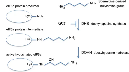
Hypusine is synthesized from the polyamine spermidine in two sequential enzymatic steps: in the first step deoxyhy-pusine synthase (DHS) catalyzes the transfer of the amin-obutyl moiety from spermidine to a specific lysine residue (Lys50 in human eIF5A) to form the deoxyhypusine inter-mediate, [Nε-(4-aminobutyl)-lysine] residue; the intermedi-ate is subsequently hydroxylintermedi-ated by deoxyhypusine hydrox-ylase (DOHH) to produce active hypusinated eIF5A (Park 2006) (Fig. 1). Two isoforms of eIF5A sharing 84 % homol-ogy exist in humans although showing distinct biological functions (Caraglia et al. 2013). eIF5A-1 is ubiquitously expressed and its level is particularly high in proliferating cells; by contrast, eIF5A-2 has a more restricted expres-sion (Jenkins et al. 2001; Guan et al. 2004). There are a lot of evidences to indicate that eIF5A is a key protein in the pathogenicity of different diseases, such as diabetes, several human cancers, malaria and HIV-1 infection (Kaiser 2012).
Although the physiological role of eIF5A-1 has not yet been fully elucidated, it has been found to function: (a) as a translation elongation factor during protein synthesis (Saini et al. 2009), (b) as a cytoplasmic shuttling protein regulat-ing mRNA transport (Liu et al. 1997; Maier et al. 2010) and (c) as a cellular cofactor of HIV-1 REV (Benne and Hershey 1978). It has also been implicated in the regula-tion of mRNA turnover (Zuk and Jacobson 1998), cell pro-liferation (Park et al. 1993, 2010), differentiation (Schnier et al. 1991; Park et al. 2010), inflammation, (Moore et al. 2008) and apoptosis (Taylor et al. 2013). Interestingly, the pro-apoptotic function of eIF5A-1 appears to be the only eIF5A activity which is independent from hypusine modifi-cation (Taylor et al. 2007, 2012; Sun et al. 2010). Growing evidence indicates that apoptosis induction is often associ-ated with decreased autophagy, underlying the existence of an interplay between these two important cellular events (Fimia and Piacentini 2010).
Autophagy is an intracellular degradation system which delivers cytoplasmic constituents to the lysosome (Xie and Klionsky 2007). This is a highly conserved process in eukaryotes and has two main physiological functions: it removes unwanted/aged/damaged constituents and recycles cytoplasmic materials to maintain macromolecular synthesis and energy homeostasis during stressful conditions includ-ing nutrient deprivation, hypoxia and low energy status.
Although Patel et al. (2009) have hypothesized that the Drosophila deoxyhypusine hydroxylase homologue Nero and its target eIF5A are involved in autophagy regula-tion, no direct evidence shows eIF5A’s involvement in the autophagic process, at least until now. On the other end, what has been recently demonstrated is spermidine’s abil-ity to stimulate autophagy in yeast, nematodes and flies, increasing the overall lifespan (Morselli et al. 2011), albeit the molecular mechanism is still unclear.
Therefore, since spermidine is directly required for eIF5A modification by hypusination, we asked whether mature eIF5A may represent the link between spermidine and autophagy. To test this hypothesis, we inhibited the conversion of native eIF5A by both pharmacological and genetic approaches and evaluated the impact on autophagy. Here we show that GC7 has an off-target effect, since its administration results in cell basal autophagy induction, independently of eIF5A activity.
Materials and methods
Materials
Mouse Anti-eIF-5a (BD 611976; dilution 1:10,000) was from BD Biosciences; rabbit anti-DHS (sc-67161; dilu-tion 1:1,000) and mouse anti-Gapdh (sc-47724; diludilu-tion
Fig. 1 Schematic
representa-tion of eIF5A post-translarepresenta-tional modification. Deoxyhypusine synthase (DHS) catalyzes the transfer of the aminobutyl moiety from spermidine to a specific lysine residue (Lys50 in human eIF5A) to form the deoxyhypusine intermediate, [Nε-(4-aminobutyl)-lysine] residue; the intermediate is subsequently hydroxylated by deoxyhypusine hydroxylase (DOHH) to produce active hypusinated eIF5A
DHS deoxyhypusine synthase
DOHH deoxyhypusine hydrolase
eIF5a protein precursor
eIF5a protein intermediate
active hypusinated eIF5a
NH3 Lys NH3 Lys NH NH3 Lys NH NH3 OH Spermidine-derived butylamino group NH2 GC7
1:1,000) were from Santa Cruz Biotechnology; rabbit anti-LC3 (NB100-2331; dilution 1:500) and Rat anti-FLAG (dilution 1:500) were from Novus Biologicals.
The anti-mouse or anti-rabbit secondary antibodies HRP-conjugated (dilution 1:5,000) and the ECL detection system (Immun-StarTM WesternTM Kit) were from Bio-Rad. Earle’s balanced salt solution (EBSS), Bafilomycin A1, chloroquine, E64D and Pepstatin A were obtained from Sigma. N1-Guanyl-1,7-diaminoheptane (GC7) was from Biosearch Technologies; D-MEM and FBS were from Invitrogen Life Technologies.
Cell culture and treatments
Human fibrosarcoma 2fTGH cells (2F) were cultured in Dul-becco’s modified Eagle’s medium (D-MEM) supplemented with 10 % foetal bovine serum, 2 mM l-glutamine, 100 mg/
ml streptomycin and 100 units/ml penicillin. Cells were grown in a humidified atmosphere containing 5 % CO2 at 37 °C.
2 × 10⁵ cells were treated with 200 µM GC7 (dissolved in 10 mM of acetic acid), 10 nM Bafilomycin A1 (BafA), 10 µg/ml of both E64d and Pepstatin A, as indicated. All compounds were dissolved in DMSO except GC7 that was dissolved in 10 mM acetic acid.
Western blotting
Cells were rinsed in ice-cold phosphate-buffered saline (PBS) and collected in lysis buffer (20 mM Tris–HCl pH 7.4, 150 mM NaCl, 1 % Triton X-100) plus protease and phos-phatase inhibitors (protease inhibitor cocktail, 1 mM sodium fluoride, 1 mM sodium orthovanadate, Sigma). Samples were centrifuged 15 min at 9,000×g and, total protein con-centration was evaluated by the DC protein Kit (Bio-Rad).
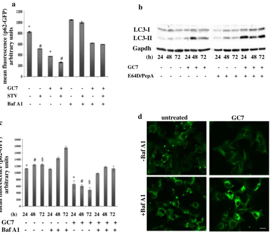
a d c mean fluorescence (p62-GFP) arbitrary unit s GC7 Baf A1 - - - + + + - - - + + +- - - + + + + + + +Baf A1 GC7 untreated -B af A1 Gapdh LC3-I (h) 24 48 72 24 48 72 24 48 72 24 48 72 (h) 24 48 72 24 48 72 24 48 72 24 48 72 GC7 - - - + + + - - - + + + E64D/PepA - - - - - - + + + + + + b LC3-II mean fluorescence (p62-GFP) arbitrary units GC7 STV Baf A1 - - + + - - + + - + - + - + - + - - - - + + + + * * # # § § * # * #
Fig. 2 Autophagy induction by GC7, an eIF5A inhibitor. a 2F cells
stably expressing a p62-GFP recombinant protein were grown in the presence or absence of 200 µM GC7 (18 h) and treated or untreated with 10 nM Bafilomycin A1 (6 h) in normal or EBSS medium. p62 degradation was evaluated by flow cytometry as the mean of fluo-rescence ± SD of three independent experiments (*,#p < 0.05). b 2F
cells were treated for 24, 48, 72 h with 200 µM GC7 and in the pres-ence or abspres-ence of 10 µg/ml E64D/Pepstatin A (the last 6 h). LC3 conversion was determined by Western blotting analysis. Gapdh was
used as a loading control. c 2F cells stably expressing a p62-GFP recombinant protein were treated or untreated with GC7, as indicated, in the presence or absence of Bafilomycin A1, and p62 degradation was evaluated by flow cytometry as the mean of fluorescence ± SD of three independent experiments (*,#,§p < 0.05). d Representative
flu-orescent micrographs of 2F cells stably expressing a p62-GFP recom-binant protein treated or untreated 24 h with GC7 and/or Bafilomycin A1. Bar 10 µm
Proteins were resolved by a 12 % SDS-PAGE and transferred onto a nitrocellulose membrane. Membranes incubated 1 h with 5 % nonfat dry milk in T-PBS contain-ing 0.05 % Tween 20 (1 h) and then incubated overnight with indicated antibodies, at 4 °C. After three washes with T-PBS, membranes were incubated 1 h with HRP-conju-gated secondary antibody, at rt. Membranes were rinsed three times with T-PBS, and the signal was detected by enhanced ECL Immunostar detection system from BioRad. Retroviral expression of GFP-p62 and mRFP-GFP-LC3 Fifteen microgram of retroviral vectors (GFP-p62 or RFP-GFP-LC3) was co-transfected with 5 μg of an expression plas-mid for the vesicular stomatitis virus G protein into 293 gp/bsr cells using the calcium phosphate method. After 48 h, the supernatant containing the retroviral particles was recovered and supplemented with polybrene (4 mg/mL). 2F cells were infected by incubation with retroviral-containing supernatant for 6–8 h, as previously described (Pagliarini et al. 2012). Autophagy analysis
For confocal microscopy analysis, 2 × 10⁵ cells were grown on glass cover slips, fixed using 4 % paraformaldehyde, and fluorescence analyzed by a Leica TCS SPII laser-scanning confocal microscope, as previously reported (Hill et al. 2009).
p62-GFP flow cytometric analysis was performed by monitoring the green-fluorescence intensity of p62 protein. Briefly, 2fp62GFP cells were fixed by 4 % paraformalde-hyde, 20,000 events were acquired by a FACScan cytome-ter (Becton–Dickinson) and data analyzed using CellQuest software (Pagliarini et al. 2012).
RNA interference
RNAi was performed using the following oligonucleotides from Ambion:
Oligos a-1 or b-1 were eIF5A Silencer Selected Pre-designed siRNA # 4392420 or Custom Selected siRNA #4390827 for eIF5A-1; siRNA #4390824 for DHS and #12935-300 as a negative control (siCtrl).
5 × 10⁵ cells/well were transfected with 100 pmol siRNA in a six-well plates using lipofectamine RNAimax (Invitrogen), as indicated by the supplier. Transfection was blocked after 24 h and cells treated as indicated.
qRT-PCR
RNA was extracted using Trizol reagent (Invitrogen) as indi-cated by the supplier. cDNA synthesis was generated using a reverse-transcription kit (Promega, Madison, WI, USA) accord-ing to the manufacturer’s recommendations. Quantitative PCRs
were performed with the Rotor-Gene 6000 (Corbett Research Ltd) thermocycler. Primer sets for all amplicons were designed using the Primer-Express 1.0 software system (Roche):
L34 forward: 5′-GTCCCGAACCCCTGGTAATAGA-3′
L34 reverse: 5′-GGCCCTGCTGACATGTTTCTT-3′
DHS forward: 5′-GTGTAAAGTGGACGCCTTCTA-3′;
DHS reverse: 5′-ACACAGGGATGTGGTTCTTC-3′;
L34 mRNA level was used as an internal control and results were expressed as previously described (Pagliarini et al. 2012).
K50A eIF5A mutant
p3XFLAG-CMV-10.1 encoding for human eIF5A-1 was kindly provided by Myung Hee Park National Institute of Dental and Craniofacial Research “NIDCR” Bethesda, MD (Clement et al. 2006). The mutant K50A was obtained in our lab using Quick Change Site-Directed Mutagenesis Kit (Stratagene). The primers used for mutate of the plasmid p3XFLAG-eIF5A-1 in the Lysine50 (p3XFLAG-K50A) were:
eIF5A K50A 5′-CTTCGAAGACTGGCGCGCACGGCC
ACGCCA–3′
eIF5A K50A antisense 5′-TGGCGTGGCCGTGCGCG
CCAGTCTTCGAAG–3′
p3XFLAG-eIF5A-1 was used as mutagenesis template. PCR amplification products were treated with restric-tion enzyme DpnI (Fermentas). An aliquot of 5 µl above PCR product was transformed into DH5a competent cells and inoculated on Luria-Beltrani (LB) plate containing 100 µg/ml ampicillin. A total of ten colonies were selected and their plasmids were isolated by mini-prep. The positive mutants were selected by DNA sequencing.
The wt and the mutant K50A of eIF5A were transiently transfected using lipofectamine LTX as indicated by the supplier.
Statistical analysis
All experiments were performed in replicate and repeated three times. Results were expressed as mean ± SD of three experiments. Data were analyzed by the t Student test and differences were considered significant when p < 0.05.
Results
The inhibitor of eIF5A activity, GC7, increases basal autophagy
To elucidate the role of eIF5A in the autophagic pathway we used the fibrosarcoma cell line 2fTGH (2F) as a model
and N1-guanyl-1,7-diamineoheptane (GC7), a spermidine analogue that competitively and reversibly inhibits deoxy-hypusine synthase (DHS; Lee and Folk 1998; Park et al. 1993; Shi et al. 1996), to inhibit eIF5A hypusination. GC7 is commonly used to block the first step of hypusination of eIF5A, resulting in the accumulation of the native protein (Landau et al. 2010). Characteristic features of early and late stages of autophagy were used to measure autophagy in 2F cells, such as the conversion of unconjugated LC3 (LC3-I) to the lipidated form (LC3-II), and the degrada-tion of p62. First, we analyzed the effect of the inhibidegrada-tion of eIF5A hypusination on basal or induced autophagy. To this end, autophagy was stimulated using EBSS medium (star-vation, stv) in 2F cells stably expressing a p62-GFP recom-binant protein (2F-p62GFP cell line); cells were treated or untreated 18 h with 200 µM GC7 in the presence or absence of 10 nM Bafilomycin A1 (BafA1), and autophagy was evaluated by measuring the degradation of p62-GFP pro-tein, by flow cytometry. As reported in Fig. 2a, we observed that the inhibition of eIF5A hypusination leads to a drastic
degradation of p62 in cells treated with GC7 alone. This potent GC7 pro-autophagic effect was also able to poten-tiate the starvation-induced p62 degradation. The positive effect of GC7 on the autophagic flux was also confirmed by the accumulation of p62 upon treatment with BafA1. To confirm these findings, 2F cells were treated for 24, 48 or 72 h with 200 µM GC7 in the presence or absence of 10 µg/ml of both E64D and PepstatinA (PepA), two inhibi-tors of autolysosome degradation activity, and autophagy induction was evaluated by monitoring the LC3 conversion by Western blotting analysis. As reported in Fig. 2b, GC7 treatment increased basal autophagy in a time-dependent manner as evidenced by the enhanced accumulation of LC3-II in the presence of E64D/PepA. Similar results were obtained in 2Fp62GFP cells in the same experimental con-ditions, by measuring the degradation of p62-GFP, by flow cytometric analysis (Fig. 2c). To further support these data the p62-GFP degradation was also evaluated by confocal analysis in 2Fp62GFP cells treated or untreated with GC7. This analysis confirmed that the GC7 administration per
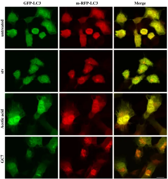
Fig. 3 GC7 does not affect the
autophagic flux. Representa-tive fluorescent micrographs of 2F cells stably expressing an RFP-GFP-LC3 recombinant protein. Cells were grown in the presence of 200 µM GC7 (24 h), vehicle (acetic acid, 24 h), EBSS (6 h) or unconditioned medium. Yellow dots represent immature autophagosomes while red dots represent active autolysosomes. Bar 10 µm
Acetic acid
untreated
se resulted in enhanced p62 degradation as evidenced by: (a) decreased GFP fluorescence (p62) compared to control (Fig. 2d, upper right panel compared to upper left panel) and (b) p62-GFP dots cytosolic accumulation in cells treated with GC7 plus BafA1 compared to cells treated with BafA1 alone (Fig. 2d, bottom panels).
Finally, to confirm that the GC7 effect resides in autophagy induction and not in the inhibition of autophagic flux, we used a GFP-RFP-tagged LC3 recombinant protein (Hill et al. 2009). To this end, 2F cells stably expressing GFP-RFP-LC3 were treated with GC7 or vehicle alone
(Acetic Acid; 24 h) and autophagy induction was evalu-ated by confocal analysis. EBSS medium (stv) was used as a positive control. Data reported in Fig. 3 clearly show a dramatic cytosolic accumulation of red-puncta LC3 dots (representing autophagolysosome, mature structures), com-pared to yellow-puncta ones (representing autophagosome, immature structures), in respect to both vehicle- alone or EBSS-treated cells, indicating a GC7-dependent enhanced complete autophagy induction, but not inhibition of autophagic flux.
eIF5A has no functional role in basal cell autophagy
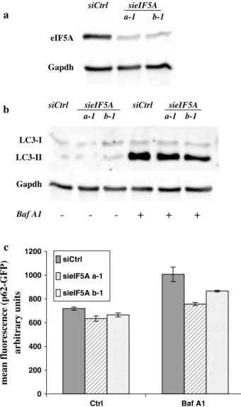
To validate by a genetic approach the consistency of our results indicating that eIF5A has a functional role in autophagy, we transiently inhibited the expression of eIF5A-1 (the isoform expressed in 2F cells), using specific siRNA oligos (sieIF5A; Fig. 4a), and cell basal autophagy was evaluated by measuring the conversion of LC3, by Western blotting analysis, in the presence or absence of BafA1 (Fig. 4b).
Although the expression of eIF5A-1 was almost com-pletely inhibited by siRNA oligos (Fig. 4a), the LC3 con-version was only marginally affected, compared to control (siCtrl; Fig. 4b).
Similar results were obtained in 2Fp62GFP cells in the same experimental conditions, by monitoring the degradation of p62-GFP in cells in which the expression of eIF5A-1 was inhibited by siRNA oligos, compared to control (siCtrl; Fig. 4c). These findings suggest that the potent pro-autophagic activity displayed by GC7 was due to an off-target effect not related to the post-transla-tional modification of eIF5A. To support this hypothesis, we decided to verify that also the eIF5A immature form (with no hypusine modification) was also not involved in modulating the autophagic process. To this end, we inhibited the modification of the native eIF5A protein by down-regulating the expression of DHS by the transient transfection of specific siRNA oligo into 2Fp62GFP cells. The inhibition was evaluated by monitoring both eIF5A protein and RNA levels (Fig. 5a). In these cells we ana-lyzed the basal autophagy in the presence or absence of BafA1, by monitoring both the LC3 conversion by West-ern blotting analysis (Fig. 5b) and p62 degradation by flow cytometry (Fig 5c). The inhibition of DHS resulted in not statistically significant effects on basal autophagy as evidenced by both LC3-II accumulation and p62 degra-dation (Fig. 5b, c).
Finally, to confirm that the accumulation of immature eIF5A resulting from inhibition of protein hypusination is not involved in autophagy induction/execution, we ectopi-cally expressed both wild type and K50A mutant eIF5A in 2F cells by transient transfection. K50A mutant codes for
mean fluorescenc e (p62-GFP ) arbitrary units c a Gapdh eIF5A siCtrl sieIF5A a-1 b-1 LC3-II Gapdh
siCtrl sieIF5A siCtrl sieIF5A a-1 b-1 a-1 b-1 Baf A1 - - - + + + b LC3-I 0 200 400 600 800 1000 1200 Ctrl Baf A1 siCtrl sieIF5A a-1 sieIF5A b-1
Fig. 4 eIF5A down-regulation does not affect basal autophagy. 2F
cells stably expressing a p62-GFP recombinant protein were tran-siently transfected with two different siRNA oligos specific for eIF5A-1 or with a scramble siRNA (siCtrl) and incubated 6 h with Bafilomycin A1, as indicated. The expression of eIF5A (a) and the conversion of LC3 (b) were determined by Western blotting analysis, while the degradation of ectopically expressed p62 (c) was evaluated by flow cytometry. Each point represents the mean of fluorescence of p62-GFP protein ± SD of three different experiments. Gapdh was used as a loading control (a, b)
an eIF5A protein in which the hypusination site (Lys50) has been abrogated (replaced with an Ala). As shown in Fig. 5d, the over-expression of both the wt and K50A eIF5A did not affect the basal autophagy of 2F cells, thus confirming the off-target effect of GC7 in the induction of autophagy.
Discussion
Spermidine plays an important role in ageing during which there is a decline of its levels in different mammalian organs (Scalabrino and Ferioli 1984). The exogenous adminis-tration of spermidine promotes longevity in many model organisms including yeast, nematodes and flies, and signifi-cantly reduces age-related oxidative protein damage in mice (Eisenberg et al. 2009; Madeo et al. 2010; Morselli et al. 2011; Tirupathi et al. 2011). This increase in longevity is linked to changes in the acetylation of nuclear histones and to a transcriptional increase of different autophagy-related genes (Eisenberg et al. 2009). Furthermore, more recent studies have shown that spermidine induces autophagy through AMPK-dependent pathway which is well known to play an anti-ageing role (Morselli et al. 2011).
Spermidine plays a pivotal role in the post-translational modification of the eukaryotic initiation factor 5A (eIF5A) since it is the essential substrate for the protein hypusina-tion (Huang et al. 2007). Under physiological conditions eIF5A is constitutively hypusinated, but its activity and subcellular localization can be conditioned by reversible acetylation (Lee et al. 2009; Ishfaq et al. 2012). PCAF is the major cellular acetyltransferase of eIF5A, and HDAC6 and SIRT2 are its major deacetylases (Ishfaq et al. 2012). Inhibition of the deacetylases or impaired hypusination increased acetylation of eIF5A, leading to its nuclear accu-mulation (Ishfaq et al. 2012). Considering that the mech-anism by which spermidine induces autophagy is not yet well elucidated, we asked whether hypusinated eIF5A can be the link in that process.
In this study, we analyzed the influence of eIF5A in the process of autophagy induced by starvation using sev-eral approaches: first, we blocked the conversion of native eIF5A into mature hypusinated protein by GC7 (N1-gua-nyl-1,7-diamineoheptane), or we used siRNA interference for knocking down the expression of native eIF5A and DHS or we over-expressed the native eIF5A or the mutated one in the hypusination site. Surprisingly, our results revealed not only that the GC7 alone can deregulate the basal autophagy, but also that this action is independent of eIF5A activity. b d a DHS Gapdh LC3-I LC3-II Gapdh Baf A1 - - + +
siCtrl siDHS siCtrl siDHS
c mean fluorescence (p62-GFP) arbitrary units 0,00 0,20 0,40 0,60 0,80 1,00 1,20 1,40 1,60 siCtrl siDHS LC3-I LC3-II Flag-eIF5A
____________
Baf A1 - + - + - + Empty wt K50A eIF5A GapdhFig. 5 Inhibiting eIF5A hypusination does not affect 2F basal
autophagy. a DHS expression was down-regulated in 2F cells stably expressing p62-GFP recombinant protein by transient transfection with a specific siRNA (siDHS) and the autophagic flux was inhib-ited by Bafilomycin A1 treatment (6 h). A scramble siRNA (siCtrl) was used as a control. The levels of both DHS protein (left panel) and mRNA (right panel) levels were evaluated by Western blotting analysis and qRT-PCR, respectively. b, c LC3 conversion (b) and p62 degradation (c) were determined by Western blotting analysis and flow cytometry, respectively. Gapdh was used as a loading control (a,
b). Values are means of fluorescence ± SD of three different
experi-ments (c). d 2F cells were transiently transfected with a Flag-tagged eIF5A-1 wild type (wt) or a K50A mutant (K50) or with empty vec-tor (Empty) and eIF5A-1 expression and LC3 conversion were evalu-ated by Western blotting analysis, in the presence or absence of Bafilomycin (6 h), as indicated
GC7 is usually used to block the first step of hypusina-tion of eIF5A which leads to the accumulahypusina-tion of the native protein (Landau et al. 2010). Interestingly, the treatment with GC7 displays an anticancer effect in various tumours such as neuroblastoma, erythroleukaemia and melanoma (Shi et al. 1996; Chen et al. 1996; Lee et al. 2002; Jasi-ulionis et al. 2007). Lee et al. (2009) demonstrated that GC7 inhibits growth and differentiation in oral cancer and immortalized keratinocytes by inducing apoptosis through the mitochondrial and the AMPK pathways (Lee et al. 2009). The results reported in this study are the first evi-dence that GC7 induces autophagy in 2fTGH cell line.
These findings are interesting considering the complex role of autophagy in cancer initiation and progression. We expected to find a block in autophagy induction in the pres-ence of the eIF5A-hypusination inhibitor GC7; surpris-ingly, by contrast, we revealed an off-target effect of the drug. In fact, the ablation of both the eIF5A itself or the enzyme mediating its hypusination, DHS, resulted in no appreciable change in the autophagic flux, thus confirming that the marked pro-autophagic effect of GC7 is not medi-ated by the hypusine pathway. In line with this conclusion, we also demonstrate that the over-expression of both wild type or K50A mutant eIF5A, in which the hypusination site (Lys50) has been replaced by an alanine, did not affect the basal autophagy in 2F cells confirming that the immature form of eIF5A (with no hypusine modification) is unrelated to the autophagic process.
In contrast to our initial hypothesis, collectively these data suggest that mature eIF5A (hypusinated form) is not involved in the autophagic pathway and that the inhibitor of deoxyhypusine synthase, GC7, has an off-target effect resulting in autophagy induction. Future studies should clarify by which mechanism GC7 is able to promote autophagy. In keeping with this assumption it is interest-ing to note that hypusine of the eIF-5A chain functions as an acyl acceptor substrate for transglutaminases (Beninati et al. 1995). Considering the structural similarity between hypusine molecule and GC7, it would be interesting to study whether this polyamine analogue can act as a sub-strate of Type 2 transglutaminase transamidating activ-ity which has been shown to play an important role in the recruitment of ubiquitinated proteins into the autophago-somes (D’Eletto et al. 2009). The GC7 off-target effect we demonstrated in this study is particularly relevant consider-ing that this drug has been proposed as an important candi-date for the therapy of cancer, diabetes and HIV infection. Indeed, eIF5A plays an important role in protein translation since disruption of the hypusination process by GC7 has been shown to inhibit the growth of many cancer cell types as well as endothelial cells (Lee et al. 2009, 2010; Cara-glia et al. 2003). For example, in hepatocellular carcinoma, over-expression of eIF5A2 was reported to be associated
with tumour features that indicate poor prognosis, such as the presence of tumour metastasis and venous infiltration (Lee et al. 2010). Furthermore, the clinical drugs ciclopirox and deferiprone, by inhibiting eIF5A hypusination, impair the transcription of the HIV-1 promoters and decrease HIV-1 gene expression (Hoque et al. 2009). Based on the considerable therapeutic interest in eIF5A as a selective target for drug development through inhibition of hypu-sination, the GC7 off-target effect described in this study acquires particular relevance and should be taken into full consideration for the use of GC7 in clinical trials.
Acknowledgments This work was supported by grants from
MIUR (PRIN 2012), AIRC (MFAG 11743 to MC; IG 2011 N.11409 to MP), FILAS, the Ministry of Health of Italy ‘‘Ricerca Corrente’’ and ‘‘Ricerca Finalizzata’’. The support of the EU Grant “Trans-path” Marie Curie project to MP is also acknowledged. We thank Prof. Myung Hee Park from National Institute of Dental and Crani-ofacial Research “NIDCR” Bethesda, MD who kindly provided the p3XFLAG-CMV-10.1 plasmid encoding for human eIF5A-1; Alessandro Lentini for his graphical support and Maria Concetta Dell’Acqua for her technical assistance.
Conflict of interest The authors have no conflicts of interest to
disclose.
References
Beninati S, Nicolini L, Jakus J, Passeggio A, Abbruzzese A (1995) Identification of a substrate site for transglutaminases on the human protein synthesis initiation factor 5A. Biochem J 305(3):725–728
Benne R, Hershey JW (1978) The mechanism of action of protein synthesis initiation factors from rabbit reticulocytes. J Biol Chem 253:3078–3087
Caraglia M, Marra M, Giuberti G, D’Alessandro AM, Baldi A, Tas-sone P, Venuta S, Tagliaferri P, Abbruzzese A (2003) The eukar-yotic initiation factor 5A is involved in the regulation of prolif-eration and apoptosis induced by interferon-alpha and EGF in human cancer cells. J Biochem (Tokyo) 133:757–765
Caraglia M, Park MH, Wolff EC, Marra M, Abbruzzese A (2013) eIF5A isoforms and cancer: two brothers for two functions? Amino Acids 44(1):103–109
Clement PM, Johansson HE, Wolff EC, Park MH (2006) Differential expression of eIF5A-1 and eIF5A-2 in human cancer cells. FEBS J 273(6):1102–1114
Cooper HL, Park MH, Folk JE (1982) Posttranslational formation of hypusine in a single major protein occurs generally in growing cells and is associated with activation of lymphocyte growth. Cell 29:791–797
D’Eletto M, Farrace MG, Falasca L, Reali V, Oliverio S, Melino G, Griffin M, Fimia GM, Piacentini M (2009) Transglutaminase 2 is involved in autophagosome maturation. Autophagy 5:1145–1154 Eisenberg T, Knauer H, Schauer A, Buttner S, Ruckenstuhl C,
Car-mona-Gutierrez D, Ring J, Schroeder S, Magnes C, Antonacci L, Fussi H, Deszcz L, Hartl R, Schraml E, Criollo A, Megalou E, Weiskopf D, Laun P, Heeren G, Breitenbach M, Grubeck-Loebenstein B, Fahrenkrog B, FroÅNhlich KU, Sinner F, Tav-ernarakis N, Minois N, Kroemer G, Madeo F (2009) Induction of autophagy by spermidine promotes longevity. Nat Cell Biol 11:1305–1314
Fimia GM, Piacentini M (2010) Regulation of autophagy in mammals and its interplay with apoptosis. Cell Mol Life Sci 67:1581–1588 Gerner EW, Meyskens FL Jr (2004) Polyamines and cancer: old
mol-ecules, new understanding. Nat Rev Cancer 4(10):781–792 Guan XY, Fung JM, Ma NF, Lau SH, Tai LS, Xie D, Zhang Y, Hu L,
Wu QL, Fang Y et al (2004) Oncogenic role of eIF-5A2 in the development of ovarian cancer. Cancer Res 64:4197–4200 Hill DS, Martin S, Armstrong JL, Flockhart R, Tonison JJ, Simpson
DG, Birch-Machin MA, Redfern CP, Lovat PE (2009) Combin-ing the endoplasmic reticulum stress-inducCombin-ing agents bortezomib and fenretinide as a novel therapeutic strategy for metastatic mel-anoma. Clin Cancer Res 15(4):1192–1198
Hoque M, Hanauske-Abel HM, Palumbo P, Saxena D, D’Alliessi Gandolfi D, Park MH, Pe’ery T, Mathews MB (2009) Inhibition of HIV-1 gene expression by Ciclopirox and Deferiprone, drugs that prevent hypusination of eukaryotic initiation factor 5A. Ret-rovirology 6:90. doi:10.1186/1742-4690-6-90
Huang Y, Higginson DS, Hester L, Park MH, Snyder SH (2007) Neuronal growth and survival mediated by eIF5A, a polyamine-modified translation initiation factor. Proc Natl Acad Sci USA 104:4194–4199
Igarashi K, Kashiwagi K (2010) Modulation of cellular function by polyamines. Int J Biochem Cell Biol 42(1):39–51
Jasiulionis MG, Luchessi AD, Moreira AG, Souza PP, Suenaga AP, Correa M, Costa CA, Curi R, Costa-Neto CM (2007) Inhibition of eukaryotic translation initiation factor 5A (eIF5A) hypusina-tion impairs melanoma growth. Cell Biochem Funct 25:109–114 Jenkins ZA, Haag PG, Johansson HE (2001) Human eIF5A2 on
chro-mosome 3q25-q27 is a phylogenetically conserved vertebrate variant of eukaryotic translation initiation factor 5A with tissue-specific expression. Genomics 71:101–109
Kaiser A (2012) Translational control of eIF5A in various diseases. Amino Acids 42(2–3):679–684
Landau G, Bercovich Z, Park MH, Kahana C (2010) The role of poly-amines in supporting growth of mammalian cells is mediated through their requirement for translation initiation and elonga-tion. J Biol Chem 285:12474–12481
Lee YB, Folk JE (1998) Branched-chain and unsaturated 1,7-diamino-heptane derivatives as deoxyhypusine synthase inhibitors. Bioorg Med Chem 6(3):253–270
Lee Y, Kim HK, Park HE, Park MH, Joe YA (2002) Effect of N1-gua-nyl-1,7 diaminoheptane, an inhibitor of deoxyhypusine synthase, on endothelial cell growth, differentiation and apoptosis. Mol Cell Biochem 237(1–2):69–76
Lee SK, Lee J, Lee SI, Bae WJ, Lee YM, Park JS, Lee SK, Park SJ, Min SK, Kim EC (2009) N1-guanyl-1,7,-diamineoheptane, an inhibitor of deoxyhypusine synthase, suppresses differentiation and induces apoptosis via mitochondrial and AMPK pathways in immortalized and malignant human oral keratinocytes. J Oral Pathol Med 38:792–800
Lee NP, Tsang FH, Shek FH, Mao M, Dai H, Zhang C, Dong S, Guan XY, Poon RT, Luk JM (2010) Prognostic significance and therapeutic potential of eukaryotic translation initiation factor 5A (eIF5A) in hepatocellular carcinoma. Int J Cancer 127(4):968–976
Liu YP, Nemeroff M, Yan YP, Chen KY (1997) Interaction of eukary-otic initiation factor 5A with the human immunodeficiency virus type 1 Rev response element RNA and U6 snRNA requires deox-yhypusine or hypusine modification. Biol Signals 6:166–174 Madeo F, Eisenberg T, Büttner S, Ruckenstuhl C, Kroemer G (2010)
Spermidine: a novel autophagy inducer and longevity elixir. Autophagy 6(1):160–162
Maier B, Ogihara T, Trace AP, Tersey SA, Robbins RD, Chakra-barti SK, Nunemaker CS, Stull ND, Taylor CA, Thompson JE, Dondero RS, Lewis EC, Dinarello CA, Nadler JL, Mirmira RG (2010) The unique hypusine modification of eIF5A promotes
islet b cell inflammation and dysfunction in mice. J Clin Invest 120:2156–2170
Mandal S, Mandal A, Johansson HE, Orialo AV, Park MH (2013) Depletion of cellular polyamines, spermidine and spermine, causes a total arrest in translation and growth in mammalians cells. PNAS 110(6):2169–2174
Moore CC, Martin EN, Lee G, Taylor C, Dondero R et al (2008) Eukaryotic translation initiation factor 5A small interference RNA-liposome complexes reduce inflammation and increase sur-vival in murine models of severe sepsis and acute lung injury. J Infect Dis 198(9):1407–1414
Morselli E, Marino G, Bennetzen MV, Eisenberg T, Megalou E, Schroeder S, Cabrera S, BeÅLnit P, Rustin P, Criollo A, Kepp O, Galluzzi L, Shen S, Malik SA, Maiuri MC, Horio Y, LoÅLpez-OtiÅLn C, Andersen JS, Tavernarakis N, Madeo F, Kroemer G (2011) Spermidine and resveratrol induce autophagy by dis-tinct pathways converging on the acetylproteome. J Cell Biol 192:615–629
Pagliarini V, Wirawan E, Romagnoli A, Ciccosanti F, Lisi G, Lippens S, Cecconi F, Fimia GM, Vandenabeele P, Corazzari M, Piacen-tini M (2012) Proteolysis of Ambra1 during apoptosis has a role in the inhibition of the autophagic pro-survival response. Cell Death Differ 19(9):1495–1504
Park MH (2006) The post-translational synthesis of a polyamine-derived amino acid, hypusine, in the eukaryotic translation initia-tion factor 5A (eIF5A). J Biochem (Tokyo) 139:161–169 Park MH, Wolff EC, Folk JE (1993) Hypusine: its post-translational
formation in eukaryotic initiation factor 5A and its potential role in cellular regulation. BioFactors 4:95–104
Park MH, Nishimura K, Zanelli CF, Valentini SR (2010) Functional significance of eIF5A and its hypusine modification in eukary-otes. Amino Acids 38:491–500
Patel PH, Costa-Mattioli M, Schulze KL, Bellen HJ (2009) The Drosophila deoxyhypusine hydroxylase homologue nero and its target eIF5A are required for cell gro wth and the regulation of autophagy. J Cell Biol 185:1181–1194
Pegg AE (2009) Mammalian polyamine metabolism and function. IUBMB Life 61(9):880–894
Saini P, Eyler DE, Green R, Dever TE (2009) Hypusine-containing protein eIF-5A promotes translation elongation. Nature 459:118– 121. doi:10.1038/nature08034
Scalabrino G, Ferioli ME (1984) Polyamines in mammalian age-ing: an oncological problem, too? A review. Mech Ageing Dev 26:149–164
Schnier J, Schwelberger HG, Smit-McBride Z, Kang HA, Hershey JW (1991) Translation initiation factor 5A and its hypusine modi-fication are essential for cell viability in the yeast Saccharomyces cerevisiae. Mol Cell Biol 11:3105–3114
Shi XP, Yin KC, Ahem J, Davis LJ, Stern AM, Waxman L (1996) Effects of N 1-guanyl-1,7-diaminoheptane, an inhibitor of deoxy-hypusine synthase, on the growth of tumorigenic cell lines in cul-ture. Biochim Biophys Acta 1310(1):119–126
Shiba T, Mizote H, Kaneko T, Nakajima T, Kakimoto Y (1971) Hypu-sine, a new amino acid occurring in bovine brain. Isolation and structural determination. Biochim Biophys Acta 244:523–531 Sun Z, Cheng Z, Taylor CA, McConkey BJ, Thompson JE (2010)
Apoptosis induction by eIF5A1 involves activation of the intrin-sic mitochondrial pathway. J Cell Physiol 223:798–809
Taylor CA, Sun Z, Cliche DO, Ming H, Eshaque B, Jin S, Hopkins MT, Thai B, Thompson JE (2007) Eukaryotic translation initia-tion factor 5A induces apoptosis in colon cancer cells and associ-ates with the nucleus in response to tumour necrosis factor alpha signalling. Exp Cell Res 313:437–449
Taylor CA, Liu Z, Tang TC, Zheng Q, Francis S, Wang TW, Ye B, Lust JA, Dondero R, Thompson JE (2012) Modulation of eIF5A expression using SNS01 nanoparticles inhibits NF-kB activity
and tumor growth in murine models of multiple myeloma. Mol Ther 20(7):1305–1314
Taylor CA, Zheng Q, Liu Z, Thompson JE (2013) Role of p38 and JNK MAPK signaling pathways and tumor suppressor p53 on induction of apoptosis in response to Ad-eIF5A1 in A549 lung cancer cells. Mol Cancer 12:35. doi:10.1186/1476-4598-12-35 Tirupathi Pichiah PB, Suriyakalaa U, Kamalakkannan S,
Kokila-vani P, Kalaiselvi S, SankarGanesh D, Gowri J, Archunan G,
Youn-Soo Cha, Achiraman S (2011) Spermidine may decrease ER stress in pancreatic beta cells and may reduce apoptosis via activating AMPK dependent autophagy pathway. Med Hypoth-eses 77:677–679
Xie Z, Klionsky DJ (2007) Autophagosome formation: core machin-ery and adaptations. Nat Cell Biol 9:1102–1109
Zuk D, Jacobson A (1998) A single amino acid substitution in yeast eIF5A results in mRNA stabilization. EMBO J 17:2914–2925