FACOLTÀ DI MEDICINA E ODONTOIATRIA
Presidente di Facoltà Prof.ssa Polimeni Antonella Dipartimento Universitario di
Scienze Odontostomatologiche e Maxillo-Facciali Direttore: Prof.ssa Ersilia Barbato
Tesi di Dottorato di Ricerca in:
Tecnologie Innovative Nelle Malattie Dello
Scheletro, Della Cute E Del Distretto Oro-Cranio-Facciale.
NEW APPROACHES IN ORTHO-SURGICAL
TREATMENTS WITH STIMULATE & INNOVATIVE
TECHNOLOGY.
Relatore
Candidato:
Chiar.ma Prof.ssa Gabriella Galluccio
Dott.ssa Alessandra Impellizzeri
Matricola: 1094512
Anno Accademico Triennio 2017/2020 Digita lly signed by IMPELL IZZERI ALESS ANDRA C=ITTABLE OF CONTENTS
INTRODUCTION ... 4
CHAPTER 1: BIOMECHANICS OF DENTAL MOVEMENT ... 8
1.1 Role of the periodontal ligament ... 8
1.2 Dental eruption mechanism ... 8
1.3 Theories on dental movement ... 9
1.4 Biology of dental movement ... 10
1.6 Biomechanics of dental movement in orthodontics ... 11
CHAPTER 2: NEW DIGITAL TECHNOLOGIES IN ORTHODONTICS ... 12
2.1 Review of the literature on digital technologies in orthodontics ... 12
2.2 CAD-CAM Technology………13 2.3 Intraoral scanner ... 14 2.3.1 Applications of intraoral scanner in orthodontics……….15 2.3.2 Comparison between conventional impression and digital scan………15 2.3.3 Advantages and disadvantages of digital impression……….16 2.2.6 Accuracy on intraoral scanner application………..17 2.4 CBCT in Orthodontics ... 18
CHAPTER 3: IMPACTED PALATALLY CANINES ... 20
3.1 Introduction on impacted canines ... 20
3.2 Etiopathogenesis of upper impacted canines ... 20
3.3 Prediction of inclusion ... 22
3.4 Diagnosis of impacted canines ... 22
3.5 Prognosis of impacted canine ... 23
3.6 Approaches of disinclusion of impacted palatally canine ... 24
3.7 Laser application on dentistry ... 28
CHAPTER 4: MATERIAL AND METHODS ... 29
4.1 Aim of the study ... 29
4.2 Study design ... 29
4.3 Analysis of radiographs ... 33
4.4 Steps of the surgery ... 35
5.1 Prognosis of the canines ... 50
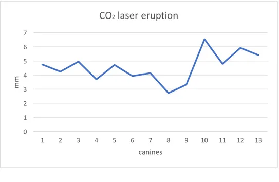
5.2 Eruption ... 51
5.3Comparison between the effect of the CO2 and Diode laser ... 53
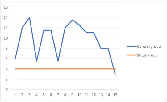
5.4 Comparison between the Study groups and the Control group ... 56
5.5 Speed of eruption ... 58
5.6 Proximity to central and lateral incisive ... 60
5.7 Palatal and Vestibular area of the canines ... 61
5.8 Conventional vs Digital monitoring ... 63
CLINICAL CASES ... 68
1. Group A – CO2 Laser ... 68
2. Group B – Diode Laser ... 82
CHAPTER 6: DISCUSSION ... 96 CHAPTER 7. CONCLUSION ... 102 REFERENCES ... 106
INTRODUCTION
Orthodontic treatment is based on the principle that the prolonged application of a pressure on a tooth causes movement, through the remodelling of the surrounding alveolar bone. The bone is reabsorbed and produced selectively. In practice, the tooth moves inside the bone bringing with it its periodontal attachment system together with the alveolar process. The bone response is mediated by the periodontal ligament and we could therefore define the dental movement as a phenomenon primarily determined by this structure. [1]
Even during the dental movement that occurs physiologically in the dental eruption, the periodontal ligament is the main characters.
The phenomenon of dental eruption reveals that forces generated inside the periodontal ligament (PDL) can induce a dental movement. The mechanism of the eruption appears to depend on metabolic events internal to the PDL. These events include, among the various phenomena, the formation of collagen fibres, the appearance of connection ties and the maturation of the fibres themselves. [2]
In this study we performed a monitoring of dental movement in a particular condition: impacted palatally canines, only surgically treated with a new laser approach, without using any type of orthodontic traction.
The upper canines are among the most frequently impacted teeth and they are often located in the palatal site. Ectopic eruption and impaction of canines are frequently encountered clinical problems. The incidence of impaction ranges from 1% to 3%. The causes of the impacted canines are multifactorial. In fact, it has been found in literature that there are both local factors that can predispose to this, both of polygenic hereditary factors. Very often multiple dental anomalies are associated in the same patient. [2]
One of the treatment options for the impacted maxillary canine is surgical exposure followed by orthodontic forced eruption.
Several surgical techniques have been introduced in the literature for the exposure of the impacted canine. Careful clinical examination and proper diagnostic imaging
approach for exposure of impacted maxillary canines should consider the anatomical position of the tooth in relation to the alveolar ridge and the amount of keratinized mucosa/gingiva. [3]
In the combined orthodontic and surgical approach, the surgery provides the placement of an anchorage means and it is followed by the second phase, the orthodontic treatment, which consists in therapy with a fixed device that has the purpose of exerting traction on the canine.
In the conventional surgery the orthodontic therapy is used for the traction of the canine and as guide for the eruption.
The new surgical approach, performed on patients in this study, is a canine exposure through laser.
Lasers are a relatively new addition to the orthodontist's armamentarium. The contact cutting mode provides enhanced bloodless site visibility and easiness in performing delicate soft tissue procedures, mostly for difficult-to-access areas. [4]
Currently, the laser method is still in the experimental phase and this type of procedure produces the spontaneous eruption of the canine, with the only need of final alignment, with fixed bonding techniques, without the need to traction it.
The purpose of this study is to monitor the movement of the impacted tooth after exposure with laser approach.
The spontaneous eruption is evaluated in a sample of 10 patients over time, in which some of these had a double inclusion and others a single canine inclusion, all in the palatal site. This monitoring lasts 4-5 months depending on the patient's response and ends with the start of the alignment phase through the direct/indirect bonding technique.
Dental monitoring was performed with intraoral scans using the CARESTREAM 3500 intraoral scanner. The scanner application allowed us to measure all dental movements and compare them over time.
Intraoral photos and analogical impressions of patients were also taken at each control visit.
Monitoring by intra-oral scans and photos was performed according to the following timing: • TIME 0: PRE-SURGERY • TIME 1: 1 WEEK • TIME 2: 8 WEEKS • TIME 3: 16 WEEKS.
With this digital method it is possible to do an initial scan that provides a three-dimensional model of the patient’s oral cavity before the surgery, that is matched with the following scans done after the surgery.
In this way, it is possible to evaluate any tooth movement expressed in angular and millimetric numbers, which are then interpreted by the orthodontist.
Another aspect of the study is focused on the differences between digital monitoring through scanner and the conventional monitoring which is based exclusively on the clinical evaluation of the photos and on the study of the plaster casts.
The popularity and availability of virtual technology in orthodontics for the replacement of hard-copy records with electronic records is growing rapidly, with a move towards a 'digital' patient for diagnosis, treatment planning, monitoring of treatment progress and results. [5]
The validity, reliability, and reproducibility of digital models obtained from the Intraoral scanner allows us to obtain dental measurements for orthodontic purposes. [6]
Making an accurate dental impression is one of the most essential and time-consuming procedures in dental practice. During this procedure, it is crucial to ensure the reproduction of the intraoral condition as accurately as possible. The accuracy of a dental impression is determined by two factors: “trueness” and “precision.” Trueness is defined as the comparison between a reference dataset and a test dataset. Precision is defined as a comparison between various datasets obtained from the same object using the same scanner. [7]
The scanner used is Carestream 3500, with it you can easily acquire precise dental coloured impressions, both 3D and 2D. The CS 3500 scanner is an open system scanner, the digital images obtained through scanning are in Standard Tessellation
The study defines the eruption time after the laser surgery and determinate a proper time to move on the next step of orthodontic therapy, which is the bonding and alignment of the full dental arch. The aim of the study is to demonstrate the validity of the monitoring through intraoral scanner of the dental movements. Successively, the aim is to demonstrate the real importance, advantages and convenience, in term of treatment time gain, to frequently monitor a patient with a scanner application, which means:
• significant reduction in the mean period of chair time, • less degree of discomfort for orthodontist and patient, • reduction of the periods of orthodontic treatment,
• help the orthodontist to make clinical decisions supported by measurable data and not just by clinical experience.
CHAPTER 1: BIOMECHANICS OF DENTAL MOVEMENT
1.1 Role of the periodontal ligament
Each tooth is attached to the surrounding alveolar bone, and at the same time separated from it by a robust supporting collagen structure: the periodontal ligament (PDL). Normally the PDL occupies a space of about 0.5 mm around the root of the tooth. The main component of the ligament is represented by a set of parallel collagen fibres that insert on one side into the root cement and on the other side in a relatively robust bone surface: the hard lamina. These support fibres, in their attachment to the dental surface, have an oblique direction in the apical direction. This arrangement allows the tooth to oppose greater resistance to the forces that occur during normal chewing activity. [1]
Much of the PDL space is occupied by the tangle of collagen fibres that constitutes the tooth fixation system, but there are two other important components of the periodontal ligament: (1) the cellular component consisting of mesenchymal cells of various types, together with neural elements and vascular; (2) tissue fluids. Both play an important role during normal activity and in orthodontic movement. During the normal activity, a continuous remodelling of the alveolar process and of the root cement occurs. The fibroblasts of the periodontal ligament have characteristics similar to those of the osteoblasts and, differentiating themselves from the local cell population, form new bone. [8]
The alveolar bone- and the cement are removed, respectively, by osteoclasts and specialized cementoclasts. These plurinucleated cells have different origins than osteoblasts and cementoblasts that produce bone and cement. [1]
1.2 Dental eruption mechanism
The phenomenon of dental eruption reveals that forces generated within the PDL itself can induce dental movement. The mechanism of the eruption appears to depend on metabolic events internal to the PDL; these include, among the various phenomena, the formation of collagen fibres, the appearance of connection ties and the maturation
during adult life. The continued presence of this mechanism indicates that it can’t only induce the eruption of the teeth in particular situations, but it can also favour its stabilization in relation to forces prolonged of slight entity. The active stabilization action implies the presence of a threshold for the orthodontic forces, below which they are ineffective to produce a dental displacement. The efficacy threshold for an external force should vary according to the pressure exerted by the tissues surrounding moles on the tooth stabilization system. It is currently believed that the active stabilization system can withstand prolonged forces of up to a few grams, perhaps up to 5-10 g / cm2; in fact, this is the magnitude of the differential pressure exerted by the soft tissues on the teeth. [1]
1.3 Theories on dental movement
The two main theories concerning the orthodontic movement hypothesize different control systems: one based on biological electrical signals and the other on a pressure-tension mechanism inside the PDL.
• The bioelectrical theory partially attributes the dental movement to changes in bone metabolism, induced by electrical currents that are generated when the alveolar bone is subjected to flexion, which causes a deformation of the crystalline structure and the migration of electrons from one area to another one. The signals generated are piezoelectric. It has now been established that the signals induced by mechanical stress are essential for maintaining the normal skeletal structure. The absence of such signals causes the loss of the mineral bone component with consequent skeletal atrophy. It has been shown that a low-voltage electric current directly applied to the alveolar bone, modifying the bioelectric potential, induces a more rapid movement of the tooth to which a spring has been applied, with respect to the control tooth. [9][10]
Electromagnetic fields can cause a change in membrane potentials and their permeability, thus inducing changes in cell activity.
• The theory of pressure-tension is based on the classical hypothesis that cellular differentiation and finally dental movement are controlled by chemical rather than electrical signals. There is no doubt that chemical messengers intervene
in the succession of events that lead to bone remodelling and dental displacement. According to the theory of pressure-tension the dental movement is due to changes in cellular activities induced by chemical messengers, probably generated by changes in blood flow within the PDL. Continuous strength induces a modification of the blood flow in the PDL through a movement of the tooth inside the alveolus; this movement involves a compression of the ligament in some areas and a pull in others. The reduction (pressure) or the increase (tension) of the diameter of the vessels, follow the application of orthodontic forces. This phenomenon changes the extent of the blood and so the blood flow decreases in the compression areas of the PDL, while it is equal or increased in the traction areas. The oxygen tension decreases in the compression zones and perhaps tends to increase in the traction zones; similarly, changes in the levels of other metabolites can occur within a few minutes. These chemical changes can stimulate a particular cell differentiation directly, or through other biologically active substances. According to this hypothesis, the dental movement is performed in three stages: alteration of the blood flow consequent to the pressure exerted on the PDL, synthesis or release of chemical messengers, activation of specialized cells. [1]
These two theories are neither incompatible nor mutually exclusive. It is currently thought that both mechanisms come into play in controlling dental movement.
1.4 Biology of dental movement
When the pressure exerted on the PDL is excessive, there is a great reduction in the blood flow inside it, up to a complete collapse of the blood vessels and consequent ischemia. This result was obtained in experimental animals in which the increase in force applied to a tooth is accompanied by a reduced perfusion of the areas of PDL under compression. [1] [11]
When a light and continuous force is applied to the tooth, a reduction of the blood flow is observed through the PDL, the fluids are pushed out of the periodontal space following the movement of the tooth into the alveolus (in a few seconds). After a few
increase in levels of cyclic adenosine monophosphate (AMPc), the "second messenger" of many functions involved in cell differentiation; this occurs approximately 4 hours after the application of a continuous pressure. [1][12]
Shortly after the application of pressure, the values of prostaglandins and beta interleukin 1, in the PDL, increase; in particular, it seems that prostaglandin E is an important mediator of cellular response. [13]
Changes in cellular shape probably play an important role. Prostaglandin release is believed to occur when cells are mechanically deformed (the release of prostaglandins may be a primary and not a secondary response to pressure stimulus). [14]
Other chemical messengers are also involved, in particular those of the cytokine family, but also nitric oxide (NO) and other regulators of cellular activity. [15]
To obtain a dental movement, osteoclasts are necessary. They remove bone tissue in the PDL compression area. It is also necessary that the osteoblasts place new bone on the tension side and intervene in the remodelling of the areas affected by the resorption on the pressure side. The cells attack the hard lamina through a process of "frontal bone resorption", which is followed by the dental movement. In the meantime, even if with some delay, osteoblasts (differentiated locally from progenitor cells present in the PDL) form new bone on the tension side and begin the remodelling activity on the compression side. [1]
The situation changes if the applied force is large enough to completely occlude the blood vessels in an area of the PDL. In this case, a sterile necrosis of the compressed area is obtained, the cellular component disappears, the avascular area in PDL is traditionally defined as area of ialinization. The osteoclasts appear in the neighbouring bone and begin to attack the outer side of the bone adjacent to the necrosis area. This process is described as indirect resorption (sub-dominant), as the attack starts from the outer wall of the hard lamina. As a consequence, the process of ialinization and the indirect reabsorption involve an inevitable delay in moving the tooth. [1]
1.6 Biomechanics of dental movement in orthodontics
A single force in practice almost never acts through the centre of resistance. Therefore, a single force results in displacement of the centre of resistance in the direction of the line of the force (tipping). This tendency for rotation is called the moment of the force
whose magnitude is equal to the magnitude of the force multiplied by its perpendicular distance from the centre of resistance of the tooth. This application of a force will cause the centre of resistance of the tooth to move parallel to, and in the same direction as, the force. The centre of rotation will be located apical to the centre of resistance. [21]
The second method by which tooth movement can be performed is through the application of a pair of equal forces which are parallel, non co-linear, and of opposite direction, termed a couple. This system, applied anywhere on a tooth, creates only a tendency for rotation referred to as the moment of the couple whose magnitude is equal to one of the forces of the couple multiplied by the inter-force distance. The centre of rotation resulting from the moment of the couple is always coincident with the centre of resistance of the tooth irrespective of its point of application.
We can describe tooth movement as rotational (tipping) and/or translational (bodily movement). [21][22]
for translation to occur there must concurrently exist a couple with an opposite sense tending to tip the root in the opposite direction as the crown.
Under these conditions, the relative amount of crown tipping (moment of the force) and root tipping (moment of the couple) expressed at any moment in determines the location of an instantaneous centre of rotation. When these two oppositely directed moments are equal in magnitude, the centre of rotation is at infinity and tooth translation occurs (Figure 3). This determinant is also expressed as the moment to force ratio.[21][22][23][24]
For a ratio of 5/1 we will have an uncontrolled tipping of the crown, as for the single force. For a ratio of 5/1 to 9/1 we will have a controlled tipping in which the crown and the root move in the same direction. For a ratio of 10/1 and 11/1 we will have a translational body movement. For values of 12/1 we will have an uprighting of the root. [25]
CHAPTER 2: NEW DIGITAL TECHNOLOGIES IN ORTHODONTICS
death and taxes. May I add a third (though not so vital): the application of the computer to orthodontic
research and diagnosis.”
Wilton Marion Krogman, (forensic anthropologist)
These were the opening words of American Association of Orthodontists’ annual meeting in New Orleans in May 1971. [26]
Digital technologies are nowadays widely used in the several branches of dentistry. By carrying out a literature review, various digital methods have emerged that have been applied in orthodontics, gradually modifying normal orthodontic practice in the last years, but the process has been slower in orthodontics than in other fields of dentistry.
These new technologies are: Invisalign or Clear Align method [27][28][29][30][31], CAD-CAM technology [26][30][32][33] associated to the manufacturing of these orthodontics devices, Cone beam technology [34][35][36][37][38], Intraoral scanner [13][14] for make impressions of the patients dental arch, new software to evaluate the dental movements [31][28][39] and to make cefalometric digital analysis [40][41][42][43][37], mobile application [37][44][45][39] for clinicians or for patients, and 3D images fusion technique [46][47][48][49][50][51][52][53].
2.2 CAD-CAM Technology
The first digital revolution took place many years ago with the production of dental devices and dental restorations using dental CAD–CAM system. New improved systems appear on the market with great rapidity.
Computer-Aided Design/Computer-Aided Manufacturing (CAD/CAM) technology, already entrenched in dentistry since 1987 when Sirona introduced its CEREC units.
CAD/CAM gave practitioners a way to digitize study model data and incorporate those data into the patient's record. In 1999, Align Technology (San Jose, Calif) was the first company to offer a digitizing service (OrthoCAD) to the orthodontic community, followed by eModels (GeoDigm, Falcon Heights, Minn) in 2001. [30][37]
The development of CAD/CAM is based around three elements: (1) data acquisition, (2) data processing and (3) manufacturing. The exponential increase in power of computers has resulted in major advances in all these areas. [32]
Trough intraoral scanner is possible to create a 3D model of the oral cavity directly with such a system, without the need to take an impression, in alternative the model is then digitize with one of the many laser scanners that are now available. The digital model can be used to design the devices, like Invisalign aligners, trough many software packages available. [33]
2.3 Intraoral scanner
The new digital method of taking impressions of entire dental arches using the intraoral scanner was introduced in 1973, associated with CAD / CAM methods. This first device introduced was the CEREC marketed by Sirona Dental System. The first use in dentistry was within the conservative branch and then extended to other areas such as dental prosthesis and orthodontics. The first orthodontic scanning system OrthoCAD developed by Cadent was introduced in 1999. The application of this devices in dentistry has grown considerably in the last few years. [54-56]
The replacement of alginate and polyvinyl siloxane (PVS) impressions with intraoral digital scanners represents a paradigm shift in orthodontics. Using this technology, orthodontists can more accurately and efficiently fabricate clear aligners, custom braces, indirect-bonding trays, and laboratory appliances without the unpleasant experience of conventional impressions. [57]
Every scanner has three major components: a wireless mobile workstation to support data entry; a monitor to enter prescriptions, approve scans, and review digital files; and a handheld camera wand to collect the scan data in the patient’s mouth.
To gather surface data points, energy from either laser light or structured light is projected from the wand onto an object and reflected to a sensor or camera within the wand. Based on algorithms, tens or hundreds of thousands of measurements are taken per inch, resulting in a 3D representation of the object’s shape. [57]
A point cloud is linked through the algorithm to form a 3D model in a standardized triangular language.
2.3.1 Applications of intraoral scanner in orthodontics
Digital scanning can be used in orthodontics for a variety of applications.
Some applications have been described for treatment planning [58], indirect bonding tray fabrication [59], palatal and lingual custom appliance design and construction [59], clear aligner technology [60], orthognathic surgery simulation and wafer construction [61,62] and more recently, the scoring of surgical outcomes in patients with Cleft, Lip and Palate abnormalities. [56,62]
New technologies are increasingly used in the field of orthodontics, given their digital impressions were found useful also for diagnosis and patient monitoring development and the increase in effectiveness.
To this end, many of the scanner companies are developing ‘comprehensive’ integrated packages as orthodontic practices and clinics change from hard copy records to the ‘digital patient’. [56]
Although there are also some disadvantages, intraoral scanning technology holds great potential to increase efficiency for orthodontic practitioners and may eventually take the place of impressions. [63]
2.3.2 Comparison between conventional impression and digital scan
Alginate and PVS impressions have been associated with problems such as pulls, tears, bubbles, voids, tray-to-tooth contact, separation from the impression tray, temperature sensitivity, limited working time, material shrinkage, inaccurate pouring, model over trimming, and breakage during shipment. [64] Impression taking also heightens anxiety and discomfort for patients of all ages, particularly those with sensitive gag reflexes. In vitro studies have shown that full-arch digital scans are as accurate as conventional impressions [65] without these drawbacks.
impression for single implant restorations. The results indicated that total treatment time was 12’29” for digital and 24’42” for conventional impressions; and time of rescan/retake was 1’40” for digital and 6’58” for conventional impressions. Although the total number of digital impressions rescans was more than that in conventional impression. This pilot study reached a conclusion that there was a significant difference in operation time between these two impression methods.
Participants were asked to answer visual analogue scale (VAS) and multiple-choice questionnaires to evaluate their perceptions of difficulty, preference, and proficiency for both impression techniques. The results showed that the grade of difficulty was lower for the digital impression than that at the conventional impression. The digital impression techniques were more acceptable and easier to grasp. [67]
According to another study the mean chairside time required to perform alginate impressions was significantly (P-value 0.0001) shorter than the mean chairside time required to perform intraoral scans. When the processing times were included, the time requirements for impressions and intraoral scans did not differ significantly (P-value 0.0649). [73]
2.3.3 Advantages and disadvantages of digital impression
For the orthodontist, advantages of digital scanning include improved diagnosis and treatment planning, increased case acceptance, faster records submission to laboratories and insurance providers, fewer retakes, reduced chair time, standardization of office procedures, reduced storage requirements, faster laboratory return, improved appliance accuracy, enhanced workflow, lower inventory expense, and reduced treatment times. Benefits to the patient include an improved case presentation and a better orthodontic experience with more comfort and less anxiety, reduced chair time, and easier refabrication of lost or broken appliances, as well as potentially reduced treatment time. [57]
In particular, young children, patients with clefts of the lip and/or palate and those with a heightened gag reflex express a dislike for traditional impressions. Recent investigations have shown 3D scanning to be more acceptable in terms of patient perception and comfort over traditional impressions. [68,69]
However, recent research has highlighted that 3D model scanners with an accuracy of at least 20 mm are suitable for assessing archform in patients with cleft lip and or palate. [56,70]
Digital scanning reduces risk of allergy when compared to the small but none-the-less pertinent risk posed by the many constituents of impression material. [71]
It has been shown in the literature that digital impression represented a remarkable superiority in efficiency over conventional impressions.
The digital impression took less time for rescans despite a larger volume required, because in this type of impression, only the missing and unacceptable areas were rescanned, contrary to the conventional ones in which, the entire arch needed to be retaken. [67,72]
However, some studies do not support these advantages, conventional impressions were favoured over intraoral scans by most patients, primarily because they were “easier” or “faster”. [63] This contrasts with other study in which most of the participants preferred the intraoral scans. [73]
Another discomfort was related to the dimensions of the scanning tip and its interference with the patient's coronoid process. As scanning technology continues to evolve, the design of a thinner scanning tip may improve comfort and increase patient acceptance of the scanning procedure. [63]
An aspect to take into consideration is the high cost of the intraoral scanner and of its updates, as well as the fact that the staff must be instructed to the correct use of the digital device.
2.2.6 Accuracy on intraoral scanner application
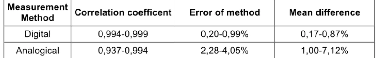
Accuracy consists of precision and trueness (ISO 5725-1). [69] Precision describes how close repeated measurements are to each other. [70] Therefore, a scanner with higher precision correlates to a more repeatable and consistent scan. [70] Trueness describes how far the measurement deviates from the actual dimensions of the measured object. [80] Therefore, a scanner with high trueness indicates that the scanner delivers a result that is close or equal to the actual dimensions of the object being scanned. [70,71]
There are two ways to create a digital impression: direct intraoral scanning or indirect extraoral scanning gypsum casts. [72]
Accuracy could be assessed as the agreement between the digital models in STL acquired using the intraoral scanner and the corresponding models that had been acquired with either the alginate impressions. [73]
A direct intraoral scanning is truly free of a physical impression so that it is able to get rid of the errors derived from the distortion of elastomeric impressions, disproportionate water/powder ratio of dental plaster and unsuitable storage conditions of physical impressions or gypsum casts. [74,75]
With digital models fabricated from alginate impressions, fine details of tooth anatomy might be lost because of the limited ability of the impression material to flow into areas with undercuts, and potential shrinkage upon desiccation can compound the problem. [76,77,78] Additional loss of information may be related to the scanning process because the accuracy of a digital model is limited by the resolution of the scanner that recorded it. [63]
However, intraoral scanning can result in digital models that more accurately represent the intraoral situation because there are fewer sources of error. It is logical to assume that when processing steps are eliminated in the production of digital models, the models will be more accurate.
2.4 CBCT in Orthodontics
The CBCT uses a cone shaped X-ray beam and one large 2D detector to capture the cone shaped beam. The 2D detector can record a large (e.g. 16 cm 22 cm) area of the face in one or two rotations (20–40 s), this type of CT reduces the effective radiation dose as compared to MSCT scanning. [34]
The use of Cone Beam Computerized Tomography (CBCT) has allowed for accurate visualization of the roots of teeth in 3 dimensions. However, a CBCT scan uses significantly more radiations than does a panoramic radiograph, so multiple CBCT scans would not be suggested clinically. An imaging technique that can be performed more times during orthodontic treatment without radiation is a digital intraoral surface
show the roots. Since neither CBCT and digital intraoral surface scans can safely and accurately visualize root positions at different stages of orthodontic treatment, root tracking might be possible with a combination of these two imaging techniques. [35]
From the CBCT it’s possible to generate a three-dimensional virtual model of each teeth, and this can be superimposed with the crown of the same teeth obtained by intraoral surface scan models.
The current standard of care suggests the use of panoramic X-rays to monitor root alignment, even though many studies have shown that panoramic X-rays do not accurately depict root positions and angulations due to their two-dimensional projection. [36] It seems to be clear that a new approach in monitoring accurately our treatments is necessary. [35]
An appealing aspect is that a CBCT scan could serve as a patient's singular data set from which to derive all the traditional views of an orthodontic workup. [37][38]
By CBCT scan of the patient it’s also possible to obtain a 3D volume model of the full arch, using many programs that can convert CBCT files to STL files.
This digital method allows, as was done in this study, to use the three-dimensional file obtained from the CBCT for overlapping with the files obtained from intraoral scans to monitor the real movement of dental eruption obtained during treatment. It also allows us to compare the position of the root of the tooth at the beginning of treatment with that at the end of the treatment obtained from the superposition with the erupted crown of the element.
CHAPTER 3: IMPACTED PALATALLY CANINES
3.1 Introduction on impacted canines
Impacted teeth are often encountered during the diagnosis and treatment of malocclusions in adolescent orthodontic patients. After the third molars, the most commonly impacted tooth is the maxillary canine with an incidence of 0.9% to 2.2%, [79-82] which varies depending on the ethnicity of the sample population.
Although the canine crown can be impacted either labially or palatally, it is more frequently positioned in the palate. [83,84]
Of all patients who have impacted maxillary canines, 8% have bilateral impactions. [80,85] There are most common in female than in male patients. [80]
Tooth impaction can be defined as the lack of presence in the oral cavity of an element that has completed its morpho structural development within the osteo-mucous structure, at the end of the period provided for its physiological eruption. The position of the tooth is intraosseous, whereas the anomalous intraosseous position of the canine before the expected time of eruption can be defined as a displacement. Most of the time, palatal displacement of the maxillary canine results in impaction. [86] In the impacted tooth the root apex is closed, with absence of the vis a tergo which is typical instead of the dental elements retained.
3.2 Etiopathogenesis of upper impacted canines
The causes of inclusion are multi-factorial. For upper canines in particular, one of the predisposing factors found is the difficult eruptive path. The canine must make a long path (about 22 mm) [80,87], being its gem located below the orbital margin, laterally to the piriform fossa, and therefore in a location far from that of eruption. [88]
During its physiological path the tooth moves downward, mesially inclined towards the root of the lateral incisor. There is a phase between 8 and 10 years that is called the ‘ugly duckling dentition’ in which appears a diastema precisely because of the push of
rotation centre of the tooth. After this phase, the canine continues its path along the distal surface of the root of the lateral incisor, thus closing the diastema. [80,88]
Another predisposing factor is that canines have the longest period of development. [80,87]
Broadbent speculated that because of the long path of eruption and so long period of development taken by the maxillary canine, it had a greater chance of inclusion. [89,90]
There are two main theories on the inclusion of these dental elements: the genetic theory and the guidance theory.
• The genetic theory states that maxillary palatal impaction has familial and hereditary component and includes other associated dental anomalies, such as missing or small lateral incisors. [91] Additionally, impaction of upper canines is correlated with enamel hypoplasia, infraocclusion of primary molars, aplasia of second premolars and small size of maxillary upper lateral incisors. [92,93]
• According to the guidance theory, the canines erupt having as their guide the distal side of the root of the lateral incisor. If it is absent or malformed there is an alteration of the eruption path of the canine.[92,94,96,97] As a matter of fact, maxillary lateral incisors are also among the most frequent with a deficient form, with small and peg-shaped crowns, and it has been confirmed as representing a microform or a lesser degree of severity of agenesis. [89]About 50 years ago, Miller and Bass independently observed that the prevalence of palatal displacement was greater when lateral incisors were congenitally missing. They concluded that the absence of the lateral incisor denied the canine its guidance, permitting it to migrate palatally. [89,97,98]
Lappin et al. [99] observed clinical and radiographic assessment of a number of impacted canine cases correlated with deciduous canines frequently over retained, often with a long and unresorbed root.
He speculated that the non-resorption of the deciduous canine was the cause of the anomaly. We know that the mechanism for root resorption require that an unerupted permanent tooth is near to the deciduous root. Thus, it is plausible that resorption has not occurred because of the distance of the permanent tooth, and so the unresorbed
root of the deciduous canine is not the cause of the displacement, but rather its result. [87] Various local factor may interfere with the normal eruption, because of the long path of the canine predisposes to a greater probability of encountering obstacles.The first entities that come to mind are the supernumerary tooth and the odontoma. [100]
When these etiologic factors are eliminated, there is a degree of autonomous correction in the eruption path that may lead to spontaneous eruption of the canine. [89]
Other local factors are a chronic periapical granuloma, that has a potent effect on deflecting or arresting the eruption of the permanent canine [89];and a trauma to the face, that can cause displacement of the unerupted canine or dilaceration of its developing root. [101,102]
Also, some skeletal abnormalities can be predisposing to the impaction of this tooth as class II division 2 malocclusion, a deep overbite, a hypodivergent profile, and abnormal maxillary width.
3.3 Prediction of inclusion
The inclusion of the canine can be predicted according to Lindauer, that modified the analysis of Ericson and Kurol, in the period of mixed dentition using the opt. On this radiography it is possible to evaluate it, dividing the frontal area into 4 sectors (I-II-III-IV), delimited by three lines:
- the tangent to the distal surface of the crown and the root of the lateral - the central axis of the lateral incisor
- the tangent to the mesial surface of the crown and the root of the lateral.
The probability of inclusion will be greater if the crown of the canine is mesio-inclinated respect to the long axis of the lateral incisor and if it is moving from the distal sectors to the mesial. This prediction is not completely reliable, but it can be considered at least indicative. [102]
3.4 Diagnosis of impacted canines
Tooth impaction is generally asymptomatic, and in most cases, the diagnosis occurs during a routine check-up by general dentists or orthodontists. [93,102]
Early detection of the included canines is essential to improve the prognosis of the dental element and to avoid all the consequences that are related to dental inclusion. We should have the suspect of the impaction in the intra-oral objective examination. Usually there is a persistence beyond the natural limits of exfoliation of the deciduous element, and a failure to erupt of the corresponding permanent. [80] It is also possible to make a comparison with the contralateral element, in the case of a single inclusion, which will be erupted.
Furthermore, the malposition such as rotations or malformation of the adjacent teeth, the absence of a normal labial canine bulge and the presence of a palatal bulge suggests the possible impaction of the canine.
After the intraoral examination, radiographic examinations are carried out to confirm the diagnostic hypothesis.
The radiographs taken are opt, periapical and occlusal endorals. Nowadays three-dimensional radiographic exams, such as computerized tomography (CT) or cone beam computerized tomography (CBCT), is considered the best diagnostic tool to assess tooth impaction. [84,103,104,105]
The radiographic check-up is crucial to evaluate the anatomy of the impacted canine and to identify its location, inclination, and position, which will condition the type of operation and the orthodontic device to be used. With intraoral radiograph we can evaluate the morphology and the seat, but being the two-dimensional examination, it is important to perform at least two radiograms with the tube shift technique to establish the mesio-distal and vestibulo-oral position of the tooth. [102]
3.5 Prognosis of impacted canine
Ericson and Kurol reported that the degree of mesial overlap of the maxillary canine relative to the adjacent lateral incisor plays a role in the severity of impaction and the probability of spontaneous eruption. In 1988, they defined number of sectors to denote different types of impaction. [106]
- Sector 1: if the cusp tip of the canine is between the inter-incisor median line and the long axis of the central incisor;
- Sector 2: if the peak of the cuspid of the canine is between the major axes of the lateral and central;
- Sector 3: if the peak of the cuspid of the canine is between the major axis of the lateral and the first premolar.
They used angle “α” to represent the angle formed between the inter-incisor midline (as the perpendicular distance respect to the occlusal plane).
Fig. 1 - Angle “α”
The localization with sector methods has a more prognostic than diagnostic value. In the year 2003, Warford et al., [107] did a study for predicting the maxillary canine impaction using sectors and angular measurement. He localised the canine according to 4 sectors and measured the angle between the bicondylar line and long axis of canine. He concluded that the probability of canine impaction increases as the angle reduces and the sector increases.
3.6 Approaches of disinclusion of palatally impacted canine
The treatment of impacted teeth requires multidisciplinary cooperation between orthodontists, oral surgeons and sometimes periodontists. Orthodontic treatment and surgical exposure of impacted teeth are performed in order to bring the impacted tooth into the line of the arch. The treatment is long, more complicated and challenging. [114]
The choice of the approach depends on a variety of factors including position of the canine, the associated malocclusion, the skill and experience of the orthodontist and
The combined surgical-orthodontic approach relocates the impacted canine in its proper place in the dental arches.
The two major surgical techniques are the open and the closed technique. [109-113]
§ The open technique involves exposing a palatally displaced canine by a circular incision (operculectomy) to remove bone and soft tissue directly overlying the impacted canine. Thus, the area of palatal mucosa which covers the crown of the tooth is cut out to create a window, through which the crown is visible. Afterward an attachment, such as an eyelet or a button, can be bonded at the time of surgery and orthodontic traction may be performed immediately. At the end, surgical pack might be used to cover the exposed area and it is removed in a few days. In this technique, the canine tooth moves into its correct position above the mucosa. [113-115] In case of bonding failure, there is no need for a second surgical exposure, but on the other side it can result in poorer periodontal outcome, increased risk of infection, greater discomfort to the patient, more extensive removal of alveolar bone, risk of closure of the exposure, increased bonding failure. [93,100]
§ The closed technique involves surgically exposing the crown of the impacted tooth raising a full muco-periostal flap, removing the bone that cover the impacted tooth with a low speed bur and eliminating the pericoronal tissue using a periodontal curette. [116] Then an orthodontic eyelet or a button is applied and bonded on the crown while a wire chain is fixed to the attaching device. The palatal flap is then replaced and sutured leaving the length of chain to exit the gingivae. A few days after the surgery, gentle orthodontic traction can be applied to the tooth to start bringing in into its correct position within the dental arch. In the closed technique, the canine tooth is orthodontically moved into its correct beneath the mucosa. This approach is usually favoured when the tooth is more deeply embedded in the bone since open surgical exposure may necessitate excessive removal of the surrounding bone. [113-115] Some advantages are less discomfort, good postoperative homeostasis, less intense functional disturbances, less extensive removal of alveolar bone, possibility of an immediate traction, applicable close to resorbing root.
Mechanical eruption of palatally impacted canines may lead to prolonged treatment. Treatment times are thought to increase further with greater vertical and mesial displacement, increased angulation of the canine and increasing age. [108,117,118]
Deleterious effects of prolonged orthodontic treatment are well-documented and include the propensity for greater root resorption and poor patient compliance. [108,119] Additionally, root resorption of teeth neighbouring the impacted tooth is thought to be accelerated if mechanical eruption is undertaken. [108, 120]
In principle, in the treatment of an impacted canine in a superficial position, the open technique can be advantageous, as it allows a complete exposure of the crown and a simple and accurate bond placement. With deep impactions, closed exposures are usually favoured as open exposures may necessitate excessive bone and soft tissue removal risking periodontal damage to the impacted tooth or neighbouring roots. Incorrect positioning of attachments in closed techniques may be undetected until the canine has been erupted, generating unwanted rotations and prolonged treatment. Before the intervention it is important to create the space to place the included tooth in the arch, or if it is already present, this space must be maintained. Bracketing the upper arch provides to increase the space needed and to obtain an adequate anchorage for traction of the impacted canine. The primary canine will be extracted at the time of surgical treatment. [116] After surgery, orthodontic treatment involves pulling the canine on the palate and then the centre of the dental arch, with several methods .Elastomeric traction is useful to initiate eruption in conjunction with fixed appliances and stainless steel archwires. Rigid stainless-steel base archwires (e.g. 0.018-inch) are desirable to minimize reactionary forces reinforcing vertical anchorage. [121] Auxiliary NiTi archwires provide inherent flexibility to continue the eruptive process. [122,123]
3.6.1 Autonomous eruption
In addition to the guided orthodontic eruption, Kokich recommend an alternative technique for treating a palatally impacted canine: to surgically uncover the tooth and allow it to erupt autonomously before beginning orthodontic treatment.
With this procedure, a mucoperiosteal, envelope flap is elevated starting apical to the gingival sulcus on the lingual surfaces of the maxillary lateral and central incisors in the area of the palatally impacted tooth. Once the flap has been elevated, there is typically a thin layer of bone covering the lingual surface of the canine crown. This
bone can be removed with either a curette or a handpiece and bur. Then, a bracket or cleat is bonded to the lingual surface of the canine crown.
The flap is repositioned over the tooth and a small hole is made in the gingival tissue over the canine crown, so the tooth will have no impediment to erupting autonomously. This open surgical defect is covered with a dressing that is mechanically attached to the lingual bracket or cleat. Then, this tooth is allowed to erupt. With this technique, if sufficient bone is removed, the palatally impacted canine will typically erupt autonomously to the level of the occlusal plane within 6 to 9 months. Then, the canine can be moved laterally toward the alveolar ridge. During this type of movement, the root is moving through the bone facilitated by the surrounding periodontal membrane. No other types of treatment for the impacted palatally canines were found in the literature on PUBMED and SCOPUS.
In this study, we monitored the dental movement in patients treated with a new approach: laser surgery. This technique is still experimental and has numerous advantages over conventional surgical technique. The laser used in this study allowed us to expose the crown of the canine and biostimulate the area around it.
We used the carbon dioxide laser and diode laser.
There are no studies in the literature on this, but the laser is widely used in the dental field for its many benefits:
- Soft-tissue excision is more precise with a laser than a scalpel. [125,126]
- A laser coagulates blood vessels, seals lymphatics, and sterilizes the wound during ablation, maintaining a clear and clean surgical field. [125,127]
- Laser surgery is routinely performed by using only topical anaesthetic. [125,128]
- There is markedly less bleeding, minimal swelling, and no need for irritating sutures or unsightly periodontal dressing.
- Post-surgically, patients report less discomfort and fewer functional complications (speaking and chewing) and require fewer analgesics than do patients treated with conventional scalpel surgery. [129]
In dentistry, the laser is used with the continuous emission and pulsed emission mode. [146]
The first allows the maximum surgical precision while with the pulsed emission are possible interventions with no use of infiltrative anaesthesia both for the brevity of the pulsations (which are hardly perceived by the nerve receptors) and because the period of thermal relaxation between a pulsation and the other it protects fabrics from strong temperature increases. [129]
3.7 Laser application on dentistry
The application of the laser on soft tissue includes wound healing, removal of tissue that surround an impacted or a partially erupted tooth (operculectomy), photodynamic therapy for malignant lesions, photostimulation of herpetic lesion. [131] Lasers can be used to ablate large areas of gingival proliferation in a totally bloodless manner (gingivectomy, gingivoplasty and other periodontal procedures), to cut maxillary and mandibular frenae (frenectomy) and also both incisional and excisional biopsies can be performed with lasers, taking into account the slight thermal damage at the margins. [153]
The laser in orthodontics find its application also because of a new and promising concept emerged: photo-biomodulation or low-level laser therapy (LLLT) can accelerate tooth movement.
According to some works, laser has a bio-stimulatory effect on cellular regeneration, which has been shown in the midpalatal suture during rapid palatal expansion [133,134] and also stimulates bone regeneration after bone fractures and extraction site. [134-136]
Laser light stimulates the proliferation of osteoclast, osteoblast, and fibroblasts, and thereby affects bone remodelling and accelerates tooth movement. The mechanism involved in the acceleration of tooth movement is due to production of ATP and activation of cytochrome C.
This stimulation of dental movement by the laser is a factor that could make the orthodontic treatment after the operation less complicated and quicker. The monitoring that we carried out after the operation evaluates the spontaneous movement of the canine after the use of the laser, without an orthodontic anchorage appliance.
CHAPTER 4: MATERIAL AND METHODS
4.1 Aim of the study
The aim of this study is to show the effectiveness of laser technology for the exposure of the palatally impacted canines, using a CO2 laser device (Smart US20D®, DEKA - Florence, Italy) and Diodi Laser device (Raffaello, DMT, Lissone, Italy, 980nm +645nm), which can stimulate the spontaneous eruption of the canine, without orthodontic traction application[137-139]. Moreover, the purpose of this study is to monitor the movement of the impacted tooth after exposure with laser approach with digital technologies.
Another aspect of this study is focused on the differences between digital monitoring through scanner and conventional monitoring which is based exclusively on the clinical evaluation of the photos and the study of the plaster casts[140-142].
4.2 Study design
The study was carried out on patients referred to the Orthodontics UOC of the Department of Odontostomatological and Maxillo-Facial Sciences of the “Sapienza” University of Rome. The period of recruitment of the patients was 1 year.
All the patients were informed about the content of the study, treatment methods, and potential risks and benefits, before providing written informed consent to take part in this study. The study received approval from the Ethical Committee of Sapienza University of Rome (#4389) and was registered in the international public register. A preliminary investigation was performed to estimate the power of the study (PS) and to establish the effect size (ES) of the population sampled for the experimental study. It was calculated a statistical significance to determine an exact number of patients to become a study.
Suppose we want to estimate the prevalence of a disease (impacted canines) in a population. Through the study of the sample we want an estimate of the prevalence with a certain precision and a chosen level of confidence. The size can be calculated, with a 95% confidence level, using the following formula:
In our case with 95% confidence level and this data, when:
P_att: 96.5% (0.965) D: 10% (0.1)
Therefore:
n = (1.96 ^ 2 * 0.965 * (1-0.965) / 0.1 ^ 2 = 12.97 That is 13
If the sample size recommended was >5% compared to the population from which it is extracted, its sample size could be reduced; with the statistical analysis, it was obtained that the sample size significance for this study was 13 canines.
The inclusion criteria considered in the experimental study design were: • patients affected by palatally impacted canines;
• male and female;
• age between 12 and 25 years; • patient will be reliable for follow-up;
• understands the protocol and can give informed consent. The Exclusion Criteria were:
• Non-cooperative patients; • inoperable patients;
• vestibular impacted canines; • mandibular impacted canines; • systemic pathologies;
These criteria resumed in the flowchart table: CONSORT FLOWCHART - Assessed for eligibility (n = 36) Excluded (n = 10) Not meeting inclusion criteria (n = 7) Randomized (n = 26) Allocated to Study Group A (n = 13) ¨ Received allocated intervention (n=13) Al lo ca ti on
En
ro
llm
en
t
Allocated to Study Group B (n = 13) ¨ Received allocated intervention (n= 13) Fo llo w u p Lost to follow up: n=0 Lost to follow up: n=0 An al ys is Analyzed (n = 13) Excluded from analysis Analyzed (n = 13) Excluded from analysis Allocated to Control Group (n = 13) ¨ Received allocated intervention (n=13) Lost to follow up: n=0 Analyzed (n = 13) Excluded from analysisThe final experimental sample was constituted of 18 patients, 9 females and 9 males; of these 8 patients showed a bilateral inclusion and 10 a mono-lateral inclusion of the canine, for a total of 26 canines. The age of the selected patients is between 12 and 22 years. Patient characteristics that were collected are: age, sex, number and location of the impacted canines.
The 18 selected patients, who make up our study group, were divided into two groups, randomly, treated with two different laser types:
• GROUP A (9 patients; 13 impacted canines): treated with CO2 laser (SmartXide®, DEKA, Florence, Italy, 10600 nm) with a power of 4.5 Watt in superpulsed mode (frequency: 80 Hz, fluency: 44.78 J / cm2, spot diameter: 400 μm.
• GROUP B (9 patients; 13 impacted canines): treated with Diode laser
(Raffaello, DMT, Lissone, Italy, 980nm + 645nm) with a power of 4 Watt in
continuous mode (fluence: from 0.1 J / cm2 to 10 J / cm2). Figure 3. Diode Laser (Raffaello, DMT)
To demonstrate the validity of the technique applied to the study group, a CONTROL GROUP, which included 9 patients with unilateral and bilateral palatally impacted canines (in total 13 canines), treated by a traditional surgical-orthodontic approach, was observed.
4.3 Analysis of radiographs
An RX orthopantomography and a CBCT were requested from patients at the beginning of therapy, to accurately evaluate the cases before surgery.[14] An evaluation of the prognosis of impacted canines was performed on OPT by two orthodontists, according to Ericson and Kurol.
Figure 4 – Rx orthopantomography
The major axis of the canine and the perpendicular to the alveolar margin were plotted on the OPT, imagining this as the axis of the canine in its presumed ideal seat. Subsequently, it was considered the height of the canine’s crown relative to the roots of the contiguous elements, which ideally should be between the third occlusal of the root of the central incisor and the mesial side of the root of the first premolar. Considered that the degree of mesial overlap of the maxillary canine, relative to the adjacent lateral incisor, plays a role in the severity of impaction and in the probability of spontaneous eruption, it was also evaluated the mesio-distal position of the canine's cusp, which could stay in four sectors.
The examination of the CT allowed to evaluate the three-dimensional morphology of the impacted tooth, its location and inclination in the three planes of space, the depth and the type of inclusion, the relationships with the other elements.
4.4 Steps of the surgery
The surgery was performed with an experimental laser disinclusion approach, using laser to make opercolectomy. The treatments were carried out by the same operator with the following procedure.
4.4.1 Local anesthesia
Before surgery, local anesthesia is performed at the level of the palatal mucosa. For group A, local anesthesia was performed using a Mepivacaine 2% solution with Adrenaline 1: 100,000, 1.8 ml solution for injection, (Pierrel Spa, Milan, Italy).
For group B, a solution of Mepivacaine Pierrel - 3% without adrenaline, 1.8 ml solution for injection (Pierrel Spa, Milan, Italy) is used.
Figure 6 – Local anesthesia
4.4.2 Operculum
For group A, the operculum was performed with CO2 laser, whose tip is used not in contact with the tissue surface during the rotary movement necessary to expose the crown of the impacted tooth.
For group B, the operculum was performed with the Diode laser, whose tip is used in contact with the tissue surface during the rotary movement necessary to open an operculum in correspondence with the crown of the impacted tooth.
The palatal mucosa overlying the tooth is then detached with a Prichard periosteal elevator and removed by the laser's cutting action.
Figure 8- Tissue incision by Diode laser
Figure 10–Operculectomy
4.4.3 Ostectomy
In both groups, in case the impacted tooth is covered by bone, it is necessary to carry out the exposure of the dental crown through a hand-piece at low speed and under abundant irrigation, with a rosette bur.
The drill is used with a tangential sliding movement to the bone tissue, in order to gradually remove it until the canine’s surface is covered, which must not be damaged.
4.4.4 Periodontal dressing
At the end of surgery, a periodontal pack (Coe-pack, periodontal dressing regular, base 90 g + catalyst 90 g) was placed to protect the treated area of the palate.
It was left on site for about seven days, blocked from a suture point in Vicryl 3.0 Ethicon V311H 3/0 SH-1 70CM, Braided suture composed of a low molecular weight polyglactin copolymer of 910 and covered with polyglactine 370 (50%) and calcium stearate (50%).
Figure 11- Application of the Coe-pack
At the end of the laser treatment, patients were informed about the need to avoid the intake of hot, spicy and hard foods in favour of cold and soft foods and to stop the brushing in the area undergoing surgery for 24 hours.
The control group was treated with traditional cold blade surgery.
In the superficially impacted canines, it was sufficient to draw an operculum with the scalpel to expose the canine crown, similarly to how it is performed with the laser in groups A and B, without the use of a mean of traction.
Figure 12 – Control group: traditional cold blade surgery in a patient with two superficially palatal impacted canines
In cases where inclusion was deeper, a flap has been performed, whose design and extension were different depending on the site and if the inclusion was unilateral or bilateral. For bilateral palatally impacted canines, a full thickness muco-periosteal flap was raised from the mesio-palatal surface of the second premolars. In unilateral cases, the flap extended to the mesio-palatal surface of the contralateral lateral incisor. The bone tissue above the canine was removed with a rosette bur on high speed handpiece and his crown was exposed.
In the deep impacted canines, the orthodontic anchorage and the relative traction thread were applied, and the flap was sutured, making an operculum in correspondence with the ostectomy, to allow the passage of the traction wire.
The anchoring applied was a button or an orthodontic bracket if the exposed dental surface allows its application.
The intensity and duration of the force were modulated for the entire disinclusion phase. The applied force was initially very light (20-30 g) and then gradually increased during traction until reaching a maximum of 60-80 g. The traction device has been appropriately activated every month.
Figure 13 – Traction device applied to disinclude two deep impacted canines
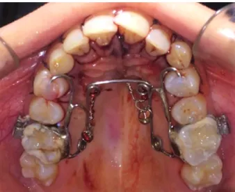
At the end of the orthodontic traction of the impacted tooth, brackets have been applied on the upper arch, in order to align the now disinclused tooth in the arch.
4.4.5 Alignment in arch with the Damon System
When the canine has erupted sufficiently into the palate, in an ideal period of four months (16 weeks), a bracket or an orthodontic button is placed on the tooth and the upper arch is bandaged with Damon System technique[143-144]. In this way the tooth can be gradually translated into the dental arch.
Figure 14 – Damon fixed appliances
The Damon System is an orthodontic method that uses passive self-ligating attacks, designed by Dr. Dweit Damon, associated with high-tech wires in copper-nickel-titanium alloy.
Self-ligating devices are a system of direct attachments that do not require elastic or metal ligatures to keep the arc in the slot, having a door or clip mechanism designed to perform this function.
If the primary canine was present, it was extracted when the permanent approaches its final position in the dental arch.
4.5 Monitoring of dental movements
Post-surgery, the patients underwent three follow-up appointments, in order to evaluate and monitor the spontaneous eruption of the canines, or the eventual early closure of the mucosa and lack of appearance of the element in the palate.
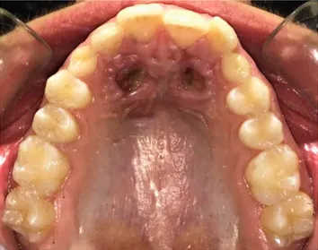
During each control visit, a clinical examination, conventional impressions and intraoral scans was carried out, photographs were taken and questions were asked to patients regarding the presence of pain, swelling, bleeding, the need to take drugs and reducing the functionality of the area involved.
Impressions and scans were made always by the same operator, with the same procedures, to standardize the data acquisition.
The Visual Analog Scale (VAS) was submitted, to the patients of the experimental group A and B and none of them registered pain on this scale, contrary to the control group which recorded post-operative pain in some cases.
Monitoring was carried out as follows: • Laser surgery
• 1 week • 8 weeks (T1) • 16 weeks (T2)
The hypothesis was that palatally impacted canines, after the laser surgery, will suffer a “spontaneous” eruption for reactivation of the physiological eruption, without any orthodontic traction (traditional method). Furthermore, this “spontaneous” eruption was monitored through different digital technology like an intraoral scanner, from the time of the laser surgery to the crown eruption. At this time, we performed the alignment in the arch with direct/indirect bonding technique.
Although, the study analysed the effectiveness of monitoring through the intraoral scanner CS3500 (Carestream Dental), which allows to acquire precise dental coloured impressions.
Digital colour scans were performed in the 1 week, 8 weeks and 16 weeks follow-up visit, through use the intraoral scanner CS3500 (Carestream Dental, USA), which made it possible to obtain scans(accuracy: 30 µm) of the dental arches in .STL format.
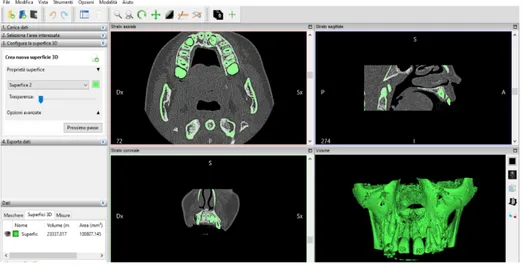
4.5.2 Conversion CT to STL model
The examination of the CT allowed to evaluate the three-dimensional morphology of the impacted tooth, its location and inclination in the three planes of space, the depth and the type of inclusion, the relationships with the other elements.
To get an accurate initial situation, the Invesalius 3.1 software was used to export the CT in STL format.
It is an open source software for reconstruction of computed tomography and RM images. The first step is to import CT files, then the range of values, that correspond to the radiopacity of the selected X-ray images that we want to be part of the 3D model, is chosen.
Figure 15 - Range of values is chosen on Invesalius 3.1
At this point a “surface” is generated and a 3D model in STL is obtained, which can be exported and meshed with the STL models of the intraoral scans.
Figure 16- 3D model generated
4.5.3 Overlap of the STL models and measurements
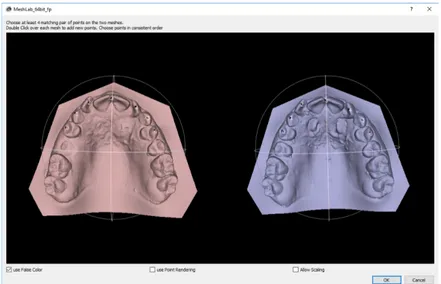
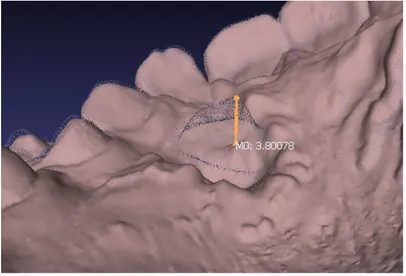
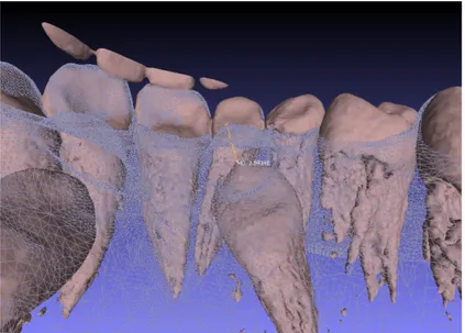
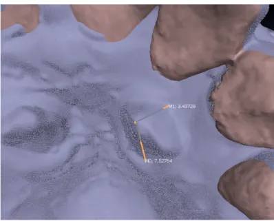
The 3D impressions were imported into the open-source software MeshLab (Visual Computing Lab, Pisa, Italy).
The three-dimensional models of the patients obtained with the scanner at 1 week, 2 months (T1) and 4 months (T2) were superimposed with the 3D models extrapolated from the pre-operative CT (T0).
After importing the scans files, the first operation was 3D data alignment. MeshLab provides a powerful tool for moving the different meshes into a common reference system, able to manage large set of range-maps.
Figure 17- Alignment of two models
Figure 18- Overlap and alignment of the two models
3D models often need to be re-oriented, or placed in a specific reference system; MeshLab provides a variety of features to manipulate the scale, positioning and orientation of a 3D model, including basic transformation operations like translation/scaling/rotation, automatic re-centering and alignment to axis, geo-referencing with reference points, interactive manipulators for rotation/translation/scaling.