UNIVERSITY OF NAPLES FEDERICO II
DEPARTMENT OF PHARMACY PhD programme in Pharmaceutical Sciences
PHARMACOLOGICAL AND NUTRITIONAL CONTROL OF DYSBIOSIS RELATED TO CNS DISORDERS: GUT-BRAIN AXIS
PhD student ADRIANO LAMA
XXXI CYCLE (2016-2018)
Coordinator Prof.
MARIA VALERIA D’AURIA
Tutor Prof.
Summary
ABSTRACT 1
INTRODUCTION 7
1. PALMITOYLETHANOLAMIDE 9
1.1. PEA: ORIGIN, STRUCTURE, AND ACTIVITY 9
1.2. PEROXISOME PROLIFERATOR-ACTIVATED RECEPTORS (PPARS): PPARa FUNCTIONS IN
THE BRAIN 11
1.3. PPARa ACTIVATION AS MECHANISM OF ACTION OF PEA 15
1.4. PHARMACOLOGICAL EFFECTS OF PPARa AGONISTS: FOCUS ON PEA IN CNS
DISORDERS 18
1.5. NEW FORMULATION OF PEA: ULTRA-MICRONIZED PEA 20
2. AUTISM SPECTRUM DISORDERS 23
2.1. DEFINITION, EPIDEMIOLOGY, DIAGNOSIS AND RISK FACTORS 23
2.2. TREATMENTS OF ASDS 27
2.3. GUT-BRAIN AXIS IN ASDS 29
3. OBESITY-INDUCED DEPRESSION 32
3.1. DEPRESSION: DEFINITION, EPIDEMIOLOGY, DIAGNOSIS AND RISK FACTORS 32
3.2. PATHOGENIC MECHANISMS OF MDD 36 3.3. DEPRESSION AND OBESITY: A BIDIRECTIONAL INTERPLAY 38 3.4. TREATMENT OF MDD 42 4. MATERIAL AND METHODS 45 4.1. SEX DIFFERENCES IN GUT MICROBIOTA OF BTBR MICE 45 4.1.1. ANIMALS 45 4.1.2. MARBLE BURYING TEST 46 4.1.3. SELF-GROOMING TEST 47 4.1.4. SOCIAL INTERACTION TEST 47 4.1.5. IN VIVO INTESTINAL PERMEABILITY ASSAY 48 4.1.6. HISTOLOGICAL ANALYSIS 49
4.1.7. MICROBIAL DNA EXTRACTION, 16S RIBOSOMAL DNA (RDNA) LIBRARY PREPARATION
AND SEQUENCING 50
4.1.8. SEQUENCING DATA ANALYSIS 52
4.1.9. QUANTIFICATION OF GENE EXPRESSION USING RT-PCR 54
4.1.10. OTHER STATISTICAL METHODS 54
4.2.1. ANIMALS 56 4.2.2. DRUGS AND TREATMENT 56 4.2.3. OPEN FIELD LOCOMOTION TEST (OFT) 58 4.2.4. MARBLE BURYING TEST 59 4.2.5. SELF-GROOMING TEST 59 4.2.6. THREE-CHAMBERED SOCIAL TEST 59 4.2.7. ELEVATED PLUS MAZE TEST (EPM) 59 4.2.8. SERUM PARAMETERS 60 4.2.9. MITOCHONDRIAL PARAMETERS 60 4.2.10. OXIDATIVE STRESS ASSAY 63 4.2.11. WESTERN BLOT ANALYSIS 64 4.2.12. QUANTIFICATION OF GENE EXPRESSION USING RT-PCR 65 4.2.13. 16S METAGENOMIC SEQUENCING AND DATA ANALYSIS 65 4.2.14. STATISTICAL ANALYSIS 66
4.3. PEA LIMITS HFD-INDUCED DEPRESSION 67
4.3.1. ANIMALS AND TREATMENTS 67
4.3.2. BODY WEIGHT AND BODY GAIN IN FAT 68
4.3.3. OPEN FIELD LOCOMOTION TEST 69
4.3.4. FORCED SWIMMING (FST) AND TAIL SUSPENSION TESTS (TST) 69 4.3.5. NOVEL OBJECT RECOGNITION TEST (NORT) 70 4.3.6. SERUM PARAMETERS 71 4.3.7. C-FOS AND IBA-1 STAINING 72 4.3.8. QUANTIFICATION OF GENE EXPRESSION USING RT-PCR 73 4.3.9. WESTERN BLOTTING 74 4.3.10. 16S METAGENOMIC SEQUENCING AND DATA ANALYSIS 75 5. RESULTS 76 5.1. SEX DIFFERENCES IN GUT MICROBIOTA OF BTBR MICE 76 5.1.1. OVERALL STRUCTURE OF GUT MICROBIOTA OF MALE AND FEMALE BTBR MICE. 76 5.1.2. GUT MICROBIOTA PROFILING OF BTBR FEMALE AND MALE MICE 78 5.1.3. KEY PHYLOTYPES DRIVING GUT MICROBIOTA PROFILES OF MALE AND FEMALE BTBR MICE 79
5.1.4. ALTERATION IN BEHAVIORAL PHENOTYPE, INTESTINAL INTEGRITY, AND IMMUNITY IN FEMALE AND MALE BTBR MICE: CORRELATION WITH GUT MICROBIOTA MODIFICATIONS 82
5.2. PEA COUNTERACTS AUTISTIC-LIKE PHENOTYPE IN BTBR MICE 88 5.2.1. PEA REDUCED REPETITIVE BEHAVIOUR AND INCREASED SOCIABILITY IN BTBR MICE 88 5.2.2. PEA IMPROVES BDNF/TRKB SYSTEM, INCREASING HIPPOCAMPAL PPAR-a
EXPRESSION 95
5.2.3. PEA TREATMENT IMPROVES HIPPOCAMPAL MITOCHONDRIAL FUNCTION AND REDUCES
OXIDATIVE STRESS 97
5.2.5. PEA EFFECTS ON GUT PERMEABILITY AND MICROBIOTA 100
5.3. PEA LIMITS HFD-INDUCED DEPRESSION 104
5.3.1. PEA LESSENS DEPRESSIVE-LIKE BEHAVIOUR AND MEMORY DEFICIT 104 5.3.2. PEA IMPROVES SERUM METABOLIC AND INFLAMMATORY PARAMETERS 105 5.3.3. ACTIVATION OF POMC NEURONS BY PEA IN ARC 106 5.3.4. PEA REDUCES NEURO-INFLAMMATION IN HYPOTHALAMUS 108 5.3.5. PEA IMPROVES BDNF/CREB SYSTEM AND REDUCES HFD-INDUCED INFLAMMATION
IN HIPPOCAMPUS 110
5.3.6. PEA AMELIORATING EFFECT ON HFD-INDUCED DEPRESSION: POSSIBLE INVOLVEMENT
OF PPAR-a 111
5.3.7. PEA RESTORES BBB INTEGRITY OF HFD-MICE HIPPOCAMPUS 112 5.3.8. PEA EFFECTS ON BDNF/CREB SYSTEM IN PREFRONTAL CORTEX OF HFD MICE 113 5.3.9. PEA EFFECTS ON GUT MICROBIOTA COMPOSITION 114
6. DISCUSSION AND CONCLUSIONS 120
6.1 GUT-BRAIN AXIS IN ASDS: FOCUS ON PEA BENEFICIAL EFFECTS 120
6.2. PEA AND OBESITY-INDUCED DEPRESSION 128
HIGHLIGHTS 134
GENERAL CONCLUSIONS: PEA AND CNS DISORDERS 135
1
Abstract
'Leaky gut' syndrome has attracted much attention in recent years, and represents now a complementary/alternative target for several complex diseases characterized by this pathological condition. It is often described as an increase in the permeability of the intestinal mucosa, allowing bacteria, toxic digestive metabolites, bacterial toxins, and small detrimental molecules to 'leak' into the bloodstream. The microbiota-gut is an integral component of the gut–brain neuroendocrine metabolic axis and any microbiota-gut disruption that can occur, could distressed homeostasis and share an inflammatory response, affecting distal organs including the brain. It has been shown, indeed, that the gut can influence the blood brain barrier (BBB) through gastrointestinal-derived hormones, small molecule and metabolic co-factor production, or through cytokine synthesis and other inflammatory mechanisms. Therefore, the CNS is under constant attack or, conversely, advantage from a wide variety of neuro-psychotropic-modulating microbes, and their metabolites. So, the proper neurodevelopment and functioning of the CNS depends from an integrated, rather than opposing, cross-talk between gut-gut microbiota and brain. Several CNS disturbances were related to gastrointestinal dysfunction and 'leaky brain', underlining the need of identifying new integrative and
multi-2
targeted approaches. These complex diseases, being multifactorial, could result, in fact, less responsive to targeted standard drugs, since poorly fits ‘one-disease one-target’ and ‘one-target one-compound’ paradigms in this context.
Here, we focused on autism spectrum disorders (ASDs) and major depressive disorder (MDD), two brain disorders linked to a dysfunction of BBB and impacted by immune and inflammatory peripheral stimuli. Our aim has been to evaluate the possible therapeutic potential of modulating several aspects of these multifactorial disorders, in order to benefit of peripheral and central contributions, converging on an improvement of overall health.
To this aim we used BTBR T+tf/J (BTBR) mice model of ASDs and high fat diet-induced MDD in young mice, and the possible pharmacological modulation by palmitoylethanolamide (PEA) was investigated.
PEA, is an endogenous N-acyl-ethanolamine, biosynthesized to maintain cellular homeostasis when this is challenged by external stressors provoking inflammation, neuronal damage and pain. The interaction and activation of PPARα has been recognized as the main mechanism of the effects evoked by this acylethanolamide. In particular, the analgesic and anti-inflammatory effects of PEA were demonstrated to be mediated by PPARα activation, since it has no effect in PPARα null mice.
3
Indeed, the discovery of PPARα in distinct areas of the brain, opened a new scenario to explore the possible activity of this acylethanolamide in the CNS. Recently it has been demonstrated that genetic inactivation of PPARα, particularly abundant in the CNS, leads to a behavioral and cognitive phenotype reminiscent of that of preclinical models of ASDs, i.e. mouse model BTBR T+tf/J (BTBR), which displays an improved repetitive behavior when autistic mice are treated with a synthetic PPARα agonist. These results, not only indicated a central role for this receptor in neurological functions associated with the behavior, but more interestingly highlighted PPARα as a potential pharmacological target to lessen ASDs symptoms. On the other side PEA activity at intestinal level has suggested a possible role of this acylethanolamide in modulating not only gut function, such as intestinal transit and permeability, but also gut-brain axis.
Before evaluating the effect of PEA on BTBR mice, in the first part of this PhD programme, we performed a study, also evaluating sex influence, on gut microbiota composition, behavioral features, and intestinal integrity, inflammatory status and architecture of adult male and female BTBR mice. These gender characterizations arise from the well-known different clinical features of male and female autistic patients and the need to identify them in a mouse model of ASD.
4
Consistently with gut-brain axis hypothesis, we showed that BTBR mice presented a profound intestinal dysbiosis compared to control strain, more marked in female than in male mice, indicating Bacteroides,
Parabacteroides, Sutterella, Dehalobacterium and Oscillospira genera as
key drivers of sex-specific gut microbiota profiles associated with altered behavior. Interestingly, we also showed that the dysbiosis was accompanied in BTBR mice by increased gut permeability and colon inflammation. Therefore, we have considered BTBR mice, as an idiopathic model of autism useful to investigate not only the correction of the autistic behavior, but also a starting point to investigate whether the reduction of intestinal inflammation and integrity, and possibly the restoration of gut microbiota balance may ameliorate pathological traits.
Based on this background, the aim of this study was to investigate the pharmacological effects of PEA on autistic-like behaviour of BTBR T+tf/J mice and to shed light on the contributing mechanisms. PEA was able to revert the altered behavior of autistic mice. This effect was contingent to PPARa activation, since was blunted by PPARa blocking or deletion. At mechanistic level, PEA restored hippocampal BDNF signaling, improved mitochondrial dysfunction and reduced serum, hippocampal and colonic inflammation. These beneficial effects were related to the reduction of leaky
5
gut in PEA-treated BTBR mice mediated by increased expression of colonic tight junctions. In addition, PEA modulated gut microbiota composition, underlining the strong link between gut and brain.
In last years, the connection between modification in gut microbiota induced by obesity and the development of MDD has become more evident. High fat feeding causes the production of several inflammatory mediators that can compromise colonic epithelial barrier function, with the translocation of bacterial metabolites in CNS, evoking deleterious events. The modulation of PPARα, mostly expressed in hippocampus, has demonstrated to improve synaptic dysfunction and HFD-related pathological events in MDD.
Here, we have addressed the effects of PEA in a mouse model of HFD-induced depression, focusing on converging mechanisms involved in its activity. In particular, we assessed PEA capability in modulating the gut-brain axis and ameliorating depressive behavior. The treatment with PEA improved the depressive-like behavior and memory deficit, shown by HFD animals, impacting on BDNF signaling pathway and reducing neuroinflammation, both in hypothalamus, hippocampus and prefrontal cortex. These beneficial effects of PEA, as PPARa agonist, were correlated to an increased expression of PPARa, its coactivator PGC1a, and the downstream gene FGF21. As in BTBR model, here PEA also modulated gut
6
microbiota composition, reducing the amount of endotoxin-producer
Desulfovibrio and increasing Clostridiales genus relative abundance, and
consistently Clostridiales-producing metabolites, including short-chain fatty acids.
In conclusion, PEA, a multifunctional compound, can represent a novel therapeutic approach for multifactorial disorders, such as ASDs and MDD, able to counteract the alteration of central and peripheral pathways involved in their onset and progression.
7
Introduction
Autism is a multifactorial disorder, where many genes converge, through multiple molecular mechanisms, onto common processes involved in the development and function of neural circuits, whose alteration leads and sustains the core symptoms of ASDs (1). Evidence based on environmental factors, such as infection, gut microbiome alteration, and immunological features are also growing, suggesting the involvement of many scenarios overlapping in this complex neurodevelopmental pathology. Obviously, even if targeted specific treatments would clarify the relevance of the altered pathways in ASDs, on the other hand the complex heterogeneity in ASDs children population could require a multi-targeted approach. Here, we analysed the modulatory effect of PEA on many of the pathways and factors, starting from the idea that the abnormalities reported to be at the onset and progression of this neurodevelopmental pathology are responsible of brain dysfunction and consistently associated in core symptoms of ASDs.
Depression, as well, has a multifactorial nature, resembling that of other complex disorders. Very recently, it has been shown that obesity, in critical periods for neurodevelopment and neuronal plasticity, may contribute to mood and cognitive disorders, therefore constituting a risk factor for developing depression and anxiety. In this case, westernized dietary patterns
8
and dysbiosis seem to contribute to obesity-related CNS complications involving gut microbiota, as a mediating factor between peripheral signals and the brain. It is conceivable to hypothesise that alteration of gut microbiota may contribute to the endocrine, neurochemical and inflammatory alterations underlying to gut-brain axis interplay. These include dysregulation of the HPA-axis with overproduction of glucocorticoids, alterations in levels of neuroactive metabolites (e.g., neurotransmitters, short-chain fatty acids) and activation of a pro-inflammatory milieu that can cause neuroinflammation. Up to date, PEA has proven to be a multi-targeted compound: the main pharmacological effects of PEA are mediated by the activation of PPARα (2, 3), a transcription factor involved in the regulation of gene networks, which control pain and inflammation (4). This mechanism is supported by the inhibition of neuronal firing (5) and by the stimulation of neurosteroid synthesis, indicating that multiple mechanisms contribute to the central effect of PEA (6).
On the other side, PEA has demonstrated to have a peripheral anti-inflammatory effect shown both on BTBR mice, reducing cytokine production, decreasing the inflammatory state at colonic and systemic levels, and in rodent models of obesity where it was able to reduce the overall low-grade inflammation (7), also impacting on the modulation of gut microbiota.
9
1. Palmitoylethanolamide
1.1. PEA: origin, structure, and activity
The acylethanolamides (AEs) are bioactive fatty molecules, synthetized “on demand” from membrane phospholipids (8). The recovery of AEs in several tissues and their pharmacological effects on multiple biological pathways, have suggested a regulator role of these endogenous molecules in paracrine or autocrine functions (9). Known their beneficial effects on nociception, lipid metabolism, and inflammation, AEs were initially considered Autacoid Local Injury Antagonism Amides (ALIAmides) (10). In last years, several research groups supported their possible role as neuromodulators, since AEs are produced in various areas of brain, both in physiological and pathological conditions (11). Indeed, both AEs are produced during neuronal damage, carrying out their neuroprotective activity through both receptor and non-receptor specific mechanisms (12).
Among all AEs, PEA is abundant in central nervous system and only recently its biological functions have been clarified (13). As shown by Figure 1.1, PEA is biosynthesized from phosphatidylethanolamine (PE) by two sequential enzymatic reactions (14, 15). The first reaction is the transfer of an acyl-group (palmitic acid) from sn-1 position of phosphatidylcholine
10
(PC) to PE by an N-acyltransferase (NAT), generating N-acyl- PE (NAPE) (16). Afterwards, PEA is released from NAPE by a phosphodiesterase of phospholipase D (NAPE-PLD) (17). PEA is rapidly catabolized by fatty acid amide hydrolase (FAAH) to palmitic acid and ethanolamine (18, 19) or N-acylethanolamine-hydrolyzing acid amidase (NAAA) in inflammatory status (20). Considering that NAAA preferentially hydrolyzes PEA rather than other AEs, the use of selective NAAA inhibitors could increase local levels of endogenous PEA, resulting anti-inflammatory and analgesic molecule (20). From the phospholipid bilayer of the plasma membrane, PEA is transported by fatty acid binding proteins or heat shock proteins (21) to degrading enzymes or target receptors, such as peroxisome PPARa (22) and transient receptor potential vanilloid (TRPV)1 (23).
11
Figure 1.1. The metabolic conversion of PEA.
1.2. Peroxisome proliferator-activated receptors
(PPARs): PPAR
a
functions in the brain
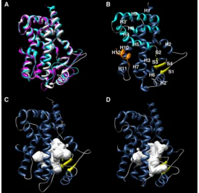
PPARs are ligand-activated transcription factors called lipid “sensors”, owning the key role in lipid and lipoprotein metabolism, energy homeostasis, cell proliferation death and differentiation. As shown by Figure 1.2, these nuclear receptors include three isotypes: PPARa (NR1C1), PPARb/d (NR1C2), and PPARg (NR1C3). PPARs, as other members of the nuclear receptor superfamily, have four domains: (i) a
N-12
terminal A/B domain, important mediator of PPAR subtype-specificity, determining PPAR subtype-specificity and containing a weak ligand-independent transactivation function called AF-1; (ii) a C domain, highly conserved, that binds DNA via a two zinc-finger motif (hallmark of the NR superfamily); (iii) a D domain, which is a hinge region; (iv) an E/F domain, which is the ligand binding domain (LBD) (24).
Figure 1.2 (A) Structural superposition of PPARα (white), PPARδ (magenta) and PPARγ
(cyan). 3D structure of (B) PPARα, (C) PPARδ and (D) PPARγ.
The conformational change of PPARs is induced by different type of fatty acids, triggering the transcription of specific genes that encode for several metabolic and cellular processes, such as fatty acid b-oxidation and
13
adipogenesis (25). To this purpose, molecules that target PPARs can modulate lipid and glucose metabolism, representing excellent therapeutic tools for metabolic disorders (26, 27).
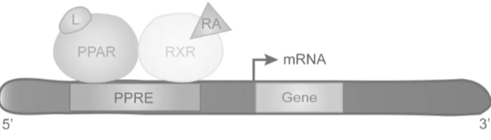
The activity of PPARs is based on the bond with specific DNA sequence elements (named PPAR response elements, PPREs), and on regulation of gene expression like heterodimers with retinoid X receptors (RXRs) (28-30). Indeed, the dimerization between specific RXR isotypes (a, b, g) and PPARs seems to influence the recognition of target gene promoters (31) (Figure 1.3)
Figure 1.3. PPARs bind RXR to constitute PPRE. Ligands can modulate the transcription
of target genes. L, ligand; RA. 9-cis-retinoic acid.
It is well known that PPAR-mediated effects are modulated by the interaction with several factors, including corepressors and coactivators (32). The steroid receptor coactivator 1 and the cAMP response element-binding (CREB) protein are two pivotal coactivators of PPARs and they own histone acetyl transferase activity that induces chromatin decondensation
14
and target gene activation. Beyond the clear interaction with PPARg, PPARg coactivator 1a (PGC-1a) binds PPARa in ligand-dependent manner, carrying out cellular functions, such as reactive oxygen species (ROS) and fatty acids (FAs) metabolism associated with mitochondrial and peroxisomal biogenesis (33-36). These functions are pivotal in counteracting CNS disorders, where redox unbalance and lipid dysmetabolism play a detrimental role in neurodegeneration. The distribution of PPARa varies in different tissues, it is generally expressed at high extent in metabolically active ones (liver, heart, skeletal muscle, intestinal mucosa and brown adipose tissue) (37), even if studies demonstrated their occurrence in brain (38, 39). In particular, Moreno et al. (40) described deeply the distribution of PPARs and RXRs in rodent brain. PPARa shows several functions in the brain: regulation of lipid metabolism (shortening of VLCFAs, use of FAs as metabolic fuel, biosynthesis of PUFAs) (41, 42); management of ROS metabolism (increase of antioxidant systems and induction of H2O2 -producing enzymes) (43-45); neural cell proliferation, differentiation and apoptosis (46-49); modulation of dopaminergic, glutamatergic and cholinergic neurotransmission (50-52); cognitive and memory function, spatial learning, emotionality (53-55); modulation of pain pathways (4,
56-15
59); modulation of fasting and satiety responses via hypothalamus-pituitary axis and peripheral glucose homeostasis (60-63).
Despite the hippocampus does not produce energy from fat metabolism, PPARα has been identified in different subfields of this tissue in rodents (64, 65). Interestingly, PPARα knockdown mice showed a reduction of several plasticity-associated molecules (NR2A, NR2B, GluR1 and Arc), but not voltage-gated ion channel ones, suggesting a crucial role of PPARα in hippocampal plasticity (64). In the same study, the authors demonstrated that PPARα agonists induced the activation of CREB (pivotal in synaptic plasticity) promoter in hippocampal neurons from wild type mice and not from PPARα knockout ones (64). All these findings highlight PPARα involvement in brain functions.
1.3. PPAR
a
activation as mechanism of action of
PEA
The anti-inflammatory, analgesic and neuroprotective effects of PEA are related to the involvement of both central and peripheral nervous system targets (2-4). In particular, different cell types involved in resolving chronic pain and inflammation (immune cells, microglia and neurons) synthetize and metabolize PEA (66).
16
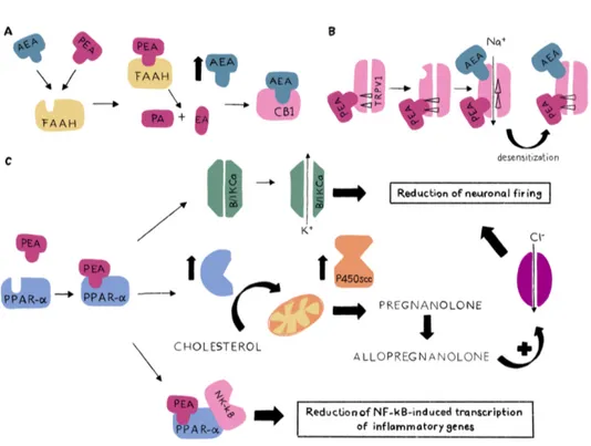
PEA is an endocannabinoid-like molecule and, as such, was originally considered a CB2 agonist (67). Furthermore, PEA activity has been also associated to its capability in increasing half-life and therefore its effects on CB or TRPV1 (68-70), the so-called “entourage effects” due to PEA competitive inhibition of AEA hydrolysis on FAAH (71) and/or direct allosteric effect on TRPV1 (72, 73). Therefore, PEA is not a merely “classical” endocannabinoid because its pharmacological effects reside outside of a strict interaction with CB receptors. Beyond the interactions of PEA with orphan receptors, such as G protein-coupled receptor (GPR) 55 and GPR119 (74-77), the main role of pharmacological activity of PEA is related to PPARα activation (13). Indeed, PEA increases properties of calcium-activated intermediate (IKCa) and big-conductance potassium (BKCa) potassium channels via a PPARα dependent non-genomic mechanism, leading to a fast reduction of neuronal firing (3). In addition, as demonstrated by Sasso et al. (78) in acute or persistent pain mice model, the activation of PPARα, through a genomic mechanism, increases the expression of steroidogenic acute regulatory protein (StAR) and cytochrome P450 side-chain cleavage (CYP450scc), leading to the transport of cholesterol into mitochondria and its metabolic conversion into pregnanolone. The subsequent increase of allopregnanolone production
17
induce a positive allosteric activation of aminobutyric acid (GABA)-A receptors, an increase in Cl- fluxes and a reinforcing effect on the reduction of neuronal firing. Furthermore, the anti-inflammatory effects of PEA seem to be correlated with PPARa capability in preventing the nuclear translocation of NF-kB and repressing the expression of pro-inflammatory proteins (i.e. TNF-a, IL-1b), limiting the recruitment of immune cells (79) (Figure 1.4)
Figure 1.4. Direct and indirect mechanisms of action of PEA. (A) the competitive
inhibition of AEA metabolism by FAAH; (B) allosteric activity on TRPV1 and its desensitization; (C) genomic and non-genomic mechanisms dependent on PPARα.
18
1.4. Pharmacological effects of PPAR
a
agonists:
focus on PEA in CNS disorders
On the basis of these observations, it is conceivable that targeting PPARa can represent the basis for novel therapies in the treatment of acute and chronic CNS pathologies. Indeed, several studies highlighted the beneficial effects of different PPARa agonists in improving neuroinflammation, underlying neurodegenerative disease and neuropsychiatric disorders (80). Among endogenous ligands of PPARa, OEA and PEA own several neuroprotective functions (81-83). In rat model of transient focal cerebral ischemia, PEA achieved a significant neuroprotective effect by reducing infarcted tissue size (84). Moreover, this molecule was able to protect neuronal cells from oxidative stress and alter the expression levels of kinases involved in neuroprotection (85). Koch et al (86) demonstrated also that PEA protected dentate gyrus granule cells from excitotoxically-induced neuronal damage, activating PPARa and not PPARg in microglial cells and hippocampal neurons. Consistently, our research group demonstrated that PEA reduced oxidative stress in astroglial cells through PPARa-triggered biosynthesis of allopregnanolone (87). The role of PEA in regulating the complex systems involved in inflammatory and adaptive immune response, acute and chronic pain is now well understood (13). Concerning pathogenic
19
mechanisms of pain, PPARa has been shown to be overexpressed in rat spinal cord after peripheral noxious stimulation (56). Furthermore, D’Agostino et al. (4) demonstrated that intracerebroventricular administration of PEA significantly reduced the expression of the pro-inflammatory mediators, such as COX-2 and iNOS, in dorsal root ganglia following carrageenan-induced intraplantar oedema. The obligatory role of PPARa was confirmed by the loss of anti-nociceptive effects in PPARa knockout mice.
In neurodegenerative disorders, such as Parkinson’s Disease (PD) and Alzheimer Disease (AD), PPARa agonists, and in particular PEA, seem to have a therapeutic effect. In MPTP-treated mice, an animal model of PD, PEA reversed MTPT-induced motor deficits in PPARa-dependent manner, reducing microglial activation, astrocyte number and S100b overexpression (88). Avagliano et al. (89) demonstrated that PEA levels are drastically reduced in post-mortem brain samples from AD patients. Consistently, both in in vitro and in vivo AD model, PEA was able to improve pathological features (90, 91).
The beneficial effects of PEA in mood disorders, especially in depression, are still overlooked. To date, only two preclinical studies and double-blind, randomized and placebo-controlled trial suggested the hypothesis that PEA
20
can reduce a depressive-like behavior (92-94). In particular, in a mouse model of anxiety/depression induced by corticosterone administration, a formulation of PEA and luteolin improved hippocampal neurogenesis and neuroplasticity. Yu et al. (92) demonstrated that oral PEA produced a significant reduction in immobility time in both tail suspension test (TST) and forced swimming test (FST). Only in a clinical study, recently published, the association of PEA with citalopram effectively improves symptoms of patients (predominantly male gender) with major depressive disorder, even if further investigations are needed (94).
Among neurodevelopmental disorders, autism spectrum disorder (ASD) is characterized by alterations in the brain's endocannabinoid system as demonstrated by Kerr et al. (95). In particular, these authors showed that in rat valproic acid model of autism there was a reduction of PPARα and GPR55 expression in the frontal cortex and PPARγ and GPR55 expression in the hippocampus while CB1 or CB2 receptor expression was not altered in all any brain regions. In another recent study, Bertolino et al (96) showed the beneficial effects of an association of PEA and luteolin in VPA mouse model and in an autistic child, even if the mechanisms of action keep still unclear.
21
Although the excellent pharmacological activity, PEA drawbacks are based on its poor solubility and bioavailability, due to its lipophilic nature. Moreover, apart from dissolution-rate-limited PEA absorption, another limit is particle size, inversely related to PEA bioavailability. All these drawbacks have prompt to improve PEA pharmacokinetic features. The micronization has been an excellent strategy to this aim, producing microparticles <10 μm (97, 98), in order to increase surface area and rate of dissolution (99), together with a reduction of absorption variability (100). Indeed, ultra-micronized PEA (PEA-um) has demonstrated an improved efficacy when administered per os compared to standard PEA powder administration. Recently, Petrosino et al. (101) have published an article focused not only on the absorbability of PEA-um and naïve PEA, but, interestingly, also on their distribution in peripheral and central tissues in physiological and inflammatory conditions. Notably, the authors demonstrated an increased plasma concentration by ultramicronization of PEA, in fact, differently from naïve PEA administration, PEA-um (30 mg/kg per os) to healthy animals resulted detectable in the bloodstream already after 5 min, with a peak plasma concentration of 5.4 ± 1.87 pmol/ml. This pharmacokinetic profile was even more favorable in inflamed animals, showing an increase of PEA-um concentration in plasma and inflamed tissues (including spinal cord).
22
Ultra-micronized PEA, provided by Epitech Group, is produced by the air-jet milling technique, where powder is slowly inserted into a air-jet-mill apparatus endowed with a chamber of 300 mm in diameter that operates with “spiral technology” driven by compressed air. The high number of collisions that occur between particles as a result of the high level of kinetic-not mechanical-energy produces sub-micron-sized crystals.
23
2. Autism spectrum disorders
2.1. Definition, epidemiology, diagnosis and risk
factors
The term autism spectrum disorders (ASDs) describes a group of early-appearing social communication deficits and repetitive sensory-motor patterns due to multiple converging causes (102). ASDs, over time, have become from barely and rare disorders of childhood onset to studied and recognized lifelong conditions with heterogeneous features. Although autistic individuals are very different from each another, the core features are based on two areas without any cultural, racial, ethnical and socio-economical differences: impaired social interaction and repetitive stereotyped motor behaviors (103). ASDs represent an economic burden, mainly for autistic patients with low functionality due to expensive provision of support: in 2014, its cost estimated $3020 for health care and $14 061 for aggregate non-health care, including $8610 for school education (104). In 2010, the global prevalence of ASDs was about 1% (105), even if a more recent review estimated an increase up to 5% in developed countries (106).
24
Since there are no reliable biomarkers, ASD diagnosis is based on American Psychiatric Association’s Diagnostic and Statistical Manual of Mental Disorders (DSM)-5 criteria, considering specific behavioral traits (102). To identify an ASD patient, a person must show evidence of complications, in each of three social communication subdomains and/or in two of the four different stereotyped and repetitive sensory-motor behaviors (Table 2.1)
Persistent deficits in social
communication and social
interaction
Restricted, repetitive patterns of behavior, interests, or activities Deficits in social–emotional
reciprocity (e.g. abnormal social approach and failure of normal back-and-forth conversation; or reduced sharing of interests, emotions, or affect)
Stereotyped or repetitive motor movements, use of objects, or speech (e.g. simple motor stereotypies, lining up toys, or flipping objects)
Deficits in non-verbal
communicative behaviors (e.g. poorly integrated verbal and
non-verbal communication,
abnormalities in eye contact and body language, or deficits in understanding and use of gestures)
Insistence on sameness, inflexible adherence to routines, or ritualized patterns of verbal and non-verbal behavior
(e.g. extreme distress at small changes, difficulties with
transitions, or rigid thinking patterns)
Deficits in developing, maintaining, and understanding
relationships (e.g. difficulties adjusting behavior to suit various social contexts; or difficulties in sharing imaginative play or making friends)
Highly restricted, fixated interests that are abnormal in
intensity or focus (e.g. strong attachment to or
preoccupation with unusual objects)
Hyperreactivity or hyporeactivity to sensory input, or unusual
25
interests in sensory aspects of the environment (e.g. apparent indifference to pain or temperature, or adverse responses to specific sounds or textures)
Table 2.1. The description of ASD signs and symptoms in DSM-5.
Early diagnosis plays a crucial role to minimize the possible intellectual disabilities, reported later in underestimated and undertreated ASD adults. In addition to screening instruments, such as several standardized assessment tools (e.g. STAT, ADOS, ADI-R and DISCO), other strategies, including an increased awareness of ASDs in the family and community and an easy access to specific services, can improve the quality of life of ASD patients. The difficulty in ASD diagnosis is getting worse by accompanying comorbidities (attention-deficit hyperactivity disorder, social anxiety, generalized anxiety and phobias) (107). This concern is mainly worth for females, rather than males, who can be underdiagnosed due to the high occurrence of comorbidities (e.g. depression and severe anxiety) that hide and mask ASD symptoms (108).
Many risk factors for ASDs have been described and they are divided in two main types: environmental and genetic. The advanced parental age (³40 years for mothers and ³50 years for fathers) and a short interpregnancy intervals have been independently associated to an increased risk for ASD
26
onset in children (106, 109). The use of antibiotics and the consequent perturbations of gut microbiota during pregnancy are a potential risk factors for infantile ASD (110). Furthermore, some non-optimal conditions of mothers (excessive weight gain, hypertension, nosocomial infections or autoimmune disease) can contribute the development of ASDs (111). Although results from different studies have to be clarified, some links with air pollutants and maternal stressors during pregnancy have been found (106). Beyond maternal conditions, preterm birth (<32 weeks), low birthweight (<1500 g), small-for-gestational-age status, and large-for-gestational age status (>95th birthweight percentile) represent other important risk factors (112, 113), even if the reasons of their involvement keep still unclear. The administration of many drugs in pregnant mothers has been associated to an increased risk of ASDs. Christensen et al. (114) demonstrated that a prenatal valproic acid exposure increased the probability of ASD in offspring. In addition, de Theije et al. (115) also in rodents demonstrated that this in utero valproic acid exposure induced gut inflammation, altered microbiota, and ASD-like behavioral abnormalities in male offspring. Other studies did not confirm the previous belief in antidepressant-induced ASD effects (116). In last years, media and no-vax organizations strongly supported the association between ASDs and
27
vaccination, but, to date, no scientific evidence has clearly demonstrated their link (117).
Beyond the environmental factors, the genetic risk has assumed an even more important role in ASD pathogenic mechanisms. Tick et al. (118) showed that ASDs can be inherited in 74-93% of cases. The first evidence of this link between ASDs and genetic was the development of autistic behavior in patients affected by rare genetic syndromes, such as fragile X syndrome (119). Following studies demonstrated also that the mutations of more than 100 genes are involved in ASD risk, indicating an objective obstacle in ASD diagnosis through genetic screening (120).
2.2. Treatments of ASDs
ASDs are a group of complex diseases and as such, its treatment represents a complicated challenge. To date, psychological interventions are the most important tools to manage these disorders (103). Weitlauf et al. (121) reported that early parent-mediated interventions to coach how to interact with ASD children give immediate beneficial effects on autistic traits. Another approach is the naturalistic behavioral developmental interventions that include the most well-known Applied Behaviour Analysis (ABA). This strategy considers the presence of adult teacher or therapist who works
one-28
to-one with ASD child and teaches some developmental abilities, such as language, imitation and cognitive skills. Several meta-analyses demonstrated the good improvements by these interventions with a duration time of 15-20 hours or more per week (122). For school-age children and adolescents, the most common behavioral interventions are social skill groups (123). Although all these psychological interventions are necessary, unfortunately, their beneficial effects are, in any case, limited.
The pharmacological treatment of ASDs is limited to reduce symptoms and not to counteract ASDs itself. In two randomized controlled trials, risperidone (124) and aripiprazole (125) showed an improvement of irritability or agitation in ASD young patients, even if these beneficial effects were not recognized in all children. In addition, both drugs are mixed with dopamine-receptor and serotonin-receptor antagonists or partial agonists, while no other medications are used in ASDs (126). The concern in using a pharmacological treatment for ASDs is the development of adverse events that include sedation, weight gain, and an increased hearth risk.
Although basic science research has discovered about pathogenic mechanisms of ASDs, the clinical implications remain few.
29
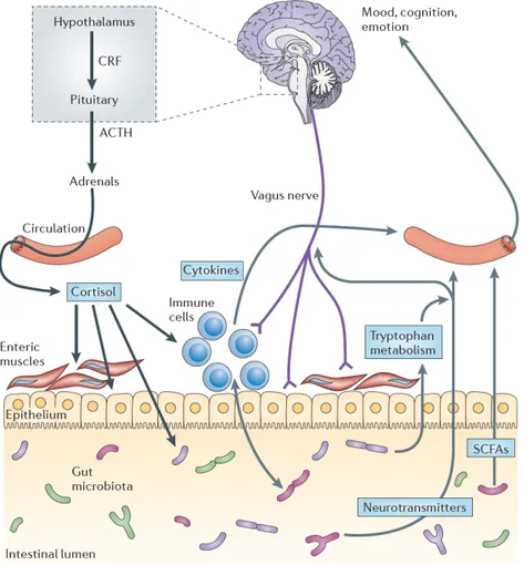
2.3. Gut-brain axis in ASDs
ASDs are neuropsychological diseases with a marked peripheral component. Indeed, beyond the social and psychological pattern, ASD patients show gastrointestinal symptoms with underlying pathogenic mechanisms that are not completely clarified yet. Alterations in gut integrity and modifications of its microbiota represent features of autistic outline and the interaction between brain and gut is achieved by influx of microbiota signals (gut hormones and microbial metabolites) in CNS, via the immune system, the vagus nerve and other host microbe interactions (127). Therefore, the scaffold of this gut-brain axis is constituted by the CNS, the autonomic nervous system (both sympathetic and parasympathetic branches), the neuroendocrine and neuroimmune systems, the enteric nervous system (ENS) and, the gut microbiota (Figure 2.1). All these features represent main characters of a complex network, where a bidirectional communication system between gut microbes and CNS played a key role for host homeostasis (128). Indeed, genes within the gut microbiota, termed the microbiome are capable of producing a myriad of neuroactive compounds, playing a pivotal role in shaping cognitive networks underlying social cognition, emotion, and behavior (129). During the development, brain neural circuits and gut microbiota co-evolve and possible alterations of gut
30
microbiota composition influence the normal growth of CNS neurotransmission, particularly for serotonergic system (130). Indeed, alterations of integrity in both gut epithelium barrier and BBB, reported in ASD patients (131, 132), can cause the translocation of bacterial metabolites, inducing immunoreactions that can activate the vagal system or directly influence CNS activity, impacting on neuronal plasticity and consequently on mood and behavior (133, 134). In rats, the lack of gut microbiota colonization leads to a reduced sociability, an increased anxiety-like behavior and alterations of neurophysiology compared to control mice with no pathogen bacteria colonization (135, 136).
Several clinical studies highlighted the link between dysbiosis and ASDs. Tomova et al. (137) showed a reduction of Bacteroidetes/Firmicutes ratio in ASD patients, while Strati et al. (138) demonstrated a significant reduction in relative abundance of phylum Bacteroidetes in autistic subjects. In contrast, Son et al. (139) did not find any alteration in gut microbiota composition in a study comparing fecal microbiota in ASD children and neurotypical siblings by qPCR. These conflicting evidences and the small scale of preclinical studies demonstrated the difficulty to outline a distinctive gut microbiota composition in ASDs.
31
Figure 2.1. The scaffold of the gut-brain axis. The interaction between brain and gut is
achieved by influx of microbiota signals, such as gut hormones and microbial metabolites, in CNS, via the immune system, the vagus nerve and other host microbe interactions.
32
3. Obesity-induced depression
3.1. Depression: definition, epidemiology, diagnosis
and risk factors
Major depressive disorder (MDD) is a debilitating disease associated to reduced quality of life, medical morbidity, and mortality (140). Indeed, in 2013, it was considered the second leading cause of disability in all countries (141). The DSM-V identifies MDD when a person suffered from at least one discrete depressive episode lasting at least two weeks, characterized by changes in mood, interests and pleasure and by alterations of cognitive and vegetative spheres (102) (see Figure 3.1).
33
Figure 3.1. Definition of MDD according to DSM-5.
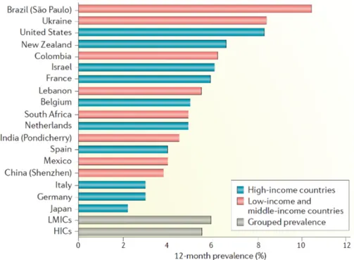
MDD represents a worldwide burden because about 6% of the adult population shows depressive episodes, mostly in women (about 2-fold) (142). In both sexes, MDD onset is approximately at 25 years of age and the range of increased risk is constituted by mid-to-late adolescence to early 40s (143). Furthermore, MDD prevalence was found to be similar between high- and low- and middle-income countries, highlighting that MDD does not represent a “modern-world” disease (Figure 3.2) (144).
34
Figure 3.2. Worldwide prevalence of MDD. The overall estimates in high-income
countries (5.5%) and low- and middle-income countries (5.9%) do not significantly differ.
The progress of MDD is essentially variable and it depends on its remission and chronicity. The prediction of an ill-fated course can be facilitated by higher symptom severity, other psychiatric comorbidities and the occurrence of childhood trauma (145). In population-based samples, the mean episode of MDD lasts between 13 and 30 weeks with a recovery of approximately 70–90% of patients within 1 year (146, 147), even if, in outpatient care settings, the remission rate can drastically decrease to 25% (148, 149). Furthermore, the recurrence of MDD is high, as about 80% of patients experiences at least another depressive episode during lifetime (150). Both in healthy people and other no-MDD patients, depression increases the
35
mortality risk by 60-80%, and its contribution of all-cause mortality is 10% (151, 152).
As mentioned above, MDD is diagnosed in patients who develop several symptoms distinguished from normal sadness or bereavement that endures at least 2 weeks (102). Beyond depressive mood, it needs only two of six symptoms such as appetite disturbances, loss of energy, reduced self-esteem, sleep disturbances, poor concentration or hopelessness. The persistent depressive disorder is a chronic disease characterized by depressive symptoms for more than 2 years. Once MDD diagnosis is made, the disorder can be classified by different specifiers (see Scheme 3.1).
Being MDD a multifactorial disease no established mechanisms can explain all aspects involved in its onset. The contribution of genetic factor is estimated to about 35%, highlighted more in family and twin-based studies rather than in single-nucleotide polymorphism-based estimates from genome-wide association studies (GWAS) (153). On the other side, environmental factors, such as sexual, physical or emotional abuse, contribute to the development of MDD. Indeed, epidemiological studies correlated the onset of MDD with stressful events (loss of employment, chronic or life-threatening health problems, financial insecurity, exposure to violence, separation and bereavement) usually occurred in the year preceding onset and disease itself (154). However, not only recent events
36
but also childhood-occurred ones, including physical and sexual abuse, psychological neglect, exposure to domestic violence or early separation from parents due to death, are clearly responsible of MDD (155).
3.2. Pathogenic mechanisms of MDD
The oldest known pathogenic mechanism of MDD is based on the lack of balance in the serotonergic, noradrenergic and dopaminergic systems (156). Indeed, for many years, MDD research focused on the metabolism of their neurotransmitters, such as noradrenaline, serotonin and dopamine, and their effect on both presynaptic and postsynaptic receptors (157).
Following studies moved their focus on connection between MDD and endocrine pathways. The hypothalamic-pituitary-adrenal (HPA) axis dysfunction represents the center of this link. Indeed, early-life stress induces the activation of corticotropin-releasing hormone (CRH)-containing neural circuits in several animal studies (158). This involvement has been confirmed by clinical studies, showing how children subjected to sexual or physical abuse showed a marked activation of HPA axis when exposed to standardized psychosocial stressors or through endocrine tests trying to stop HPA activity (159). Moreover, increased levels of cortisol, whose production is generally controlled by the hypothalamus, represented a risk factor for the onset of MDD in clinical studies (160, 161).
37
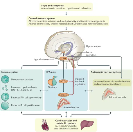
The immune system is another major character involved in the physiological stress-sensing pathways and interacts directly and indirectly with HPA, the autonomic nervous system and CNS in pathogenic mechanisms of MDD. Indeed, once peripheral cytokines reach and cross the BBB, they activate CNS-resident cells, such as astrocytes, microglia and neurons. In a clinical study, patients who received cytokine treatments, such as IL-2 or interferon-g, for hepatitis or cancer therapy, showed higher rate to develop depressive episodes (162). Other meta-analyses confirmed these findings, showing high levels of serum cytokines, such as tumor necrosis factor (TNF)-a and IL-6 in MDD patients (163, 164). The increased levels of circulating cortisol and inflammatory cytokines can affect brain function, through the disruption of neuroplasticity and the reduction of neurogenesis, as demonstrated by low levels of the neurotrophin, brain-derived neurotrophic factor (BDNF) (165). Taken together, all these evidences underline how all those biological pathways, taking part into the so-called psycho-immune-neuroendocrine (PINE) network (Figure 3.3) are involved in MDD.
38
Figure 3.3. The psycho-immune-neuroendocrine (PINE) network of MDD. Beyond the
impairment of CNS, the hyperstimulation of HPA axis is another pathological feature of MDD. Moreover, this pathological condition activates the immune system, increasing levels of circulating cytokines and triggering a low-grade chronic inflammation. Once MDD becomes chronic, both HPA hyperactivity and inflammation might converge towards an alteration of autonomic nervous system (ANS), contributing to MDD comorbidities, such as cardiovascular and metabolic disorders.
3.3. Depression and obesity: a bidirectional interplay
Among several risk factors, different studies highlighted the bidirectional association between obesity and depression (166, 167). Faith et al. (168) examined different studies to understand whether the association “depression-to-obesity” is stronger than “obesity-to-depression” or vice
39
versa. These authors reported a more significant influence of obesity to depression than the opposite (80% vs 53%).
The general mechanisms that try to explain the obesity-induced depression are two:
• Obesity and MDD share fundamental biological mechanisms (inflammatory, neuroendocrine, metabolic and gut-related patterns), and the alterations of these systems induced by obesity can lead to MDD;
• Chronic psychological stress related to lack of self-confidence induced by body-image consciousness leads to dysregulation of PINE network, triggering the onset of MDD.
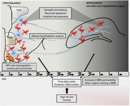
According to this paradigm, the pathophysiological features of obesity, such as low-grade inflammation, ANS unbalance, leptin and ghrelin changes, and dysbiosis play a pivotal role as underlying pathogenic mechanisms of MDD. The obesity-induced systemic inflammation is characterized by the increase of serum levels of pro-inflammatory cytokines, such as IL-6 and TNF-a, that stimulate the microglial proliferation and astrocyte dysfunction with consequent neuroinflammation, mainly in areas that lack an effective BBB, including hypothalamic arcuate nucleus (ARC) (169). The ARC is strongly connected not only with other regions of the hypothalamus, but also with
40
other brain areas, such as mesolimbic dopamine system, hippocampus, orbitofrontal cortex, nucleus accumbens, striatum and prefrontal cortex, regulating motivation and reward pathways (170). Thus, beyond the effects on feeding signals, the inflammation in ARC can potentially impair cognitive function (Figure 3.4).
Moreover, neuroinflammation can trigger the PINE network to reach the critical threshold for MDD onset, even if molecular mechanisms keep still unclear.
Leptin and ghrelin are central regulators in the PINE network. Beyond their key role in food intake, these molecules are implicated in mood regulation (171-173). Leptin interacts with its receptor, particularly abundant in ARC, and, activating the proopiomelanocortin (POMC) neurons and suppressing neuropeptide Y (NPY) and agouti-related protein (AgRP) ones, reduces feeding behavior. HFD-induced obesity induces a leptin resistance, characterized by high systemic levels of leptin and reduced central leptin sensitivity. This detrimental condition can lead to a reduced neuroprotection and onset of MDD (174-176).
41
Figure 3.4. Possible mechanism of cognitive dysfunction induced by obesity.
HFD/obesity induce the HPA dysfunction that can alter other brain areas, such as hippocampus, reducing neurogenesis and inducing neuronal apoptosis.
The contribution of the change in gut microbiota induced by obesity in the development of MDD has been analyzed in several studies. As mentioned above, high fat diet consumption causes the production of several inflammatory mediators that can compromise colonic epithelial barrier function, inducing the so-called “leaky gut” (177). This increased permeability allows the entrance of immunogenic molecules, such as LPS (178), in the systemic circulation, whose occurrence has been recognized in both animal (179) and human (180) affected by depression. Moreover, long-term consumption of a Western-style HFD, constituted by low levels of
42
fiber, may result in profound reduction of short chain fatty acids (SCFAs), endogenous molecules with marked anti-inflammatory and neurogenesis activity (181). The intestinal dysbiosis induced by obesity, may promote depression through the modulation of hippocampal BDNF. Indeed, Bercik et al. (182) demonstrated that the administration of antimicrobials was responsible of BDNF reduction in mice hippocampus, while colonization of germ-free mice with gut microbiota from other specific pathogen free mice increased the expression of neurotrophin.
3.4. Treatment of MDD
Given the complexity and high prevalence of MDD, its prevention is fundamental to limit it. These strategies include the strengthening protective factors, such as the increase of social support, and the reduction of prodromal symptoms, limiting them before that they overcome the disease threshold. Indeed, a meta-analysis demonstrated the reduction of depressive symptoms (21%) in MDD patients who received a preventive intervention (183). The treatment of MDD depends on the severity of symptoms and include two main strategies: psychotherapy and pharmacotherapy. Mild depressive episodes could be treated only with psychotherapy, but when the grade worsens, a pharmacological treatment alone or in combination with
43
medication and psychotherapy should be adopted, two weeks after the beginning of symptoms (184). Several meta-analyses did not show any consistent difference among different types of psychotherapy (185-187). The pharmacological treatment of MDD comprises the use of monoamine-based antidepressant drugs, capable to stimulate an adaptive neuronal response against MDD central perturbations (Figure 3.5), although the exact biomolecular mechanisms of action are not completely clarified. In addition, the wider choice of selective serotonin reuptake inhibitors (SSRIs) or serotonin–norepinephrine reuptake inhibitors (SNRIs) than that of classical antidepressant drugs is due to better adverse-effect profile rather than efficacy. However, SSRIs and SNRIs are also characterized by several adverse effects, such as headache, insomnia, gastrointestinal symptoms, dizziness, sexual dysfunction, weight gain and sleep disturbances (188). Among new therapeutic approaches for MDD, several compounds are under investigation, such as neurokinin 1 antagonists, glutamatergic system modulators, anti-inflammatory agents, opioid tone modulators and opioid κ antagonists, hippocampal neurogenesis-stimulating treatments and anti-glucocorticoid therapies (140).
44
45
4. Material and Methods
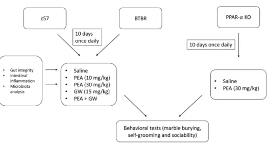
4.1. Sex differences in gut microbiota of BTBR mice
4.1.1. Animals
C57Bl/6J (C57) and BTBR T + tf/J (BTBR) inbred strains of mice were purchased from The Jackson Laboratory (Bar Harbor, ME, USA) and a colony was established and maintained. For the study, 24 fully symptomatic BTBR mice (12 months of age, 12 for gender) and 24 C57 control mice (12 months of age, 12 for gender) were housed in the same room under standard 12-h light/12-h dark cycle with free access to water and standard laboratory chow diet. Mice were from different litters and housed by gender separately 2/3 per cage. Before killing and prior to serum and sample collection, animals, kept overnight fasted, were anesthetized by enflurane and euthanized by an intraperitoneal injection of a cocktail of ketamine/xylazine. As suggested by the animal welfare protocol, all efforts were made to minimize animal suffering and to use only the number of animals necessary to produce reliable scientific data. All procedures involving animals and their care were conducted in conformity with international and national law and policies (EU Directive 2010/63/EU for animal experiments, ARRIVE
46
guidelines and the Basel declaration including the 3R concept). The procedures reported here were approved by the Institutional Committee on the Ethics of Animal Experiments (CSV) of the University of Naples “Federico II” and by the Ministero della Salute under protocol no. 0022569-P-20/12/2010.
4.1.2. Marble burying test
For the performing of all behavioral tests, we considered the book of Bayley et al. (189). Male and female BTBR and C57 (n=12 each group) were individually placed in a plastic container (46 cm long x 24 cm wide x 21 cm deep) with 5 cm of clean woodchip bedding (Northeastern Products, NY). The plastic container was placed in a room used for behavioral testing. Once a mouse was gently allocated into the test container, a wire lid was placed on top and undisturbed mice were allowed to freely explore the container for 30 min, as habituation time. Twenty-four hours later, 20 glass marbles (1.5 cm in diameter) were placed on top of clean bedding, arranged in five rows of four and mice located into plastic cages for 30 minutes. After this experimental time, each mouse was removed from the testing container and replaced to their home cage. When a threshold of 75% coverage for each marble was observed, it was considered buried and recorded. After the test,
47
the marbles were thoroughly cleaned and new bedding was used for each mouse. The same animals tested for marble burying test were also examined for grooming behavior.
4.1.3. Self-grooming test
Mice were individually placed in an empty plastic cage (28 cm wide × 17 cm long × 12 cm high) and were freely to explore the arena for 20 minutes. The first 10 min were necessary for a habituation time. During the second phase, a trained observer, sat approximately 1.6 m from the test cage, timed the cumulative period that mice spent in self-grooming. Grooming behavior included head washing, body grooming, genital/tail grooming and paw and leg licking. After the test, the cage was thoroughly cleaned. After self-grooming test, the same animals were tested for sociability test.
4.1.4. Social interaction test
Social interaction was examined using the three-chambered apparatus as previously described (190-192). The apparatus (60 x 40 cm) has two doorways that divide it into a three chambers apparatus (20 x 40 cm each). Number of entries and time spent in each chamber were automatically
48
detected by a video camera coupled with a video-tracking software (Any-maze, Stoelting). The sociability test was preceded by 5-min habituation session where each mouse is restrained in the center of the middle chamber. After this phase, a novel sex, strain and age matched mouse (not used in later testing and previously habituated) is placed in one side of the chamber under an enclose cup while the other side contained an empty cup. During this sociability phase, walls between the compartments were removed and the tendency to approach a novel mouse was compared with tendency to approach a novel object is monitored and recorded. Each mouse was free to explore all three chambers for 10 minutes and both sides were alternated between the left and right chambers across subjects.
4.1.5. In vivo intestinal permeability assay
In vivo intestinal permeability assay was performed for a subset of mice
using fluorescein isothiocyanate-labeled dextran (FITC-dextran) method, as previously described (193). Before the beginning of the test, food and water were withdrawn for 6 hours, after which mice (n=5, each group) were administrated by gavage with FITC labeled dextran 4000 (Sigma-Aldrich, Milan, Italy), as permeability tracer (60 mg/100 g body weight). After 24 hours blood of all animals was collected by intracardiac puncture and
49
centrifuged (3000 rpm for 15 min at RT). Then plasma FITC-dextran concentration was determined (excitation, 485 nm; emission, 535 nm; HTS-7000 Plus-plate-reader; Perkin Elmer, Wellesley, MA, USA), using a standard curve generated by serial dilution of the tracer.
4.1.6. Histological analysis
Colonic tissues of male and female mice of both strains (n=3) were removed, washed and then fixed in paraformaldehyde (4% v/v; Carlo Erba, Italy) for 12 hours. These samples were dehydrated, embedded in paraffin and cut into 5 μm thick sections before being stained with hematoxylin-eosin (H&E; Carlo Erba, Italy). Images were obtained by a Leica DFC320 video camera (Leica, Milan, Italy) connected to a Leica DM RB microscope using the Leica Application Suite software V2.4.0.
Colon sections were analyzed by the same pathologist in a blinded manner to evaluate their structure and architecture. Histopathology was quantified following: a) the severity of inflammatory cell infiltration was evaluated by percentage of leukocyte density in lamina propria area and estimated in a high-power field (HPF) representative of the section (0 for no signs of inflammation, 1 for minimal < 10%, 2 for mild 10–25% with scattered neutrophils, 3 for moderate 26–50%, 4 for marked > 51% with dense
50
infiltrate); b) The extent of the inflammation was estimated as expansion of leukocyte infiltration (0 for none, 1 for mucosal, 2 for mucosal and submucosal and 3 for mucosal, submucosal and transmural level).
4.1.7. Microbial DNA extraction, 16S ribosomal DNA (rDNA) library preparation and sequencing
Freshly evacuated fecal pellets were kept directly in a sterile microtube one day before the sacrifice of mice and stored at -80 °C until assayed. Bacterial genomic DNA was extracted from frozen fecal samples using the QIAamp DNA Stool Mini Kit (Qiagen) according to manufacturer’s instructions. DNA concentration was measured fluorometrically using Qubit dsDNA BR assay kit (Invitrogen) and quality was assessed by spectrophotometric measurements with NanoDrop (ThermoFisher Scientific Inc). Samples were stored at -20 °C until processed for amplification. It is well documented that various compartments of the gastro-intestinal tract harbour different bacterial populations. We chose to analyze readly accessible fecal samples for gut microbiome analyses mainly because fairly representative of the whole gastro-intestinal tract, with exception of some surface-adherent bacterial species. Sequencing samples were prepared according to the protocol 16S Metagenomic Sequencing Library Preparation for Illumina
51
Miseq System with some modifications. The V3–V4 regions of the 16S rDNA gene were amplified starting from 200 ng of DNA template in a reaction volume of 50 μ L containg 1x Fast start High Fidelity Reaction Buffer, 5 μ M of each primer, 0.2 nM of dNTPs, 3 mM MgCl2, and 2 U FastStart High Fidelity PCR System (Roche Applied Science). PCR was performed using the following cycles conditions: an initial denaturation step at 95 °C for 2 min, followed by 30 cycles of 95 °C for 30 s, 55 °C for 45 s, 72 °C for 55 s and ended with an extension
step at 72 °C for 5 minutes; products were visualized by electrophoresis on 1.2% agarose gel. After a purification step with Agencourt AMPure XP (Beckman Coulter Inc), the amplicons were indexed with 10 subsequent cycles of PCR using the Nextera XT Index Kit (Illumina). Each PCR reaction contained 10 μ L of amplicons from first PCR, 5 μl index 1 primer (N7xx), 5 μl index 2 primer (S5xx), 5 μ l 1x Fast start High Fidelity Reaction Buffer, 6 μL MgCl2 (3 mM), 1 μ L dNTPs (0.2 nM), 0.4 μ L FastStart High Fidelity PCR System (2U) and 17.6 μ l PCR grade water. PCRs were carried out, visualized using gel electrophoresis and subsequently cleaned as described above.
Library sizes were assessed using a Bioanalyzer DNA 1000 chip (Agilent technologies) and quantified with Qubit. Normalized libraries were pooled,
52
denatured with NaOH, then diluted to 10pM and combined with 25% (v/v) denatured 10pM PhiX, according to Illumina guidelines. Sequencing run was performed on an Illumina Miseq system using v3 reagents for 2 × 281 cycles.
4.1.8. Sequencing data analysis
V3-V4 16S rDNA FASTQ paired-end reads were quality filtered and assembled using PEAR (194). Only sequences showing average PHRED score ≥ 30, read length between 400 and 500 bp and overlapping regions between mate-pair end of at least 40 nucleotides were retained in this step. Passing filter sequences were then processed with PRINSEQ (195) in order to obtain FASTA and quality files for further analyses. Metagenomic analyses on the resulting data were conducted using Quantitative Insights Into Microbial Ecology (QIIME, version 1.8.0) (196). 16S sequences were used to pick OTUs at 97% of sequences similarity from Greengenes 16S gene database (GG, may 2013 version) (197) with a closed reference-based OTU picking method. The GG database was used to taxonomically classify the identified OTUs and to compute their distribution across different taxonomic levels. To avoid sample size biases in subsequent alpha and beta diversity analyses, a sequence rarefaction procedure was applied using a
53
maximum depth of 32,228 sequences/sample. To assess sampling depth coverage and species heterogeneity in each sample, alpha diversity metrics were employed on rarefied OTU table using Good’s coverage, Observed species and Shannon’s diversity index. A two-sample permutation t-test, using 999 Monte Carlo permutations to compute p-value, was performed to compare the alpha diversities between sample groups. OTUs diversity among sample communities (beta diversity) was assessed by applying unweighted Unifrac distances. Statistical significance of beta diversities was assessed on unweighted UniFrac distances matrixes using ANOSIM method (198) with 999 permutations. Statistical differences in OTUs frequencies across sample groups at different taxonomic levels were assessed using nonparametric Kruskal-Wallis test. Next, two analyses were applied on OTU tables generated by QIIME to identify key OTUs that discriminate female and male BTBR mice from their respective controls: Metastats comparison using the online interfaces (199) and LDA Effect Size analysis (LEfSe) (200). Only those OTUs reported by both methods to be significantly different between the two groups (p < 0.05 for Metastats, LDA > 2 and p < 0.05 for LEfSe) have been considered as key discriminatory OTUs. Key genera that discriminate female and male BTBR mice from their respective controls were identified applying only LEfSe.