Università Politecnica delle Marche
DOCTORAL THESIS
Self-assembled nanomaterials: an extended
structural characterization of lipid nanoparticles and
guanosine-based hydrogels
PhD student: Federica Carducci
Supervisor: Prof. Paolo Mariani
Department of Life and Environmental Sciences (DiSVA)
XXX cycle
Al mio papà, che è stato il primo a credere in me
e a mia madre, perché la sua forza è unica al mondo
Una corrente di eventi ha inizio dalla decisione, facendo sorgere a nostro favore ogni tipo
di incidenti imprevedibili, incontri e assistenza materiale, che nessuno avrebbe sognato
potessero venire in questo modo.
Tutto quello che puoi fare, o sognare di poter fare, incomincialo. Il coraggio ha in sé genio,
potere e magia. Incomincia adesso.
Declaration of Authorship
Abstract
List of Abbreviations
Thesis organization
Chapter 1
Lipid nanoparticles
1.1 Introduction: The Drug Delivery revolution in science 1.1.1 Drug Delivery Systems (DDS)
1.2 Lipid Nanoparticles
1.2.1 Solid Lipid Nanoparticles (SLN) 1.2.2 Nanostructured Lipid Carriers (NLC) 1.2.3 Monoolein Aqueous Dispersions (MAD) 1.3 Polymorphism of Lipid/Water Systems: a brief overview 1.4 Structural characterization using X-Ray Diffraction (XRD)
1.5 Phase identification of lipid-water systems
1.6 Determination of subcrystalline structure of lipidic phase
Chapter 2
Structural characterization of NPs: case studies
2.1 Introduction
2.2 Characterization of Nanostructured Lipid Dispersion (NLD)
2.2.1 Lipid nanoparticles for the delivery of crocin (Crocus sativus L.)
2.3 Characterization of Solid Lipid Nanoparticles (SLN) and Nanostructured Lipid Carriers (NLC)
2.3.1 Functionalization of nanoparticles with 1,3,5-triaza-7-phosphaadamantane (PTA) platinum (II) carboxylates: a structural study
2.3.2 Nafion® containing SLN as Tool for Anticancer Pt delivery
2.3.3 Characterization of SLN and NLC for the delivery of poorly water-soluble neuroactive drugs 2.4 Characterization of Monoolein Aqueous Dispersions (MAD)
2.4.1 Structural Characterization of MAD kept in contact with quercetin
2.5 High Pressure Homogenization (HPH) vs Ultrasonication (U): characterization of NPs stability and validation of method using Small Angle X-Ray Scattering (SAXS)
2.5.1 SLN and NLC
2.5.2 Lipid-based nanoparticles for the delivery of progesterone
2.6 An extended characterization of Monoolein/water Phase Diagram: choosing the best way to solubilize crocetin
Chapter 3
DNA derivatives
3.1 Introduction
3.2 Guanosine self-assembly
3.3 Lyotropic Liquid Crystalline behavior of DNA and DNA derivatives 3.3.1 DNA derivatives: columnar mesophases
3.3.2 Phase identification of columnar mesophases of DNA derivatives 3.3.3 Stacking analysis
Chapter 4
Guanosine-based hydrogels: macroscopical characterization, XRD analysis and
Phase Diagrams determination
4.1 Introduction: guanosine-based hydrogels
4.2 Macroscopical characterization of guanosine-based hydrogels 4.2.1 Phase Diagram determination
4.2.2 Gel-to-sol transition: determination using Polarized-light optical microscopy (POM) 4.3 Characterization of hydrogels using X-ray Diffraction (XRD)
4.3.1 The less-hydrated systems phase diagrams: low-angle region analysis
4.3.2 Stacking of quartets in the quadruplex architecture: high-angle region analysis
Chapter 5
G/GMP Hydrogels: analysis of anisotropicity and SAXS results
5.1 The more-hydrated systems: Small-angle X-ray Scattering (SAXS) analysis 5.2 Analysis of anisotropicity
5.2.1 Structure factor calculation: circularly averaged 1D-profiles analysis 5.2.1.1 G/GMP 1:1
5.2.1.2 G/GMP 1:2 5.3 G/(2’/3’)-GMP hydrogels
Chapter 6
Possible applications of G/GMP hydrogels
6.1 Introduction
6.2 G/GMP inner structural organization: AFM observation 6.3 Interaction of G/GMP hydrogel and model proteins
6.3.1 Bovine Serum Albumin (BSA) 6.3.2 Cytochrome C
Chapter 7
Guanosine derivatives: Osmotic Stress Technique
7.1 Introduction
7.2 The osmotic stress technique: a brief introduction 7.3 Guanosine 2’/3’-Monophosphate
7.3.1 Analysis of lateral forces using osmotic stress technique: salt concentration 7.3.1.1 (2’/3’)-GMP disodium salt
7.3.1.2 (2’/3’)-GMP potassium salt 7.3.1.3 (2’/3’)-GMP ammonium salt
7.3.2 Analysis of lateral forces using osmotic stress technique: curve fitting 7.4 Guanosine/Guanosine 5’-Monophosphate Hydrogels
Chapter 8
Conclusions and future insights
Chapter 9
Materials and Methods
9.1 Lipidic nanoparticles 9.2 Guanosine derivatives
9.2.1 Acid-base titration of 5’-GMP free acid form
9.2.2 Ion-exchange chromatography of GMP disodium salt 9.2.3 Guanosine hydrogels
9.2.3.1 G/GMP less-hydrated samples (XRD) 9.2.3.2 G/GMP less-hydrated samples (SAXS-POM) 9.2.3.3 G/GMP more-hydrated samples (SAXS-POM) 9.2.3.4 G/GMP hydrogels (AFM observations)
9.2.4 Osmotic Stress Technique: 2’/3’-GMP sample preparation 9.3 X-ray Diffraction (XRD)
9.3.1 Huygens’ Principle
9.3.2 Braggs’ Law and Lattice Theory 9.4 Small Angle X-ray Scattering (SAXS)
9.4.1 General Equation of SAXS 9.4.2 Cylinder Model
9.4.3 Core-shell cylinder model 9.5 Polarized-light optical microscopy
9.5.1 POM observations using hot-stage plate (Mettler ToledoÒ)
Bibliography
Declaration of Authorship
I, Federica Carducci, declare that this thesis titled “Self-assembled nanomaterials: an extended structural characterization of lipid nanoparticles and guanosine-based hydrogels” and the work presented it are my own and has been generated by me as the result of my own original research.
I confirm that:
- This work was done wholly or mainly while in candidature for a research degree at this University; - where any part of this thesis has previously been submitted for a degree or any other qualification at
the University or any other institution, this has been clearly stated;
- where I have consulted the published work of others, this is always clearly attributed;
- where I have quoted from the work of others, the source is always given. With the exception of such quotations, this thesis is entirely my own work;
- where the thesis is based on work done by myself jointly with others, I have made clear exactly what done by others and what I have contributed myself.
- parts of this work have been published in the following papers:
- Elisabetta Esposito, Maddalena Sguizzato, Markus Drechsler, Paolo Mariani, Federica Carducci, Claudio
Nastruzzi, Rita Cortesi “Data on Scaling up and in vivo human study of progesterone lipid nanoparticles”, Data in Brief 14 (2017d) 639-642
- Elisabetta Esposito, Maddalena Sguizzato, Markus Drechsler, Paolo Mariani, Federica Carducci, Claudio
Nastruzzi, Rita Cortesi “Progesterone lipid nanoparticles: Scaling up and in vivo human study”, Eur J
Pharm Biopharm 119 (2017c) 437-446
- Elisabetta Esposito, Markus Drechsler, Paolo Mariani, Federica Carducci, Michela Servadio, Francesca
Melancia, Patrizia Ratano, Partizia Campolongo, Viviana Trezza, Rita Cortesi, Claudio Nastruzzi “Lipid nanoparticles for administration of poorly water soluble neuroactive drugs”, Biomed Microdevices
(2017b) 19:44 DOI 10.1007/s10544-017-0188-x
- Rita Cortesi, Enrica Cappellozza, Markus Drechsler, Catia Contado, Anna Baldisserotto, Paolo Mariani,
Federica Carducci, Alessandra Pecorelli, Elisabetta Esposito, Giuseppe Valacchi “Monoolein aqueous dispersions as delivery system for quercetin”, Biomed Microdevices (2017) 19:41 DOI 10.1007/s10544-017-0185-0;
- Maddalena Sguizzato, Elisabetta Esposito, Markus Drechsler, Eleonora Gallerani, Riccardo Gavioli, Paolo
Mariani, Federica Carducci, Rita Cortesi, Paola Bergamini “Nafion®-Containing Solid Lipid Nanoparticles
- Maddalena Sguizzato, Rita Cortesi, Eleonora Gallerani, Markus Drechsler, Lorenza Marvelli, Paolo Mariani,
Federica Carducci, Riccardo Gavioli, Elisabetta Esposito, Paola Bergamini “Solid lipid nanoparticles for the delivery of 1,3,5- triaza-7-phosphaadamantane (PTA) platinum (II) carboxylates”, Mater Sci Eng C
74, (2017a), 357-364;
- Elisabetta Esposito, Markus Drechsler, Paolo Mariani, Anna Maria Panico, Venera Cardile, Lucia Crascì,
Federica Carducci, Adriana Carol Elena Graziano, Rita Cortesi, Carmelo Puglia “Nanostructured lipid dispersions for topical administration of crocin, a potent antioxidant from saffron (Crocus sativus L.)”,
Mater Sci Eng C 71 (2017a), 669-677.
Signed...………... Date...………...
Abstract
This thesis presents an extended work concerning the structural characterization of Drug Delivery Systems (DDS) made by lipidic nanoparticles and hydrogels based on DNA-derivatives. The link between these two completely different DDS is represented by the phenomenon of self-assembly, at the base of the formation of these extraordinarily versatile materials.
Self-assembly can be defined as the autonomous organization of components into patterns or structures, without the human intervention (Whitesides and Grzybowsky, Science, 2002). In the present case, and both for lipids and DNA-derivatives, the obtainment of complex supramolecular structures leads to the possibility to use these systems as powerful tools in the controlled delivery of drugs. For example, fundamental is the role played by lipidic nanoparticles in the delivery of anticancer drugs in which the biggest challenge is to avoid the unwanted side-effects and thus to develop always well-tolerated formulations.
In the first part of the thesis I present a thorough structural characterization of several nanoparticle systems tested to transport different antioxidants, anticancer drugs and other active principles. In the second part attention will be focused into hydrogels prepared by using guanosine derivatives. An almost completed characterization made by coupling macroscopical (e.g. microscopy observations, inverted-vial test) with microscopic techniques (e.g. Small-Angle X-ray Scattering and “classical” X-ray diffraction), lead us to move towards possible applications of these self-assembled hydrogels in the biological field, not yet explored for this specific kind of nanomaterials.
List of Abbreviations
DDS: Drug Delivery Systems EE: Encapsulation Efficiency EM: Electron Microscopy G: Guanosine
GMP: Guanosine 5’-MonoPhosphate HPH: High-Pressure Homogenization IEC: Ion-Exchange Chromatography LC: Loading Capacity
LDC: Lipid Drug Conjugates
MAD: Monoolein Aqueous Dispersions NLC: Nanostructured Lipid Carriers NLD: Nanostructured Lipid Dispersions NPs: NanoParticles
OM: Optical Microscopy
PCS: Photon Correlation Spectroscopy SAFiN: Self-Assembled Fibrillary Network SAN: Self-Assembled Nanomaterials SAS: Small-Angle Scattering
SAXS: Small Angle X-ray Scattering SLN: Solid Lipid Nanoparticles
SWAXS: Small- and Wide-Angle X-ray Scattering TAGs: TriacylGlicerols
U: Ultrasonication
UH: Ultra-Homogenization XRD: X-Ray Diffraction
Thesis organization
This thesis is organized into 9 chapters. Chapter 1 provides at first an introduction to DDS, with an emphasis on lipid self-assembling nanoparticles. In the second part, I report basic principles about Lipid/Water polymorphism and phase identification using X-ray diffraction (a preamble about this extraordinary technique is given in "Material and Methods", Chapter 9). Chapter 2 summarizes main results obtained considering different lipid nanoparticles. Each analysed system has been discussed in separated sections, focusing attention to the specific kind of nanoparticle used, active principle tested and method of production. In each section, as declared at the beginning of this thesis, reference to the published paper (if any) is always given. In Chapter 3 a brief introduction about guanosine self-assembly and Lyotropic Liquid-Crystalline behavior is given. Details about the phase identification of columnar mesophases observed in such derivatives can be also found. Chapter 4 introduces to guanosine-based hydrogels. Here, after a brief introduction, results about the macroscopical characterization can be found, together with the X-ray diffraction analysis of less-hydrated guanosine hydrogels. Results on fully hydrated hydrogels obtained by Small-Angle X-Ray Scattering are presented and discussed in Chapter 5, in which attention is focused on the analysis of circularly averaged 1D-profiles. Finally, in Chapter 6, a possible application of guanosine hydrogels is presented. Chapter 7 summarizes results obtained on two different guanosine derivatives by osmotic stress measurements, performed in order to provide information on force effects on hydrogel formation and stability. Chapter 8 provides conclusions and insights into the ongoing and future works concerning DDS and finally, in Chapter
Chapter 1
Lipid nanoparticles
1.1 Introduction: The Drug Delivery revolution in science
Wanted to find a starting point in the development of controlled drug delivery systems, this can be the 1952, when Smith Kline & French developed the first sustained release formulation for 12-hour delivery of dextroamphetamine (Dexedrine). Many efforts have been done in the last two decades in the development of DDS, and now the next goal will be the third generation of DDS (Park, 2014). The necessity to develop homogeneous formulations of nanoparticles started a revolution in science. In DDSs and in general in nanoscience, self-assembled nanomaterials (SAN) occupy a special place. SAN can be defined as matter structured rationally at scale less than 100nm (Whitesides et al., 2005) in which the nanoscale order is provided by the spontaneous assembly of biomolecules.
1.1.1 Nanoparticle-based Drug Delivery Systems (DDS)
A Nanoparticle-based drug delivery system is defined as a formulation or device of nanometric size, produced using lipids or natural and synthetic polymers, that enables the introduction of a therapeutic substance into the body. The process of drug delivery consists in the improvement of the efficacy and safety controlling the rate, time and place of release of drugs in the body. The prefix nano- in nanoparticles refers to solid or colloidal particles consisting of macromolecular substance that vary in dimensions from 10nm to 1000nm (Kreuter, 1994)
Nanoparticle-based DDS can be classified into two main typologies:
- produced using natural or synthetic polymers (e.g. poly- lactic acid, PLA or poly-lactide-co-glycolide, defined as PGLA) in which can be found Hydrogel-nanoparticles, Micelles and Liposomes (made in this case using amphiphilic copolymer), Nanomaterial formulations, Nanosystems, Nanocells, Dendrimers, Nanotubes, Polymersomes (block copolymer vesicles) and Quantum dots;
- Metal Nanoparticles (e.g. Iron-oxide Nanoparticles, Gold Nanoparticles, Nanoshells and Nanocages, Silver Nanoparticles, (Mody et al., 2010));
- lipidic-based NPs produced using homolipids, lipids containing only carbon (C), hydrogen (H) and oxygen (O); heterolipids, containing nitrogen (N) and phosphorous (P) in addition to C, H, O and finally complex lipids, more complex lipids such as lipoproteins, phospholipids.
Nanoparticulate systems made using natural or synthetic polymers represent an alternative and exciting way to overcome limitations such the facilitation of transport of drugs across the Blood Brain Barrier (BBB). What remains a challenge is the precise characterization of molecular targets, ensuring the action of the drug only to the targeted organ (Singh and Lillard, 2009).
widely discussed, together with the first tentative to approach them in the field of biological applications, starting from the study of interaction of those DNA-based systems with model proteins.
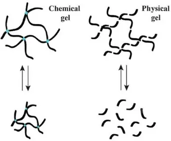
The first report on the potential use of hydrogels in medical science is probably due to Wichterle and Lim (Wichterle and Lim, 1960). A gel is a substance characterized by a continuous microscopic structure with macroscopic dimensions (Peters and Davis, 2016). The solid-like network (gel) has the ability to entrap and retain the solution, which analyzing gel constituents is the predominant (in weight, often accounting for > 97-99% of the material). When gels are made using organic solvents, those materials are referred to the so-called organogel, whereas gels formed with water are defined as hydrogels. A distinct classification can be made about gels: physical or “reversible” gels, in which the 3D-network is held by noncovalent interaction such as Van der Waals forces, hydrogen bonds and Coulombic interactions (Peters and Davis, 2016) and chemical or “permanent” gels, where the 3D network is constitute of covalent crosslinks.
Figure 1.1 difference between chemical and physical gels. Figure taken form the paper of Peters and Davis, 2016 “Supramolecular gels
made from nucleobase, nucleoside and nucleotide analogs”
Weak forces, at the base of the generation of the 3D-network of physical gels, confer to them the characteristic of stimuli-responsiveness, thus the ability to respond when exposed to variation of pH, temperature and pressure, ionic strength and enzymatic ability. A more detailed classification can be made in the case of physical gels, which are given by:
- simple entanglement systems, in which the network is held together by molecular entanglements or crystallites;
- ion-mediated or “ionotropic” networks, in which the network is stabilized by interaction between polyelectrolyte and multivalent ions of opposite charges (e.g. alginate);
- thermally induced networks, in which the heating (or the cooling) induces the structure formation; - self-assembly process.
- synthetic polymers (PLA, PEG, PGLA, acrylates etc…); - mixture of synthetic and natural polymers;
- DNA (Kahn et al.,2017; Bomboi et al., 2016) and DNA derivatives (Peters et al., 2015a; Peters et al., 2015b; Adhikari et al., 2014).
The spontaneous assembly at the base of the formation of the structure of lipid nanoparticles and DNA-based systems is what makes those compounds comparable.
The development of innovative supramolecular materials obtained by self-assembling of biomolecules is an extremely attractive field for nanotechnology.
1.2 Lipid Nanoparticles
Lipid nanoparticles drug delivery systems are an accepted, proven, commercially viable strategy to formulate pharmaceuticals for topical, oral, pulmonary or parenteral delivery (Attama et al., 2012).
Tailoring lipid formulations, a very wide range of different drug delivery systems can be listed: - Solid Lipid Nanoparticles (SLN);
- Nanostructured Lipid Carriers (NLC); - Monoolein Aqueous Dispersions (MAD); - Lipid drug conjugates (LDC)-nanoparticles; - Liposomes;
- Transfersomes; - Niosomes;
- Liquid Crystal Drug Delivery systems; - Nanoemulsions.
During my PhD work, attention has been focused on the structural characterization of SLN and NLC and MAD. I report here a brief introduction and description of these systems.
1.2.1 Solid Lipid Nanoparticles (SLN)
Developed in 1990s as alternative carrier systems to the existing liposomes, SLN can be defined as a solid matrix nanodisperse phase of crystalline solid lipids (Esposito et al., 2016) which can be composed of biocompatible lipids, well-tolerated when administered in vivo. One of the main advantage is the possibility to prepare SLN without using organic solvent. Other benefits are the modulation of the release of drug, the preservation of degradation of molecules and the enhancement of the specificity toward cells with the consequent increase of bioavailability of drug (Siekmann and Westesen, 1992; Muller et al., 2000; Lippacher et al., 2001) and the ability to incorporate lipophilic drugs. Major limitations are instead particle growth, gelation tendency, instability in terms of tendency of polymorphic transitions and low incorporation rate due to the crystalline structure of these NPs (Attama, 2008). The latter is probably the major limitation of SLN. Muller (Muller et al., 2000) conducted many investigations to overcome this limitation developing NLC, defined by Esposito (Esposito et al., 2016) as an improvement of SLN.
Figure 1.2 figures have been obtained using Cryo-TEM and taken from the paper “Progesterone lipid nanoparticles, scaling up and in
vivo human study” (Esposito et al., 2017) are showed empty-SLN (C) and empty-NLC (D). In this latter, in particular in microphotography, it is possible to observe bulges corresponding to the oil fraction typical of NLC composition. Bar corresponds to 150 nm
1.2.2 Nanostructured Lipid Carriers (NLC)
The main components of a NLC are a solid lipid matrix and a liquid lipid phase with a mean particle size in the nanometer range. The unique nanostructure of the lipid matrix (Muller et al., 2000) makes these nanoparticles able to improve drug loading and firmly retain drug during storage. Examples of studies made using NLC are the topical administration of Retinol/Coenzym Q10 (Müller et al., 2002; Pardeike et al., 2009) or the parenteral route of administration of bromocriptine (Esposito et al., 2008)
1.2.3 Monoolein Dispersions
Two different kinds of monoolein dispersions can be distinguished: the first one, Monoolein Aqueous Dispersions (MAD) can be defined as aqueous nanostructured dispersion of an amphiphilic lipid of complex lyotropic liquid crystalline phases (Cortesi et al., 2017); monoolein is able to adopt different mesophases simply varying lipid and water concentration (see section 1.3). The nature of MAD dispersed phase is influenced by the type of emulsifier (Worle et al., 2007). Poloxamer 407 copolymer is one of the most used emulsifier in the case of monoolein. This compound leads to aqueous solution composed by hexasomes, cubosomes ad vesicles (Gustafsson et al., 1996; Larsson, 2000; Siekmann, 2002). The possibility to explore the monoolein:water phase diagram make those DDS able to incorporate poorly water-soluble drugs, enhancing encapsulation efficacy.
The second one, NLD, can be defined as Nanostructured Lipid Dispersions. A work has been published recently using those nanoparticles and crocin as active principle (Esposito et al., 2017a) and it will be discussed widely in section 2.2. Nanostructured Lipid dispersion (NLD) is a particular kind of dispersion made using monoolein, together with sodium cholate and caseinate as stabilizing agents (Srinivasan et al.,1996; Jeongh et al., 2014)
1.3 Polymorphism of Lipid/Water Systems: a brief overview
Lipids can organize into a variety of structures (such as lamellar, hexagonal and cubic phase) due to their amphiphilic nature and thus to their Lyotropic Liquid Crystalline behavior.
Simply varying composition (concentration of emulsifier, kind of lipid) and temperature, a wide range of structures can be obtained.
This paragraph wants to give a brief description of different mesophases observable in lipid-water systems. In Figure 1.4 it is possible to appreciate how large is the number of lipid morphologies achievable simply varying the content of water and temperature, while in table 1.1 the structural and topological properties of the different lipidic phases have been summarized.
The first mesophase observed in a temperature-composition phase diagram (as the one reported in Figure 1.3) in the highly hydrated region is the micellar phase, constituted by spheres or ellipsoids, in which lipids are organized with polar heads kept in contact with aqueous medium and aliphatic chains direct within the core of micelle (note that in the less hydrated side of the phase diagram a second micellar phase, the inverse micellar phase, can be found, in which lipids organize exactly in the opposite way, with water inside the micelles and the hydrocarbon chains that form a continuous hydrocarbon moiety). Micelles form above a critical value of concentration, defined as critical micelle concentration (CMC). Below this value lipid molecules are presents as monomers, dimers and small aggregates. The addition of other lipidic molecules to micelle solution can’t provoke the increase of number of monomers in solution but the formation of new micelles. Bile salts can form micelles, they play a key role because of their ability of incorporate in their cavity high quantity of lipids and of consequence cholesterol.
Lamellar phase is a 1D-lipid phase constituted of a bilayer (the same structure of biological membranes). Most of lipids are able to adopt this structure.
Considering 2D (two-dimensional) structure of lipid, hexagonal phase can be mentioned. In this case, long cylinders are localized at the vertex and the center of a hexagon with polar head directed in the aqueous medium and with hydrocarbon chains in the middle of the cylindrical structure (in Type I, direct). In Type II the orientation of lipids is inverted: as aforementioned for the micellar phase, in the inverted-hexagonal phase the way in which lipids organize is exactly the opposite of the direct one. Phases adopted by lipids can be related also to the geometry of lipid molecule itself (Micellar: conical configuration of lipid; Hexagonal (I, direct): truncated conical configuration; Lamellar: cylindrical; Hexagonal (II, inverse): inverted conical truncated. The organization of lipids in a cubic phase is more complicated. Bicontinuous cubic phase can be imagined as an interconnection of rods reciprocally intertwined and unconnected (Mariani et al., 1988). Among the seven cubic phases so far identified in lipids, of greatest interest are the inverted bicontinuous cubic phases, where Q224 (Pn3m), Q230 (Ia3d) and Q229 (Im3m) can be mentioned. The bicontinuous phases can be imagined as the paradigm of the IPMS (Infinite Periodic Minimal Surface).
The Q224 (Pn3m) can be related to the diamond D-surface in which rods are tetrahedrally joined 4 by 4; Q229 (Im3m) 6 by 6, cubically, related to a IPMS gyroid G-surface and finally the coplanarly joined 3 by 3, related to a primitive P-surface Q230 (Ia3d) (Esposito et al., 2016). As showed in Table 2 Ia3d can be found both in type
Table 1.1 structural and topological properties of some of the lipidic phases
PHASE STRUCTURAL UNIT CLASS TYPE
Lamellar (1D) Lamellae - -
Hexagonal (2D) Infinite long rods Rod-like I (direct) or II (inverse)
Cubic P4332 (3D)
Rod network and micelles
Mixed rod-like and
micellar II
Cubic Pm3n (3D) Micelles Micellar I
Cubic Pn3m (3D) Interwined rod networks Bicontinous II
Cubic Fd3m (3D) Micelles Micellar II
Cubic Im3m (3D) Interwined rod networks Bicontinous II
Cubic Ia3d (3D) Interwined rod networks Bicontinous I or II
Figure 1.5 examples of various lipid phases I: different types of gel (A) subgel, Lc; (B) gel, untilted chains, Lβ; (C) gel, tilted chains Lβ;(D)
rippled gel, Pβ’; (E) fully interdigitated gel Lβint; (F) partially interdigitated gel; (G) mixed interdigitated gel; (H) liquid crystalline, Lα. II Liquid
crystalline morphologies: (A) spherical micelles; (B) cylindrical micelles (tubules); (C) disks; (D) inverted micelles; (E) part of a rhombohedral phase; (F) lamellae; (G) inverted hexagonal phase; (H) inverted micellare cubic; (J) bilayer cubic Im3m phase; (K) bilayer cubic Pn3m phase; (L) bilayer cubic Ia3d phase. (Figure taken from “Lipid, Phase Transitions of”, Rumiana Koynova and Boris Tenchov, 2008)
1.4 Structural characterization using X-Ray Diffraction (XRD)
Structural characterization of lipid and DNA derivatives polymorphism can be made using both X-Ray Diffraction (XRD) and Polarized-light Optical Microscopy (POM), fundamental techniques to provide details about shape, inner organization and distribution of dispersions. Before starting to examine in depth results obtained for each kind of nanomaterial tested, is necessary to give a brief overview about the theory at the base of phase identification, considering each kind of investigated systems. A brief introduction to XRD can be found in Chapter 9, section 9.3.
In our laboratory, XRD measurements have been performed without previous filtration, using a 3.5 kW Philips PW 1830 X-ray generator (Amsterdam, The Netherlands) equipped with a Guinier-type focusing camera (homemade design and construction, Ancona, Italy) operating with a bent quartz crystal monochromator (λ= 1.54Å).
Diffraction patterns are recorded on GNR Analytical Instruments Imaging Plate system (Novara, Italy). Samples were held in a tight vacuum cylindrical cell provided with thin mylar windows. Diffraction data have been collected at room temperature (25°C), using a Haake F3 thermostat (ThermoHaake, Karlsruhe, Germany) with an accuracy of 0.1°C.
Further measurements have been made at:
- Elettra Synchrotron (Basovizza, Trieste), the national synchrotron radiation facility, using a SWAXS setup, a wavelength (λ) of 1.54Å and a q range comprised between 0.01< q< 0.6Å-1;
- European Synchrotron Radiation Facility (ESRF-Grenoble, France). In this case the wavelength λ used has been of 0.99Å, using and a q range between 0.01< q< 0.5Å-1.
1.5 Phase identification of lipid-water systems
The spacing of the Bragg peaks detected in the low-angle X-ray diffraction region (q< 0.6Å-1), being q the modulus of the scattering vector defined by q = 4π sinθ/λ, where 2θ is the scattering angle) has been measured and peak indexing performed considering different symmetries commonly observed in lipid phases. To explain how the data treatment relative to samples measured using X-ray diffraction has been made, is necessary to consider diffraction profile in two distinct regions: the low-angle region (q=0.01-0.6Å-1) and the wide-angle region (0.6-2.0Å-1), see Figure 1.5
Peak indexing has been made using the software IgorPRO®. Reciprocal spacing of the Braggs reflections can be calculated using different equations.
For the 1D lamellar phase only one indice, h, of the three Miller indices h, k, l can be defined. The related equation for this phase is 𝑄"##= 2𝜋
"
' where 𝑑 is the 𝑑 spacing and h=1,2,3… and of consequence peak ratio will be 1: 2: 3: 4 … .
The relevant equation for 2D dimensional organization (hexagonal phase) is given definying the two Miller indices h and k and is 𝑄"6#= 4𝜋
"8968:"6
(< =) with h,k=1,2,3… . The ratio between peaks in this case will be 1: 3: 4: 7: 9. Finally, for the 3D cubic mesophase, h,k,l can be defined. In this case the relative equation
Using the appropriate equation, the lattice and related unit cell value can be derived and consequently phase diagram can be determined. A theoretical phase diagram has been reported in Figure 1.3.
Table 1.2 equations relative to the reciprocal spacing of Bragg’s reflections for a 1D-lamellar, 2D-hexagonal and 3D-cubic phases
phase symmetry
equation
peak ratio
1D-lamellar
𝑄
"##= 2𝜋
" ' , with h = 1,2,3,… 1, 2, 3, 4, … 2D-hexagonal𝑄
"6#= 4𝜋
"8968:"6 (< =) with h,k = 1,2,3,… 1, Ö3, Ö4, Ö7, Ö9, … 3D-cubic𝑄
"6D= 2𝜋
"8968:D8 < with h,k,l = 1,2,3,… 1, Ö2, Ö3, Ö4, Ö5, …Figure 1.6 definition of the low- and the high-angle region of diffraction in a typical XRD profile obtained for nanoparticles
Lamellar phase
HIGH-ANGLE REGION
LOW-ANGLE REGION
1.6 Determination of the subcrystalline structure of lipidic phase
Long-chain compounds, such as fatty acids and their esters, may exist in different crystals forms. Crystalllization, in the case of oil and fats, occurs in a spontaneous way, e.g. in TAGs the formation of physical or chemical links between the triacylglycerol molecules lead to a total or partial restriction of movement and of consequence to the formation of crystals, (Kawamura, 1979) characterized by the packing of molecules in a fixed pattern, defined as lattice. The structural complexity of these compounds justify the possibility to found different kind of molecule arrangements (polymorphism). In material science, a polymorph can be thought as a solid that can be described in more than one crystal forms (Domingues et al., 2015). Morphology describes how the set of the faces determines the regular structure in which crystal is defined. In fats, crystal are solids with atoms arranged in a three-dimensional pattern.
The unit cell is the repeating unit that makes up the complete structure of a crystal. The sub-cell, in turn, is the smallest structure in the real unit of the cell, defined by the transverse packing of aliphatic chains in the long-chain compounds. The polymorphism of these compounds (and in our case of lipids used in the production of nanoparticles) is defined by the sub-cell structure (Boistelle, 1988).
Considering measurements made about nanoparticles (results will be disccused widely in the next chapter), attention has to be focused to reflections in the wide-angle region of the X-ray diffraction images acquired, that is related to atomic distance.
The analysis of the position given in q, the scattering vector, in Å-1, enabled us to determine the sub-cellular organization of lipids in the nanoparticles.
In lipids, three are the different kinds of sub-cell structures that predominate: the a, b and the b’ forms. The a is related to the metastable hexagonal chain packing, the b to the triclinic parallel organization (with the higher stability) and finally the b’ to the intermediate orthorhombic perpendicular packing. The increase of stability and of consequence of melting point is in that order aàb’àb (Figure 1.7).
Figure 1.7 polymorphism in crystalline forms (a) a hexagonal metastable form (b) intermediate b’ orthorhombic perpendicular packing (c) b triclinic parallel, the most stable form. Here is reported the spatial projections of crystalline form for long-chain compounds. The figure has been taken from the Advances in Lipids Crystallization Technology 111 http://dx.doi.org/10.5772/59767, Chapter 5, Domingues et al.,2015.
The determination of sub-cellular structure assumed by lipids in NPs formulations represent an additional information about the stability of nanoparticles, due to the inner organization of lipid chains.
Considering the field of industrial processing of food products, oil and fats crystallization is probably the biggest limitation that can be found. The different composition of TAGs determines the physical properties of oil and fats. The formation of crystals (also related to climatic differences between countries and long distances transport of foods) influences structure, flavor, stability, storage quality and visual characteristics of foods (O’Brien, 2008).
Thus, X-ray diffraction technique provides a unique tool to identify the polymorphism of crystals by determining the dimensions of the crystalline unit- and sub-cell, analyzing the wide-angle region of diffraction profile.
because they are softer and provide good aeration and creaminess properties. Therefore, the
’ form is the polymorph of interest for the production of fat-rich foods such as margarine and
confectionary and baking products. For the production of chocolates with good physical and
sensory characteristics the V form is the desirable polymorph, since it is associated with
properties such as brightness, uniformity, snap characteristic and improved shelf life [ 8].
X-ray diffraction is an analytical technique used to identify the polymorphism of crystals by
determining the dimensions of the crystalline unit and sub-cells. Due to different geometrical
configurations, polymorphs diffract x-rays at different angles. In fats, high diffraction angles
correspond to short spacings distances between parallel acyl groups in the TAG of sub-cells
and allow for verifying the different polymorphs [4 ].
3.1.6. Microstructure
The lipid composition and crystallization conditions influence the crystal habit, i.e., different
crystal morphologies are possible. Crystals aggregate into larger structures forming a lattice,
which characterizes the microstructural level of a fat. The microstructure concept includes
information regarding the state, quantity, shape, size, and spatial and interaction relationship
between all components of the crystal lattice and has tremendous influence on the macroscopic
Figure 1. Spatial projections of the crystalline forms , 'and . Packings a H hexagonal b orthorhombic c T triclinic [4 ].
Advances in Lipids Crystallization Technology http://dx.doi.org/10.5772/59767 111
Chapter 2
Structural characterization of NPs: case studies
2.1 Introduction
A very important part of the work made during my PhD thesis concerned the structural characterization of lipid nanoparticles, which are gaining an ever-growing interest as novel drug carriers. Lipid systems and dispersions have been widely studied in the last 40 years (Luzzati et al.,1968; Luzzati et al.,1997; Yeagle, 2010). The capability of lipids to organize in suprastructures is due first to their amphiphilic nature: this unique characteristics makes those biomolecules able to segregate in water their polar and paraffinic moieties into distinct regions, which they will define a periodically ordered long-range organization of the structural elements (in 1 dimension, up to 3) combined with an highly disordered short-range conformation of hydrocarbon chains; second, a key role is done by the interplay of different parameters such concentration of water and temperature, and by the nature of lipid itself in terms of molecular structure and shape (chain length, branching and unsaturation, backbone and head group structure).
Four have been the main lipid nanoparticles characterized (a brief description has been made in the previous Chapter 1, section 1.2.1-3): SLN (Solid Lipid Nanoparticles), a colloidal lipid emulsions and solid matrix nanoparticle, NLC (Nanostructured Lipid Carriers) defined as second generation SLNby Esposito (Esposito et al., 2016), composed of a solid lipid matrix and a liquid lipid phase, MAD (Monoolein Aqueous Dispersions) that can be defined as dispersions of an amphiphilic lipid in water and finally NLD (Nanostructured Lipid Dispersions), a particular kind of dispersions made using monoolein, together with sodium cholate and caseinate as stabilizing agents. MAD can be made taking advantage of the complex and unique Lyotropic Liquid Crystalline behavior of this magic lipid.
In the following chapter, results obtained about structural characterization of nanoparticles are presented. Measurements have been made using XRD, together with POM. Results obtained using other techniques, together with details about sample preparation, have been added in the discussion of XRD/POM results to give an exhaustive comprehension of each work made. In every section is clearly declared what has been done by others.
2.2 Characterization of Nanostructured Lipid Dispersion (NLD)
2.2.1 Lipid nanoparticles for the delivery of crocin (Crocus sativus L.)
The majority of this section is published in the reference Esposito et al., 2017a
(Esposito E., M. Drechsler, P. Mariani, A. M. Panico, V. Cardile, L. Crascì, F. Carducci, A. C. E. Graziano, R. Cortesi, C. Puglia, Mater. Sci. Eng. C, 2017a, 669-677)
Has to be underlined that I made the structural characterization using X-ray diffraction of NLD. The preparation of samples has been made by the group of the University of Ferrara, Department of Life Sciences and Biotechnologies. Observations using Cryo-TEM has been made by Markus Drechsler of the University of Bayreuth and biological experiments by the groups of the University of Catania, Departments of Drug Sciences and Biomedical Sciences.
Crocin (CRO), in addition to safranal, is one of the most relevant biological constituents of saffron. These compounds show an important antioxidant and anticancer activity in vitro.
However, crocin is characterized by unfavorable physicochemical features that make easier its degradation. In the work presented here and published in the paper of Esposito (Esposito et al., 2017a) the preparation, structural characterization and biological aspect in the use of nanostructured lipid dispersions as drug delivery systems are presented, with the aim to highlight that NLD can provide a useful system to control the rate of CRO diffusion through the skin and the photodegradation of this active principle.
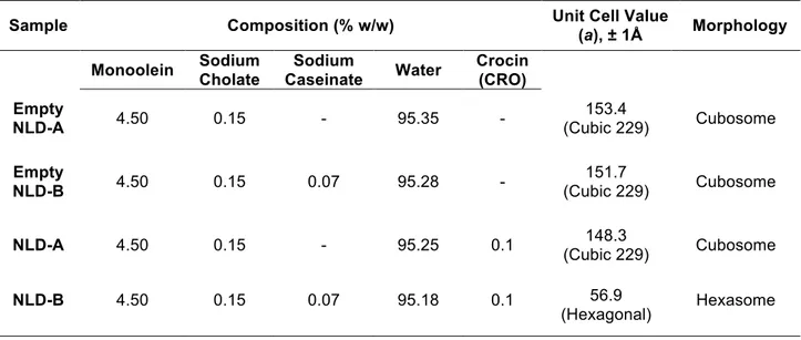
In table 2.1 has been summarized the composition of NLD and structural organization obtained by X-ray diffraction experiments.
Table 2.1 composition of NLD and structural organization obtained by X-ray diffraction experiments on NLD
Sample Composition (% w/w) Unit Cell Value
(a), ± 1Å Morphology Monoolein Sodium Cholate Sodium Caseinate Water Crocin (CRO) Empty NLD-A 4.50 0.15 - 95.35 - 153.4 (Cubic 229) Cubosome Empty NLD-B 4.50 0.15 0.07 95.28 - 151.7 (Cubic 229) Cubosome
NLD-A 4.50 0.15 - 95.25 0.1 (Cubic 229) 148.3 Cubosome
NLD-B 4.50 0.15 0.07 95.18 0.1 56.9
NLD have been prepared by the group of Ferrara, using mixtures of monoolein with sodium cholate and sodium caseinate. To give a brief summary of results obtained, the presence of naturally derived materials as sodium cholate and caseinate lead to:
- NLD-A: transparent dispersions
- NLD-B: milky dispersions
The addition of CRO confers a characteristic yellow coloration. In both cases, the presence of CRO leads to an increase of the mean diameter. Photon Correlation Spectroscopy (PCS) enabled to check the Z average diameter of nanoparticles, which is between 150 and 250 nm. In addition:
- EE (Encapsulation Efficiency) and LC (Loading Capacity) have been evaluated.
EE value is higher in NLD-B, probably the heterogeneous inner structure of nanoparticles facilitates the encapsulation of CRO. The use of mixtures of sodium cholate and caseinate, together with monoolein, lead to the formation of a most heterogeneous system (composed of cubic phases, uni-lamellar, bi-uni-lamellar, invaginated vesicles and sponge-like structures).
Figure 2.1 X-ray diffraction profiles observed on NLD before filtration. From the bottom: empty A, empty B, A and
NLD-B. The vertical black lines indicate the expected peak positions: Im3m, spacing ratios as √2:√4:√6:√8:√10:√12…; HII, spacing ratios as
√1:√3:√4… q=4π sinθ/λ, where 2θ is the scattering angle
- MTT assay performed on Melanoma Human Cell line A375 cultured in DMEM (Dulbecco’s Modified Eagle’s Medium) shows an antiproliferative activity of this compound (respectively 86+2% in NLD-A and 90+1% in NLD-B). Empty and CRO-loaded vesicles do not interfere with cell viability in human fibroblasts, used as model of normal cells.
Using X-ray diffraction (without previous filtration) inner structural organization of NLD has been investigated, both in presence and absence of CRO. Considering NLD-A, the presence of the cubic 229 (Im3m) has been observed in both cases. The addition of CRO doesn’t provoke modifications in terms of morphology. Different is the case of NLD-B: the presence of CRO induces a transition from cubosome to hexasome morphology of NPs. An additional confirmation of the inner phase transition has been given by the PCS (Photon Correlation Spectroscopy) measurements: in the case of NLD-B a decrease in the mean diameter of NPs has been reported, in line with the transition from cubic phase 229 to a hexagonal phase.
The use of NLD (in this case produced with naturally derived compound instead of copolymers such Pluronic 127®) represents a new strategy in the case of the delivery of crocin. An increase in terms of NLD lifetime
Inte
nsity (a.u.)
0.25 0.20 0.15 0.10 0.05Q (Å
-1) NLD-B NLD-A empty NLD-B empty NLD-A2.3 Characterization of Solid Lipid Nanoparticles (SLN) and Nanostructured Lipid Carriers (NLC)
2.3.1 Functionalization of nanoparticles with 1,3,5-triaza-7-phosphaadamantane (PTA) platinum (II) carboxylates: a structural study
The majority of this section is published in the reference Sguizzato et al., 2016
(Sguizzato M., R. Cortesi, E. Gallerani, M. Drechsler, L. Marvelli, P. Mariani, F. Carducci, R. Gavioli, E. Esposito, P. Bergamini Mater. Sci. Eng., C, 357-364, 2016)
For clarity has to be remarked that I performed the structural characterization using X-ray diffraction of SLN. The preparation of samples has been made by the group of the University of Ferrara, Department of Life Sciences and Biotechnologies; the synthesis of Pt-PTA and other assays by the group of the University of Ferrara, Department of Chemical and Pharmaceutical Sciences. Observations using Cryo-TEM has been made by Markus Drechsler of the University of Bayreuth.
A new way to functionalize SLN with Pt-PTA complexes is presented in this work. Stearic acid based-SLN seems to be a promising route for the delivery of PTA, after an initial screening of several fatty acid (oleic acid, palmitic acid, castor oil, myristic acid and stearic acid) made by the group of the University of Ferrara (Department of Life Sciences and Biotechnology).
A scheme of the chemical approach used to functionalize stearic based SLN with Pt-PTA is reported here: taking advantage of the presence of carboxylic groups on the surface of SLN, these can be first deprotonated and then coordinated to Platinum-PTA complexes as carboxylates. This part has been made by the other group of Ferrara (Department of Chemical and Pharmaceutical Sciences). The formation of the complex has been proved using 31P-NMR.
Figure 2.2 formation of Pt carboxylates on the surface of SLN. Figure has been taken from the paper Sguizzato et al., 2016
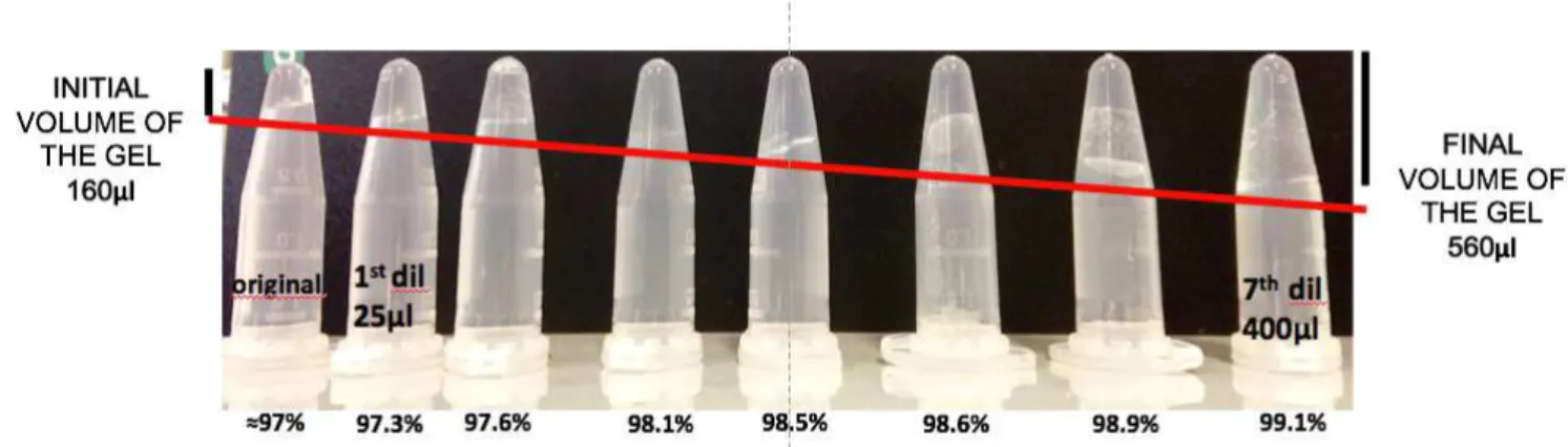
SLN have been produced using the emulsion-dilution method. This approach leads, in the case of stearic-acid based NPs, to the formation of a solution of vesicles characterized by great stability (up to 2 months) and homogenous aspect (in absence of aggregates and flocculates). PCS measurements show a Z-average value comprised between 600 and 800nm. The polydispersity index is between 0.21 and 0.41nm. Results obtained using X-ray diffraction are reported in the following table.
Table 2.2 composition of SLN and structural organization obtained by X-ray diffraction
Samples Composition (%w/w) Unit Cell Value
(a) ± 1 Å Morphology Stearic acid Tween®
20 Tween® 80 Water PTA
SLN T20 2 3.34 - 94.66 - 42.7 Lamellar
SLN T80 2 - 3.34 94.66 - 41.8 Lamellar
This work provides a demonstration of the fact that the use of a different kind of polysorbate is not relevant, considering NPs morphology. In this case SLN have been produced using T20 and T80 (ST20 produced with Tween® 20 and ST80 with Tween® 80). For both SLN two low-intensity Bragg’s peaks have been detected.
Figure 2.3 X-ray diffraction profiles of SLN T20 (ST20) and SLN T80 (ST80).
Q= 4πsinθ/λ, where 2θ is the scattering angle
The determination of the inner structure of Solid Lipid Nanoparticles has been made indexing the two major peaks. Reflections have been calculated to be in a ratio 1:2 so the inner structure of nanoparticles can be attributed to a 1-dimensional lamellar phase. The distance between the mid-plane of two opposing lipid bilayers has been calculated using Bragg’s Law and it’s 42.7Å for ST20 and 41.8Å for ST80. The practically similar value of unit cell and X-ray profile confirm that the inner structure is lamellar for both SLN.
In addition to the structural characterization, analysis on the potential antiproliferative activity of Pt-PTA have been made by the group of Ferrara (department of Life Sciences and Biotechnology), using two different cancer cell lines: A2780 (ovarian) and K562 (erythroleukemic). ST20 and ST80 show no toxicity towards the two cell lines and considering the in vitro antiproliferative effect of ST20 and 80 containing Pt-PTA complexes cell growth inhibition is comparable of that of Cisplatin. These results confirm the promising use of Pt-PTA functionalized SLN as anticancer drugs with the advantage of reduced side-effects, very heavy in the case of Cisplatin (first generation of Pt-based anticancer drugs).
2.3.2 Nafion® containing SLN as Tool for Anticancer Pt delivery
The majority of this section is published in the reference Sguizzato et al., 2017
(Sguizzato M., E. Esposito, M. Drechsler, E. Gallerani, R. Gavioli, P. Mariani, F. Carducci, R. Cortesi, P. Bergamini, Hindawi Journal of Chemistry, DOI 10.1155/2017/3206298, 2017)
Has to be remarked that I performed the structural characterization using X-ray diffraction of SLN. The preparation of samples has been made by the group of the University of Ferrara, Department of Life Sciences and Biotechnologies and observations using Cryo-TEM has been made by Markus Drechsler of the University of Bayreuth.
As alternative to the stearic acid based SLN for the coordination of Pt-PTA complexes, in this work has been proposed in this work the use of Nafion®
, a perfluorosulfonic acid resin. In this case Nafion® has been used to
establish an ionic connection with the protonable phosphine PTA (1,3,5-triaza-7-phosphaadamantane), applicable in the case of Pt coordination.
What plays an essential role is the fact that Nafion® is a strong proton donor and PTA is characterized by proton acceptor nitrogen atoms. The fact that NAF/PTAH+ complexes are able to maintain the ability to coordinate Pt via phosphorus group (demonstrated using 31P-NMR) makes these compounds optimal candidates for the coordination of Pt as anticancer drug.
Figure 2.4 X-ray diffraction profiles observed for nanoparticles dispersions without (black dots) and with NAF (white dots) at 37°C. Red
lines indicate positions of reflections expected for a 1D lamellar phase. The presence of a lamellar phase organization of nanoparticles for both samples is observable. In the case of SLN-NAF the second order of a lamellar phase can be appreciated (at very low intensity value) confirming the higher inner organization of NPs
Inte nsity (a.u.) 0.4 0.3 0.2 0.1 Q (Å-1) SLN NAF
Table 2.3 composition of SLN and structural organization obtained by X-ray diffraction
Samples Composition (%w/w) Unit Cell
Value (a) ± 1Å Morphology Tristearin Poloxamer 188 Nafion ® Water Pt SLN 3.35 2.50 - 94.15 - 46.0 Lamellar SLN NAF 3.35 2.50 1.65 92.15 - 49.0 Lamellar
The production of solid lipid nanoparticles has been made by the group of Ferrara, using the method of homogenization stirring followed by ultrasonication. In table 2.3 details about the composition and structural organization have been summarized. This process leads to a white milky emulsion, with a uniform macroscopic appearance (absence of aggregates and adherence to the vial’s walls). SLN-Nafion have been proved to be stable within 90 days. For the structural characterization Cryo-TEM, PCS and X-ray diffraction measurements have been performed. Electron microscopy (EM) shows a discoid shape particles; PCS gave a Z-average value of 250nm with a polydispersity index comprised between 0.202 and 0.235. Using X-Ray diffraction, characterization in terms of morphology formulations made both in the presence and absence of Nafion® has been made. Figure 2.4 shows results obtained for nanoparticles (SLN without Nafion have been used for comparison). Diffraction patterns are very similar, with only one peak resolved in the low-angle region (q<0.6 Å-1) that can be attributed to an inner organization of nanoparticles into a 1-D lamellar phase. Values of unit cell parameter are reported in table x and suggest that the addition of Nafion® doesn’t provoke a modification in the inner organization of NPs. Fundamental to evaluate the real possibility to use SLN-Nafion for the delivery of Pt-PTA has been evaluated performing in vitro cytotoxicity tests with two different cell lines, the same of the previous work presented in this thesis, A2780 (ovarian cancer cells) and K562 (erythroleukemic cancer cell line). No toxic effect of NAF has been reported, underlying their promising use as vehicles for the Pt-PTA complexes.
2.3.3 Characterization of SLN and NLC for the delivery of poorly water-soluble neuroactive drugs
The majority of this section is published in the reference Esposito et al., 2017b
Esposito E., M. Drechsler, P. Mariani, F. Carducci, V. Trezza, M. Servadio, F. Melancia, P. Ratano, R. Cortesi, C. Nastruzzi 2017 19:44 DOI 10.1007/s10544-017-0188-x
Has to be remarked that I made the structural characterization using X-ray diffraction of SLN. Other results have been reported for clarity to give an extended comprehension of the work. When experiments have been performed by other, this is clearly indicated.
known about the low solubility level in water of these molecules and this represent the major limitation in the use of these molecules.
To give a brief overview about the spectrum of action of these drugs, DMF acts remitting multiple sclerosis. Is employed also in psoriasis treatment, early brain injury and learning deficits(Bomprezzi, 2015; Linker and Gold, 2013); RP supplementation can be considered as a new approach in the multiple sclerosis prevention and treatment and at the same time to a correct maintenance of CNS(Saboor-Yaraghi, 2015; Maden, 2007). PRG is a neurosteroid with neuroprotective effects; its action is essential during pregnancy(Webster et al., 2015). Finally, URB597 in animals has potential effects against depression, anxiety and autism(Piomelli et al., 2006; Servadio et al., 2016).
Nanoencapsulation strategy allowed to obtain biocompatible and non-toxic vehicles. Tristearin (T) has been employed as unique component for the production of SLN and in addition of glyceril monoolein (Miglyol) for the production of NLC.
As first step, solubility of drugs has been evaluated using water, mixture of water/ethanol and pure ethanol. The higher solubility has been observed using pure ethanol. Due to the fact that this organic solvent is not appropriate for the use and toxic in human (because induces behavioral effects) and rats (due to its toxicity), has been evaluated the possibility to encapsulate drug in lipid nanoparticles, evaluating in this case drug solubility in SLN, NLC and PEG400/P80/saline 5:5:90. This part has been made by other authors.
Figure 2.5 solubility level of different drugs tested using in black: SLN, in grey: NLC, in white: PEG400/P80/ saline 5:5:90. Appreciable
variations can be appreciated in the case of DMF, PRG and RP. Maddalena Sguizzato et al.,2017
Figure 2.5 shows the difference in terms of solubility for PRG, RP, DMF and URB597 when incapsulated in SLN, NLC or in PEG400/P80/saline 5:5:90. The solubilization seems to be sensibly higher when drugs are encapsulated in SLN and NLC. For URB597, efficiency is low in all cases.
To improve encapsulation of URB597, the group of Ferrara tried the encapsulation of URB597 in polysorbate 80 (P80) modified nanoparticles. P80 exerts a specific role in brain targeting(Göppert and Müller, 2005). Usually unmodified NPs are captured from opsonin and eliminated from the body.
In table 2.4, results related to the structural organization of nanoparticles made with X-ray diffraction and CryoTEM have been reported.
Table 2.4 structural characterization of nanoparticles. Results obtained with X-ray diffraction and Cryo-TEM
NPs samples X-ray diffraction Interlamellar distance (Å) Cryo-TEM morphology
empty SLN 37.4 flat discoid
SLN/P80 45.5 bicellar
SLN-DMF 45.5 capped multilamellar
SLN-RP 38.1 bicellar
SLN-PRG 44.9 capped multilamellar
SLN-URB597 46.0 bicellar
SLN/P80-URB597 46.8 capped multilamellar
empty NLC 38.0 flat discoid
NLC-DMF 45.2 capped multilamellar
NLC-RP 45.0 flat discoid
NLC-PRG 44.8 bicellar
NLC-URB597 46.9 capped multilamellar
Graphs reported in Figure 2.6 show one major peak relative to the inner lamellar structure of nanoparticles, at low-angle value (around 0.18Å-1) and three secondary reflections (at 1.37, 1.63 and 1.71Å-1) relative to the subcrystalline organization of lipid chains. In this work, an evidence of a triclinic parallel organization of lipid chains (b form) is given, as reported by Sato and Ueno in Polymorphism in Fats and Oil, 2005 John Wiley & Sons, Inc. and by Jenning et al., 2000 (Sato and Ueno, 2000). Attention has to be focused also to the fact that solubilization of lipophilic molecules into lipid nanoparticles avoiding the use of organic solvents has been achieved. For RP and PRG the solubility level has been increased respectively 4- and 8-fold using lipid nanoparticles. The use of SLN/P80 instead of PEG400/P80/saline allowed to improve the solubility level of URB597 of 1.5-fold. At the same time, they tested the intranasal administration of SLN/P80-URB597, suggesting that this can be a great alternative to the intraperitoneal administration of this drug.
Figure 2.6 X-Ray diffraction profiles relative to A) SLN-URB compared with NLC-URB B) two different batches of SLN URB C) SLN URB
made with Tween in comparison with SLN URB. Black lines indicate the position of reflections in the high-angle region relative to the subcrystalline organization of nanoparticles in a triclinic parallel geometry
250 200 150 100 50 I(q) 2.0 1.5 1.0 0.5 q(Å-1) 250 200 150 100 50 I(q) 2.0 1.5 1.0 0.5 q(Å-1) 250 200 150 100 50 I(q) 2.0 1.5 1.0 0.5 q(Å-1) NLC URB SLN URB 22/9 SLN URB 22/9 SLN URB 29/04 SLN URB tween SLN URB 22/9
2.4 Characterization of Monoolein Aqueous Dispersions (MAD)
2.4.1 Structural Characterization of MAD kept in contact with quercetin
The majority of this section is published in the reference Cortesi et al., 2017
Cortesi R., E. Cappellozza, M. Drechsler, C. Contado, A. Baldisserotto, P. Mariani, F. Carducci, A. Pecorelli, E. Esposito, G. Valacchi Biomed Microdevices (2017) 19:41 DOI 10.1007/s10544-017-0185-0
Has to be remarked that I made the structural characterization using X-ray diffraction of MAD. Other results have been reported for clarity to give an extended comprehension of the work.
Quercetin (3,3’,4’,5-7-pentahydroxyflavone) is one the most powerful antioxidant present in nature. In addition to its key role in preventing oxidation (e.g. QT received considerable attentions regarding its activity to prevent Reactive Oxygen Species-mediated damage in skin, (Casagrande et al., 2006, 2007; Vicentini et al., 2008), it has many pharmacological potentials: decreasing lipid levels in blood, preventing anemia, as anti-platelet aggregation agent (Hollman and Katan, 1999).
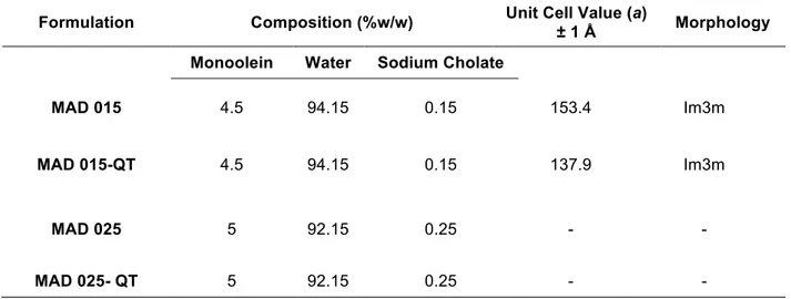
Table 2.5 composition of SLN in % w/w and structural organization results obtained by X-ray diffraction
Formulation Composition (%w/w) Unit Cell Value (a)
± 1 Å Morphology Monoolein Water Sodium Cholate
MAD 015 4.5 94.15 0.15 153.4 Im3m
MAD 015-QT 4.5 94.15 0.15 137.9 Im3m
MAD 025 5 92.15 0.25 - -
MAD 025- QT 5 92.15 0.25 - -
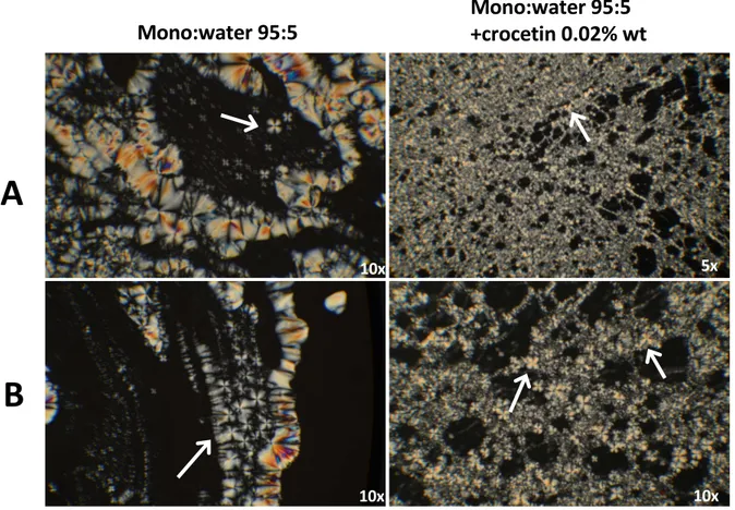
One of the main limitations in the use of this biological compound is its low-solubility in water. Monoolein Aqueous Dispersions (MAD) represent a way to solve this problem. In this work is reported the development of MAD encapsulating QT using sodium cholate as emulsifier. Two different kind of samples have been prepared, MAD015 (with sodium cholate at 0.15% w/w, as described in table 2.5) and MAD025 (with higher concentration of sodium cholate, 0.25% w/w). Samples have been prepared using the method of emulsification of lipid and emulsifier, followed by homogenization. QT has been added to MAD before addition of water in a concentration of 0.25mg/ml (0.5% by weight with respect to the content of monoolein and 0.025% by weight with respect to the dispersing phase). Characterization has been performed using CryoEM, X-Ray Diffraction (XRD), PCS and SdFFF (Sedimentation Field Flow Fractionation) as techniques. In addition, drug content evaluation in MAD, stability studies, In vitro release profiles (Franz cells) studies and biological tests on
relevant differences have been reported relatively to the content of emulsifier used in the dispersions formulation. The 0.15% (w/w) of sodium cholate (MAD015) leads to the obtainment of a mixture of vesicles and cubic structures; in the case of 0.25% (w/w) of emulsifier (MAD025) only unilamellar invaginated vesicles have been observed. This is in line with X-ray diffraction observations, summarized in graph 2.7 and table 2.5. From diffraction profiles is clearly observable that peaks detected are related to a cubic phase inner organization of MAD-015, both in the presence and in the absence of quercetin. Experiments have been performed at 37°C, both for MAD015 and MAD025, in the presence and absence of QT.
Figure 2.7 low-angle X-ray diffraction profiles of Monoolein Aqueous Dispersions, black lines indicate the expected position of Im3m cubic
phase. From the diffraction profiles, it is possible to deduce that quercetin doesn’t provoke changes in the nanoparticles organization
Unit cell value calculated from XRD profiles and reported in table 2.5 suggests that QT probably induces a small shrinking of water channels due to surface dehydration. No signals have been detected in the case of MAD025 and this is justifiable by the predominance of unilamellar structures observed using CryoEM, not detectable using XRD. Information about dimension have been obtained instead thanks to PCS and SdFF, performed by other groups. The addition of QT leads to a sensible increase of mean diameter of MAD015 and 025 with the respect of that one of empty MAD. For example, in the case of MAD015, the average value of empty dispersions is of 227.0nm, consequently to the addition of QT this became 402.3nm. Probably QT locates within lipids. This study let to comprise the importance of structural characterization in the development of new MAD DDS. The comprehension of lipid NPs inner organization (especially in the case of use of different emulsifiers and active principles) is a fundamental step that has to be made to obtain stable and efficient systems to be used in the delivery of drugs.
300 250 200 150 100 Inte nsity (a.u.) 0.20 0.16 0.12 0.08 0.04 q (Å-1)
Im3m Symmetry Peaks
MAD sodium cholate 0.15
2.5 High Pressure Homogenization (HPH) vs Ultrasonication (U): characterization of NPs stability and validation of method using Small Angle X-Ray Scattering (SAXS)
Measures have been made in May 2017 at the beamline BM29 (BioSAXS), European Synchrotron Radiation Facility (ESRF), Grenoble (FR). Data have not yet been published.
2.5.1 SLN and NLC
Due to the always growing interest of NPs production across industries, High Pressure Homogenization (HPH) has becoming the preferred method to be used for large-scale production of NPs.
Preliminary results are reported here, relatively to samples made by the group of Ferrara (Dept. of Life Sciences and Biotechnology) but using two different methods of production of NPs: Ultrasonication method (U) and High-Pressure Homogenization (HPH), an ongoing process widely used in pharmaceutical and food industry. This method is innovative due to its ability to lead and uniform droplets size in lipid emulsion and to avoid the presence of aggregates (Esposito et al., 2017a), but the real advantage in the use of this kind of homogenization is the possibility to achieve scale-up nanoparticle volumes from lab to pilot and industrial scale. Measures have been performed in Grenoble, ESRF (European Synchrotron Radiation Facility), Beamline BM29, using small angle X-ray scattering (SAXS) technique.
Table 2.6 composition of samples related to SLN. Three columns have been reported: in the first one has been reported type of
nanoparticles made, in the second one different lipid used and related concentration and finally, in the third one, method of production used
Nanoparticle Lipid_ Concentration (% wt) Method of production
SLN Tristearin (T)_ 5 HPH SLN Suppocire (S)_5 HPH SLN Compritol I_5 HPH SLN Precirol (P)_5 HPH SLN Tristearin (T)_ 5 U SLN Suppocire (S)_5 U SLN Compritol I_5 U SLN Precirol (P)_5 U SLN Suppocire (S)_8 HPH SLN Suppocire (S)_9 HPH SLN Suppocire (S)_10 HPH SLN Suppocire (S)_8 U SLN Suppocire (S)_9 U SLN Suppocire (S)_10 U
Graphs reported in Figure 2.8 and organized in two columns indicate the two different temperatures tested (column1: 30°C, column 2: 37°C). In all graphs, black curves are related to nanoparticles produced using High Pressure Homogenization method (H). In column 1 (30°C), NPs made using the canonical method of Ultrasonication (U) are reported in green, in column 2 (37°C), NPs produced using ultrasonication U are showed in orange.
As it is possible to observe in Figure 2.8, lines A (Tristearin) and D (Precirol), the use of those lipids in the production of NPs does not seem to produce variations in lipid morphology, at both temperatures tested.
Layout 1:
Figure 2.8 SAXS profiles related to solid lipid nanoparticles (SLN) made using different lipids (Line A: Tristearin (T), B: Suppocire (S), C:
Compritol (C), D: Precirol (P)) and different method of production: High-pressure homogenization (HPH, in graphs indicated as H, black
curves, both for 30°C (left column) and 37°C (right column)) and ultrasonication U, (green curves for the 30°C, orange curves in the
case of 37°C)
In the case of Suppocire (line B), at 37°C, it is relevant the fact that the peak related to the lamellar phase disappears only in the case of the orange curve, that one produced using the method of Ultrasonication. Probably the High-Pressure Homogenization (HPH) is able to confer a major stability in terms of inner organization of the NPs.
General considerations about the production of NPs using H method can be made for the lipid Compritol (Line C). The characteristic morphology of the peak (large at the bottom and very narrow and defined at the top) suggest us the possibility of the presence of a double phase in the inner organization of NPs.
In addition, is very curious that the presence of higher number of peaks relative to the lamellar phase (up to the 3rd order) has been evidenced only at 37°C (Figure 2.8, column 2, line C_ in both profiles).