R E S E A R C H
Open Access
A ruthenium(II)-curcumin compound
modulates NRF2 expression balancing the
cancer cell death/survival outcome
according to p53 status
Alessia Garufi
1,2, Silvia Baldari
1,3, Riccardo Pettinari
4, Maria Saveria Gilardini Montani
5, Valerio D
’Orazi
6,
Giuseppa Pistritto
7, Alessandra Crispini
8, Eugenia Giorno
8, Gabriele Toietta
1, Fabio Marchetti
9, Mara Cirone
5and
Gabriella D
’Orazi
1*Abstract: Background: Tumor progression and tumor response to anticancer therapies may be affected by
activation of oncogenic pathways such as the antioxidant one induced by NRF2 (nuclear factor erythroid 2-related
factor 2) transcription factor and the pathways modified by deregulation of oncosuppressor p53. Often, oncogenic
pathways may crosstalk between them increasing tumor progression and resistance to anticancer therapies.
Therefore, understanding that interplay is critical to improve cancer cell response to therapies. In this study we
aimed at evaluating NRF2 and p53 in several cancer cell lines carrying different endogenous p53 status, using a
novel curcumin compound since curcumin has been shown to target both NRF2 and p53 and have anti-tumor
activity.
Methods: We performed biochemical and molecular studies by using pharmacologic of genetic inhibition of NRF2
to evaluate the effect of curcumin compound in cancer cell lines of different tumor types bearing wild-type (wt)
p53, mutant (mut) p53 or p53 null status.
Results: We found that the curcumin compound induced a certain degree of cell death in all tested cancer cell
lines, independently of the p53 status. At molecular level, the curcumin compound induced NRF2 activation,
mutp53 degradation and/or wtp53 activation. Pharmacologic or genetic NRF2 inhibition further increased the
curcumin-induced cell death in both mutp53- and wtp53-carrying cancer cell lines while it did not increase cell
death in p53 null cells, suggesting a cytoprotective role for NRF2 and a critical role for functional p53 to achieve an
efficient cancer cell response to therapy.
(Continued on next page)
© The Author(s). 2020 Open Access This article is licensed under a Creative Commons Attribution 4.0 International License, which permits use, sharing, adaptation, distribution and reproduction in any medium or format, as long as you give appropriate credit to the original author(s) and the source, provide a link to the Creative Commons licence, and indicate if changes were made. The images or other third party material in this article are included in the article's Creative Commons licence, unless indicated otherwise in a credit line to the material. If material is not included in the article's Creative Commons licence and your intended use is not permitted by statutory regulation or exceeds the permitted use, you will need to obtain permission directly from the copyright holder. To view a copy of this licence, visithttp://creativecommons.org/licenses/by/4.0/. The Creative Commons Public Domain Dedication waiver (http://creativecommons.org/publicdomain/zero/1.0/) applies to the data made available in this article, unless otherwise stated in a credit line to the data.
* Correspondence:[email protected]
1Department of Research and Advanced Technologies, IRCCS Regina Elena
National Cancer Institute, Rome, Italy
(Continued from previous page)
Conclusions: These findings underline the prosurvival role of curcumin-induced NRF2 expression in cancer cells
even when cells underwent mutp53 downregulation and/or wtp53 activation. Thus, NRF2 inhibition increased cell
demise particularly in cancer cells carrying p53 either wild-type or mutant suggesting that p53 is crucial for efficient
cancer cell death. These results may represent a paradigm for better understanding the cancer cell response to
therapies in order to design more efficient combined anticancer therapies targeting both NRF2 and p53.
Keywords: p53, NRF2, Curcumin, (arene)ruthenium(II) compound, Brusatol, Cancer therapy, Oxidative stress,
Chemoresistance, Autophagy
Background
The oncosuppressor p53 plays a key role in cell growth
and apoptosis in response to various stress signals [
1
].
Given its central role in maintaining genomic stability
and preventing oncogenesis, p53 is the most inactivated
oncosuppressor in human tumors by gene mutations or
by protein deregulation [
2
]. Mutant (mut) p53 proteins
may acquire a misfolded hyperstable conformation [
3
]
that may be achieved by binding heat shock proteins
(HSP) such as HSP90, a cellular chaperone that is crucial
for the stability of many client proteins including
mutp53 [
4
,
5
]. Besides loss of function and
dominant-negative effect on the wild-type (wt) p53 activity, the
hotspot p53 mutants may also acquire new oncogenic
functions, contributing to cancer progression, invasion
and resistance to therapies [
6
]. Thus, targeting mutp53
is a challenging strategy to halt cancer growth [
7
]. In this
regard, several different approaches have been taken in
the last years developing small molecule or using
phyto-chemicals from nature to induce mutp53 degradation or
conformational changes, providing new insight on
mutp53 reactivation [
8
,
9
], as also demonstrated by our
studies [
10
–
13
]. Autophagy has been shown to be
in-volved in mutp53 degradation [
14
–
23
], suggesting the
use of autophagy stimulators to counteract mutp53
oncogenic activity. Thus, mutp53 has been shown to
counteract autophagy mechanism to likely halt its own
degradation [
24
]. Finally, mutp53 degradation by
au-tophagy has been shown to increase the cytotoxic effects
of chemotherapeutic drugs [
17
]. Mutp53 oncogenic
ac-tivities ma also depend by modifications of the tumor
microenvironment altering the secretion of
inflamma-tory cytokines that affect the crosstalk between cancer
and stromal cells [
25
,
26
] or by interaction with other
transcription factors such as NRF2 (nuclear factor
eryth-roid 2-related factor 2, encoded by NFE2L2 gene) or
HIF-1 (hypoxia-inducible factor 1) to support tumor
progression and resistance to therapies [
27
]. Therefore,
understanding the interplay between these oncogenic
pathways may have an impact on the development of
more efficient targeted anticancer therapies.
NRF2 is the main regulator of cellular antioxidant
re-sponse [
28
] and is activated in response to oxidative
and/or electrophilic stress, the so-called canonical
condi-tions. Following activation, NRF2 detaches from its
negative regulator KEAP1 (Kelch-like ECH-associated
protein 1), stabilizes and moves to the nucleus where it
binds to sequence-specific responsive elements of
anti-oxidant target genes promoters. Among these genes
there are catalase, superoxide dismutase (SOD), HO-1
(heme-oxygenase 1), NAD(P)H quinone oxidoreductase
1 (NQO1), and glutathione (GSH), that help to restore
the cellular redox homeostasis [
29
]. Constitutive
activa-tion of NRF2 is found in several different tumors also by
gaof-function mutations of the NFE2L2 gene or by
in-activating mutations of the KEAP1 gene. These
muta-tions are considered drivers of cancer progression,
metastasis, and resistance to therapies [
30
]. NRF2
non-canonical activation may depend by
p62/SQSTM1-medi-ated KEAP1 degradation [
31
], or by p21Cip1/WAF1
(target of p53) that binds to KEAP1 to interrupt the
KEAP1/NRF2 complex [
32
]. NRF2 may have both tumor
suppressive and tumor-promoting actions and is
there-fore considered a
“double face” molecule [
33
]. Thus,
while NRF2 transient activation is certainly considered
cytoprotective, its continual activation may support
tumor progression and tumor resistance to therapies.
Therefore, NRF2 overexpression in cancer cells may be
considered a marker of chemoresistance [
34
].
Curcumin is considered a chemopreventive molecule
with anti-oxidant, anti-apoptotic and anti-inflammatory
properties and with an excellent safety profile although
it presents low solubility and bioavalibility [
35
].
Curcu-min has been shown to activate the NRF2 pathway
trig-gering cellular protection against oxidative injury that, in
advanced stage cancers, may induce chemoresistance
[
36
]. On the other hand, curcumin may induce mutp53
degradation through autophagy or convert mutp53
pro-tein to transcriptionally active wtp53, reversing the
therapeutic resistance and improving cancer cell death,
as also demonstrated by our studies [
16
–
18
,
37
,
38
].
In this study we aimed at evaluating the effect of a
novel curcumin compound in several cancer cell lines
carrying different endogenous p53 status. We used a
water-soluble ruthenium(II)-curcumin (RuCUR)
com-pound that presents high solubility and cytotoxic effect
]compared to other similar curcumin compounds [
39
–
46
].
Moreover, the compound show selectivity toward cancer
cells over nontumorous cells, suggesting an in vivo use [
42
].
We found that RuCUR induced cancer cell death in all
tested cell lines and independently of the p53 status. At
molecular level we found NRF2 activation, mutp53
degrad-ation and/or wtp53 activdegrad-ation. Pharmacologic or genetic
NRF2 inhibition further increased the RuCUR-induced cell
death in both mutp53- and wtp53-carrying cancer cell lines
while did not increase cell death in p53 null cells. In the
present study, we found that activation of the NRF2
anti-oxidant pathway contributed to resistance to RuCUR
treat-ment, despite mutp53 degradation and activation of wtp53.
Furthermore, inhibition of NRF2 could overcome this
re-sistance in particular in cancer cells carrying p53 either
wild-type or mutant, indicating the crucial role for a
func-tional p53 pathway to efficiently induce cell death in
re-sponse to treatments.
Methods
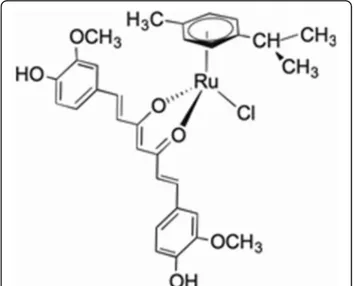
Ruthenium(II)-curcumin compound (RuCUR) synthesis
The water-soluble ruthenium(II)-curcumin compound
(RuCUR) was synthesized as described previously [
42
,
43
]. In brief, for the synthesis of RuCUR, equimolar
amounts of curcumin and [Ru(6-p-cymene)Cl2]2 were
stirred in methanol in the presence of KOH, the mixture
was filtered to remove potassium chloride, then the
solu-tion was concentrated and stored at 4 °C, from which
red crystals of RuCUR slowly formed (Fig.
1
). The purity
of the prepared product was confirmed through
elemen-tal analysis (on a Fisons Instruments 1108 CHNSO
elemental analyzer), melting point (on a STMP3 Stuart
scientific instrument and on a capillary apparatus), IR
spectral analysis (on a PerkinElmer Frontier FT-IR
in-strument) and 1H and 13C NMR spectroscopy (on a 500
Bruker Ascend operating at 500 MHz for 1H and 125
MHz for 13C relative to TMS). The RuCUR compound
was
dissolved
in
water
and
used
at
different
concentration.
Cell culture and reagents
The cell lines used in this study were: human SKBR3
(breast cancer, carrying R175H p53 mutation), T98MG
(glioblastoma, carrying M237I p53 mutation), MCF7
breast cancer, U87 glioblastoma, HCT116 and RKO
colon cancer cell lines (all carrying wild-type p53), and
HCT116 p53 null. The cells were cultured in either
DMEM (Dulbecco modified Eagle’s medium) (Life
Technologies-Invitrogen),
or
RPM1
–1640
(Life
Technologies-Invitrogen),
with
10%
heat-inactivated
foetal bovine serum (FBS) (Corning, NY, USA, #35–079)
and L-glutamine/streptomycin (100
μg/mL) (Corning,
NY, USA, #30
–002), in 5% CO2 at 37 °C. They were all
mycoplasma negative. The NRF2 inhibitor Brusatol [
47
]
(Sigma-Aldrich) was used at 100 nM, as previously
re-ported [
48
].
Viability assay
For viability assay cells were plated at subconfluence in
35 mm multiwall Petri dishes and the day after treated
as reported. Both floating and adherent cells were
col-lected and cell viability was determined by Trypan blue
(Sigma-Aldrich, #72571) staining and direct counting
with a Neubauer haemocytometer, as previously
re-ported [
13
].
3D spheroids proliferation assay
The 3D spheroids, were obtained into ultra-low
attach-ment cell culture multiwell plates (96 well, Corning), as
previously reported [
49
,
50
]. When spheroids reached
approximate size of 300
–500 um (about 4 days after
pla-tinga), they were treated with RuCUR (10, 50, 100
μM).
Spheroids images were acquired with a Nikon Eclipse
TS100 microscope equipped with a Nikon ELWD
cam-era (Nikon Instrument Europe BV, Amsterdam, The
Netherlands) every 24 h. The formula to evaluate the
spheroids volume was: V = a x (b2)/2, where a and b are,
respectively, length and width.
Measurement of intracellular Reactive Oxygen Species
(ROS)
Reactive oxygen species (ROS) production was assessed
by
using
the
2′,7′-dichlorofluorescein
diacetate
(DCFDA; Sigma–Aldrich) staining, as previously
re-ported [
51
]. Cell pellets were collected and analyzed in
the FL-1 channel of a FACScalibur flow cytometer (BD
Transduction Laboratories), using CELLQuest Pro
soft-ware (version 6.0, BD Biosciences). DCF fluorescence
values were expressed as mean fluorescence intensity
(MFI).
Western blot analysis
Cell pellets were lysed in lysis buffer (50 mM Tris–HCl,
pH 7.5, 150 mM NaCl, 5 mM EDTA, 150 mM KCl, 1
mM dithiothreitol, 1% Nonidet P-40) (all from
Sigma-Aldrich) and a mix of protease inhibitors (Complete™,
Mini Protease Inhibitor Cocktail, Merk, Life Science
S.r.l., Milan, Italy, #11836153001) and protein
concentra-tion determined with the BCA Protein Assay (BioRad,
Hercules, CA, USA) to load equal amount of total cell
extracts on 9–18% SDS-polyacrilamide gels (PAGE)
(Bio-Rad, #456–1095) electrotrophoreses. After blotting
to polyvinylidene difluoride membranes (PVDF,
Milli-pore Corporation, Billerica, MA, USA, #IPVH 00010).
Unspecific sites were blocked with 5% non-fat powdered
milk or with 3% bovine serum albumin (BSA, both from
Sigma-Aldrich) in 0.05% Tween-20 (v/v in TBS).
Mem-branes were then incubated with primary antibodies.
The antibodies we used in this study were: mouse
monoclonal anti-p53 (DO-1) (1:1000) (sc-126), rabbit
polyclonal anti-p21 (C-19) (1:500) (sc-397), mouse
monoclonal anti-HO-1 (A-3) (1:1000) (sc-136,960),
mouse monoclonal anti-p62/SQSTM1 (D-3) (1:1000)
(sc-28,359), all from Santa Cruz Biotechnology; rabbit
polyclonal anti-LC3B (1:2000) (Sigma, #L7543); mouse
monoclonal anti-NQO1 (A180) (ThermoFisher, #39–
3700), mouse monoclonal anti-HSP90 (1:1000) (BD
Bioscience, #610418), rabbit polyclonal anti-NRF2 (1:
1000) (Abcam, #ab62352), mouse monoclonal
anti-poly(ADP-ribose) polymerase (1:1000) (PARP, cleavage
site-214-215, Sigma, #AB3565), mouse monoclonal
anti-phospho-Histone
H2AX
(1:1000)
(Ser139,
clone
JBW301) (Sigma, #05–636), and rabbit polyclonal anti
phospho-4E-BP1 (236B4) (Thr37/46, Cell Signaling
Technology, #2855). Mouse monoclonal actin
anti-body (Ab-1) (JLA20) (1:10.000) (Calbiochem, #CP01)
was used as loading control.
Anti-imunoglobulin-G-horseradish peroxidise (HRP) secondary antibodies that
we used were: anti-mouse IgG-HRP (BioRad, #172–
1011) and anti-rabbit IgG-HRP (BioRad, #172–1019).
Enzymatic signals were visualized by chemiluminescence
(ECL Detection system, Amersham GE Healthcare,
Milan, Italy, #RPN2106), according to the
manufac-turer’s protocol.
RNA extraction and semi-quantitative reverse
transcription (RT)-PCR analysis
Total RNA extraction was performed by using TRIzol
Reagent
(Life
Technology-Invitrogen),
cDNA
was
synthesized by using MuLV reverse transcriptase kit
(Applied Biosystems, Foster City, CA, USA) and
semi-quantitative Reverse-Transcribed (RT)-PCR was carried
out by using Hot-Master Taq polymerase (Eppendorf,
Milan, Italy), as previously reported [
13
]. Densitometric
analysis allowed to quantify mRNA levels compared to the
control 28S gene expression. Primer sequences are as
fol-low: NRF2 For: TCCATTCCTGAGTTACAGTGTCT;
Rev.:
TGGCTTCTGGACTTGGAACC.
HO-1
For:
AAGATTGCCCAGAAAGCCCTGGAC; Rev.: AACTGT
CGCCACCAGAAAGCTGAG. DRAM For: TCAAAT
ATCACCATTGATTTCTGT;
Rev.:
GCCACATACG
GATGGTCATCTCTG. PUMA For: TGTGAATCCT
GTGCTCTGCC; Rev.: TTCCGGTATCTACAGCAGCG.
P21 For: CCCCTTCGGCCCGGTGGAC; Rev.: CCGTTT
TCGACCCTGAGAG. 28S For: GTTCACCCACTAAT
AGGGAACGTGA; Rev.: GATTCTGACTTAGAGGCG
TTCAGT.
siRNA interference
For RNA interference, cells were transfected with the
Nrf2 siRNA (sc-3703, Samta Cruz Biotechnology) and
control siRNA (sc-37,007, Santa Cruz Biotechnology)
using LipofectaminePLus reagent (Invitrogen, #11514–
015), following the manufacturer’s instructions, as
previ-ously reported [
12
].
Densitometric analyis
Quantification of the protein bands was assessed by
densitometric analysis using the ImageJ software (
http://
imagej.nih.gov
) and relative band intensity normalized to
β-actin signals.
Statistical analysis
Data are presented as mean ± standard deviation (S.D.)
of at least three independent experiments. Two-tailed
Student’s t-test and one-way ANOVA analysis were used
for statistical significance of, respectively, two o more
sample comparisons. Difference was considered
statis-tical significant when
p-value was ≤0.05.
Results
RuCUR compound induces mutp53 downregulation and
cell death
We first evaluated the effect of RuCUR compound on
cancer cell lines carrying mutp53 by performing a
three-dimensional (3D) culture spheroids proliferation assay
[
49
,
50
]. Cells cultured in ultralow attachment plates
were treated with different doses of RuCUR compound
and spheroids proliferation was recorded acquiring
im-ages every 24 h. Figure
2
a shows that increasing doses of
RuCUR significantly inhibited spheroids proliferation,
indicative of the capacity of the compound to reach cells
even in 3D culture conditions, and, accordingly, induced
cell death, as assessed by viability assay (Fig.
2
b). At
bio-chemical level, RuCUR reduced mutp53 protein levels at
almost all doses although the highest dose was more
efficient (Fig.
2
c). Therefore, we used the highest dose
for the next experiments. We found that
RuCUR-induced mutp53 downregulation correlated with strong
reduction of HSP90 levels and with induction of
autoph-agic marker LC3II (Fig.
2
d); moreover, RuCUR induced
p21 expression, indicative of wtp53 reactivation and
in-creased the expression of pro-apoptotic and
DNA-damage response proteins (respectively, cleaved PARP
and
γH2AX) (Fig.
2
d). Altogether, these findings suggest
that the RuCURC-triggered mutp53 downregulation,
likely through autophagy, correlated with unbalance of
the cell death/survival pathways toward cell death.
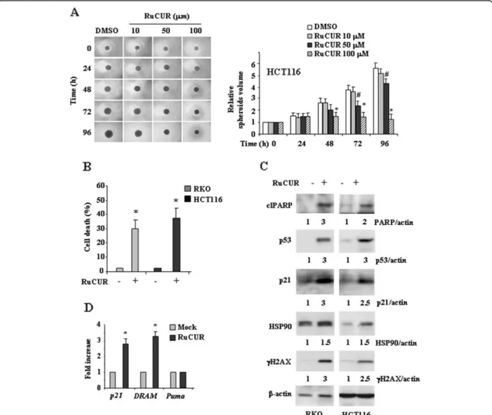
RuCUR compound induces cell death in wild-type (wt)
p53-carrying cancer cells
Next we evaluated the effect of RuCUR compound on
can-cer cell lines carrying wtp53. We found that increasing doses
of RuCUR compound significantly inhibited spheroids
pro-liferation (Fig.
3
a) and, accordingly, induced cell death as
assessed by viability assay (Fig.
3
b). At biochemical level
RuCUR induced cleavage of PARP and p53 stabilization
with up-regulation of the levels of its targets p21 and
DRAM, although it did not induce the pro-apoptotic Puma
target gene (Fig.
3
c, d), suggesting a selective and not
com-pletely apoptotic p53 activation, as previously reported [
52
].
RuCUR induced histone H2AX phosphorylation, producing
γH2AX, indicative of DNA damage response, responsible of
p53 activation (Fig.
3
c). Finally, differently from what
ob-served for mutp53 cells in Fig.
2
d, RuCUR slightly increased
HSP90 levels in wtp53-carrying cells (Fig.
3
c), in agreement
with the finding that HSP90 can promote p53
transcrip-tional activity [
53
] and that curcumin may indirectly target
HSP90 according to cell type [
54
]. Altogether, these results
indicate that RuCUR-induced cell death in wtp53 cells
cor-related with wtp53 activation.
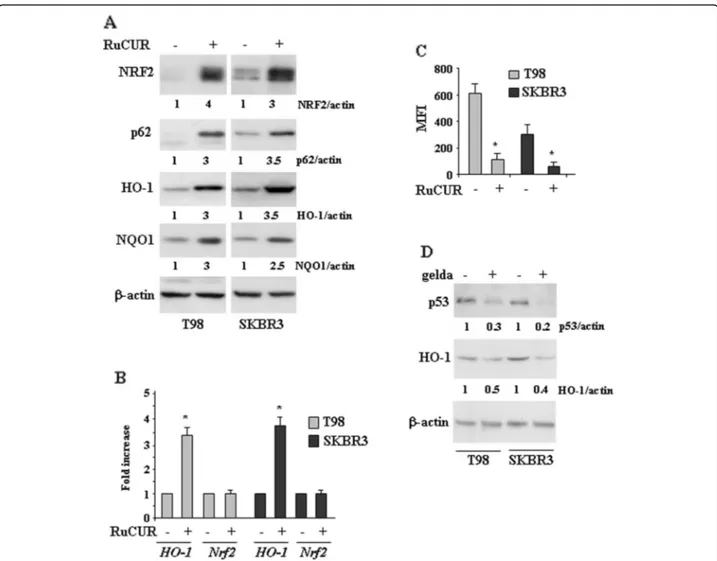
RuCUR compound induces NRF2 pathway in
mutp53-carrying cancer cells
Since curcumin has been shown to activate NRF2 [
36
]
we evaluated the effect of RuCUR on NRF pathway in
mutp53 cancer cells. We found that RuCUR strongly
stabilized the levels of NRF2 protein and induced the
ex-pression of its antioxidant targets, including HO-1 and
NQO1 by western blot (Fig.
4
a) and RT-PCR (Fig.
4
b)
analyses. Interestingly, increased NRF2 levels correlated
with increased p62 levels (Fig.
4
a), in according with the
notion that p62 competitively binds to Keap1 to activate
NRF2 in a noncanonical manner [
55
]. In support of this,
NRF2 gene did not undergo modification following
RuCUR treatment (Fig.
4
b). Finally, NRF2 stabilization
correlated with a strong decrease of intracellular ROS in
both cell types (Fig.
4
c), in line with the antioxidant
ef-fect of NRF2. Since NRF2 and mutp53 may regulate
each other [
56
] we attempted to evaluate the NRF2/
mutp53 interplay by using the HSP90 inhibitor
geldana-mycin [
57
]. We found that geldanamycin while strongly
reduced mutp53 protein levels it did not induce NRF2
pathway but rather decreased HO-1 levels (Fig.
4
d),
sug-gesting that targeting mutp53 may inhibit also NRF2
pathway. This did not happen with RuCUR that instead
exerted a strong dual effect by both inducing NRF2 and
reducing mutp53, suggesting how different molecular
approaches may differently modulate the two oncogenic
pathways.
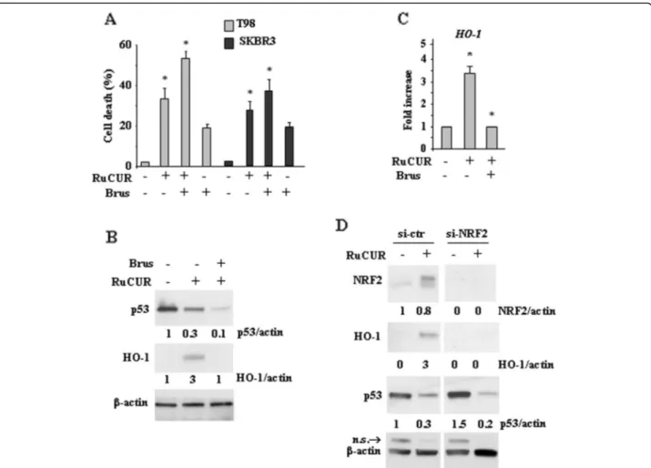
Since NRF2 is an important pro-survival molecule
we attempted to inhibit it. We found that NRF2
pharmacologic inhibition with brusatol [
47
,
48
]
sig-nificantly increased RuCUR-induced cell death of
mutp53 cancer cells, as assessed by viability assay
(Fig.
5
a). Pharmacologic inhibition of NRF2 activity
indeed impaired the RuCUR-induced HO-1 protein
levels (Fig.
5
b) and gene expression (Fig.
5
c).
Interest-ingly, the RuCUR-reduced mutp53 protein levels were
further reduced following NRF2 inhibition (Fig.
5
b),
strengthening the notion that NRF2 and mutp53 may
regulate each other [
56
,
58
]. Similar results were
ob-tained with genetic NRF2 silencing using specific
siRNA (Fig.
5
d). These findings indicate that
inhibit-ing
the
NRF2
survival
pathway
further
reduces
mutp53 levels and increases cancer cell death.
(See figure on previous page.)
Fig. 2 RuCUR (ruthenium-curcumin) compound reduced mutant (mut) p53 levels that correlated with reduced proliferation and increased cell death. (a) Mutp53-carrying T98 and SKBR3 cells were seeded on ultra-low attachment multiwell plates allowing for tumor spheroid formation. Four days after seeding, spheroids were formed (approximate size 300–500 μm) and then treated with different doses of RuCUR (10, 50, 100 μM) for the indicated times. Tumor spheroids volume was quantified according to the formula: V = a x (b2)/2, where a and b are, respectively, length and width. Representative images of spheroids derived from both cell lines are shown in the upper panels. Spheroids volumes are reported on the bottom panels. Histograms represent the fold increase quantified with respect to controls set to 1.0, ± SD. * (p ≤ 0.01), # (p ≤ 0.05) (single treatments compared to untreated spheroids). (b) Cell viability was measured by trypan blue exclusion assay in T98 and SKBR3 cells treated for 24 h with different doses of RuCUR (1, 10, 50, 100μM) and expressed as cell death percentage ± S.D. * (p ≤ 0.01), # (p ≤ 0.05) (single treatments compared to untreated cells). (c) Western blot analysis of p53 protein was performed in T98 and SKBR3 cells untreated or treated with RuCUR (1, 10, 50, 100μM) for 24 h. The ratio of p53 levels vs β-actin, following densitometric analysis using ImageJ software, is shown. (d) Western blot analysis of the indicated protein levels was performed in T98 and SKBR3 cells untreated or treated with RuCUR (100μM) for 24 h. Actin was used as protein loading control. The ratio of the protein levels vsβ-actin, following densitometric analysis using ImageJ software, is reported. n.s = not specific
RuCUR compound induces NRF2 pathway in
wtp53-carrying cancer cells
Finally, we evaluated the effect of RuCURC on NRF2
pathway in wtp53 cells. We found that RuCUR reduced
the intracellular ROS generation (Fig.
6
a) that correlated
with increased NRF2 pathway (Fig.
6
b). Inhibition of
NRF2 activity by brusatol strongly impaired the
RuCUR-induced NQO1 and HO-1 levels (Fig.
6
b and c).
Intri-guingly, brusatol co-treatment impaired the
RuCUR-induced p21 and DRAM expression, while increased
Puma expression (Fig.
6
d), suggesting that inhibiting
NRF2 could reestablish p53 apoptotic activity. In line
with this effect, NRF2 inhibition further increased
RuCUR-induced cell death (Fig.
6
e).
Fig. 3 RuCUR compound reduces proliferation and induces cell death in wild-type (wt) p53-carrying cancer cells. (a) Colon cancer HCT116 cell, were seeded on ultra-low attachment multiwell plates allowing for tumor spheroid formation. Four days after seeding, spheroids were formed (approximate size 300–500 μm) and then treated with different doses of RuCUR (10, 50, 100 μM) for the indicated times. Tumor spheroids volume was quantified according to the formula: V = a x (b2)/2, where a and b are, respectively, length and width. Representative images of spheroids are shown in left panel. Spheroid volume are reported on the right panel. Histograms represent the fold increase quantified with respect to controls set to 1.0, ± SD. * (p ≤ 0.01), # (p ≤ 0.05) (single treatments compared to untreated spheroids). (b) Cell viability was measured by trypan blue exclusion assay in RKO and HCT116 cells treated with RuCUR (100μM) for 24 h and expressed as cell death percentage ± S.D. * (p ≤ 0.01) (single treatments compared to untreated cells). (c) Western blot analysis of the indicated protein was performed in RKO and HCT116 cells untreated or treated with RuCUR (100μM) for 24 h. Actin was used as protein loading control. The ratio of the protein levels vs β-actin, following densitometric analysis using ImageJ software, is reported. (d) Total mRNA was extracted from RKO and HCT116 cells treated with RuCUR (100μM) for 24 h. The indicated gene expression was assayed by the polymerase chain reaction (PCR) of reverse-transcribed cDNA. Densitometric analysis was performed using ImageJ software to calculate the gene/28S ratio. Histograms represent the fold increase quantified with respect to controls set to 1.0, ± SD. * (p ≤ 0.01)
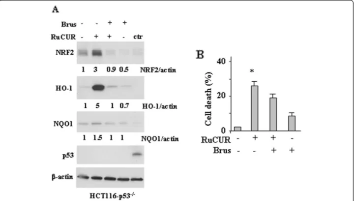
As a final point, to evaluate the role of endogenous
p53 we used HCT116 p53 null cells. We found that
RuCUR activated NRF2 pathway also in p53 null cells
(Fig.
7
a); however, analysis of cell viability show that the
RuCUR-induced cell death was slightly reduced by
NRF2 inhibition with brusatol (Fig.
7
b), opposite to what
observed in mutp53 and wtp53 cells (Figs.
5
and
6
a, e).
Altogether, these findings suggest that reducing NRF2
pathway in wtp53-carrying cells may unbalance the
pro-survival/pro-death axis toward cell death and restore
p53 apoptotic activity that indeed is crucial for cancer
cell death; on the other hand, reducing NRF2 pathway in
p53 null cells did not significantly increase cancer cell
death, underlining the critical role of p53 in cell death.
Discussion
The findings of the present study revealed that a novel
water-soluble
ruthenium(II)-curcumin
compound
(RuCUR) [
42
] was able to induce cancer cell death that
correlated with mutp53 downregulation and with
activa-tion of wtp53; they also revealed a resistance mechanism
via the NRF2-induced antioxidant system likely
en-hanced by ROS reduction. In addition, our findings
sug-gest that NRF2 inhibitors can overcome the RuCUR
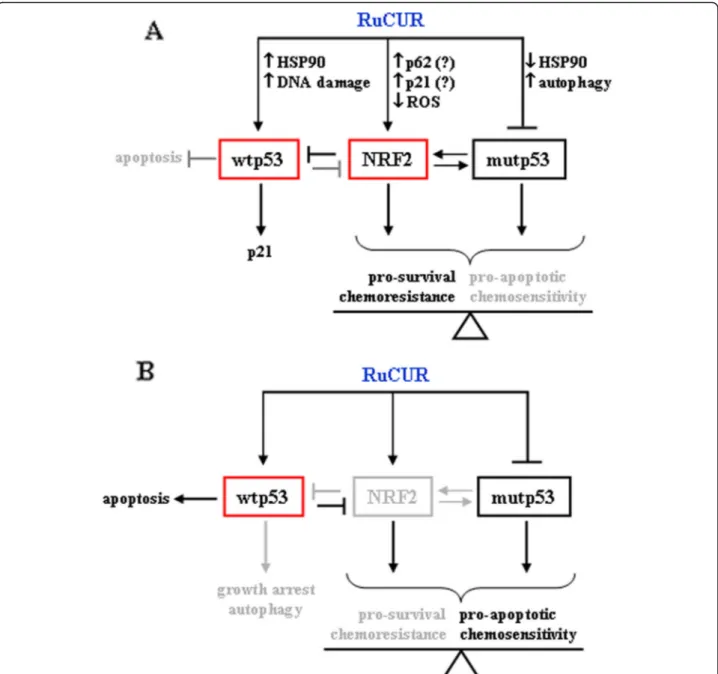
Fig. 4 RuCUR compound induces NRF2 pathway in mutp53-carrying cancer cells. (a) Western blot analysis of the indicated protein was performed in T98 and SKBR3 cells untreated or treated with RuCUR (100μM) for 24 h. Actin was used as protein loading control. Densitometry was performed using ImageJ software. Relative band intensities value were normalized toβ-actin (loading control) and finally quantified with respect to untreated control arbitrarily set to 1.0. (b) Total mRNA was extracted from T98 and SKBR3 cells untreated or treated with RuCUR (100μM) for 24 h. The indicated gene expression was assayed by semi-quantitative polymerase chain reaction (PCR) of reverse-transcribed cDNA. Densitometric analysis was performed using ImageJ software to calculate the gene/28S ratio. Histograms represent the fold increase quantified with respect to controls set to 1.0, ± SD. * (p ≤ 0.01). (c) Oxidant species production in T98 and SKBR3 cells after RuCUR (100 μM) treatment for 16 h evaluated by 2′,7′-dichlorofluorescein diacetate (DC-FDA) staining and assessed by Fluorescence-Activated Cell Sorting (FACS) analysis. Histograms of the mean fluorescence intensity (MFI) are the mean ± SD of three independent experiments. * (p ≤ 0.01). (d) Western blot analysis of the indicated protein levels was performed in T98 and SKBR3 cells untreated or treated with geldanamycin (gelda) (100 nM) for 24 h. The ratio of p53 and HO-1 levels vsβ-actin, following densitometric analysis using ImageJ software, is shown. Representative images are shown
resistance via inhibition of the antioxidant system
(Fig.
8
a, b). However, the lack of p53 did not contribute
to counteract the death resistance even following NRF2
inhibition, underlying the important role of p53
(re)acti-vation for cancer cells demise.
Mutp53 proteins may promote tumor invasion,
metas-tasis and chemoresistance, disclosing mutp53 as a
guard-ian of the cancer cells [
59
]. Restoration of wild-type p53
function prompts the rapid elimination of human
can-cers carrying a functional loss of p53, often by gene
mu-tation [
7
–
9
]. Many p53-reactivating compound have
been shown to induce anti-tumor effects in several
tumor types [
60
]. A number of evidences are now
sup-porting the role of autophagy in mutp53 degradation.
Autophagy is a catabolic process that helps to eliminate
unfolded proteins or damaged organelles, promoting cell
survival [
61
,
62
] even if, in some instances, it may induce
cell death [
63
]. Besides mutp53, other oncogenic
pro-teins are degraded via autophagy including BCR-ABL,
PML-RAR, Ret, KIT and Myc, consistent with the
tumor-suppressive activity of autophagy [
64
,
65
].
There-fore, inducing autophagy may contribute to cancer cell
death and to disable oncogenes but it can also have
pos-sible disadvantages due to autophagy crosstalk with the
immune response which is fundamental for the success
of anticancer therapies [
65
,
66
].. Interestingly, a mutual
interplay between autophagy and mutp53 may occur:
while autophagy degrades mutp53, mutp53 inhibits
au-tophagy counteracting its own elimination as a
self-protecting mechanism, thus promoting chemoresistance
Fig. 5 Targeting NRF2 pathway increases cell death of mutp53-carrying cancer cells. (a) Cell viability was measured by trypan blue exclusion assay in T98 and SKBR3 cells pre-treated with brusatol (100 nM for 4 h) prior to adding RuCUR (100μM) for 24 h and expressed as cell death percentage ± S.D. * (p ≤ 0.01) (single treatments compared to untreated cells). (b) Western blot analysis of the indicated protein in T98 cells pre-treated with brusatol (100 nM for 4 h) prior to adding RuCUR (100μM) for 24 h. Actin was used as protein loading control. The ratio of protein levels vsβ-actin, following densitometric analysis using ImageJ software, is reported. (c) Total mRNA was extracted from T98 cells pre-treated with brusatol (100 nM for 4 h) prior to adding RuCUR (100μM) for 24 h. HO-1 gene expression was assayed by the polymerase chain reaction (PCR) of reverse-transcribed cDNA. Densitometric analysis was performed using ImageJ software to calculate the gene/28S ratio. Histograms represent the fold increase quantified with respect to controls set to 1.0, ± SD. * (p ≤ 0.01). (d) Western blot analysis of the indicated protein in NRF2-silenced and in siRNA-control (ctr) T98G cells, treated with RuCUR (100μM) for 24 h. Actin was used as protein loading control. The ratio of the protein levels vsβ-actin, following densitometric analysis using ImageJ software, is reported
[
67
]. Curcumin has been shown to induce autophagy
[
38
] and to contribute to mutp53 degradation [
16
–
18
,
37
]. In line with this findings, here we found that
RuCUR triggered mutp53 downregulation likely through
autophagy; in addition, it also induced HSP90 reduction,
a molecular chaperone critical for maintaining mutp53
stability [
4
] protecting mutp53 from MDM2-induced
degradation [
68
]. Curcumin has been shown to have an
indirect effect on HSP90 downregulation [
54
], although
the exact mechanism of HSP90 reduction in our setting
needs to be further explored. Interestingly, HSP90
chap-eroning activity can target also wtp53, promoting its
transcriptional activity [
53
], underscoring the two-faced
role of molecular chaperones in p53 activity [
69
]. Thus,
here we found that RuCUR increased HSP90 levels in
wtp53 cells that correlated with p53 activation, although
the mechanism of HSP90/wtp53 regulation has not been
clearly elucidated yet.
Curcumin may induce NRF2 pathway activation [
36
].
NRF2 induces cytoprotective genes that, on one hand,
(See figure on previous page.)
Fig. 6 RuCUR compound induces NRF2 pathway in wtp53-carrying cancer cells. (a) Oxidant species production in RKO and HCT116 cells after RuCUR (100μM) treatment for 16 h evaluated by 2′,7′-dichlorofluorescein diacetate (DC-FDA) staining and assessed by Fluorescence-Activated Cell Sorting (FACS) analysis. Histograms of the mean fluorescence intensity (MFI) represent the mean ± SD. * (p ≤ 0.01). (b) Western blot analysis of the indicated protein levels was performed in (left panel) RKO and HCT116 cells untreated or treated with RuCUR (100μM) for 24 h and in MCF7 and U87 cells (right panel) cells pre-treated with brusatol (100 nM for 4 h) prior to adding RuCUR (100μM) for 24 h. Actin was used as protein loading control. The ratio of the protein levels vsβ-actin, following densitometric analysis using ImageJ software, is reported (c) Western blot analysis of the indicated protein levels in RKO and HCT116 cells pre-treated with brusatol (100 nM for 4 h) prior to adding RuCUR (100μM) for 24 h. Actin was used as protein loading control. The ratio of protein levels vsβ-actin, following densitometric analysis using ImageJ software, is reported. (d) Total mRNA was extracted from RKO and HCT116 cells pre-treated with brusatol (100 nM for 4 h) prior to adding RuCUR (100μM) for 24 h. The indicated gene expression was assayed by the polymerase chain reaction (PCR) of reverse-transcribed cDNA. Densitometric analysis was performed using ImageJ software to calculate the gene/28S ratio. Histograms represent the fold increase quantified with respect to controls set to 1.0, ± SD. * (p ≤ 0.01). (e) Cell viability was measured by trypan blue exclusion assay in RKO and HCT116 cells pre-treated with brusatol (100 nM for 4 h) prior to adding RuCUR (100μM) for 24 h and expressed as cell death percentage ± S.D. * (p ≤ 0.01) (single treatments compared to untreated cells)
Fig. 7 RuCUR compound treatment in p53 null cells. (a) Western blot analysis of the indicated protein levels was performed in HCT116 p53 −/−cells pre-treated with brusatol (100 nM for 4 h) prior to adding RuCUR (100 μM) for 24 h. Actin was used as protein loading control. The ratio of the protein levels vsβ-actin, following densitometric analysis using ImageJ software, is reported. Ctr: positive control for p53. (b) Cell viability was measured by trypan blue exclusion assay in HCT116 p53−/−cells pre-treated with brusatol (100 nM for 4 h) prior to adding RuCUR (100 μM) for 24 h and expressed as cell death percentage ± S.D. * (p ≤ 0.01) (single treatments compared to untreated cells)
protect cells from the oxidative stress [
28
] and, on the
other hand, induce cancer cells proliferation, resistance
to drugs, and apoptosis inhibition [
70
]. NRF2 may also
interact with mutp53 [
56
,
58
] suggesting a criminal
alli-ance to sustain calli-ancer cells [
71
]. Here, we found that
RuCUR induced NRF2 stabilization in all cell lines,
re-gardless of the p53 status. Several mechanisms can
cor-relate with NRF2 stabilization such as ROS inhibition,
increased p62/SQSTM1 [
31
] or p21
Cip1/WAF1levels [
32
].
We found that RuCUR reduced ROS generation in both
wtp53 and mutp53 cells, it also induced p62 in mutp53
cells and p21 in wtp53 cells, although the exact
mechanisms of NRF2 stabilization in our setting need to
be further explored. Interestingly, NRF2 inhibition
in-creased cell death in both wtp53 and mutp53 cells,
underscoring the resistance effect of the antioxidant
sys-tem. At molecular level, NRF2 inhibition further
de-creased mutp53 levels, underscoring the link between
NRF2 and mutp53 to sustain cancer cell survival (Fig.
8
).
NRF2 inhibition in wtp53 cells impaired the
RuCUR-induced p21 and DRAM expression, while increased
Puma expression, suggesting that inhibiting NRF2 could
reestablish p53 apoptotic activity [
48
,
52
]. The
mechan-ism of NRF2 inhibition of the p53 apoptotic function is
Fig. 8 Schematic representation of the effect of RuCUR in cancer cells. (a) RuCUR induced NRF2, degraded mutp53 and induced wtp53, although not the apoptotic activity. (b) NRF2 inhibition restored wtp53 apoptotic activity and further degraded mutp53, unbalancing the cell-fate toward cell death
an interesting filed of study and we hypothesize that
NRF2 and the oxidative system may impair the function
of the p53 apoptotic activator homeodomain-interacting
protein kinase 2 (HIPK2) [
72
–
75
], although further
stud-ies are needed to demonstrate this hypothesis. It is
worth to note that p53 apoptotic activation in turn
in-hibits NRF2 cytoprotective function that may hamper
the p53-induced apoptosis [
76
], in a complex regulatory
loop between NRF2 and p53 (Fig.
8
). Finally, reduction
of the antioxidant system increased cell death only in
mutp53 and wtp53 cells while it did not have this effect
in p53 null cells. These findings underscore the critical
role of p53 for efficient cancer cell death, although the
exact mechanisms need to be further explored.
Conclusions
This study suggests that the overexpression of the
anti-oxidant system is involved in the mechanism of cancer
cell resistance to RuCUR treatment. The elimination of
mutp53 and the activation of wtp53 induced cell death
although only after inhibition of the antioxidant system
cell death greatly improved. Interestingly, p53 null cells
did not undergo increased cell death following inhibition
of the NRF2 pathway, underscoring the important role
of p53 in cancer cell death. These findings also suggest
that the use of phitochemicals that can increase the
anti-oxidant system and, for this reason, being useful
antiag-ing agents, may not be beneficial for the demise of
tumors, despite autophagy induction targeting
onco-genes such as mutp53, underlining the complex
relation-ship between autophagy, antioxidant system and tumor
cell death. In conclusion, these findings may represent a
paradigm for better understanding the complex interplay
between NRF2 and p53 molecular pathways, in order to
design more efficient anticancer therapies. Further
pre-clinical and pre-clinical investigations of RuCUR /NRF2
in-hibition combination should be performed in order to
explore the anticancer activities.
Abbreviations
3D:Three-dimensional; BSA: Bovine serum albumin;
DCFDA: Dichlorofluorescein diacetate; DMEM: Dulbecco modified Eagle’s medium; FBS: Foetal bovine serum; GSH: Glutathione; HIF-1: Hypoxia-inducible factor 1; HIPK2: Homeodomain-interacting protein kinase 2; HO-1: Heme-oxygenase 1; HSP: Heat shock protein; KEAPHO-1: Kelch-like ECH-associated protein 1; MFI: Mean fluorescence intensity; mTOR: Mammalian target of rapamycin; mutp53: Mutant p53; NQO1: NAD(P)H quinone oxidoreductase 1; NRF2: Nuclear factor erythroid 2-related factor 2; PARP: Poly(ADP-ribose) polymerase; PVDF: Polyvinylidene difluoride; ROS: Reactive oxygen species; RT-PCR: Reverse-Transcribed Polymerase Chain Reaction; SD: Standard deviation; SOD: Superoxide dismutase; wtp53: Wild-type p43
Acknowledgements
We thank S. Soddu and A. Verdina for sharing reagents and for critical discussion.
Authors’ contributions
GD and MC designed the study. AG, SB, MSGM, VD, GP, RP, and EG performed experiments. GD, MC, GT, AC, FM analysed data. GD wrote the paper. All authors revised and approved the final manuscript.
Funding
This research was funded by the Italian Association for Cancer Research (AIRC) (Grant IG 2015, Id16742) to GD; by PRIN 2017 (2017K55HLC) and by AIRC (Grant IG 2019, Id23040) to MC; and by the University of Camerino (Fondo di Ateneo per la Ricerca 2018) to FM.
Availability of data and materials
All data generated or analysed during in this study are included in this published article.
Ethics approval and consent to participate Not applicable.
Consent for publication Not applicable. Competing interests
The authors declare that they have no competing interests. Author details
1Department of Research and Advanced Technologies, IRCCS Regina Elena
National Cancer Institute, Rome, Italy.2University“G. D’Annunzio”, School of Medicine, Chieti, Italy.3Department of Medical, Surgical Sciences, and
Biotechnologies, Sapienza University, Latina, Italy.4School of Pharmacy, Chemistry Section, University of Camerino, Camerino Macerata, Italy.
5
Department of Experimental Medicine, Sapienza University, laboratory affiliated to Pasteur Institute Italy Foundation Cenci Bolognetti, Rome, Italy.
6
Department of Surgical Sciences, Sapienza University, Rome, Italy.7Italian medicines agency-Aifa, centralized procedure office, Rome, Italy.
8
Department of Chemistry and Chemical Technologies, laboratory MAT-IN LAB, Calabria University, Rende, Italy.9School of Science and Technology,
Chemistry Section, University of Camerino, Camerino Macerata, Italy.
Received: 4 June 2020 Accepted: 24 June 2020
References
1. Vousden HK, Prives C. Blinded by the light: the growing complexity of p53. Cell. 2009;137:413–31.
2. Muller PA, Vousden KH. Mutant p53 in cancer: new functions and therapeutic opportunities. Cancer Cell. 2014;25:304–17.
3. Lukashchuk N, Vousden KH. Ubiquitination and degradation of mutant p53. Mol Cell Biol. 2007;27:8284–95.
4. Alexandrova EM, Marchenko ND. Mutant p53-heat shock response oncogenic cooperation: a new mechanism of cancer cell survival. Front Endocrinol. 2015;6:53.
5. Alexandrova EM, Moll UM. Depleting stabilized GOF mutant p53 proteins by inhibiting molecular folding chaperones: a new promise in cancer therapy. Cell Death Differ. 2017;24:3–5.
6. Schulz-Heddergott R, Moll UM. Gain-of-function (GOF) mutant p53 are actionable therapeutic target. Cancers. 2018;10:1–16.
7. Di Agostino S, Fontemaggi G, Strano S, Blandino G, D’Orazi G. Targeting mutant p53 in cancer: latest insight. J Exp Clin Cancer Res. 2019;38:290. 8. Wiman KG. Pharmacological reactivation of mutant p53: from protein
structure to the cancer patient. Oncogene. 2010;29:4245–52.
9. Brown CJ, Cheok CF, Verma CS, Lane DP. Reactivation of p53: from peptides to small molecules. Trends Pharmacol Sci. 2011;32:53–62.
10. Puca R, Nardinocchi L, Porru M, Simon AJ, Rechavi G, Leonetti C, et al. Restoring p53 active conformation by zinc increases the response of mutant p53 tumor cells to anticancer drugs. Cell Cycle. 2011;10:1679–89. 11. Margalit O, Simon AJ, Yabukov E, Puca R, Yosepovich A, Avivi C, et al. Zinc
supplement augments in vivo antitumor effect of chemotherapy by restoring p53 function. Int J Cancer. 2012;131:E562–8.
12. Garufi A, Ubertini V, Mancini F, D'Orazi V, Baldari S, Moretti F, et al. The beneficial effect of zinc(II) on low-dose chemotherapeutic sensitivity
involves p53 activation in wild-type p53-carrying colorectal cancer cells. J Exp Clin Cancer Res. 2015;34:87.
13. Garufi A, Trisciuoglio D, Porru M, Leonetti C, Stoppacciaro A, D'Orazi V, et al. A fluorescent curcumin-based Zn(II)-complex reactivates mutant (R175H and R273H) p53 in cancer cells. J Exp Clin Cancer Res. 2013;32:72.
14. Rodriguez OC, Choudhury S, Kolukula V, Vietsch EE, Catania J, Preet A, et al. Dietary downregulation of mutant p53 levels via glucose restriction. Cell Cycle. 2012;23:4436–46.
15. Vakifahmetoglu-Norberg H, Kim M, Xia HG, Iwanicki MP, Ofengeim D, Coloff JL, et al. Chaperone-mediated autophagy degrades mutant p53. Genes Dev. 2013;27:1718–30.
16. Garufi A, Pucci D, D'Orazi V, Cirone M, Bossi G, Avantaggiati ML, et al. Degradation of mutant p53H175 protein by Zn(II) through autophagy. Cell Death Dis. 2014;5:e1271.
17. Garufi A, D'Orazi V, Crispini A, D'Orazi G. Zn(II)-curc targets p53 in thyroid cancer cells. Int J Oncol. 2015;47:1241–8.
18. Thongrakard V, Titone R, Follo C, Morani F, Suksamrarn A, Tencomnao T, et al. Turmeric toxicity in A431 epidermoid cancer cells associates with autophagy degradation of anti-apoptotic and anti-autophagic mutant p52. Phytother Res. 2014;28:1761–9.
19. Garufi A, Pistritto G, Cirone, D’Orazi G. Reactivation of mutant p53 by capsaicin, the major constituent of peppers. J Exp Clin Cancer Res 2013;35:136. 20. Baldari S, Ubertini V, Garufi A, D'Orazi G, Bossi G. Targeting MKK3 as a novel
anticancer strategy: molecular mechanisms and therapeutical implications. Cell Death Dis. 2015;6:e1621.
21. Aggarwal M, Saxena R, Sinclair E, Fu Y, Jacobs A, Dyba M, et al. Reactivation of mutant p53 by a dietary-related compound phenethyl isothiocyanate inhibits tumor growth. Cell Death Diff. 2016;23:1615–27.
22. Foggetti G, Ottaggio L, Russo D, Monti P, Degan P, Fronza G, et al. Gambogic acid counteracts mutant p53 stability by inducing autophagy. Biochi Biophys Acta. 1864;2017:382–92.
23. Garufi A, Federici G, Gilardini Montani MS, Crispini A, Cirone M, D’Orazi G. Interplay between endoplasmic reticulum (ER) stress and autophagy induces mutant p53H373 degradation. Biomolecules. 2020;10.pii:E392. 24. Cordani M, Butera G, Pacchiana R, Donadelli M. Molecular interplay between
mutant p53 proteins and autophagy in cancer cells. Biochim Biophys Acta. 1867;2017:19–28.
25. Cordani M, Pacchiana R, Butera G, D'Orazi G, Scarpa A, Donadelli M. Mutant p53 proteins alter cell secretome and tumor microenvironment:
involvement in cancer invasion and metastasis. Cancer Lett. 2016;376:303–9. 26. Butera G, Brandi J, Cavallini C, Scarpa A, Lawlor RT, Scupoli MT, et al. The
mutant p53-driven secretome has oncogenic functions in pancreatic ductal adenocarcinoma cells. Biomolecules. 2020;10:E884.
27. D'Orazi G, Cirone M. Mutant p53 and cellular stresses pathways: a criminal alliance that promotes cancer progression. Cancers. 2019;11:E614. 28. Vomund S, Schäfer A, Parnham MJ, Brüne B, von Knethen A. Nrf2, the
master regulator of anti-oxidative responses. Int J Mol Sci. 2017;18:2772. 29. Zhu M, Fahl WE. Functional characterization of transcription regulators that
interact with the electrophile response element. Biochem Biophys Res Commun. 2001;289:212–9.
30. de la Rojo VM, Chapman E, Zhang DD. NRF2 and hallmarks of cancer. Cancer Cell. 2018;34:21–43.
31. Jiang T, Harder B, de la Rojo VM, Wong PK, Chapman E, Zhand DD. p62 links autophagy and Nrf2 signaling. Free Rad Biol Med. 2015;88:199–204. 32. Chen W, Sun Z, Wang XJ, Jiang T, Huang Z, Fang D, et al. Direct interaction
between Nrf2 and p21(Cip1/WAF1) upregulates the Nrf2-mediated antioxidant response. Mol Cell. 2009;34:663–73.
33. Wu S, Lu H, Bai Y. Nrf2 in cancers: a double-edged sword. Cancer Med. 2019;8:2252–67.
34. Telkoparan-Akillilar P, Suzen S, Saso L. Pharmacological applications of Nrf2 inhibitors as potential antineoplastic drugs. Int J Mol Sci. 2019;20:E2025. 35. Patel SS, Acharya A, Ray RS, Agrawal R, Raghuwanshi R, Jain P. Cellular and
molecular mechanisms of curcumin in prevention and treatment of disease. Crit Rev Food Sci Nutr. 2020;60:887–939.
36. Ashrafizadeh M, Ahmadi Z, Mohammadinejad R, Farkhondeh T,
Samarghandian S. Curcumin activates the Nrf2 pathway and induces cellular protection against oxidative injury. Curr Mol Med. 2020:20116–33. 37. Madan E, Parker TM, Bauer MR, Dhiman A, Pelham CJ, Nagane M, et al. The
curcumin analog HO-3867 selectively kills cancer cells by converting mutant p53 protein to transcriptionally active wild-type p53. J Biol Chem. 2018;293: 4262–76.
38. Shakeri A, Cicero AFG, Panahi Y, Mohajeri M, Sahebkar A. Curcumin: a naturally occurring autophagy modulator. J Cell Physiol. 2019;234:5643–54. 39. Caruso F, Rossi M, Benson A, Opazo C, Freedman D, Monti E, et al.
Ruthenium-arene complexes of curcumin: X-ray and DFT structure, synthesis and spectroscopic (NMR, ESI-MS) characterization, in vitro antitumor activity and DNA docking studies of (p-cymene) Ru(curcuminato)chloro. J Med Chem. 2012;55:1072–81.
40. Bonfili L, Pettinari R, Cuccioloni M, Cecarini V, Mozzicafreddo M, Angeletti M, et al. Arene-RuII complexes of curcumin exert antitumor activity via proteasome inhibition and apoptosis induction. Chem Med Chem. 2012;7: 2010–20.
41. Antonyan A, Vitali LA, Pettinari R, Marchetti F, Gigliobianco MR, Pettinari C, et al. Evaluation of (arene)Ru(II) complexes of curcumin as inhibitors of dipeptidyl peptidase IV. Biochimie. 2014;99:146–52.
42. Pettinari R, Marchetti F, Condello F, Pettinari C, Lupidi G, Scopelliti R, et al. Ruthenium(II)−Arene RAPTA type complexes containing curcumin and bisdemethoxycurcumin display potent and selective anticancer activity. Organometallics. 2014;33:3709–15.
43. Caruso F, Pettinari R, Rossi M, Monti E, Gariboldi MB, Marchetti F, et al. The in vitro antitumor activity of arene-ruthenium(II) curcuminoid complexes improves when decreasing curcumin polarity. J Inorg Biochem. 2016;162:44–51.
44. Pettinari R, Marchetti F, Pettinari C, Condello F, Petrini A, Scopelliti R, et al. Organometallic rhodium(III) and iridium(III) cyclopentadienyl complexes with curcumin and bisdemethoxycurcumin co-ligands. Dalton Trans. 2015; 44:20523–31.
45. Pettinari R, Condello F, Marchetti F, Pettinari C, Smoleński P, Riedel T, et al. Dicationic ruthenium(II)–arene–curcumin complexes containing methylated 1,3,5-Triaza−7-phosphaadamantane: synthesis, structure, and cytotoxicity. Eur J Inorg Chem. 2017:2905–10.
46. Pettinari R, Marchetti F, Di Nicola C, Pettinari C, Cuccioloni M, Bonfili L, et al. Novel osmium(II)–cymene complexes containing curcumin and
bisdemethoxycurcumin ligands. Inorg Chem Front. 2019;6:2448–57. 47. Ren D, Villeneuve NF, Jiang T, Wu T, Lau A, Toppin HA. Brusatol enhances
the efficacy of chemotherapy by inhibiting the Nrf2-mediated defense mechanism. Proc Natl Acad Sci U S A. 2011;108:1433–8.
48. Garufi A, Traversi G, Gilardini Montani MS, D'Orazi V, Pistritto G, Cirone M, et al. Reduced chemotherapeutic sensitivity in high glucose condition: implication of antioxidant response. Oncotarget. 2019;10:4691–702. 49. Vinci M, Gowan S, Boxall F, Patterson L, Zimmermann M, Court W, et al.
Advances in establishment and analysis of three-dimensional tumor spheroid-based functional assays for target validation and drug evaluation. BMC Biol. 2012;10:29.
50. Baldari S, Di Rocco G, Heffern MC, Su TA, Chang CJ, Toietta G. Effects of copper chelation on BRAFV600E positive colon carcinoma cells. Cancers. 2019;11:E659.
51. Puca R, Nardinocchi L, Starace G, Rechavi G, Givol D, D’Orazi G. Nox1 is involved in p53 deacetylation and suppression of its transcriptional activity and apoptosis. Free Rad Biol Med. 2010;48:1338–46.
52. Garufi A, Pistritto G, Baldari S, Toietta G, Cirone M, D’Orazi G. p53-dependent PUMA to DRAM antagonistic interplay as a key molecular switch in cell-fate decision in normal/high glucose conditions. J Exp Clin Cancer Res. 2017;36:126.
53. Walerych D, Kudla G, Gutkowska M, Wawrzynow B, Muller L, King FW, et al. Hsp90 chaperones wild-type p53 tumor suppressor protein. J Biol Chem. 2004;279:48836–45.
54. Li Y, Zhang D, Xu J, Shi J, Jiang L, Yao N, et al. Discovery and development of natural heat shock protein 90 inhibitors in cancer treatment. Acta Pharm Sin B. 2012;2:238–45.
55. Komatsu M, Kurokawa H, Waguri S, Taguchi K, Kobayashi A, Ichmura Y, et al. The selective autophagy substrate p62 activates the stress response transcription factor Nrf2 through inactivation of Keap1. Nat Cell Biol. 2010; 12:213–23.
56. Lisek C, Campaner E, Ciani Y, Walerych D, Del Sal G. Mutant p53 tunes the NRF2-dependent antioxidant response to support survival of cancer cells. Oncotarget. 2018;9:20508–23.
57. Miyata Y. Hsp90 inhibitor geldanamycin and its derivatives as novel cancer chemotherapeutic agents. Curr Pharm Des. 2005;11:1131–8.
58. Roblat B, Melino G, Knight RA. NRF2 and p53: Januses in cancer? Oncotarget. 2012;3:1272–83.
59. Mantovani F, Collavin L, Del Sal G. Mutant p53 as a guardiano f the cancer cell. Cell Death Diff. 2019;26:199–212.
60. Yu X, Narayanan S, Vazquez A, Carpizo DR. Small molecule compounds targeting the p53 pathway: are we finally making progress? Apoptosis. 2014;19:1055–68.
61. Granato M, Santarelli R, Lotti LV, Di Renzo L, Gonnella R, Garufi A, et al. JNK and macroautophagy activation by bortezomib has a pro-survival effect in primary effusion lymphoma cells. PLoS One. 2013;8:e75965.
62. White E. The role for autophagy in cancer. J Clin Invest. 2015;125:42–6. 63. Doherty J, Baehrcke EH. Life, death and autophagy. Nat Cell Biol. 2018;20:
1110–7.
64. Granato M, Rizzello C, Romeo MA, Yadav S, Santarelli R, D’Orazi G, et al. Concomitant reduction of c-Myc expression and PI3K/AKT/mTOR signaling by quercetin induces a strong cytotoxic effect against Burkitt’s lymphoma. Int J Biochem Cell Biol. 2016;79:393–400.
65. Cirone M, Saveria GMM, Granato M, Garufi A, Faggioni A, D’Orazi G. Autophagy manipulation as a strategy for efficient anticancer therapies: possible consequences. J Exp Clin Cancer Res. 2019;38:262.
66. Masuelli L, Granato M, Benvenuto M, Mattera R, Bernardini R, Mattei M, et al. Chloroquine supplementation increases the cytotoxic effect of curcumin against Her2/neu overepressing breast cancer cells in vitro and in vivo in nude mice while counteracts it in immune competent mice. Oncoimmunol. 2017;6:e1356151.
67. Cordani M, Oppici E, Dando I, Butturini E, Dalla Pozza E, Nadal-Serrano M, et al. Mutant p53 proteins counteract autophagic mechanism sensitizing cancer cells to mTOR inhibition. Mol Oncol. 2016;10:1008–29.
68. Li D, Marchenko ND, Schulz R, Fischer V, Velasco-Hernandez T, Talos F, et al. Functional inactivation of endogenous MDM2 and CHIP by HSP90 causes aberrant stabilization of mutant p53 in human cancer cells. Mol Cancer Res. 2011;9:577–88.
69. Wawrzynow B, Zylicz A, Zylicz M. Chaperoning the guardian of the genome. The two-faced role of molecular chaperones in p53 tumor suppressor action. Biochim Biophys Acta. 2018;2:161–74.
70. DeNicola GM, Karreth FA, Humpton TJ, Gopinathan A, Wei C, Frese K, et al. Oncogene-induced Nrf2 transcription promotes ROS detoxification and tumorigenesis. Nature. 2011;475:106–9.
71. D'Orazi G, Cirone M. Mutant p53 and cellular stress pathways: A criminal alliance that promotes cancer progression. Cancers (Basel). 2019;11:614. 72. Torrente L, Sanchez C, Moreno R, Chowdhry S, Cabello P, Koseki H, et al.
Crosstalk between NRF2 and HIPK2 shapes cytoprotective responses. Oncogene. 2017;36:6204–12.
73. de la Vega L, Grishina I, Moreno R, Kruger M, Braun T, Schmitz ML. A redox-regulated SUMO/acetylation switch of HIPK2 control the survival threshold to oxidative stress. Mol Cell. 2012;46:1–12.
74. Nardinocchi L, Puca R, Givol D, D’Orazi G. HIPK2-a therapeutical target to be (re)activated for tumor suppression: role in p53 activation and HIF-1alpha inhibition. Cell Cycle. 2010;9:1–6.
75. D’Orazi G, Garufi A, Cirone M. NRF2 interferes with HIPK2/p53 activity to impair solid tumors chemosensitivity. IUBMB Life. 2020; in press. 76. Faraonio R, Vergara P, Di Marzo D, Pierantoni MG, Napolitano M, Russo T,
et al. p53 suppresses the Nrf2-dependent transcription of antioxidant response genes. J Biol Chem. 2006;52:39776–84.
Publisher
’s Note
Springer Nature remains neutral with regard to jurisdictional claims in published maps and institutional affiliations.