www.nature.com/scientificreports
The antiplatelet agent revacept
prevents the increase of systemic
thromboxane A
2
biosynthesis
and neointima hyperplasia
Sara Alberti
1,2,8, Qianqian Zhang
3,8, Ilaria D’Agostino
1,2,8, Annalisa Bruno
1,2,
Stefania Tacconelli
1,2, Annalisa Contursi
1,2, Simone Guarnieri
2, Melania Dovizio
1,2,
Lorenza Falcone
2, Patrizia Ballerini
2,4, Götz Münch
5, Ying Yu
6,7& Paola Patrignani
1,2* Neointima hyperplasia is a crucial component of restenosis after coronary angioplasty. We have hypothesized that enhanced generation of platelet-derived thromboxane (TX)A2 in response tovascular damage plays a critical role in neointimal hyperplasia and that antiplatelet agents may mitigate it. In cocultures of human platelets and coronary artery smooth muscle cells (CASMC), we found that platelets induced morphologic changes and enhanced the migration of CASMC. The exposure of platelets to Aspirin [an inhibitor of cyclooxygenase (COX)-1] reduced the generation of TXA2 and prevented the morphological and functional changes induced by platelets in CASMC.
Platelet-derived TXA2 induced COX-2 and enhanced prostaglandin (PG)E2 biosynthesis in CASMC, a
known mechanism promoting neointimal hyperplasia. COX-2 induction was prevented by different antiplatelet agents, i.e., Aspirin, the TP antagonist SQ29,548, or Revacept (a dimeric soluble GPVI-Fc fusion protein). The administration of the novel antiplatelet agent Revacept to C57BL/6 mice, beginning three days before femoral artery denudation, and continuing up to seven days after injury, prevented the increase of the systemic biosynthesis di TXA2 and reduced femoral artery
intima-to-media area and the levels of markers of cell proliferation and macrophage infiltration. Revacept might serve as a therapeutic agent for percutaneous coronary angioplasty and stent implantation.
Percutaneous transluminal coronary angioplasty (PTCA) with or without vascular stenting is commonly used for the treatment of coronary heart disease1.
However, restenosis occurs in 30–50% of patients undergoing simple balloon angioplasty, and in 10–30% of patients who receive an intravascular stent1.
Collagen exposure at the vascular damage site leads to platelet adhesion and aggregation mainly via the Glycoprotein (GP) VI signaling2. Thromboxane (TX)A
2, a primary product of arachidonic acid metabolism in
activated platelets via the cyclooxygenase(COX)-1 pathway, promotes migration and proliferation of vascular smooth muscle cells(VSMC)3–5. TXA
2 operates through the binding to TXA2 receptors (TP)6, which by
cou-pling to Gα 12/13 lead to YAP/TAZ activation; they are the major downstream effectors of the Hippo signaling pathway7. Injury-induced vascular proliferation is reduced in mice lacking the TP receptor or treated with a TP
antagonist8. In patients undergoing PTCA, enhanced systemic biosynthesis of TXA
2 (as assessed by measuring
the urinary levels of the major enzymatic metabolites, TXM) was largely suppressed by low-dose Aspirin9,10, thus
suggesting that this biochemical change reflects platelet activation at the site of vascular damage10.
Restenosis is an excessive response of the coronary artery to damage during angioplasty. It consists of plate-let activation, inflammatory cell recruitment, VSMC proliferation, and migration to the intima. VSMC change
OPEN
1Department of Neuroscience, Imaging and Clinical Science, “G. D’Annunzio” University, Chieti, Italy. 2CAST
(Center for Advanced Studies and Technology) (Ex CeSI-MeT), “G. D’Annunzio” University, Via dei Vestini 31, 66100 Chieti, Italy. 3International Peace Maternity and Child Health Hospital of China Welfare Institution, Shanghai,
China. 4Department of Innovative Technologies in Medicine and Dentistry, “G. D’Annunzio” University,
Chieti, Italy. 5AdvanceCOR GmbH, Martinsried, Germany. 6Shanghai Institute for Biological Sciences, Chinese
Academy of Science, Shanghai, China. 7Department of Pharmacology, School of Basic Medical Sciences, Tianjin
their phenotype from contractile to synthetic, which promotes the extracellular matrix synthesis1. These events
constitute the neointimal hyperplasia, which contributes to postprocedural lumen loss. In animal models, peak neointimal growth is observed 28 days following bare-metal stents placement, whereas in humans, it is identi-fied at 6–12 months11.
COX-2, an inducible enzyme that mediates the generation of prostanoids in inflammation3, plays a role in
restenosis progression12. COX-2 is markedly upregulated in vascular inflammation, such as atherosclerosis and
balloon-injured arteries13,14. Pharmacological inhibition or specific deletion of COX-2 in VSMC reduces
vascu-lar neointima hyperplasia in response to mechanical injury12,14. COX-2 mediates these effects by enhancing the
generation of prostaglandin (PG)E2 that contributes to vascular restenosis pathogenesis through the activation of
the PGE2 receptor subtype EP3α/β and its signaling pathways cAMP/protein kinase A and phosphatidylinositol
3-kinase12. Moreover, genetic disruption of microsomal PGE
2 synthase-1 attenuates neointima formation after
vascular injury15. However, the pharmacological inhibition of vascular COX-2 by the use of selective COX-2
inhibitors (named coxibs) is not recommended in this setting for their cardiovascular hazard16.
In the early phase of restenosis, platelets are the chief cellular players17 through their adhesion to VSMC,
at the site of the vascular damage, facilitated by subendothelial collagen exposure, and to the release of soluble factors, such as lipid mediators derived from arachidonic acid (especially TXA2)18.
Revacept, a novel antiplatelet agent in clinical development, is a soluble form of the platelet GPVI receptor that binds specifically to collagen at vascular damage sites, thus inhibiting platelet adhesion and aggregation without affecting general hemostasis in humans19. Using fluorescence-labeled glycoprotein VI (GPVI)-Fc in
ApoE-deficient [ApoE(−/−)-mice, fed with a 1.25% cholesterol diet over 16 weeks], the mechanical-induced carotid
injury was associated with increased GPVI-Fc-binding to injured carotids compared to intact carotids20.
In cocultures of human platelets and coronary artery smooth muscle cells (CASMC), we addressed the hypothesis that platelet-derived TXA2 is the trigger of the induction of COX-2-derived-PGE2, a pathway involved
in the development of neointimal hyperplasia12. Then, we tested whether Revacept attenuates neointimal
for-mation and prevents enhanced systemic TXA2 biosynthesis in mice after arterial injury. Our findings suggest
that the inflammatory response to vascular damage after coronary angioplasty might be mitigated by avoiding platelet-vessel wall interaction and preventing enhanced TXA2 biosynthesis using Revacept.
Methods
Coculture experiments with human coronary smooth muscle cells (CASMC) and washed
human platelets isolated from venous blood.
CASMC (at passage 5) (Lonza Walkersville Inc, MD, USA) were cultured in flasks coated with collagen type I from rat tail (BD Bioscience Discovery Labware, Bed-ford MA, USA), at 37 °C in a humidified atmosphere of 5% CO2 in air, in the culture system BulletKit (Lonza)[containing SmBM (Basal Medium) and SmGM-2 (Smooth Muscle Cell Growth Medium-2) SingleQuots sup-plements] and 5% fetal bovine serum (FBS). Then, CASMC (0.8 × 10⁵ cells) were seeded in a six-multiwell plate
coated with collagen type I from rat tail in 2 ml of culture system containing 5% FBS. After 48 h (h), the medium was changed with Dulbecco’s modified Eagle’s medium (DMEM) containing 0.75% bovine serum albumin (BSA) (Sigma-Aldrich, Milan, Italy) and 1% polymyxin-B sulphate (Sigma-Aldrich) and human washed plate-lets (0.5 × 10⁸ cells in 100 μl), freshly isolated from concentrated buffy coats (obtained from the blood bank of SS
Annunziata Hospital, Chieti, Italy), as previously described21, were added. This study was carried out following
the recommendations of the Declaration of Helsinki. Healthy volunteers (23–45 years) who had not taken any nonsteroidal antiinflammatory drug (NSAID) in the 2 weeks before blood donation were enrolled. Informed consent was obtained from each subject. All experimental protocols were approved by the Blood Center (Asl2 Lanciano-Vasto-Chieti, Italy), local Ethics Committee of "G. d’Annunzio" University of Chieti-Pescara. Separate experiments (n) were performed using different buffy coats.
Immunofluorescence.
CASMC (0.8 × 10⁵ cells) were cultured alone or cocultured with platelets (0.5 × 10⁸)for 8 h; then, cells were washed twice with phosphate buffer solution (PBS) pH 7.4 and fixed in acetone/metha-nol (40:60) for 20 min at room temperature. Cells were blocked with a filtrated solution of BSA (3%) in PBS for 30 min at room temperature. Cells were incubated overnight at 4 °C with polyclonal antibodies anti-COX-1 or anti-COX-2 (1:200, Cayman Chemical, Ann Arbor, MI, USA) and anti-α-SMA (1:200, Santa Cruz Biotech-nology, Dallas, Texas, USA). Cells were then washed three times with PBS and incubated with the secondary antibodies, donkey anti-rabbit Alexa Fluor 488 and goat anti-mouse Alexa Fluor 546 (1:1000, Life Technologies, Waltham, MA, USA) for 1 h at room temperature. Cells were washed three times with PBS and incubated for 5 min with 4′,6-diamidino-2-phenylindole (DAPI; 300 nM, Sigma Aldrich) to label nuclear DNA. Finally, cells were washed and mounted in slides with Diamond antifade mounting media (Life Technologies). Slides were observed with a Zeiss LSM 510 meta microscope (Carl Zeiss, Jena, Germany), and confocal images were ana-lyzed using Zeiss LSM 5 series software (Carl Zeiss).
Pharmacological treatments in vitro.
The selective COX-1 inhibitor Aspirin (acetylsalicylic acid, ASA, Sigma-Aldrich), the selective COX-2 inhibitor Rofecoxib (Witega Laboratorien, Berlin, Germany), and the TP antagonist SQ 29,548 (Sigma-Aldrich) were dissolved in dimethylsulfoxide (DMSO, Sigma-Aldrich). Rofecoxib (0.3 μM) or SQ 29,548 (10 μM), or DMSO were added to CASMC for 30 min before the addition of platelets. In other experiments, platelets were pretreated with Aspirin (100 μM) or DMSO before the incubation with CASMC; briefly, platelet-rich plasma was incubated with Aspirin (100 µM) or DMSO for 30 min at room tem-perature; then platelets were isolated21, washed twice, and either cultured alone or cocultured with CASMC for20 h. Finally, Revacept19 or vehicle [PBS/4% mannitol/1% sucrose (Sigma Aldrich)] were added to CASMC for
www.nature.com/scientificreports/
CASMC or platelets cultured alone or cocultured were lysed in 1%Triton and 1 mM phenylmethylsulphonyl fluoride (Sigma-Aldrich) and stored at − 80 °C until assayed by Western blot. In some experiments, the super-natants were collected, centrifuged at 700×g for 5 min, and stored at − 80 °C until assayed for prostanoid levels.
Migration assay.
Cell migration of confluent CASMC cultured alone or with platelets, pretreated, or not with Aspirin, was evaluated by scrape wounding assay, as previously described22,23. Briefly, after 20 h ofplatelet-CASMC cocultures, platelets were washed away. Cells, seeded on a 6-multiwell dish, were scraped using a 200 μl pipette tip, simulating a wound, incubated in DMEM 0.75% BSA, and monitored periodically by light micro-scope up to 24 h. Image processing was performed using Image J 1.44 software (NIH, USA), and the percentage of cell-free area and cell covered area were calculated using the analysis particle tool.
Biochemical analyses.
TXB2 and 6-keto-PGF1α [the nonenzymatic hydrolysis product of TXA2 andprostacyclin (PGI2), respectively], PGD2, PGE2, and PGF2α, were measured in cell culture media by previously
described and validated immunoassay techniques23,24.
Western blot analyses.
Cell lysates were mixed with sodium dodecyl sulfate (SDS)(Sigma-Aldrich) sam-ple buffer and heated to 95 °C for 5 min. Proteins (15 μg) were analyzed by SDS polyacrylamide gel electropho-resis (SDS-PAGE)21,23. Anti-COX-2, anti-COX-1 polyclonal antibodies (1:1000, Cayman Chemical), GAPDHpolyclonal antibody (1:1000, Santa Cruz Biotechnology), or anti-β-actin polyclonal antibody (1:1000, Santa Cruz Biotechnology) were used21,23. Quantification of optical density (OD) of different specific bands was
car-ried out using Alliance 4.7 and Alliance 1 D software (UVITEC, Cambridge, UK) and normalized to the OD of β-actin or GAPDH.
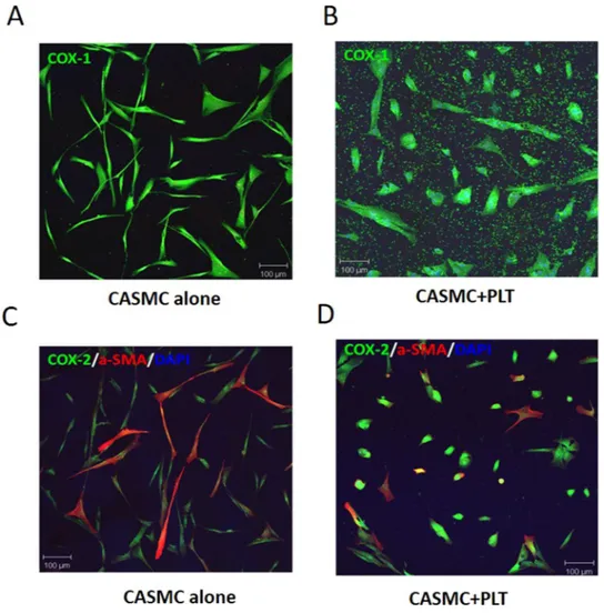
Figure 1. Effects of platelets on phenotypic modulation of CASMC. Human CASMC (0.8 × 10⁵ cells) were
cultured alone or cocultured with human platelets (0.5 × 10⁸). COX-1 (green) (A,B) or COX-2 (green) and
Animals.
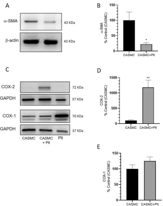
Male C57BL/6 mice (8–10 weeks old, n = 20) were purchased from Shanghai SLAC Laboratory Animal Co., Ltd. (Shanghai, China). All animals were maintained and used according to the veterinary guide-lines of the Institutional Animal Care and Use Committee of the Institute for Nutritional Sciences, Chinese Academy of Sciences, Shanghai (China). Experimental protocols were approved by the Institutional Animal Care and Use Committee of the Institute for Nutritional Sciences, Chinese Academy of Sciences, Shanghai (China). Every effort was made to minimize the number of animals used and any pain or discomfort experi-enced by the animals.Figure 2. Effects of platelets on α-SMA, COX-2, and COX-1 protein levels in CASMC cultured alone or
cocultured with platelets. (A) α-SMA levels were evaluated by Western blot in CASMC cultured alone or cocultured with platelets for 24 h; (B) the protein bands were quantified, and the optical density (OD) was calculated using laser densitometry and normalized to the OD of β-actin; data are reported as % of control (CASMC cultured alone), mean ± SEM, n = 5 (separate experiments); *P < 0.05 versus CASMC cultured alone (using Student’s t-test). (C–E) COX-2 and COX-1 levels were evaluated by Western blot in platelets (Plt) or CASMC cultured alone or in CASMC cocultured with platelets (CASMC + Plt) for 20 h; (D,E) the protein bands were quantified and OD was calculated and normalized to the OD of GAPDH, data are reported as % of control (CASMC cultured alone), mean ± SEM, n = 5 and n = 3 (separate experiments), respectively; (D) **P < 0.01 versus CASMC cultured alone (using Student’s t-test).
www.nature.com/scientificreports/
In vivo study design and surgical procedures.
The femoral artery wire injury model was described previously elsewhere25,26. A contralateral (left femoral arteries) sham surgery was used as a control byperform-ing the protocol without the wire injury. Right femoral arteries were exposed by blunt dissection and subjected to denudation. Briefly, arteries were exposed by blunted dissection, dilated by topical application of one drop of 1% lidocaine hydrochloride (Sigma-Aldrich) and transverse arteriotomy was performed in the muscular branch; a straight spring wire (0.38 mm in diameter, COOK, Bloomington, IN, USA) was carefully inserted into the femoral artery for more than 5 mm toward the iliac artery and left in place for 1 min to denude and dilate the artery. Then, the wire was removed, and blood flow in the femoral artery was restored. Animals were randomly assigned to two different treatment groups. One group (n = 10) received Revacept, the protein obtained by fusing the extracellular domain of GPVI with the human immunoglobulin (IgG) Fc domain19 (at the dose of 2 mg/kg/
day), via tail-vein injection, for three days before bilateral femoral artery denudation was performed. The treat-ment was continued up to day 10. Control group mice (n = 10) were treated with recombinant human IgG1 Fc (at the dose of 2 mg/kg/day) (Sino Biological Inc., China) via tail-vein injection. The compounds were dissolved in a solution of PBS/mannitol(4%)/sucrose(1%). The concentration of Revacept and Fc were selected based on previously published studies27. For all surgical procedures, the animals were ventilated with isoflurane and
oxygen to maintain anesthesia throughout the experiment, and anesthetic depth was assessed by observing the reflex responses to paw pinches. Histology and immunohistochemistry were performed on specimens collected at day 32 (i.e., 28 days after surgery). Moreover, 24 h-urine collections were performed on day 0 (baseline), day 3, day 7, and 32 (i.e., before injury and 3 and 28 days after injury, respectively) for the assessment of 11-dehydro-TXB2 (TXM) levels, as a marker of the systemic TXA2 biosynthesis, mainly derived from the activity of platelet
COX-128,29.
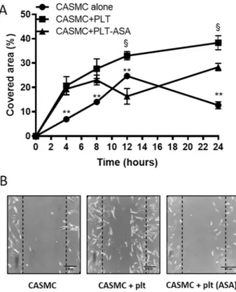
Figure 3. Effects of platelets on CASMC migration. (A) Human CASMC (0.8 × 10⁵ cells) were cultured alone
or cocultured with human platelets (0.5 × 10⁸) (pretreated or not with Aspirin, ASA, 100 µM) for 20 h and cell
migration of confluent cells was evaluated at different time-points (up to 24 h) by scrape wounding assay, using an inverted microscope and a digital camera (original magnification × 10), the dashed lines indicate the starting position of the cell migration occurring toward the center of the wound; % of covered area values are reported as mean ± SEM, n = 3 (separate experiments), the scale bar is of 100 µm. In each experimental condition, % covered area was significantly different (P < 0.01) at each time-point versus time 0 (using two-way ANOVA, Dunnett’s multiple comparisons test), not shown. At 4 h, **P < 0.01, CASMC alone versus CASMC + PLT and CASMC + PLT-ASA; at 8 h, **P < 0.01, CASMC alone versus CASMC + PLT and CASMC + PLT-ASA; at 12 h, **P < 0.01, CASMC alone versus CASMC + PLT and CASMC + PLT-ASA, §P < 0.01, CASMC + PLT versus
CASMC + PLT-ASA; at 24 h, **P < 0.01, CASMC alone versus CASMC + PLT and CASMC + PLT-ASA, §P < 0.01,
CASMC + PLT versus CASMC + PLT-ASA (using two-way ANOVA, Tukey’s multiple comparisons test). (B) The results of cell migration obtained at 24 h are shown.
Histology and immunohistochemistry.
At the end of the in vivo experiments (i.e., 28 days after sur-gery), all animals were euthanized and intracardially perfused with 10 ml of 4% paraformaldehyde (PFA, Sigma Aldrich). Femoral arteries were fixed with 4% PFA for 10–15 min and then embedded in paraffin. Paraffin-embedded Sections (7 μm) were stained with hematoxylin and eosin (HE, Sigma-Aldrich), and the intima-to-media area (I/M) ratio was assessed as previously described12. Briefly, the intimal area is the area encircled by theinternal elastic lamina minus lumen area. The medial area is calculated as the area encircled by the external elas-tic lamina minus intima area. The intima-to-media ratio was calculated as the intimal area divided by the medial area. For immunohistochemistry, tissue sections were incubated with primary antibodies against Ki67 (1:500, Abcam, Cambridge, UK), CD68 (1:200, AbD Serotec, Kidlington, UK), followed by incubation with horserad-ish peroxidase(HRP)–conjugated secondary antibody [anti-rabbit IgG (Cell Signaling Technology, Leiden, The Netherlands)1:2000; anti-rat IgG (ProteinTech Group, Rosemont, IL, USA) 1:2000]. After three washings with PBS, samples underwent 3′-Diaminobenzidine (DAB, Sigma-Aldrich) staining, hematoxylin restaining, dehy-dration, and cover glass mounting. Slides were observed with a Zeiss Axiovision microscope (Carl Zeiss) and Image J 1.44 software was used for the analysis.
Assessment of urinary levels of TXM.
Two-hundred μl of 24 h-urine collection samples were extracted, and the levels of the TXM, i.e., an enzymatic urinary metabolite of TXB2, were measured by previously validatedimmunoassay techniques28,29. The urinary TXM levels were corrected for creatinine excretion and reported as
ng/mg of creatinine.
Statistical analysis.
All values were reported as mean ± SD (standard deviation) in the text, while shown as mean ± SEM (standard error of the mean) in the Figures; the n values (indicating the number of separate experiments) were reported in Figure legends. Statistical analysis among two groups was performed by Student’s t-test; for 3 and more groups, ANOVA (one-way or two-way) or Mixed-effects analysis for repeated measures (when some values were missed) were carried out using GraphPad PRISM software (version 8.00 for Windows; GraphPad, San Diego, CA, USA). P values < 0.05 were considered statistically significant.Results
Effects of platelets on CASMC phenotype and morphology in vitro.
The changes induced by plate-lets on the morphology and the phenotype of the CASMC were assessed using confocal microscopy by the labe-ling of COX-1(a constitutive protein) and α-SMA [typical of a contractile phenotype]30. CASMC cultured aloneappeared as spindle-shaped cells (Fig. 1A). The co-incubation with platelets led to epithelioid cell morphology, yielding a cobblestone pattern31 (Fig. 1B) associated with the downregulation of α-SMA vs. CASMC cultured
alone (Fig. 1C,D and Supplementary Fig. 1); these changes describe the induction of the synthetic phenotype of vascular smooth muscle cells32. COX-2 expression was induced in CASMC cocultured with platelets (Fig. 1D
and Supplementary Fig. 1). The downregulation of α-SMA and upregulation of COX-2 induced by platelets in CASMC were confirmed by Western blot analysis (Fig. 2A–D). In contrast, CASMC COX-1 expression was not affected by their coculture with platelets (Fig. 2C,E).
Platelets enhanced the migratory capacity of CASMC.
Cell migration of CASMC cultured alone or with platelets for 20 h was evaluated by scrape wounding assay23. As shown in Fig. 3A,B, platelets enhanced themigration of CASMC. CASMC cultured with platelets that were pretreated with Aspirin (to cause a selective inhibition of platelet-COX-1 activity and TXA2 generation) showed a significantly lower migratory capacity
versus CASMC cultured with platelets untreated with Aspirin, at 8–24 h (Fig. 3A,B).
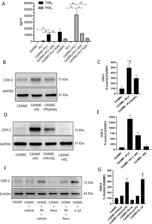
Prostanoid biosynthesis in CASMC-platelet cocultures.
In platelets cultured alone, TXB2 was theprimary product of arachidonic acid metabolism (1014 ± 817 pg/ml) (Fig. 4A) while PGE2, PGF2α, and PGD2
were minor products of COX-1 activity (all < 250 pg/ml, not shown). In CASMC cultured alone, PGE2 (Fig. 4A)
and 6-keto-PGF1α (not shown) were the most abundant prostanoids (3903 ± 3335 and 1310 ± 1271 pg/ml,
respec-tively). In the coculture of platelets and CASMC, PGE2 and TXB2 were significantly enhanced (Fig. 4A).
6-keto-PGF1α did not significantly change in CASMC-platelet cocultures versus CASMC cultured alone (not shown). To
verify the COX-1 or COX-2 origin of the increased biosynthesis of TXB2 and PGE2 in the coculture of CASMC
and platelets, we assessed the impact of the exposure of platelets to Aspirin which was washed away before the incubation with CASMC (to target only platelet COX-1) or Rofecoxib (a selective COX-2 inhibitor). As shown in Fig. 4A, the increase of TXB2 detected in platelet-CASMC cocultures was entirely prevented by the
treat-ment of platelets with Aspirin, suggesting that enhanced TXB2 derived from platelets. This is further sustained
by Rofecoxib inability to affect TXB2 production in the coculture (Fig. 4A). Rofecoxib completely prevented
the increased production of PGE2 measured in platelet-CASMC cocultures, implying that COX-2 induction in
CASMC played a central role in enhanced PGE2 biosynthesis (Fig. 4A). PGE2 levels detected in the coculture
medium were significantly reduced when aspirinated platelets were incubated with CASMC (Fig. 4A).
Effect of aspirin on platelet-induced COX-2 expression in CASMC.
As shown in Fig. 4B,C, the pre-treatment of platelets with Aspirin attenuated the capacity of platelets to induce COX-2 in CASMC. These results suggest that platelets induced COX-2-dependent PGE2 via an Aspirin sensitive mechanism.Effect of TXA
2receptor (TP) blockage on COX-2 induction in platelet-CASMC coculture.
Wewww.nature.com/scientificreports/
Figure 4. Effects of platelets on prostanoid generation and COX-2 expression of CASMC. (A) Levels of
PGE2 and TXB2 in the conditioned medium of CASMC and platelets cultured alone, or cocultured for 20 h,
assessed by immunoassays; the effect of Rofecoxib (Rofe, 0.3 µM) or aspirinated platelets (ASA, 100 µM was added during platelet isolation and then washed away) was assessed. **P < 0.01, *P < 0.05; n = 3–4 (separate experiments) (using one-way ANOVA, Tukey’s multiple comparisons test). (B,E) The effect of ASA (100 µM) or of the TP antagonist SQ 29,548 (SQ, 10 µM) on COX-2 expression in cocultures of CASMC and platelets, was assessed by Western Blot. (F,G) CASMC were cultured alone or cocultured with platelets in the presence of Revacept (Reva, 40 µg/ml) or vehicle (PBS/4% mannitol/1% sucrose) and COX-2 levels were analyzed by Western blot; interleukin (IL)-1β was used as positive control. (C,E,G) COX-2 protein bands were quantified, and the optical density (OD) was calculated using laser densitometry and normalized to the OD of GAPDH or β-actin; data are reported as % of control (CASMC cultured alone), mean ± SEM. (C) **P < 0.01 versus CASMC and §P < 0.05 versus CASMC + PLT(ASA), n = 3 (separate experiments); (E) **P < 0.01 versus all other conditions,
n = 4 (separate experiments); (G) **P < 0.01 versus all other conditions, except CASMC-IL1β, §P < 0.01 versus all
other conditions, except CASMC-vehicle-PLT, n = 3 (separate experiments); all analyses were conducted using one-way ANOVA and Tukey’s multiple comparisons test.
selective TP antagonist SQ 29,548. As shown in Fig. 4D,E, TP blockage mitigated the induction of COX-2 in CASMC by the interaction with platelets. These results show that platelet-derived TXA2 is the trigger of COX-2
induction in CASMC.
Effect of the antiplatelet agent Revacept on COX-2 induction in CASMC-platelet
cocul-tures.
Vascular smooth muscle cells synthesize and secrete collagen33, contributing to the interaction withplatelets and their activation. Thus, we tested whether Revacept, which affects the binding of platelets to collagen binding sites19, may prevent the induction of COX-2 in CASMC. As shown in Fig. 4F and G, Revacept mitigated
platelet capacity to induce COX-2 in CASMC.
Revacept prevented the increase of systemic TXA
2biosynthesis in response to injury in
mice.
We used the femoral artery wire injury model25,26 in C57BL/6 mice to verify the occurrence of plateletactivation in response to vascular damage by assessing the urinary levels of TXM, an enzymatic metabolite of TXB2, that is a marker of the systemic biosynthesis of TXA2 mainly derived from activated platelets28,29.
Moreo-ver, we studied whether the administration of Revacept (2 mg/kg/day) to mice prevented the increase of TXA2
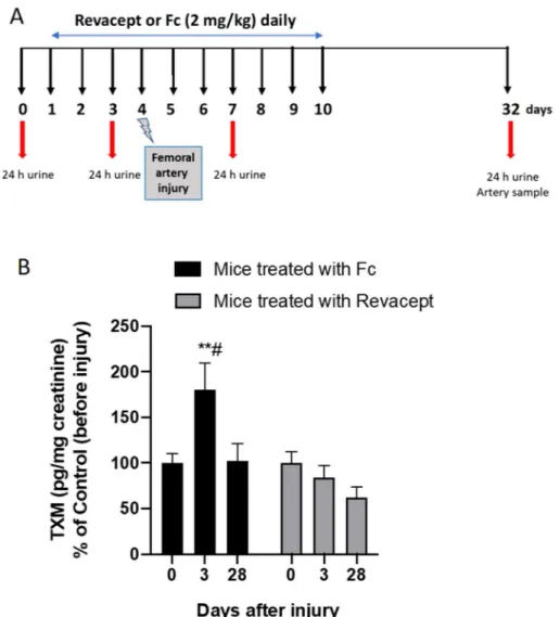
biosynthesis in response to vascular injury as compared with mice treated with recombinant human IgG1 Fc (2 mg/kg/day) (control group). The experimental design of the study is reported in Fig. 5A.
At baseline, urinary TXM levels were comparable in the two groups of mice (Fc-control: 1.2 ± 0.6 ng/mg creatinine; Revacept: 1.3 ± 0.5 ng/mg creatinine; n = 10) (not shown). As shown in Fig. 5B, in mice treated with
Figure 5. Effects of Revacept administration on urinary levels of TXM in mice in response to injury. (A) Study
design of Revacept or Fc-control (2 mg/kg/day) administration to mice using a model of neointima formation caused by the damage of femoral arteries via transluminal wire injury. (B) Urinary levels of TXM were assessed at different time-points, before (0) and after vascular injury (3 and 28 days) in mice treated with Fc control or Revacept (2 mg/kg/day); data are expressed as mean ± SEM, n = 9–10; **P < 0.01 versus the other Fc conditions,
#P < 0.05 versus the conditions of Revacept after injury (using mixed-effects analysis for repeated measures data
www.nature.com/scientificreports/
Fc-control, the urinary levels of TXM significantly (P < 0.05) increased at 3 days after injury versus the values detected before vascular damage. At 28 days after injury (remodeling phase), TXM values were comparable to those measured at pre-injury condition (Fig. 5B). These results show that platelet activation occurs in response to damage because of the endothelial denudation and subintimal component exposure. In contrast, in the remod-eling phase, platelet function returns to the baseline condition.
The administration of Revacept, 2 mg/kg daily, prevented the increase of TXM levels (P < 0.05) versus Fc control at three days after vascular injury (Fig. 5B).
Revacept reduced the expression of markers of vascular neointima proliferation and
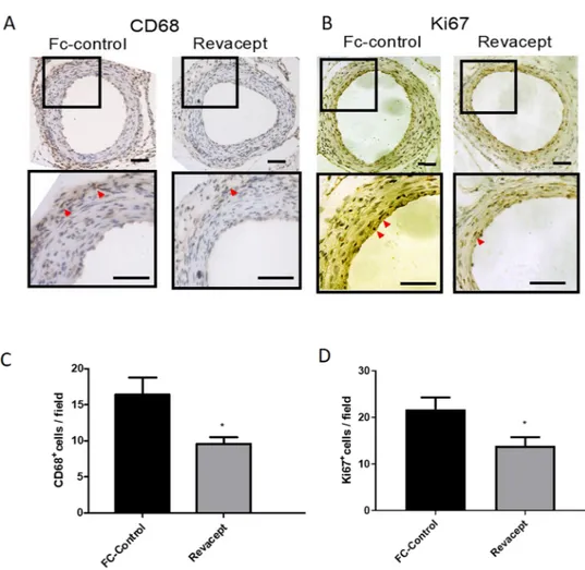
mac-rophage infiltration.
In the remodeling phase (at 28 days after injury), we studied the expression levels of proteins, which are markers of macrophages infiltration and cell proliferation, such as CD68 and Ki67, respec-tively, in paraffin-embedded section of femoral arteries. The vascular injury was associated with an increased expression of CD68 (Fig. 6A,C) and Ki67 (Fig. 6B,D). The administration of Revacept mitigated the enhanced expression of these markers (Fig. 6A–D).Altogether these results show that the inhibition of platelet activation in response to vascular injury by Reva-cept translates into the prevention of the proliferative and inflammatory events which feature the remodeling phase.
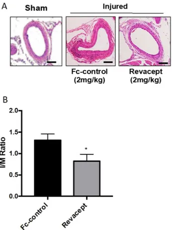
Revacept reduced vascular neointima formation in response to injury in mice.
In this model of neointima hyperplasia induced by mechanical damage, we evaluated the effect of the administration of Revacept on the arterial media thickness in paraffin-embedded sections of femoral arteries. As reported in Fig. 7A and B, 28 days after arterial injury, Revacept caused a significant (P < 0.05) average reduction of I/M ratio by 41.36% versus the Fc-control group.Figure 6. Effects of Revacept administration to mice on CD68 and Ki67 expression in femoral artery sections.
(A,B) Immunohistochemistry of femoral artery sections to assess CD68 and Ki67 at 28 days after surgery in mice treated with Revacept or Fc. (C,D) CD 68+ and Ki67+ cells per field are presented as mean ± SEM, n = 4
Discussion
Platelet activation represents an early response to vascular damage34. Platelets adhere to extracellular matrix
proteins exposed in the injured vasculature and release numerous soluble mediators, including TXA2. It acts
in an autocrine or paracrine manner to stimulate adjacent platelets, thus generating more TXA2 and
amplify-ing other agonist action6. TXA
2 also enhances lymphocyte and macrophage functions35–37 and stimulates the
biosynthesis of extracellular matrix proteins38. For all these actions, TXA
2 might play a crucial role in initiating
and accelerating intimal hyperplasia development.
In a femoral artery wire injury model25,26 in C57BL/6 mice, we assessed the time-course of the systemic
bio-synthesis of TXA2 by measuring the urinary levels of TXM, an enzymatic metabolite of TXB2, which is mainly
derived from activated platelets28,29. Our results show a rapid increase in the systemic biosynthesis of TXA
2 in
response to vascular damage, which returned to pre-injury values at a later phase on neointimal hyperplasia (i.e., 28 days after injury). The enhanced generation of platelet TXA2 may induce numerous cellular events that
contribute to neointimal hyperplasia. By performing coculture experiments between platelets and CASMC, we have shown that platelet-derived TXA2 contributes to COX-2 induction and enhanced PGE2 generation by
CASMC. We exploited Aspirin capacity to cause an irreversible inhibition of platelet COX-1-dependent TXA2
generation, persisting when the drug is washed away. The incubation of CASMC with Aspirin-treated platelets mitigated the induction of COX-2. Comparable results were obtained by TP blockage.
Zhang et al.12 have previously shown that COX-2-derived PGE
2 promotes vascular neointimal hyperplasia in
response to mechanical injury. Moreover, COX-2-derived PGE2 regulates polarization and directional migration
of vascular smooth muscle cells via the activation of the PGE2 receptor subtype EP3α/β12. Here, we show that
platelets enhanced the migration of CASMC; this response was mitigated by the selective inhibition of platelet COX-1 by Aspirin.
Collectively these results suggest that platelets are activated by the interaction with vascular smooth muscle cells and extracellular matrix proteins, such as collagen, and release TXA2, which contributes to enhanced
COX-2-dependent PGE2 biosynthesis. Our findings imply the effectiveness of pharmacological inhibition of platelet
TXA2 biosynthesis by Aspirin to restrain the development of injury-induced vascular neointimal hyperplasia.
However, the results of clinical trials with Aspirin suggest a small reduction in the restenosis after coronary angioplasty but a significant reduction in the frequency of ischemic complications39. The weakness of these
studies is that different doses of Aspirin were used; furthermore, the drug capacity to cause a virtually complete suppression of platelet COX-1-dependent TXB2 biosynthesis was not verified40. The possible beneficial effect of
Figure 7. Effects of Revacept administration neointima formation following vascular injury in mice. (A)
Hematoxylin and eosin staining of cross-sections of injured femoral arteries from mice treated with Fc-control or Revacept (2 mg/kg/day), harvested at 4 weeks after transluminal wire injury. (B) Quantification of intima-to-media (I/M) ratio, data are reported as mean ± SEM, n = 6–10; *P < 0.05 versus Fc-control (using Student’s t-test).
www.nature.com/scientificreports/
low-dose Aspirin in patients undergoing percutaneous coronary intervention through the inhibition of vascular inflammation triggered by activated platelets remains to be verified in appropriate clinical studies.
Aspirin exerts antiplatelet effects by inhibiting TXA2‐dependent aggregation responses and amplifying these
responses by released ADP41. Conversely, Revacept combines inhibitory effects on both collagen-mediated
plate-let adhesion and subsequent aggregation at the site of vascular injury19. These concomitant inhibitory effects on
platelet function by Revacept are proper to limit the consequences of vascular damage. We show that the adminis-tration of Revacept mitigated vascular neointima hyperplasia in a femoral artery injury mouse model. This effect was associated with reduced vascular expression of markers of macrophages infiltration and cell proliferation. Revacept administration prevented the rapid increase of the systemic biosynthesis of TXA2 after vascular injury
derived mainly from platelets9,10. These data support the hypothesis that platelet activation in response to vascular
injury represents an early event contributing to the inflammatory response involved in vascular remodeling. Interestingly, the interaction of platelets with CASMC in vitro led to morphological changes typical of a synthetic phenotype with downregulation of α-SMA and upregulation of COX-2. These changes were associated with the enhanced migratory capacity of CASMC. Revacept prevented the induction of COX-2 by the interac-tion of platelets with CASMC.
Collectively our findings sustain the distinct roles of platelets beyond their fundamental participation in primary hemostasis. As previously shown in cocultures with myofibroblasts, platelet-derived TXA2 induces
mor-phological changes and enhances their capacity to proliferate and migrate, thus contributing to tissue fibrosis23.
Moreover, we have shown that the interaction of platelets with colon cancer cells translates into the overex-pression of COX-2, a hallmark of malignancy, and the induction of marker genes of epithelial-mesenchymal transition21. Inhibition of platelet-cancer cell interactions by Revacept prevented platelet-induced COX-2
induc-tion and changes of epithelial-mesenchymal transiinduc-tion markers21.
The main limitation of the present study is that the effect of Revacept on neointimal formation was not compared with Aspirin given alone or co-administered with Revacept. Low-dose Aspirin could cause additional efficacy by preventing the amplification of platelet response induced by other platelet agonists, in addition to collagen. This is an important point that needs to be explored in a specific study.
In conclusion, we show that Revacept, an inhibitor of the binding of platelet collagen receptors (mainly GPVI) to collagen exposed in areas of damaged endothelium19, constrains the release of TXA
2 from activated platelets.
These distinct effects played by Revacept make it a promising therapeutic strategy to prevent restenosis in patients with coronary artery disease treated with percutaneous transluminal coronary angioplasty and stent implantation. Importantly, Revacept was found to affect platelet function in the absence of bleeding complications19. A phase
II randomized, double blind trial (www.clini caltr ials.gov; NCT03312855) is ongoing to assess the efficacy and safety of Revacept in patients undergoing elective percutaneous coronary intervention (ISAR-PLASTER Trial42.
Received: 27 April 2020; Accepted: 13 November 2020
References
1. Weintraub, W. S. The pathophysiology and burden of restenosis. Am. J. Cardiol. 100, 3K-9K (2007).
2. Nieswandt, B. & Watson, S. P. Platelet-collagen interaction: Is GPVI the central receptor? Blood 102, 449–461 (2003). 3. Ricciotti, E. & FitzGerald, G. A. Prostaglandins and inflammation. Arterioscler. Thromb. Vasc. Biol. 31, 986–1000 (2011). 4. Hanasaki, K., Nakano, T. & Arita, H. Receptor-mediated mitogenic effect of thromboxane A2 in vascular smooth muscle cells.
Biochem. Pharmacol. 40, 2535–2542 (1990).
5. Yokota, T. et al. Thromboxane A2 receptor stimulation promotes closure of the rat ductus arteriosus through enhancing neointima formation. PLoS ONE 9, e94895 (2014).
6. Smyth, E. M. Thromboxane and the thromboxane receptor in cardiovascular disease. Clin. Lipidol. 5, 209–219 (2010). 7. Feng, X. et al. Thromboxane A2 activates YAP/TAZ protein to induce vascular smooth muscle cell proliferation and migration. J.
Biol. Chem. 291, 18947–18958 (2016).
8. Cheng, Y. et al. Role of prostacyclin in the cardiovascular response to thromboxane A2. Science 296, 539–541 (2002).
9. Ciabattoni, G. et al. Aspirin, but not heparin, suppresses the transient increase in thromboxane biosynthesis associated with cardiac catheterization or coronary angioplasty. J. Am. Coll. Cardiol. 21, 1377–1381 (1993).
10. Braden, G. A., Knapp, H. R. & FitzGerald, G. A. Suppression of eicosanoid biosynthesis during coronary angioplasty by fish oil and aspirin. Circulation 84, 679–685 (1991).
11. Virmani, R., Kolodgie, F. D., Farb, A. & Lafont, A. Drug eluting stents: Are human and animal studies comparable? Heart 89, 133–138 (2003).
12. Zhang, J. et al. Cyclooxygenase-2-derived prostaglandin E2 promotes injury-induced vascular neointimal hyperplasia through the
E-prostanoid 3 receptor. Circ. Res. 113, 104–114 (2013).
13. Grosser, T., Yu, Y. & Fitzgerald, G. A. Emotion recollected in tranquility: lessons learned from the COX-2 saga. Annu. Rev. Med. 61, 17–33 (2010).
14. Yang, H. M. et al. Celecoxib, a cyclooxygenase-2 inhibitor, reduces neointimal hyperplasia through inhibition of Akt signaling.
Circulation 110, 301–308 (2004).
15. Wang, M. et al. Microsomal prostaglandin E2 synthase-1 modulates the response to vascular injury. Circulation 123, 631–639 (2011).
16. Grosser, T., Fries, S. & FitzGerald, G. A. Biological basis for the cardiovascular consequences of COX-2 inhibition: therapeutic challenges and opportunities. J. Clin. Investig. 116, 4–15 (2006).
17. Chandrasekar, B. & Tanguay, J. F. Platelets and restenosis. Am. Coll. Cardiol. 35, 555–562 (2000).
18. Kearney, D., Byrne, A., Crean, P., Cox, D. & Fitzgerald, D. J. Optimal suppression of thromboxane A(2) formation by aspirin dur-ing percutaneous transluminal coronary angioplasty: no additional effect of a selective cyclooxygenase-2 inhibitor. J. Am. Coll.
Cardiol. 43, 526–531 (2004).
19. Ungerer, M. et al. Novel antiplatelet drug revacept (Dimeric Glycoprotein VI-Fc) specifically and efficiently inhibited collagen-induced platelet aggregation without affecting general hemostasis in humans. Circulation 123, 1891–1899 (2011).
20. Schönberger, T. et al. The immunoadhesin glycoprotein VI-Fc regulates arterial remodelling after mechanical injury in ApoE-/- mice. Cardiovasc. Res. 80, 131–137 (2008).
21. Dovizio, M. et al. Pharmacological inhibition of platelet-tumor cell cross-talk prevents platelet-induced overexpression of cyclooxy-genase-2 in HT29 human colon carcinoma cells. Mol. Pharmacol. 84, 25–40 (2013).
22. Terzuoli, E. et al. Bradykinin B2 receptor contributes to inflammatory responses in human endothelial cells by the transactivation of the fibroblast growth factor receptor FGFR-1. Int. J. Mol. Sci. 6, 9pii:E2638 (2018).
23. Sacco, A. et al. Platelet-specific deletion of cyclooxygenase-1 ameliorates dextran sulfate sodium-induced colitis in mice. J.
Phar-macol. Exp. Ther. 370, 416–426 (2019).
24. Patrignani, P. et al. Biochemical and pharmacological characterization of the cyclooxygenase activity of human blood prostaglandin endoperoxide synthases. J. Pharmacol. Exp. Ther. 271, 1705–1712 (1994).
25. Sata, M. et al. A mouse model of vascular injury that induces rapid onset of medial cell apoptosis followed by reproducible neoin-timal hyperplasia. J. Mol. Cell Cardiol. 32, 2097–2104 (2000).
26. Roque, M. et al. Mouse model of femoral artery denudation injury associated with the rapid accumulation of adhesion molecules on the luminal surface and recruitment of neutrophils. Arterioscler. Thromb. Vasc. Biol. 20, 335–342 (2000).
27. Ungerer, M. et al. The GPVI–Fc fusion protein revacept reduces thrombus formation and improves vascular dysfunction in ath-erosclerosis without any impact on bleeding times. PLoS ONE 8, e71193 (2013).
28. Ciabattoni, G. et al. Fractional conversion of thromboxane B2 to urinary 11-dehydrothromboxane B2 in man. Biochim. Biophys. Acta 992, 66–70 (1989).
29. Catella, F. & FitzGerald, G. A. Paired analysis of urinary thromboxane B2 metabolites in humans. Thromb. Res. 47, 647–656 (1987). 30. Louis, S. F. & Zahradka, P. Vascular smooth muscle cell motility: From migration to invasion. Exp. Clin. Cardiol. 15, e75-85 (2010). 31. Li, S. et al. Innate diversity of adult human arterial smooth muscle cells: cloning of distinct subtypes from the internal thoracic
artery. Circ. Res. 89, 517–525 (2001).
32. Ross, R. & Glomset, J. A. Atherosclerosis and the arterial smooth muscle cell: proliferation of smooth muscle is a key event in the genesis of the lesions of atherosclerosis. Science 180, 1332–1339 (1973).
33. Rocnik, E. F., Chan, B. M. & Pickering, J. G. Evidence for a role of collagen synthesis in arterial smooth muscle cell migration. J.
Clin. Investig. 101, 1889–1898 (1998).
34. Gawaz, M., Langer, H. & May, A. E. Platelets in inflammation and atherogenesis. J. Clin. Investig. 115, 3378–3384 (2005). 35. Leung, K. H. & Mihich, E. Prostaglandin modulation of development of cell mediated immunity in culture. Nature 288, 597–600
(1980).
36. Ceuppens, J. L., Vertessen, S., Deckmyn, H. & Vermylen, J. Effects of thromboxane A2 on lymphocyte proliferation. Cell. Immunol. 90, 458–463 (1985).
37. Thomas, D. W. et al. Proinflammatory actions of thromboxane receptors to enhance cellular immune responses. J. Immunol. 171, 6389–6395 (2003).
38. Bruggeman, L. A., Horigan, E. A., Horikoshi, S., Ray, P. E. & Klotman, P. E. Thromboxane stimulates synthesis of extracellular matrix proteins in vitro. Am. J. Physiol. 261, F488–F494 (1991).
39. Smith, S. C. Jr. et al. American College of Cardiology/American Heart Association task force on practice guidelines (Committee to revise the 1993 guidelines for percutaneous transluminal coronary angioplasty); Society for Cardiac Angiography and Interven-tions. ACC/AHA guidelines for percutaneous coronary intervention (revision of the 1993 PTCA guidelines)-executive summary: a report of the American College of Cardiology/American Heart Association task force on practice guidelines. Circulation 103, 3019–3041 (2001).
40. Steinhubl, S. R. & Berger, P. B. Aspirin following PCI: Too much of a good thing? Eur. Heart J. 30, 882–884 (2009).
41. Weiss, H. J. The discovery of the antiplatelet effect of aspirin: a personal reminiscence. J. Thromb. Haemost. 1, 1869–1875 (2003). 42. Schüpke, S. et al. Revacept, a novel inhibitor of platelet adhesion, in patients undergoing elective PCI-design and rationale of the
randomized ISAR-PLASTER Trial. Thromb. Haemost. 119, 1539–1545 (2019).
Author contributions
Concept or design of the work: S.A., P.P., Y.U.; performed the acquisition, analysis or interpretation of data for the work; S.A., Q.Z., A.B., I.D.A., L.F., S.T., A.C.; draft the work or revising it critically for important intellectual content; P.P., Y.Y., M.D., P.B., G.M., S.G. All authors approve the final version to be published and agreed to be accountable for all aspects of the work in ensuring that questions related to the accuracy or integrity of any part of the work are appropriately investigated and resolved.
Funding
The study was supported by a MIUR Grant (ex 60%) to PP.
Competing interests
All authors declare none conflict of interest except Götz Münch, who is CEO of AdvanceCOR GmbH.
Additional information
Supplementary information is available for this paper at https ://doi.org/10.1038/s4159 8-020-77934 -x.
Correspondence and requests for materials should be addressed to P.P.
Reprints and permissions information is available at www.nature.com/reprints.
Publisher’s note Springer Nature remains neutral with regard to jurisdictional claims in published maps and
www.nature.com/scientificreports/
Open Access This article is licensed under a Creative Commons Attribution 4.0 International
License, which permits use, sharing, adaptation, distribution and reproduction in any medium or format, as long as you give appropriate credit to the original author(s) and the source, provide a link to the Creative Commons licence, and indicate if changes were made. The images or other third party material in this article are included in the article’s Creative Commons licence, unless indicated otherwise in a credit line to the material. If material is not included in the article’s Creative Commons licence and your intended use is not permitted by statutory regulation or exceeds the permitted use, you will need to obtain permission directly from the copyright holder. To view a copy of this licence, visit http://creat iveco mmons .org/licen ses/by/4.0/.