21. Møller J, Rasmussen K, Christensen L. External quality assessment of methylmalonic acid and total homocysteine. Clin Chem 1999;45:1536 – 42. 22. Pfeiffer CM, Huff DL, Smith SJ, Miller DT, Gunter EW. Comparison of plasma total homocysteine measurements in 14 laboratories: an international study. Clin Chem 1999;45:1261– 8.
23. Ubbink JB, Delport R, Riezler R, Hayward Vermaak WJ. Comparison of three different plasma homocysteine assays with gas chromatography-mass spectrometry. Clin Chem 1999;45:670 –5.
DOI: 10.1373/clinchem.2003.028472
Increased S100B in Cerebrospinal Fluid of Infants with Bacterial Meningitis: Relationship to Brain Damage and Routine Cerebrospinal Fluid Findings, Diego Gazzolo,1 Dariusz Grutzfeld,2Fabrizio Michetti,3*Amelia Toesca,3Mario Lituania,1Matteo Bruschettini,1Anna Dobrzanska,2and Pier-luigi Bruschettini1(1Department of Pediatrics and Obstet-rics and Gynecology, Giannina Gaslini Children’s Univer-sity Hospital, Genoa, Italy;2Department of Neonatology, Children’s Memorial Health Institute, Warsaw, Poland; 3Institute of Anatomy and Cell Biology, Catholic Univer-sity, Rome, Italy; * address correspondence to this author at: Institute of Anatomy and Cell Biology, Catholic Uni-versity, Largo Francesco Vito, 1, I-00168 Rome, Italy; fax 39-06-30154813, e-mail [email protected]) Perinatal infections such as bacterial meningitis (BM) are one of the major factors associated with perinatal brain damage (1– 4 ). Despite accurate monitoring, the early stages of meningitis are crucial because brain damage may occur at a subclinical stage when ultrasound assess-ment is still silent (5, 6 ). Laboratory assessassess-ment is based on chemical analysis of cerebrospinal fluid (CSF) and the detection of bacteria, and the possibility of detecting cases at risk of brain damage is to date limited. The measure-ment of brain constituents able to diagnose subclinical lesions at this stage could therefore be useful.
S100B is a calcium-binding protein primarily present in nervous tissue (7–9 ). Increased S100B in biological fluids has been shown to be a marker of brain damage both in adults and during the antenatal and postnatal periods (10 –16 ).
The present case– control study is aimed at investigat-ing whether the measurement of S100B in CSF could also be useful in infants with BM for the early detection of cases at risk of encephalitis.
Samples of CSF were collected from infants consecu-tively admitted between April 1998 and June 2000 to our tertiary referral center for intensive care for infectious diseases. For the present study we identified from our database 44 patients with BM and matched them for gestational age at sampling with 44 patients without BM (1 BM case vs 1 control). We retrieved clinical, laboratory, and routine CSF test data and CSF S100B concentrations. Eligibility criteria for infants with BM were as follows: clinical (respiratory distress, lethargy, presence/absence of minor/major neurologic symptoms, feeding and ab-dominal distension problems, temperature instability or
increases, unexplained recurrent hypoglycemia, poor vas-cular perfusion) and laboratory signs of septicemia with altered CSF results (leukocyte count, protein, glucose, visible bacteria) (17 ). Causative bacteria were gram-posi-tive cocci (Streptococcus agalactiae) in 19 cases, unidentified gram-positive cocci in 12 cases, and gram-negative rods (Haemophilus influenzae or Escherichia coli) in 13.
For ethical reasons, the healthy group consisted of infants in whom CSF samples had been collected to investigate confirmed or probable meningitis. Infants were included in the control group when CSF findings of meningeal inflammation such as CSF leukocyte count and protein and glucose concentrations were normal and CSF culture or bacterial antigen test results were negative (17 ). An additional criterion for admission to the control group was that the ultrasound patterns were negative for en-cephalitis and for central nervous system diseases. Exclu-sion criteria were fetal or neonatal central nervous system malformations, chromosomal abnormalities, perinatal as-phyxia, and congenital heart disease.
In the control group septicemia was identified (15 of 44 with unidentified gram-positive cocci, 22 of 44 with Staphylococcus epidermidis, and 7 of 44 with Staphylococcus species).
All recruited infants were delivered at term without apparent perinatal complications and/or clinical history of neurologic abnormalities or comorbidities between birth and admission to our neonatal intensive care units. Cerebral ultrasound (US) was assessed at the time of CSF sampling, at 72 h after admission, and on discharge from hospital. Cerebral computerized tomography (CT) was performed, in the infants with BM plus encephalitis (BME), for the detection of the presence and extension of encephalitis when US was already suggestive of brain lesion.
Local Ethics Committees approved the study protocol, and the parents of the infants gave signed and informed consent that an aliquot of the CSF obtained at the time of CSF collection for clinical indications could be used for research purposes.
At sampling, 100 L of CSF for S100B measurement was obtained by lumbar puncture for study. Samples were immediately centrifuged at 900g for 10 min, and the supernatants were stored at⫺70 °C.
The S100B protein concentration was measured by use of a commercially available immunoluminometric assay (Lia-mat Sangtec 100; AB-Sangtec Medical) (10, 18, 19 ). Each measurement was performed in duplicate according to the manufacturer’s recommendations. The limit of detection of the assay was 0.02 g/L, the intraassay imprecision (CV) was ⬍5%, and the interassay CV was ⬍10%. S100B measurements were performed by a single expert who did not know the infants’ clinical conditions. S100B concentrations are reported as medians and interquartile ranges. Statistical analyses were performed with the Kruskal–Wallis one-way ANOVA for compari-sons between groups and the Mann–Whitney U-test when data did not follow a gaussian distribution. The Fisher exact test was used to compare the incidences of abnormal
cerebral US and CT results. The correlation between the S100B concentrations in CSF and routine CSF patterns was assessed by linear regression analysis. Multiple logis-tic regression analysis was performed with the occurrence of BME as the independent variable to analyze the influ-ence of various laboratory variables (S100B and CSF variables). S100B in CSF as a diagnostic test for BME was assessed by ROC curves. A P value⬍0.05 was considered significant.
The clinical findings recorded at sampling are shown in Table 1. As expected, CSF patterns were higher in infants with BM than in controls (P⬍0.05). US patterns recorded on admission to neonatal intensive care units were nega-tive in all infants examined (P ⬎0.05). At 72 h after admission, the US patterns showed the presence of en-cephalitis in 18 of 44 infants with BM. The abnormalities were ventriculitis (n ⫽ 9) and abnormal parenchymal echogenicities (periventricular, n ⫽ 5; focal, n ⫽ 4). Identical patterns were obtained in the CT analyses. According to the presence/absence of encephalitis, the BM group was subdivided into the BMO group (BM only; n ⫽ 26) and the BME group (with encephalitis; n ⫽ 18). Routine CSF patterns did not differ significantly between the two groups (P⬎0.05).
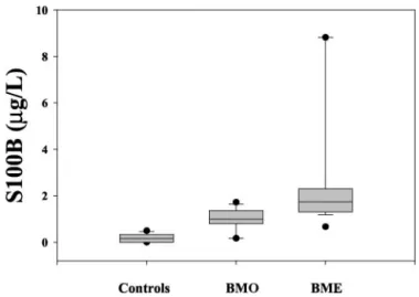
The concentrations of S100B in CSF were significantly higher in the total BM group (median, 1.34 g/L; 25th percentile, 0.84g/L; 75th percentile, 1.78 g/L) than in the controls (median, 0.16 g/L; 25th percentile, 0.11 g/L; 75th percentile, 0.33 g/L; P ⬍0.001). CSF S100B concentrations were significantly higher in the infants of the BME group than in the BMO group and the controls (P ⬍0.001; Fig. 1). The difference between the BMO group and controls was also significant (P⬍0.01).
CSF S100B correlated with CSF variables such as leu-kocyte count (r ⫽ 0.44; P ⬍0.001), glucose (r ⫽ 0.25; P ⬍0.01), and protein (r ⫽ 0.29; P ⬍0.01).
Multiple logistic regression analysis showed a positive correlation only between CSF S100B and the occurrence of BME (P ⬍0.001; odds ratio, 7.0). ROC curve analysis showed that higher S100B values were diagnostic for early BME detection, with a S100B cutoff of 1.0g/L, a sensitivity of 91% (95% confidence interval, 71–98.6%), a specificity of 82% (95% confidence interval, 70 –91.4%), and an area under the curve of 0.918.
The present findings suggest the potential usefulness of S100B measurements in CSF as a possible tool for the early detection of infants at risk of brain lesions from BM. BM represents a major cause of perinatal mortality and mor-bidity, and approximately one-third of affected infants develop neurologic sequelae (20 ).
Increased S100B has been reported in CSF, cord blood, amniotic fluid, and urine in preterm/term infants with cerebral bleeding (10, 14 –16 ), in high-risk fetuses (13, 21 ), in asphyxiated newborns (16, 22 ), and in children sub-jected to extracorporeal membrane oxygenation (23 ) and cardiopulmonary by-pass (12 ). In these conditions, S100B concentrations have been shown to detect individuals at high risk when routine monitoring procedures are silent. S100B cutoff values in urine and blood differ from that in CSF. The discrepancy can reasonably be attributed to the
Table 1. Maternal, clinical, and laboratory characteristics of the infants admitted to the study.
BM (nⴝ 26) BME (nⴝ 18) Controls (nⴝ 44) Maternal age,a years 27 27 28
Gestational age,aweeks 38 38 38
Birth weight,a
g 3198 3267 3209
Apgar score⬍7, yes/total tested
1st min 0.0/26 0/18 0/44
5th min 0/26 0/18 0/44
Gender, M/F 12/14 10/8 24/20
Age at sampling,adays 10 10 9
Maternal chorioamnionitis, % 7.7b
11.1b
0
Maternal fever, % 15 17 18
Initial clinical examination, %
Asymptomatic 0 0 0
Septicemia 100 100 100
Some clinical signs of BM 54 50 0
Critically ill 46 50 0
Blood analytes on admission Leukocyte count,a 106 /mm3 18.7b 20.9b 14.1 C-Reactive protein,amg/L 3.1b 3.4b 0.4 CSF findings
Leukocyte count,acells/mm3 80b 291b 43
Protein,a mg/L 1280b 1510b 410 Glucose,amg/L 900b 950b 440 Cerebral US (normal/abnormal), % At time of CSF sampling 100/0 100/0 100/0 72 h after admission 100/0 0/100b 100/0 At discharge 100/0 0/100b 100/0 Cerebral CT (normal/abnormal), % 100/0 0/100b Not performed aMedian value. bP⬍0.05.
Fig. 1. S100B protein concentrations in CSF of control infants and infants with BM only (BMO) and BM with encephalitis (BME). The lower and upper bars represent the 10th and 90th percentiles; F indicate the 5th and the 95th percentiles; the box indicates the interquartile range; and the horizontal line within the box indicates the median. S100B was significantly higher in infants with BME than in infants with BM or in controls (P⬍0.001 for both).
difference in biological fluids and differences in the peri-natal disease and study population investigated.
Increased S100B concentrations in CSF are a reasonably direct manifestation of damage to the central nervous system, which is known to contain protein at this stage.
Higher S100B concentrations may reflect a bacterial infection because such infections are known to induce a cascade of events, including activation of proinflamma-tory cytokines, which in the brain trigger exaggerated activation of glial cells, which produce S100B (24 ). High extracellular concentrations of S100B have been shown to be neurotoxic, leading to apoptosis and neuronal death via a nitric oxide-mediated pathway (10, 25 ). We think it is possible that at least part of the S100B measured in the CSF of infants with encephalitis derives from this process and participates in the pathologic events accompanying parenchymal damage.
Standard monitoring procedures performed when in-fants were admitted to our neonatal intensive care units were unable to indicate which infants would develop encephalitis. At this stage, S100B was significantly higher in the infants who later developed encephalitis than in those who did not develop encephalitis or in controls. S100B was also higher in BM cases without encephalitis than in controls. One explanation involves the possibility that some of the S100B in the CSF of infants without apparent brain damage may derive from changes in brain– blood barrier permeability (a common finding in meningitis). Another possible explanation is subclinical brain damage not detectable by standard diagnostic pro-cedures.
The present findings support the notion that assess-ment of S100B in CSF may provide additional information to physicians. Measurement of a brain constituent could be particularly useful for monitoring the effectiveness of therapeutic strategies. It is even possible that S100B as-sessment in other biological fluids may help in the early diagnosis of BM and encephalitis in infants with infec-tious diseases. The availability of longitudinal S100B measurements [half-life of⬃1 h (26)] in other biological fluids (e.g., peripheral blood and urine) has been already reported (14 –16 ).
The correlation between S100B and CSF patterns is intriguing. The correlation between S100B and polymor-phonuclear leukocytes, a manifestation of the severity of an infection, offers additional support to the notion that a more severe of infection is associated with a higher risk of neurologic sequelae (20 ). This concept is supported by the correlation between S100B and other CSF variables.
In conclusion, the present investigation provides a new perspective for the clinical study of S100B, with special reference to neurologic sequelae after bacterial infections. Although CSF remains the biological fluid of choice, further investigations are needed in other biological fluids to improve the care of newborns.
This work was supported in part by grants to Fabrizio Michetti from Universita` Cattolica del S. Cuore, from the
Ministero dell’Universita` e Ricerca Scientifica e Tecno-logica, and from the Ministero della Salute and to Diego Gazzolo from the “Let’s Improve Prenatal Life” Founda-tion. We also thank Sangtec Medical (Bromma, Sweden) and Byk Goulden Italia for supplying analysis reagents. We offer a special thanks to the Nursery Team of the Department of Neonatology of the Giannina Gaslini Chil-dren’s University Hospital (Genoa, Italy) and the Depart-ment of Neonatology, Warsaw Medical University Hos-pital (Warsaw, Poland) for their constant enthusiasm and participation during this research.
References
1. Nelson KB, Ellenberg JH. Obstetric complications as risk factors for cerebral palsy or seizure disorders. JAMA 1984;251:1843– 8.
2. Nelson KB, Grether JK. Maternal infection and cerebral palsy in infants of normal birthweight. JAMA 1997;278:201–11.
3. Nelson KB, Grether JK. Potentially asphyxiating conditions and spastic cerebral palsy in infants of normal birth weight. Am J Obstet Gynecol 1998;179:507–13.
4. Yoon BH, Romero R, Park JS, Kim CJ, Kim SH, Choi JH, et al. Fetal exposure to an intra-amniotic inflammation and the development of cerebral palsy at the age of three years. Am J Obstet Gynecol 2000;182:675– 81. 5. Dammann O, Leviton A. Maternal intrauterine infection, cytokines, and brain
damage in the preterm newborn. Pediatr Res 1997;42:1– 8.
6. Duncan JR, Cock ML, Scheerlinck JPY, Westcott KT, McLean C, Harding R, et al. White matter injury after repeated exposure in the preterm ovine fetus. Pediatr Res 2002;52:941–9.
7. Moore BW. A soluble protein characteristic of the nervous system. Biochem Biophys Res Commun 1965;19:739 – 44.
8. Rickmann M, Wolff JR. S100 protein expression in a subpopulation of neurons of rat brain. Neuroscience 1995;67:977–91.
9. Yang Q, Hamberger A, Hyden H, Wang S, Stigbrand T, Haglid K. S100 has a neuronal localization in the rat hindbrain revealed by an antigen retrieval method. Brain Res 1995;696:49 – 61.
10. Michetti F, Gazzolo D. S100B protein in biological fluids: a tool for perinatal medicine. Clin Chem 2002;48:2097–104.
11. Michetti F, Massaro A, Russo G, Rigon G. The S100 antigen in cerebrospinal fluid as a possible index of cell injury in the nervous system. J Neurol Sci 1980;44:731– 43.
12. Gazzolo D, Vinesi P, Geloso MC, Marcelletti CF, Iorio FS, Marianeschi SM, et al. S100 blood concentrations in children subjected to cardiopulmonary by-pass. Clin Chem 1998;44:1058 – 60.
13. Gazzolo D, Marinoni E, Di Iorio R, Lituania M, Bruschettini PL, Michetti F. Circulating S100B protein is increased in intrauterine growth retarded fetuses. Pediatr Res 2002;51:215–9.
14. Gazzolo D, Vinesi P, Bartocci M, Geloso MC, Bonacci W, Serra G, et al. Elevated S100 blood level as an early indicator of intraventricular hemor-rhage in preterm infants. Correlation with cerebral Doppler velocimetry. J Neurol Sci 1999;15;170:32–5.
15. Gazzolo D Bruschettini M, Lituania M, Serra G, Bonacci W, Michetti F. Increased urinary S100B protein as an early indicator of intraventricular hemorrhage in preterm infants: correlation with the grade of hemorrhage. Clin Chem 2001;47:1636 – 8.
16. Gazzolo D, Di Iorio R, Marinoni E, Masetti P, Serra G, Giovannini L, et al. S100B Protein is increased in asphyxiated term infants developing intraven-tricular hemorrhage. Crit Care Med 2002;30:1356 – 60.
17. Johnson CE, Whitwell JK, Pethe K, Saxena K, Super DM. Term newborns who are at risk of sepsis: are lumbar punctures necessary? Pediatrics 1997;4: 1–5.
18. Jensen R, Marshak DR, Anderson C, Lukas TJ, Watterson DM. Character-ization of human brain S100 protein fraction: amino acid sequence of S100. J Neurochem 1985;45:700–5.
19. Baudier J, Glasser N, Haglid K, Gerard D. Purification, characterization and ion binding properties of human brain S100b protein. Biochim Biophys Acta 1984;790:164 –73.
20. Escobar GJ, Li DK, Armstrong MA, Gardner MN, Folk BF, Verdi JE, et al. Neonatal sepsis workshop in infantsⱖ 2000 grams at birth: a population-based study. Pediatrics 2000;106:256 – 63.
21. Gazzolo D, Bruschettini M, Di Iorio R, Marinoni E, Lituania M, Marras M, et al. Maternal nitric oxide supplementation decreases cord blood S100B in intrauterine growth retarded fetuses. Clin Chem 2002;48:647–50. 22. Nagdyman N, Komen W, Ko HK, Muller C, Obladen M. Early biochemical
indicators of hypoxic-ischemic encephalopathy after birth asphyxia. Pediatr Res 2001;49:502– 6.
23. Gazzolo D, Masetti P, Meli M, Grutzfeld D, Michetti F. Elevated S100B protein as an early indicator of intracranial haemorrhage in infants subjected to extracorporeal membrane oxygenation. Acta Paediatr 2002;91:218 –21. 24. Yang GY, Gong C, Qin Z, Liu XH, Lorris Betz A. Tumor necrosis factor␣ expression produces increased blood-brain barrier permeability following temporary focal cerebral ischemia in rat. Brain Res Mol Brain Res 1999; 69:135– 43.
25. Hu J, Ferreira A, Van Eldik LJ. S100b induces neuronal death through nitric oxide release from astrocytes. J Neurochem 1997;69:2294 –301. 26. Jonsson H, Johnsson P, Hoglund P, Alling C, Blomquist S. Elimination of
S100B and renal function after cardiac surgery. J Cardiothorac Vasc Anesth 2000;14:698 –701.
DOI: 10.1373/clinchem.2003.021048
Influence of Practicable Virus Inactivation Procedures on Tests for Frequently Measured Analytes in Plasma, Martin Hersberger,1* Charly Nusbaumer,2 Andre´ Scholer,2 Verena Kno¨pfli,2and Arnold von Eckardstein1 (1Institute of Clinical Chemistry, University Hospital Zurich, Raemis-trasse 100, CH-8091 Zurich, Switzerland;2Department of Laboratory Medicine, Kantonsspital Basel, University Hospital, Petersgraben 4, Basel, Switzerland; * author for correspondence: fax 41-1-255-4590, e-mail hmr@ikc. unizh.ch)
Clinical specimens from patients carrying highly infec-tious agents such as corona, Lassa, Ebola, or Marburg viruses may present a biohazard to laboratory workers. Although specialized medical microbiology laboratories amplify and analyze such viruses under the required biosafety measures, few general clinical laboratories have the equipment to perform their analyses without putting their personnel at risk. The recent spread of severe acute respiratory syndrome (SARS) has heightened concern about personnel safety.
Although the WHO (1 ) and the CDC (2 ) recommend that blood samples from patients in whom SARS is suspected should be analyzed under biosafety level II conditions, most high-throughput analyzers in the clinical chemistry laboratory use open tubes and therefore do not meet the biosafety level II standards. In such cases, the CDC recommends administrative measures and/or addi-tional personal protective equipment to reduce risk. Some manufacturers announced that their analyzers should not be used for samples from patients in whom SARS is suspected, citing possible aerosol production during anal-ysis. Hence, laboratory directors and biosafety officers are in the dilemma of how to offer urgent diagnostic and surveillance tests to clinicians and at the same time protect their coworkers from potential biohazards.
Gamma irradiation and heat inactivation procedures have been investigated for the mentioned lipid-enveloped viruses (3, 4 ). Gamma irradiation seems not to affect some tests, but the intensity necessary to inactivate viruses is usually not available in hospitals, and the procedure decreases activities of enzymes and results of coagulation tests (3, 5 ). Heat inactivation can be done simply in a biological safety cabinet with a waterbath at 60 °C,
treat-ing samples for 30 min to inactivate corona viruses or for 60 min to inactivate Lassa, Ebola, or Marburg viruses (3 ). Only limited and nonquantitative data are available on the effects of these procedures on tests for common, clinically important analytes (3, 4, 6 ). We examined the effect of these two heat inactivation procedures as well as the effect of a virus-envelope-destroying detergent on tests used in intensive and emergency care.
Residual routine blood samples were selected to cover a wide range of concentrations (Table 1). Blood samples anticoagulated with either lithium heparinate or citrate (Vacutainer; Becton Dickinson) were centrifuged at 2500g for 10 min, and plasmas were transferred to plastic vials. The sealed plastic vials were totally immersed in a 60 °C waterbath for either 30 or 60 min, and the plasmas were analyzed together with a nontreated aliquot on a Roche-Hitachi Clinical Chemistry and Immunoassay Analyzer (Modular) with commercial tests from Roche Diagnostics GmbH and on a CA7000 Blood Coagulation Analyzer with commercial tests from Dade-Behring.
After either heat inactivation procedure, measured con-centrations ranged between 90% and 110% of pretreat-ment concentrations for electrolytes, creatinine, urea, uric acid, bilirubin, glucose, lactate, total protein, albumin, C-reactive protein, troponin T, the N-terminal fragment of pro-B-type natriuretic peptide, and the-chain of human chorionic gonadotropin (Table 1). For thyroid-stimulating hormone, aspartate aminotransferase, and pancreatic amylase, measured concentrations were⬃90% of values in untreated samples after the 30-min inactivation proce-dure and decreased to 70 – 80% after the 1-h incubation. In contrast, creatine kinase, myoglobin, alanine aminotrans-ferase,␥-glutamyl transferase, lactate dehydrogenase, al-kaline phosphatase, and the blood coagulation indicators were virtually inactivated, and free thyroxine was in-creased 2.4-fold after the shorter incubation procedure.
We searched for another inactivation method with minimal interference in these tests. Because Triton X-100 treatment of plasma samples inactivates other lipid-envel-oped viruses, such as HIV and Berne viruses (7, 8 ), we investigated it. The inactivation procedure consisted of mixing (on a Vortex-type mixer) 1 mL of plasma and 10 L of a solution containing 100 mL/L Triton X-100 and incubating the mixture for 60 min at room temperature. Measured concentrations ranged from 91% to 107% of untreated values for electrolytes, metabolites, enzymes, proteins, and hormones (Table 1), offering a virus inacti-vation procedure for heat-labile analytes such as creatine kinase, myoglobin, alanine aminotransferase,␥-glutamyl transferase, lactate dehydrogenase, alkaline phosphatase, and free thyroxine. More importantly, even the results for blood coagulation indicators were little affected by the Triton X-100 inactivation procedure, although there was a 20% increase in the International Normalized Ratio and a 10% increase in the activated partial thromboplastin time. To investigate whether Triton X-100 inactivation inter-feres with assays from other suppliers, we measured electrolytes, metabolites, and proteins on a RXL