Normalization of adiponectin
concentrations by leptin
replacement in ob/ob mice is
accompanied by reductions in
systemic oxidative stress and
inflammation
Gema Frühbeck
1,2,3,4, Victoria Catalán
1,2,3, Amaia Rodríguez
1,2,3, Beatriz Ramírez
1,2,3, Sara
Becerril
1,2,3, Piero Portincasa
5& Javier Gómez-Ambrosi
1,2,3The circulating concentrations of adiponectin, an antidiabetic adipokine, have been shown to be reduced in obesity, in relation to an increase in inflammation. The aim of the present work was to assess the effect of leptin replacement on adiponectin levels and expression as well as on markers of oxidative stress and inflammation in leptin-deficient ob/ob mice. Twelve-week-old male mice (n = 7–10 per group) were treated with either saline (wild type and ob/ob mice) or leptin (ob/ob mice) for 18 days. A third group of ob/ob mice was treated with saline and pair-fed to the amount of food consumed by the leptin-treated group. Leptin replacement restored values of adiponectin (P < 0.001), reduced circulating 8-isoprostane and serum amyloid A (SAA) levels (P < 0.05 for both), and significantly downregulated the increased gene expression of osteopontin (Spp1, P < 0.05), Saa3 (P < 0.05), Cd68 (P < 0.01), Il6 (P < 0.01) and NADPH oxidase (Nox1 and Nox2, P < 0.01) in the perirenal WAT and Spp1 (P < 0.05) in the liver of ob/ob mice. In cultured adipocytes from ob/ob mice, leptin increased (P < 0.05) the mRNA expression and secretion of adiponectin. We concluded that circulating concentrations of adiponectin are positively regulated by leptin and ameliorate obesity-associated oxidative stress and inflammation in mice.
Over the last decades, changes in lifestyle have caused a progressive increase in the incidence of obesity, being
one of the most prevalent metabolic disorders1, 2. Excess adiposity favors the development of cardiometabolic
alterations such as type 2 diabetes (T2D), cardiovascular disease, dyslipidemia, steatohepatitis and cancer1. Given
the increasing prevalence of obesity and its comorbidities, further research to unravel the metabolic mechanisms
involved in its etiopathogenesis is necessary3.
Adipose tissue secretes a wide variety of biologically active molecules representing an extremely active
endo-crine organ3. These secreted proteins, collectively called adipokines, such as leptin and adiponectin, are known
to be involved in the pathophysiological link between increased adiposity and cardiometabolic abnormalities3.
Leptin is a 16 kDa protein primarily produced by adipose tissue in proportion to the amount of body adiposity
functioning as a lipostat signal informing the hypothalamus on the size of fat deposits in the body3. Circulating
leptin concentrations are higher in obese individuals, which has led to the concept of leptin resistance4. Besides
1Metabolic Research Laboratory, Clínica Universidad de Navarra, Pamplona, Spain. 2CIBER Fisiopatología de la Obesidad y Nutrición (CIBEROBN), Instituto de Salud Carlos III, Pamplona, Spain. 3Obesity and Adipobiology Group, Instituto de Investigación Sanitaria de Navarra (IdiSNA), Pamplona, Spain. 4Department of Endocrinology & Nutrition, Clínica Universidad de Navarra, Pamplona, Spain. 5Clinica Medica “A. Murri”, Department of Biomedical Sciences and Human Oncology, University of Bari Medical School, Policlinico Hospital, Bari, Italy. Correspondence and requests for materials should be addressed to J.G.-A. (email: [email protected])
Received: 27 February 2017 Accepted: 18 April 2017 Published: xx xx xxxx
its regulatory function of energy homeostasis, leptin is involved in the regulation of neuroendocrine function,
haematopoiesis, angiogenesis, and reproduction, among others5.
Adiponectin is another important adipokine of 30 kDa expressed almost exclusively in adipose tissue6. Plasma
adiponectin concentrations are decreased in obese patients as well as in people with cardiovascular disease7.
Adiponectin increases insulin sensitivity and prevents lipid accumulation in skeletal muscle and liver,
stimu-lating fatty acid oxidation. Adiponectin has been also reported to exert anti-inflammatory actions8. The
benefi-cial effects of adiponectin are mediated primarily via the action of two receptors, AdipoR1 and AdipoR2 which
exhibit overlapping and distinct effects9.
Obesity may induce systemic oxidative stress in relation with and altered adipokine secretion10. Oxidative
stress plays a critical role in the development of obesity associated comorbidities favoring the appearance of T2D,
hypertension, liver steatosis and atherosclerosis11. Moreover, systemic and local inflammation is also increased in
obesity leading to an increased risk of the development of a wide number of comorbidities12, 13. Leptin-deficient
ob/ob mice exhibit massive obesity, dyslipidemia and insulin resistance. These mice have been proposed as a
model of the metabolic syndrome since they exhibit many of the pathophysiologic alterations most frequently
associated with obesity, including increased oxidative stress14–16 and elevated systemic inflammation14, 17.
The effect of leptin on circulating adiponectin concentrations has been previously studied with contradictory
results. In this sense, leptin-deficient ob/ob mice, in which leptin’s effect are absent, exhibit either decreased18, 19,
increased20, 21, or unchanged22 adiponectin circulating concentrations. Moreover, many previous studies have
used supraphysiological or even pharmacological doses of leptin18, 23 that may overestimate its effects or,
alterna-tively, exert no effect since it has been reported that pharmacological doses of leptin may lose its effect due to
sat-uration or downregulation of receptors24 or even elicit opposite effects to those observed after the administration
of a low leptin dose around the physiological level25.
The aim of the present work was to assess the effect of exogenous administration of leptin at a low dose on circulating concentrations and adipose tissue expression of adiponectin in leptin-deficient ob/ob mice as well as its impact on systemic and adipose tissue and liver inflammation and oxidative stress.
Results
Leptin administration restores adiponectin concentrations in ob/ob mice in association with a
decrease in oxidative stress and markers of inflammation.
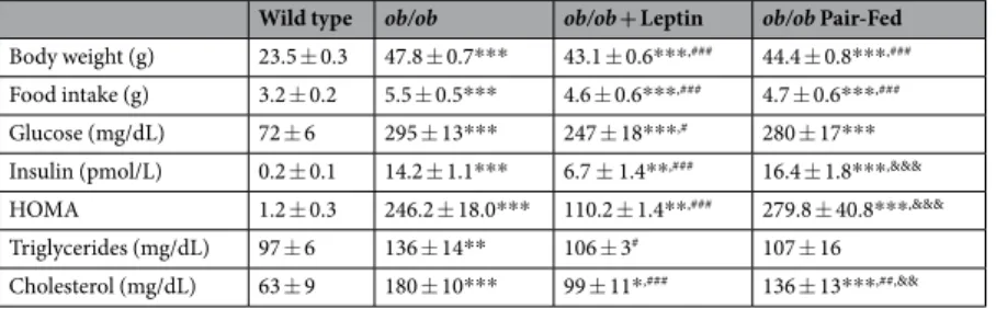
As expected, leptin-deficient obese ob/ob mice showed increased body weight and food intake as well as higher serum glucose, insulin, triglyceride and
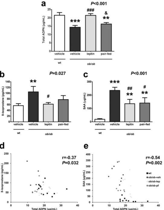
total cholesterol concentrations as compared to wt mice (Table 1). Serum concentrations of total adiponectin
were significantly reduced in ob/ob mice as compared to wt littermates (14.4 ± 1.1 vs 21.6 ± 1.7 μg/mL; P < 0.001,
Fig. 1a). Leptin treatment significantly reduced body weight, food intake as well as serum variables after 18 days
of treatment (Table 1). Serum total adiponectin levels were normalized after leptin replacement (21.5 ± 1.1 μg/
mL; P < 0.001 vs untreated ob/ob) while the PF group exhibited very similar levels to vehicle-treated ob/ob mice (16.3 ± 0.9 μg/mL; P < 0.05 vs leptin-treated ob/ob mice). Systemic oxidative stress was significantly higher in
ob/ob mice, as evidenced by 8-isoprostane levels (105 ± 19 vs 57 ± 7 pg/mL; P < 0.01) with leptin administration
normalizing its levels (62 ± 6 pg/mL; P < 0.05 vs untreated ob/ob). Pair-fed ob/ob mice exhibited an improvement in 8-isoprostane concentrations (78 ± 14 pg/mL), showing a half-way phenotype between leptin- and
saline-treated ob/ob mice (Fig. 1b). Leptin administration significantly reduced circulating concentrations of the acute
phase reactant serum amyloid A (SAA), which were dramatically increased in the ob/ob mice, to a similar extent than pair-feeding (wt: 19 ± 5; ob/ob-vehicle: 236 ± 23; ob/ob-leptin: 136 ± 33; ob/ob pair-fed: 141 ± 40 μg/mL;
P < 0.001, Fig. 1c). Serum total adiponectin concentrations were negatively correlated with the circulating levels
of 8-isoprostane (r = −0.37, P = 0.032; Fig. 1d) and SAA (r = −0.54, P = 0.002; Fig. 1e).
Leptin raises adiponectin concentrations increasing its expression and secretion in adipose
tis-sue.
The amount of adiponectin protein in EWAT and SCWAT was significantly reduced in all ob/ob groupsas compared to wt mice (P < 0.01) with leptin or pair-feeding exerting no effect (Fig. 2a and b). Adiponectin
released to the media was significantly lower (P < 0.05) in EWAT differentiated adipocytes from ob/ob mice than
that from wt mice after 48 h of culture (Fig. 2c). After 48 h of culture leptin significantly upregulated (P < 0.05)
adiponectin secretion to the media in differentiated adipocytes from ob/ob mice (Fig. 2d). Adipoq mRNA
expres-sion was downregulated in ob/ob mice as compared to wt (P < 0.01). The in vitro increased secretion and the
Wild type ob/ob ob/ob + Leptin ob/ob Pair-Fed Body weight (g) 23.5 ± 0.3 47.8 ± 0.7*** 43.1 ± 0.6***,### 44.4 ± 0.8***,###
Food intake (g) 3.2 ± 0.2 5.5 ± 0.5*** 4.6 ± 0.6***,### 4.7 ± 0.6***,###
Glucose (mg/dL) 72 ± 6 295 ± 13*** 247 ± 18***,# 280 ± 17***
Insulin (pmol/L) 0.2 ± 0.1 14.2 ± 1.1*** 6.7 ± 1.4**,### 16.4 ± 1.8***,&&&
HOMA 1.2 ± 0.3 246.2 ± 18.0*** 110.2 ± 1.4**,### 279.8 ± 40.8***,&&&
Triglycerides (mg/dL) 97 ± 6 136 ± 14** 106 ± 3# 107 ± 16
Cholesterol (mg/dL) 63 ± 9 180 ± 10*** 99 ± 11*,### 136 ± 13***,##,&&
Table 1. Body weight, food intake and serum analysis in ob/ob mice. Effect of leptin replacement. HOMA, homeostatic model assessment. Data presented as mean ± SEM. Differences were analyzed by ANOVA followed
by LSD tests. *P < 0.05, **P < 0.01 and ***P < 0.001 vs wild type; #P < 0.05, ##P < 0.01 and ###P < 0.001 vs ob/ob;
absence of changes in the amount of protein in WAT after in vivo leptin administration was accompanied by a leptin-induced upregulation in adiponectin mRNA expression in the ob/ob mice, showing no difference with the
wt mice (Fig. 3a).
Leptin reduces the expression of genes involved in inflammation and oxidative stress in
adi-pose tissue.
No effect of leptin on the expression of adiponectin receptors in adipose tissue was observed(Fig. 3b and c). Adipose tissue from ob/ob exhibited a dramatic increase (2.5- to 12-fold, P < 0.001) in the
expres-sion of genes involved in inflammation. Leptin administration significantly reduced (P < 0.05) the expresexpres-sion
of Saa3, Cd68, Il6 and Spp1 in ob/ob as compared to vehicle-treated littermates (Fig. 3d,e,g and i). Tnf mRNA
showed the same trend, although the differences with the vehicle group were only marginal (P = 0.073) (Fig. 3f),
while no changes were observed in the expression levels of Il1b (Fig. 3h). In a similar way, adipose tissue from
Figure 1. Leptin replacement increases total adiponectin (ADPN) concentrations, which were reduced in ob/ob mice, reducing systemic inflammation and oxidative stress. Serum levels of total adiponectin (a), 8-isoprostane (b) and serum amyloid A (SAA) (c) in wild type (wt) and ob/ob mice receiving vehicle, leptin or receiving vehicle and pair-fed to the leptin group. Data presented as mean ± SEM. Differences between groups were
analyzed by one way ANOVA followed by LSD test. **P < 0.01 and ***P < 0.001 vs wt; #P < 0.05, ##P < 0.01
and ###P < 0.001 vs ob/ob treated with vehicle; &P < 0.05 vs ob/ob treated with leptin. Scatter diagrams showing
the negative correlation found between the circulating concentrations of total adiponectin and the levels of 8-isoprostane (d) and SAA (e). Pearson’s correlation coefficient and P values are indicated. n = 6–10 per group.
leptin-deficient ob/ob mice exhibited an increase (7- to 8-fold, P < 0.001) in the expression of genes related to oxidative stress. Leptin replacement significantly reduced (P < 0.01) the expression of Nox1 and Cybb in ob/ob as
compared to vehicle-treated ob/ob mice (Fig. 3j and k)
Leptin ameliorates liver steatosis and reduces the expression of genes involved in
inflamma-tion in the liver.
Liver weight was higher in ob/ob mice and significantly reduced (P < 0.001 as compared toob/ob mice receiving vehicle) by leptin treatment (Fig. 4a), showing no differences in relative terms with wt mice
(Fig. 4b). Analysis of intrahepatic TG content showed elevated TG levels in ob/ob mice and that leptin
replace-ment significantly (P < 0.001) reduced them (Fig. 4c). This reduction in TG was accompanied by a decrease in
AST levels in the leptin-treated group, showing no statistical differences with the wt group, that was not observed
after pair-feeding (Fig. 4d). No effect of leptin was found on ALT levels (Fig. 4e). Pair-fed ob/ob mice exhibited a
halfway liver phenotype between leptin- and saline-treated ob/ob mice (Fig. 4a–c). Lack of leptin was associated
with an increase in the mRNA levels of the lipogenic transcription factor Srebf1, whose expression was slightly
reduced after leptin treatment, showing significant differences only with the pair-fed group (Fig. 4f). As observed
in adipose tissue, no effect in the expression of adiponectin receptor after leptin administration was shown in
the liver (Fig. 4g and h). Increased expression of genes involved in inflammation such as Cd68 and Spp1 in ob/ob
mice was observed, which was normalized after leptin administration only in the case of Spp1 (Fig. 4i and k). No
changes were observed in the hepatic expression levels of Il6 and Cybb (Fig. 4j and l).
Discussion
The major findings of the present study are that leptin replacement: (1) restored serum concentrations of adi-ponectin, which were decreased in ob/ob mice, through the increase in its expression and secretion, (2) reduced the levels of markers of oxidative stress, such as 8-isoprostane, and inflammation, such as SAA levels, and (3) downregulated the increased expression of genes involved in inflammation such as Spp1 (Opn), Cd68, Il6 and
Saa3 in the WAT and Spp1 in the liver, and oxidative stress, such as NADPH oxidase (Nox1 and Cybb) in the WAT
of ob/ob mice.
The analysis of the serum concentrations of adiponectin in ob/ob mice has produced discrepant results, with
reported lower18, 19, higher20, 21, or unchanged22 levels. Data from the present study and from previous studies of
Figure 2. Protein amount of adiponectin is decreased in white adipose tissue of ob/ob mice and leptin stimulates its secretion from adipocytes. Protein expression levels of total adiponectin (ADPN) in mouse epididymal (EWAT, a) and subcutaneous (SCWAT, b) adipose tissues from wild type (wt) and ob/ob mice receiving vehicle, leptin or receiving vehicle and pair-fed to the leptin group. The blot densitometry data were normalized with β-actin values (upper panels). Values are the mean ± SEM. The expression of ADPN in the wt group was assumed to be 100. Differences between groups were analyzed by ANOVA followed by LSD tests. **P < 0.01 vs wt. (c) Concentrations of adiponectin in secreted media from EWAT adipocytes differentiated
from wt and ob/ob mice after 48 h of culture (n = 3–7 mice). *P < 0.05 vs wt by two-tailed unpaired Student’s
t test. (d) Effect of leptin at a concentration of 10−8 mol/L on the amount of adiponectin in the secreted media
in adipocytes differentiated from ob/ob mice up to 48 h of culture. The effect of treatment was analyzed by unpaired Student’s t tests. *P < 0.05 vs unstimulated adipocytes.
our group19 clearly show that ob/ob mice exhibit reduced serum adiponectin levels. One of the factors that may
explain these discrepancies is the genetic background of the ob/ob mice, which has been shown to strongly
influ-ence the metabolic impact of the leptin deficiency26, although most of the reported studies have used ob/ob mice
on a B6 background27. Other factors such as the age of the animals, diet or fasting conditions offer alternative
explanations.
Figure 3. Leptin replacement in ob/ob mice regulates the expression of genes involved in inflammation and oxidative stress in adipose tissue. Gene expression levels of Adipoq (a), Adipor1 (b), Adipor2 (c), Saa3 (d),
Cd68 (e), Tnf (f), Il6 (g), Il1b (h), Spp1 (i), Nox1 (j) and Cybb (k) in perirenal adipose tissue in wild type (wt)
and ob/ob mice receiving vehicle, leptin or receiving vehicle and pair-fed to the leptin group. Data presented as mean ± SEM of 6–10 animals. Differences between groups were analyzed by one way ANOVA followed by
LSD test. *P < 0.05, **P < 0.01 and ***P < 0.001 vs wt; #P < 0.05 and ##P < 0.01 vs ob/ob treated with vehicle;
Leptin replacement restored serum adiponectin to normal levels, in agreement with a previous study with a
higher subcutaneous dose of leptin in a shorter period of time (6 d)18. This effect was beyond the inhibitory action
of leptin on food intake, since pair-fed animals showed adiponectin levels closer to the vehicle-treated than to the Figure 4. Leptin replacement in ob/ob mice reduces liver steatosis and regulates the expression of genes involved in inflammation. Liver weight (a), relative liver weight (b), triglyceride content in the liver (c) and serum AST (d) and ALT (e) levels of the animals from the different experimental groups. Mean ± SEM of 7–10 animals. (f) Expression of the lipogenic gene Srebf1 in the liver. Gene expression levels of Adipor1 (g), Adipor2 (h), Cd68 (i), Il6 (j), Spp1 (k) and Cybb (l) in the liver in wild type (wt) and ob/ob mice receiving vehicle, leptin or receiving vehicle and pair-fed to the leptin group. Data presented as mean ± SEM of 6–10 animals. Differences between groups
were analyzed by one way ANOVA followed by LSD test. *P < 0.05, **P < 0.01 and ***P < 0.001 vs wt; #P < 0.05,
leptin-treated ob/ob mice. This effect is in contrast to another work where central leptin administration reduced
adiponectin concentrations28. Further studies might elucidate whether leptin regulates central or peripherally
adiponectin levels differentially.
Leptin administration induced an increase in Adipoq mRNA expression in WAT. Previous studies have shown
lower adiponectin mRNA expression in leptin-deficient ob/ob and leptin receptor-deficient db/db mice29–31, as
well as in obese Zucker fa/fa rats lacking functional leptin receptors32, and that leptin significantly increases
Adipoq expression in WAT18, 23, 33, 34 and in the heart35. Interestingly, we found lower amounts of adiponectin
protein in WAT of ob/ob mice consistent with other studies30, 36, 37, but leptin replacement failed to normalize its
levels. This in vivo observation prompted us to hypothesize that the restored circulating levels of adiponectin with an increase in Adipoq mRNA expression but without changes in the protein levels in WAT is due to an increased expression and secretion in WAT. Our in vitro studies evidenced a reduced adiponectin secretion in adipocytes from ob/ob mice and that leptin significantly increased adiponectin secretion after 48 h of exposure confirming our hypothesis. A recent study did not observe the same stimulatory effect of leptin in adiponectin secretion in
human white adipocytes, but the authors studied leptin effects only after 24 h of treatment38, a period of time in
which we also did not observe a significant effect of leptin. It seems that more prolonged periods of time (up to 48 h) are needed to observe such an effect. We conclude that leptin regulates adiponectin concentrations stimu-lating its expression and secretion from WAT.
The restoration of adiponectin concentrations after leptin replacement in ob/ob mice was accompanied by a decrease in systemic inflammation and oxidative stress. Since obese ob/ob mice exhibit increased inflammation
and oxidative stress compared with their lean littermates14, we aimed to study systemic levels of 8-isoprostane
and SAA as well as genes involved in inflammatory and oxidative stress in WAT and liver of obese ob/ob mice. Oxidative stress is defined as an imbalance in the redox state resulting in an increased production of reactive oxy-gen species (ROS), ultimately leading to oxidative damage of cellular components. Oxidative stress was increased in ob/ob mice as evidenced by the elevated levels of 8-isoprostane together with the increase in the expression of
the NADPH oxidase subunits Nox1 and Nox2 (Cybb) in WAT, which encode the major enzymes generating ROS10.
These observations are in agreement with a large number of studies related to increased serum oxidative stress in
obesity both in animal models and humans10, 11, 39. Leptin administration restored 8-isoprostane levels to
normal-ity and significantly reduced the expression of Nox1 and Cybb. However, the relationship between leptin and
oxi-dative stress has not been fully elucidated. Leptin stimulates in vitro ROS production in different cell types40, 41 and
systemic oxidative stress in animal models42, suggesting a stimulatory role of leptin. On the contrary, oxidative
stress is increased in obese Zucker fa/fa rats39, while leptin administration reduces the oxidative stress in different
cellular43 or rodent models44, 45. In the present study, exogenous administration of leptin to ob/ob mice repressed
the increased expression of Nox1 and Cybb in WAT. These findings are in line with previous observations showing
that leptin restores the defective antioxidant enzyme capacity in serum of leptin-deficient mice46 and humans47,
but disagree with the reported increased p47phox (Ncf1) expression after leptin replacement in the livers of ob/
ob mice48. It is possible that the much higher leptin dose used in the latter study explains the discrepant findings.
Noteworthy, another study has reported in vitro a protective effect of leptin against ethanol-induced oxidative
stress49 and our group has evidenced that leptin replacement reduces the expression of genes related to oxidative
stress in the skeletal muscle of ob/ob mice14.
Acute-phase reactants have been suggested to contribute to the maintenance of the obesity-associated
low-grade chronic inflammation50, 51. Interestingly, our study provides evidence that the acute-phase response was
increased in ob/ob mice as evidenced by the increased serum concentrations of SAA, which were counteracted, at least in part, by exogenous leptin administration. Saa3 has been involved in an inflammatory paracrine loop between adipocytes and activated macrophages in mice, being proposed as an index of the number of activated
macrophages monitoring the adipose tissue inflammatory state52. Tnf has been proposed as one of the major
stimulators of adipose tissue production of Saa3 in this loop. However, the reduction of Tnf expression in WAT after leptin replacement in our study was only marginal, which suggests that the downregulation of other
proin-flammatory cytokines may be involved in this effect53, 54. Leptin administration reduced the elevated gene
expres-sion of Saa3, Cd68 and Opn (Spp1) in WAT and Spp1 in the liver, which are upregulated in obesity-associated
inflammation in mice and humans51, 55, 56. A proinflammatory role has been attributed to leptin57. However, the
consideration of leptin as a proinflammatory factor derives mostly from in vitro studies and may be in relation to
its proliferative effect and its ability to stimulate the metabolism58. In this sense, leptin administration at
physio-logical or pharmacophysio-logical levels does not modify circulating inflammatory marker levels in humans59, 60. Taken
together, our data suggest that leptin prevent the obesity-associated inflammatory state and the increased oxida-tive stress in leptin-deficient ob/ob mice. The stimulatory effect of leptin on circulating adiponectin levels in ob/ob mice could be involved in the amelioration in oxidative stress and inflammation observed in these animals, since a negative correlation between adiponectin and 8-isoprostane and SAA after leptin replacement was found. An increase in the expression of adiponectin receptors Adipor1 and Adipor2, which were reduced in ob/ob in
agree-ment with a previous work37, is unlikely to mediate these improvements, since their expression was unaffected
after leptin administration.
Systemic concentrations of 8-isoprostane and SAA was reduced in pair-fed ob/ob mice (although not reaching statistical significance in the case of 8-isoprostane), showing that weight loss contributes to the improvement in
oxidative stress and inflammation as previously reported11, 51. However, the effect of leptin on the expression of
genes involved in inflammation and oxidative stress in adipose tissue and liver was beyond the leptin-induced inhibitory effect on food intake, evidencing a direct positive effect of leptin at the dose used. Similar findings were
previously reported by our group in the skeletal muscle14. The effect of leptin on the expression of these genes in
other organs merits further research.
The leptin-induced reduction in the expression of proinflammatory genes in the liver was accompanied by a significant reduction in TG accumulation and serum AST levels. This observation suggests that leptin
replacement in ob/ob mice, even at the low dose employed, exerts anti-steatotic effects in the liver in agreement
with previous reports from our group61 and others62 using higher doses of leptin. Our data point out that the
reduction in inflammation and oxidative stress may contribute, together with the reduction in lipogenesis and
increase in fatty-acid oxidation63, to the improvement of hepatic function after leptin replacement.
In summary, obesity is accompanied by a chronic pro-inflammatory state and increased oxidative stress. Findings of our study provide evidence that systemic oxidative stress and inflammation, together with the ele-vated expression of genes involved in oxidative stress and inflammation of ob/ob mice are reversed by leptin replacement. A rise in adiponectin concentrations seems to be involved in the metabolic improvement observed. The translational value of the present study is that subjects with adipose tissue dysfunction, characterized by a lower secretion of adiponectin in relation to leptin levels, may have increased systemic oxidative stress and inflammation. Therefore, therapeutic tools targeted to improve this dysfunctional adipokine secretion pattern in adipose tissue may render a lower oxidative and inflammatory profile and, consequently, a lower cardiometabolic risk.
Materials and Methods
Animals and treatments.
Wild type (wt, C57BL/6) and leptin- deficient (B6.Cg-Lepob/J, ob/ob, JAX™
MiceStock Number 000632) mice were purchased from Charles River (Saint-Germain-sur-l′Arbresle, France). Mice were housed at an ambient temperature of 22 ± 2 °C on a 12:12 h light-dark cycle (lights on at 08:00 h) under path-ogen-free conditions and given a standard laboratory diet (Rodent Toxicology Diet, B&K Universal Ltd., Hull, UK) and tap water ad libitum. Twelve-week-old male mice were treated with saline or leptin (0.1 mg/kg body weight) i.p. twice daily for 18 days; recombinant mouse leptin was purchased from PeproTech EC Ltd. (London, UK). In addition, another group of ob/ob mice was treated with saline and pair-fed to the amount of food con-sumed by the leptin-treated group in order to dissociate the well-known appetite reducing effect of leptin from
other effects. Mice were killed by CO2 inhalation. Epididymal white adipose tissue (EWAT), subcutaneous WAT
(SCWAT) and perirenal WAT (PRWAT) were snap-frozen in liquid nitrogen and stored at −80 °C until extraction of RNA. Plasma was separated and stored at −80 °C for subsequent measurements. All experimental procedures conformed to the European Guidelines for the Care and Use of Laboratory Animals and the study was approved by the Ethical Committee for Animal Experimentation of the University of Navarra.
Serum measurements.
Blood samples were collected by cardiac puncture. Serum glucose was determinedby using a Blood Glucose Meter (Ascensia ELITE
®
, Bayer, Barcelona, Spain). Serum insulin was measured bymeans of an Ultra Sensitive Rat Insulin ELISA kit using mouse insulin as standard (Crystal Chem Inc., Chicago, IL, USA). Intra-and inter-assay coefficients of variation (CV) were 5.5% and 4.8%, respectively. Serum aspartate
aminotransferase (AST) and alanine aminotransferase (ALT) were measured by enzymatic tests (Infinity
™
ASTor ALT Liquid Stable Reagent Thermo Fisher Scientific, Waltham, MA, USA). Serum adiponectin was quantified by ELISA (Biovendor, Modrice, Czech Republic). Intra-and inter-assay CV were 4.2% and 5.9%, respectively. Circulating levels of 8-isoprostane (8-iso PGF2a), a proposed marker of antioxidant deficiency and oxidative stress, were measured by an immunoenzymatic assay (Cayman Chemicals, Ann Arbor, MI, USA). SAA con-centrations were assessed using an ELISA kit (BioSource International Inc., Camarillo, CA, USA). Intra-and inter-assay CV were 5.3% and 8.1%, respectively.
RNA extraction and Real-Time PCR.
Total RNA was extracted from 100 mg of PRWAT samples byhomogenization with an ULTRA-TURRAX
®
T 25 basic (IKA®
Werke GmbH, Staufen, Germany) using TRIzolreagent (Invitrogen, Carlsbad, CA, USA) and the RNeasy Lipid Tissue Mini Kit (Qiagen, Valencia, CA, USA). The RNA concentration was determined from absorbance at 260 nm. Transcript levels were quantified by Real-Time PCR (7300 Real-Time PCR System, Applied Biosystem, Foster City, CA, USA). Primers and probes (Sigma-Aldrich, Madrid, Spain) were designed using the software Primer Express 2.0 (Applied Biosystems).
Primers and probes used to amplify the cDNA are described in Table 2. The cDNA was amplified at the
follow-ing conditions: 95 °C for 10 min, followed by 45 cycles of 15 s at 95 °C and 1 min at 59 °C, usfollow-ing the TaqMan
®
Universal PCR Master Mix (Applied Biosystems). The primer and probe concentrations for gene amplification were 300 nmol/L and 200 nmol/L, respectively. All results were normalized to the levels of 18 S rRNA (Applied
Biosystems) and relative quantification was calculated using the ΔΔCt formula64. Relative mRNA expression
was expressed as fold expression over the calibrator sample (average of gene expression corresponding to the wt group). All samples were run in triplicate and the average values were calculated.
Protein extraction and Western-blot.
Similar amounts of EWAT or SCWAT were homogenized andprotein content was measured as previously described65. Equal amounts of protein (30 μg) were run out in 10%
SDS-PAGE, subsequently transferred to nitrocellulose membranes (Bio-Rad Laboratories, Inc., Hercules, CA, USA) and blocked in Tris-buffered saline with Tween 20 (TBS-T) containing 5% nonfat dry milk for 1 h at room temperature (RT). Blots were then incubated overnight at 4 °C with a monoclonal anti-mouse adiponectin mice antibody (MAB3608, Chemicon, Temecula, CA, USA) diluted 1:1,000 in blocking solution. The antigen-antibody complexes were visualized using peroxidase-conjugated anti-rabbit IgG antibody (1:5,000) and the enhanced chemiluminescence ECL detection system (Amersham Biosciences, Buckinghamshire, UK). The intensity of the
bands was determined by densitometric analysis with the Gel DocTM EQ gel documentation system and Quantity
One 4·5·0 software for quantitation of images (Bio-Rad)66.
Intrahepatic lipid content.
Tissue were homogenized and diluted in saline at a final concentration of50 mg/mL as previously described61, 65. Homogenates were diluted (1:1) in 1% deoxycholate (Sigma-Aldrich) and
incubated at 37 °C for 5 min. For triglyceride measurements, samples were diluted 1:100 in the reagent (Infinity
™
dye was measured based on its absorbance at 550 nm with a Sunrise ELISA plate reader (Tecan, Männedorf,
Switzerland). Concentrations were determined compared with a standard curve of triglycerides (Infinity
™
Gene (GenBank accession) Oligonucleotide sequence (5′–3′)
Adipoq (NM_009605)
Forward AAGGAGATGCAGGTCTTCTTGGT
Reverse CACTGAACGCTGAGCGATACAT
TaqMan Probe FAM-TGGAATGACAGGAGCTGAAGGGCCA-TAMRA
Adipor1 (NM_028320)
Forward TCATCTACCTCTCCATCGTCTGTGT
Reverse CAAGCCAAGTCCCAGGAACA
TaqMan Probe FAM-CATTGTGGCACAGTGGGACCGGTT-TAMRA
Adipor2 (NM_197985)
Forward TTTGCCACCCCTCAGTATCG
Reverse TGACATAATGCAAGGTAGGGATGAT
TaqMan Probe FAM-TGTTCGTGGGCTTAGGCCTGAGTGG-TAMRA
Cd68 (NM_001291058)
Forward TACATGGCGGTGGAATACAATG
Reverse GAACAGCTGGAGAAAGAACTATGCTT
TaqMan Probe FAM-AGGCAGCACAGTGGACATTCATGGC-TAMRA
Cybb (NM_007807)
Forward TGTGTCGAAATCTGCTCTCCTTT
Reverse AAAGTGAGGTTCCTGTCCAGTTGT
TaqMan Probe FAM-AGTGCGTGTTGCTCGACAAGGAT-TAMRA
Il1b (NM_008361)
Forward TGACAGTGATGAGAATGACCTGTTC
Reverse TGATGTGCTGCTGCGAGATT
TaqMan Probe FAM-ACCCCAAAAGATGAAGGGCTGCTTCC-TAMRA
Il6 (NM_031168)
Forward CGGAGGCTTAATTACACATGTTCTC
Reverse CAGTTTGGTAGCATCCATCATTTCT
TaqMan Probe FAM-CGTGGAAATGAGAAAAGAGTTGTGCAATGG-TAMRA
Nox1 (NM_172203)
Forward TTATCGCTCCCAGCAGAAGGT
Reverse CATGCTAAAGCCTCGCTTCCT
TaqMan Probe FAM-ATTACCAAGGTTGTCATGCACCCA-TAMRA
Saa3 (NM_011315)
Forward AGTCATCAGCGATGCCAGAGA
Reverse CCCCACTCATTGGCAAACTG
TaqMan Probe FAM-CACGGGACATGGAGCAGAGGACTCA-TAMRA
Spp1 (NM_009263)
Forward TTTGCCGTTTGGCATTGC
Reverse TGGGTGCAGGCTGTAAAGCT
TaqMan Probe FAM-TCCTCCCTCCCGGTGAAAGT-TAMRA
Srebf1 (NM_011480)
Forward TCCCAAGAGCCCTGCACTT
Reverse GTCCACAAAGAAACGGTGACCTA
TaqMan Probe FAM-TTGACACGTTTCTTCCTGAGCAGCGC-TAMRA
Tnf (NM_013693)
Forward CCAGACCCTCACACTCAGATCAT
Reverse ACTCCAGCTGCTCCTCCACTT
TaqMan Probe FAM-CCTGTAGCCCACGTCGTAGCAAACCA-TAMRA
Table 2. Sequences of the primers and TaqMan probes. Adipoq, adiponectin; Adipor1, adiponectin receptor 1; Adipor2, adiponectin receptor 2; Cd68, macrophage antigen CD68; Cybb, superoxide-generating NADPH oxidase heavy chain subunit (Nox2); Il1b, interleukin-1β; Il6, interleukin-6; Nox1, NADPH Oxidase 1; Saa3, serum amyloid A3; Spp1, secreted phosphoprotein 1 (osteopontin), Srebf1, sterol regulatory element binding transcription factor 1; Tnf, tumor necrosis factor-α.
Triglycerides Standard, Thermo Fisher Scientific). The protein content of the preparations was measured by the Bradford method, using BSA (Sigma-Aldrich) as standard. All assays were performed in duplicate.
Culture of adipocytes and analysis of adiponectin secretion.
Mice stromovascular fraction cells (SVFCs) were isolated from EWAT from wt or ob/ob mice. Briefly, small pieces (≈500 mg) of adipose tissue were minced and incubated for 1 h at 37 °C with constant shaking in 5 mL of adipocyte medium (DMEM/F-12 [1:1] (Invitrogen, Paisley, UK) supplemented with 16 μmol/L biotin, 18 μmol/L panthotenate, 100 μmol/L ascor-bate and antibiotic-antimycotic (Invitrogen)) supplemented with 2% BSA (Sigma-Aldrich) and 2 mg/mL col-lagenase (Roche). The digestion was stopped adding adipocyte medium supplemented with 10% newborn calf serum (NCS) (Sigma-Aldrich) and suspended cells were then filtered through a 100-μm nylon cell strainer (BD Biosciences, Erembodegem, Belgium) and centrifuged for 10 min at 600 g. Adipocytes were decanted and stromo-vascular pellets were resuspended in adipocyte medium supplemented with 10% NCS. After a filtration through a 70-μm nylon cell strainer (BD Biosciences), cells were spun at 400 g for 5 min. SVFCs were resuspended ineryth-rocyte lysis buffer (0.154 mol/L NH4Cl, 10 mmol/L KHCO3, 0.1 mmol/L EDTA) and allowed to settle for 10 min
at RT, followed by a centrifugation for 10 min at 400 g. The cell pellet was resuspended in adipocyte medium
supplemented with 10% NCS. SVFCs were grown in six-well plastic plates (5 × 10⁵ cells per well) and
main-tained at 37 °C in a humidified atmosphere with 95% air-5% CO2. Adipocyte differentiation was initiated when
cells reached about 80–90% confluence. Cells were stimulated with differentiation medium I (adipocyte medium supplemented with 10% NCS, 0.5 mmol/L 3-isobutyl-1-methylxanthine, 0.1 μmol/L dexamethasone and 10 μg/ mL insulin) for 2 days. Cells were then switched to differentiation medium II (adipocyte medium supplemented with 10% NCS and 10 μg/mL insulin) for another 6 days and media were changed every 2 days. Adipocytes were
70–75% differentiated (as determined by morphology) in the 8th day of differentiation. Adipocytes were
incu-bated for 2 h in serum-free adipocyte medium and, then, stimulated with mouse recombinant leptin (PeproTech)
at a concentration of 10−8 mol/L for 48 h. One sample per experiment was used to obtain control responses in the
presence of solvent. Concentrations of adiponectin in the media were measured by ELISA (Biovendor).
Statistical analysis.
Data are presented as mean ± SEM and differences between groups of mice were lyzed by one-way ANOVA followed by Fisher’s LSD tests. The effect of group/treatment in cell cultures was ana-lyzed by unpaired Student’s t tests. Correlation between two variables were computed by Pearson’s correlation coefficients (r). A P value lower than 0.05 was considered statistically significant. The calculations were performed using SPSS 23 (SPSS, Chicago, IL, USA) and GraphPad Prism 6 (GraphPad Software, Inc., La Jolla, CA, USA).References
1. Bray, G. A., Frühbeck, G., Ryan, D. H. & Wilding, J. P. Management of obesity. Lancet 387, 1947–1956, doi: 10.1016/S0140-6736(16)00271-3 (2016).
2. Frühbeck, G. et al. Obesity: the gateway to ill health - an EASO position statement on a rising public health, clinical and scientific challenge in Europe. Obes Facts 6, 117–120, doi:10.1159/000350627 (2013).
3. Lancha, A., Frühbeck, G. & Gómez-Ambrosi, J. Peripheral signalling involved in energy homeostasis control. Nutr Res Rev 25, 223–248, doi:10.1017/S0954422412000145 (2012).
4. Myers, M. G., Cowley, M. A. & Münzberg, H. Mechanisms of leptin action and leptin resistance. Annu. Rev. Physiol. 70, 537–556, doi:10.1146/annurev.physiol.70.113006.100707 (2008).
5. Blüher, M. & Mantzoros, C. S. From leptin to other adipokines in health and disease: facts and expectations at the beginning of the 21st century. Metabolism. 64, 131–145, doi:10.1016/j.metabol.2014.10.016 (2015).
6. Scherer, P. E., Williams, S., Fogliano, M., Baldini, G. & Lodish, H. F. A novel serum protein similar to C1q, produced exclusively in adipocytes. J. Biol. Chem. 270, 26746–26749, doi:10.1074/jbc.270.45.26746 (1995).
7. Scheid, M. P. & Sweeney, G. The role of adiponectin signaling in metabolic syndrome and cancer. Rev Endocr Metab Disord 15, 157–167, doi:10.1007/s11154-013-9265-5 (2014).
8. Kadowaki, T. et al. Adiponectin and adiponectin receptors in insulin resistance, diabetes, and the metabolic syndrome. J. Clin. Invest.
116, 1784–1792, doi:10.1172/JCI29126 (2006).
9. Keshvari, S., Henstridge, D. C., Ng, C., Febbraio, M. A. & Whitehead, J. P. Muscle-specific overexpression of AdipoR1 or AdipoR2 gives rise to common and discrete local effects whilst AdipoR2 promotes additional systemic effects. Sci Rep 7, 41792, doi:10.1038/ srep41792 (2017).
10. Furukawa, S. et al. Increased oxidative stress in obesity and its impact on metabolic syndrome. J. Clin. Invest. 114, 1752–1761, doi:10.1172/JCI21625 (2004).
11. Vincent, H. K. & Taylor, A. G. Biomarkers and potential mechanisms of obesity-induced oxidant stress in humans. Int. J. Obes. 30, 400–418, doi:10.1038/sj.ijo.0803177 (2006).
12. Catalán, V., Gómez-Ambrosi, J., Rodríguez, A. & Frühbeck, G. Adipose tissue immunity and cancer. Front Physiol 4, 275, doi:10.3389/fphys.2013.00275 (2013).
13. Gómez-Ambrosi, J. et al. Increased cardiometabolic risk factors and inflammation in adipose tissue in obese subjects classified as metabolically healthy. Diabetes Care 37, 2813–2820, doi:10.2337/dc14-0937 (2014).
14. Sáinz, N. et al. Leptin administration downregulates the increased expression levels of genes related to oxidative stress and inflammation in the skeletal muscle of ob/ob mice. Mediators Inflamm. 2010, 784343–15, doi:10.1155/2010/784343 (2010). 15. Zhu, X. et al. Tumour necrosis factor-a inhibition with lenalidomide alleviates tissue oxidative injury and apoptosis in ob/ob obese
mice. Clin. Exp. Pharmacol. Physiol. 41, 489–501, doi:10.1111/cep.2014.41.issue-7 (2014).
16. Liu, M. et al. Potent effects of dioscin against obesity in mice. Sci Rep 5, 7973, doi:10.1038/srep07973 (2015).
17. Mirsoian, A. et al. Adiposity induces lethal cytokine storm after systemic administration of stimulatory immunotherapy regimens in aged mice. J. Exp. Med. 211, 2373–2383, doi:10.1084/jem.20140116 (2014).
18. Delporte, M. L., El Mkadem, S. A., Quisquater, M. & Brichard, S. M. Leptin treatment markedly increased plasma adiponectin but barely decreased plasma resistin of ob/ob mice. Am. J. Physiol. Endocrinol. Metab. 287, E446–453, doi:10.1152/ajpendo.00488.2003
(2004).
19. Sáinz, N. et al. Leptin administration favors muscle mass accretion by decreasing FoxO3a and increasing PGC-1a in ob/ob mice.
PLoS ONE 4, e6808, doi:10.1371/journal.pone.0006808 (2009).
20. Sennello, J. A. et al. Regulation of T cell-mediated hepatic inflammation by adiponectin and leptin. Endocrinology 146, 2157–2164, doi:10.1210/en.2004-1572 (2005).
21. Sloan, C. et al. Central leptin signaling is required to normalize myocardial fatty acid oxidation rates in caloric-restricted ob/ob mice.
Diabetes 60, 1424–1434, doi:10.2337/db10-1106 (2011).
22. Singhal, N. S., Patel, R. T., Qi, Y., Lee, Y. S. & Ahima, R. S. Loss of resistin ameliorates hyperlipidemia and hepatic steatosis in leptin-deficient mice. Am. J. Physiol. Endocrinol. Metab. 295, E331–338, doi:10.1152/ajpendo.00577.2007 (2008).
23. Zhang, W., Della-Fera, M. A., Hartzell, D. L., Hausman, D. & Baile, C. A. Adipose tissue gene expression profiles in ob/ob mice treated with leptin. Life Sci. 83, 35–42, doi:10.1016/j.lfs.2008.04.021 (2008).
24. Frühbeck, G., Aguado, M. & Martínez, J. A. In vitro lipolytic effect of leptin on mouse adipocytes: evidence for a possible autocrine/ paracrine role of leptin. Biochem. Biophys. Res. Commun. 240, 590–594, doi:10.1006/bbrc.1997.7716 (1997).
25. Hoffmann, A. et al. Leptin within the subphysiological to physiological range dose dependently improves male reproductive function in an obesity mouse model. Endocrinology 157, 2461–2468, doi:10.1210/en.2015-1966 (2016).
26. Haluzik, M. et al. Genetic background (C57BL/6J versus FVB/N) strongly influences the severity of diabetes and insulin resistance in ob/ob mice. Endocrinology 145, 3258–3264, doi:10.1210/en.2004-0219 (2004).
27. Frühbeck, G. & Gómez-Ambrosi, J. Control of body weight: a physiologic and transgenic perspective. Diabetologia 46, 143–172, doi:10.1007/s00125-003-1053-4 (2003).
28. Ueno, N., Dube, M. G., Inui, A., Kalra, P. S. & Kalra, S. P. Leptin modulates orexigenic effects of ghrelin and attenuates adiponectin and insulin levels and selectively the dark-phase feeding as revealed by central leptin gene therapy. Endocrinology 145, 4176–4184, doi:10.1210/en.2004-0262 (2004).
29. Makimura, H., Mizuno, T. M., Bergen, H. & Mobbs, C. V. Adiponectin is stimulated by adrenalectomy in ob/ob mice and is highly correlated with resistin mRNA. Am. J. Physiol. Endocrinol. Metab. 283, E1266–1271, doi:10.1152/ajpendo.00227.2002 (2002). 30. Ye, J., Gao, Z., Yin, J. & He, Q. Hypoxia is a potential risk factor for chronic inflammation and adiponectin reduction in adipose
tissue of ob/ob and dietary obese mice. Am. J. Physiol. Endocrinol. Metab. 293, E1118–1128, doi:10.1152/ajpendo.00435.2007 (2007). 31. Okada, T. et al. URB is abundantly expressed in adipose tissue and dysregulated in obesity. Biochem. Biophys. Res. Commun. 367,
370–376, doi:10.1016/j.bbrc.2007.12.164 (2008).
32. Oana, F. et al. Physiological difference between obese (fa/fa) Zucker rats and lean Zucker rats concerning adiponectin. Metabolism.
54, 995–1001, doi:10.1016/j.metabol.2005.02.016 (2005).
33. Wendel, A. A., Purushotham, A., Liu, L. F. & Belury, M. A. Conjugated linoleic acid fails to worsen insulin resistance but induces hepatic steatosis in the presence of leptin in ob/ob mice. J. Lipid Res. 49, 98–106, doi:10.1194/jlr.M700195-JLR200 (2008). 34. Hoffmann, A. et al. Leptin dose-dependently decreases atherosclerosis by attenuation of hypercholesterolemia and induction of
adiponectin. Biochim. Biophys. Acta 1862, 113–120, doi:10.1016/j.bbadis.2015.10.022 (2016).
35. Takahashi, T. et al. Impaired expression of cardiac adiponectin in leptin-deficient mice with viral myocarditis. Int Heart J 47, 107–123, doi:10.1536/ihj.47.107 (2006).
36. Seo, J. B. et al. Adipocyte determination- and differentiation-dependent factor 1/sterol regulatory element-binding protein 1c regulates mouse adiponectin expression. J. Biol. Chem. 279, 22108–22117, doi:10.1074/jbc.M400238200 (2004).
37. Favero, G. et al. Melatonin reduces obesity and restores adipokine patterns and metabolism in obese (ob/ob) mice. Nutr. Res. 35, 891–900, doi:10.1016/j.nutres.2015.07.001 (2015).
38. Singh, P. et al. Differential effects of leptin on adiponectin expression with weight gain versus obesity. Int J Obes (Lond) 40, 266–274, doi:10.1038/ijo.2015.181 (2016).
39. Bankoglu, E. E. et al. Impact of weight loss induced by gastric bypass or caloric restriction on oxidative stress and genomic damage in obese Zucker rats. Free Radic. Biol. Med. 94, 208–217, doi:10.1016/j.freeradbiomed.2016.02.033 (2016).
40. Bouloumié, A., Marumo, T., Lafontan, M. & Busse, R. Leptin induces oxidative stress in human endothelial cells. FASEB J. 13, 1231–1238 (1999).
41. Blanca, A. J. et al. Leptin induces oxidative stress through activation of NADPH oxidase in renal tubular cells: Antioxidant effect of L-carnitine. J. Cell. Biochem. 117, 2281–2288, doi:10.1002/jcb.25526 (2016).
42. Beltowski, J., Wojcicka, G., Marciniak, A. & Jamroz, A. Oxidative stress, nitric oxide production, and renal sodium handling in leptin-induced hypertension. Life Sci. 74, 2987–3000, doi:10.1016/j.lfs.2003.10.029 (2004).
43. Zheng, J. et al. Leptin protects cardiomyocytes from serum-deprivation-induced apoptosis by increasing anti-oxidant defence. Clin.
Exp. Pharmacol. Physiol. 37, 955–962, doi:10.1111/j.1440-1681.2010.05415.x (2010).
44. Zhang, J. Y. Jr. et al. Leptin administration alleviates ischemic brain injury in mice by reducing oxidative stress and subsequent neuronal apoptosis. J Trauma Acute Care Surg 72, 982–991, doi:10.1097/TA.0b013e3182405459 (2012).
45. Gülen, S. & Dincer, S. Effects of leptin on oxidative stress in healthy and streptozotocin-induced diabetic rats. Mol. Cell. Biochem.
302, 59–65, doi:10.1007/s11010-007-9426-5 (2007).
46. Watson, A. M., Poloyac, S. M., Howard, G. & Blouin, R. A. Effect of leptin on cytochrome P-450, conjugation, and antioxidant enzymes in the ob/ob mouse. Drug Metab. Dispos. 27, 695–700 (1999).
47. Ozata, M., Uckaya, G., Aydin, A., Isimer, A. & Ozdemir, I. C. Defective antioxidant defense system in patients with a human leptin gene mutation. Horm. Metab. Res. 32, 269–272, doi:10.1055/s-2007-978634 (2000).
48. Chatterjee, S. et al. Leptin is key to peroxynitrite-mediated oxidative stress and Kupffer cell activation in experimental non-alcoholic steatohepatitis. J. Hepatol. 58, 778–784, doi:10.1016/j.jhep.2012.11.035 (2013).
49. Balasubramaniyan, V., Shukla, R., Murugaiyan, G., Bhonde, R. R. & Nalini, N. Mouse recombinant leptin protects human hepatoma HepG2 against apoptosis, TNF-a response and oxidative stress induced by the hepatotoxin-ethanol. Biochim. Biophys. Acta 1770, 1136–1144, doi:10.1016/j.bbagen.2007.04.009 (2007).
50. Gómez-Ambrosi, J. et al. Involvement of leptin in the association between percentage of body fat and cardiovascular risk factors.
Clin. Biochem. 35, 315–320, doi:10.1016/S0009-9120(02)00320-X (2002).
51. Gómez-Ambrosi, J. et al. Increased serum amyloid A concentrations in morbid obesity decrease after gastric bypass. Obes. Surg. 16, 262–269, doi:10.1381/096089206776116525 (2006).
52. Sanada, Y. et al. Serum amyloid A3 gene expression in adipocytes is an indicator of the interaction with macrophages. Sci Rep 6, 38697, doi:10.1038/srep38697 (2016).
53. Rodríguez, A., Catalán, V., Gómez-Ambrosi, J. & Frühbeck, G. Aquaglyceroporins serve as metabolic gateways in adiposity and insulin resistance control. Cell Cycle 10, 1548–1556, doi:10.4161/cc.10.10.15672 (2011).
54. Crewe, C., An, Y. A. & Scherer, P. E. The ominous triad of adipose tissue dysfunction: inflammation, fibrosis, and impaired angiogenesis. J. Clin. Invest. 127, 74–82, doi:10.1172/JCI88883 (2017).
55. Gómez-Ambrosi, J. et al. Plasma osteopontin levels and expression in adipose tissue are increased in obesity. J. Clin. Endocrinol.
Metab. 92, 3719–3727, doi:10.1210/jc.2007-0349 (2007).
56. Wellen, K. E. & Hotamisligil, G. S. Obesity-induced inflammatory changes in adipose tissue. J. Clin. Invest. 112, 1785–1788, doi:10.1172/JCI20514 (2003).
57. Loffreda, S. et al. Leptin regulates proinflammatory immune responses. FASEB J. 12, 57–65 (1998).
58. Scotece, M. et al. Adiponectin and leptin: new targets in inflammation. Basic Clin Pharmacol Toxicol 114, 97–102, doi:10.1111/ bcpt.2013.114.issue-1 (2014).
59. Chan, J. L. et al. Recombinant methionyl human leptin administration to achieve high physiologic or pharmacologic leptin levels does not alter circulating inflammatory marker levels in humans with leptin sufficiency or excess. J. Clin. Endocrinol. Metab. 90, 1618–1624, doi:10.1210/jc.2004-1921 (2005).
60. Farooqi, I. S. & O’Rahilly, S. Is leptin an important physiological regulator of CRP? Nat. Med. 13, 16–17, doi:10.1038/nm0107-16
(2007).
61. Rodríguez, A. et al. Leptin administration restores the altered adipose and hepatic expression of aquaglyceroporins improving the non-alcoholic fatty liver of ob/ob mice. Sci Rep 5, 12067, doi:10.1038/srep12067 (2015).
62. Harris, R. B. et al. A leptin dose-response study in obese (ob/ob) and lean (+/?) mice. Endocrinology 139, 8–19, doi:10.1210/ endo.139.1.5675 (1998).
63. Lee, Y. et al. Liporegulation in diet-induced obesity. The antisteatotic role of hyperleptinemia. J. Biol. Chem. 276, 5629–5635, doi:10.1074/jbc.M008553200 (2001).
64. Catalán, V. et al. Validation of endogenous control genes in human adipose tissue: relevance to obesity and obesity-associated type 2 diabetes mellitus. Horm. Metab. Res. 39, 495–500, doi:10.1055/s-2007-982502 (2007).
65. Lancha, A. et al. Osteopontin deletion prevents the development of obesity and hepatic steatosis via impaired adipose tissue matrix remodeling and reduced inflammation and fibrosis in adipose tissue and liver in mice. PLoS ONE 9, e98398, doi:10.1371/journal. pone.0098398 (2014).
66. Ezquerro, S. et al. Acylated and desacyl ghrelin are associated with hepatic lipogenesis, b-oxidation and autophagy: role in NAFLD amelioration after sleeve gastrectomy in obese rats. Sci Rep 6, 39942, doi:10.1038/srep39942 (2016).
Acknowledgements
The authors wish to thank all the staff of the animal housing facilities for their technical expertise in animal care and handling. Supported by the project grants FIS PI13/00460, PI14/00950 and PI16/01217, integrated in the Plan Estatal I + D + I 2013–16 from the Spanish Instituto de Salud Carlos III–Subdirección General de Evaluación y Fomento de la investigación–FEDER, Fundación Caja Navarra (20–2014) and Centro de Investigación Biomédica en Red Fisiopatología de la Obesidad y Nutrición, CIBEROBN, ISCIII, Spain.
Author Contributions
G.F. and J.G.-A. designed the study and obtained the funds. G.F. and J.G.-A. analyzed and interpreted the data. G.F. and J.G.-A. drafted the manuscript. G.F., V.C., A.R., B.R., S.B., P.P. and J.G.-A. provided study materials or performed experiments. G.F., V.C., A.R., B.R., S.B., P.P. and J.G.-A. critically revised the article for important intellectual content. All authors read and approved the final version of the article.
Additional Information
Competing Interests: The authors declare that they have no competing interests.
Publisher's note: Springer Nature remains neutral with regard to jurisdictional claims in published maps and institutional affiliations.
Open Access This article is licensed under a Creative Commons Attribution 4.0 International License, which permits use, sharing, adaptation, distribution and reproduction in any medium or format, as long as you give appropriate credit to the original author(s) and the source, provide a link to the Cre-ative Commons license, and indicate if changes were made. The images or other third party material in this article are included in the article’s Creative Commons license, unless indicated otherwise in a credit line to the material. If material is not included in the article’s Creative Commons license and your intended use is not per-mitted by statutory regulation or exceeds the perper-mitted use, you will need to obtain permission directly from the
copyright holder. To view a copy of this license, visit http://creativecommons.org/licenses/by/4.0/.