Nailfold Capillaroscopy Characteristics of
Antisynthetase Syndrome and Possible Clinical
Associations: Results of a Multicenter International
Study
Marco Sebastiani, Konstantinos Triantafyllias, Andreina Manfredi, Miguel Angel González-Gay,
Natalia Palmou-Fontana, Giulia Cassone, Ulrich Drott, Christiane Delbrück,
Jorge Rojas-Serrano, Chiara Bertolazzi, Laura Nuño, Margherita Giannini, Florenzo Iannone,
Esther F. Vicente, Santos Castañeda, Albert Selva-O’Callaghan, Ernesto Trallero Araguas,
Giacomo Emmi, Annamaria Iuliano, Jutta Bauhammer, Nikolaus Miehle, Simone Parisi,
Lorenzo Cavagna, Veronica Codullo, Carlomaurizio Montecucco, Francisco Javier Lopez-Longo,
Julia Martínez-Barrio, Juan Carlos Nieto-González, Silvia Vichi, Marco Confalonieri,
Paola Tomietto, Raoul Bergner, Alberto Sulli, Francesco Bonella, Federica Furini,
Carlo Alberto Scirè, Alessandra Bortoluzzi, Christof Specker, Simone Barsotti, Rossella Neri,
Marta Mosca, Marzia Caproni, Julia Weinmann-Menke, Andreas Schwarting, Vanessa Smith
and Maurizio Cutolo, and the American and European Network of Antisynthetase Syndrome
Collaborative Group
ABSTRACT. Objective. To describe nailfold videocapillaroscopy (NVC) features of patients with antisynthetase
syndrome (AS) and to investigate possible correlations with clinical and serological features of the disease.
Methods. We retrospectively analyzed NVC images of 190 patients with AS [females/males 3.63,
mean age 49.7 ± 12.8 yrs, median disease duration 53.7 mos (interquartile range 82), 133 anti-Jo1 and 57 non–anti-Jo1-positive patients]. For each patient, we examined number of capillaries, giant capillaries, microhemorrhages, avascular areas, ramified capillaries, and the presence of systemic sclerosis (SSc)-like pattern. Finally, we correlated NVC features with clinical and serological findings of patients with AS. Concomitantly, a historical cohort of 75 patients with antinuclear antibody–negative primary Raynaud phenomenon (RP) and longterm followup was used as a control group (female/male ratio 4.13/1, mean age 53.9 ± 17.6 yrs) for NVC measures.
Results. NVC abnormalities were observed in 62.1% of AS patients compared with 29.3% of primary
RP group (p < 0.001). An SSc-like pattern was detected in 67 patients (35.3%) and it was associated with anti-Jo1 antibodies (p = 0.002) and also with a longer disease duration (p = 0.004). Interestingly, there was no significant correlation between the presence of SSc-like pattern and RP, and only 47% of patients with SSc-like pattern had RP.
Conclusion. NVC abnormalities are commonly observed in AS, independently from the occurrence
of RP. The presence of an SSc-like pattern could allow identification of a more defined AS subtype, and prospective studies could confirm the association with clinical and serological features of AS. (First Release November 15 2018; J Rheumatol 2019;46:279–84; doi:10.3899/jrheum.180355) Key Indexing Terms:
ANTISYNTHETASE SYNDROME ANTISYNTHETASE ANTIBODIES RAYNAUD PHENOMENON SYSTEMIC SCLEROSIS PATTERN NAILFOLD VIDEOCAPILLAROSCOPY
Ismael Cosío Villegas, Mexico City, Mexico; Servicio de Reumatología, Hospital Universitario La Paz, Madrid, Spain; Interdisciplinary Department of Medicine (DIM), Rheumatology Unit, University of Bari, Bari, Italy; Rheumatology Department, Hospital Universitario de la Princesa, Instituto de Investigación Sanitaria (IIS) Princesa, Madrid; Unidad de Enfermedades Autoinmunes Sistémicas, Servicio de Medicina Interna, Universidad Autonoma de Barcelona, on behalf of the GEAS From the Rheumatology Unit, Azienda Policlinico of Modena, University
of Modena and Reggio Emilia, Modena, Italy; ACURA Rheumatology Center, Bad Kreuznach, Germany; Rheumatology Division, Hospital Universitario Marqués de Valdecilla, IDiVAL, University of Cantabria, Santander, Spain; Rheumatology Division, University Hospital of Frankfurt, Frankfurt, Germany; Interstitial Lung Disease and Rheumatology Unit, Instituto Nacional de Enfermedades Respiratorias,
group, Barcelona, Spain; Department of Experimental and Clinical Medicine, and Department of Medical and Surgical Critical Care, Section of Dermatology, University of Florence, Florence; Unità Operativa Complessa (UOC) Reumatologia, Ospedale San Camillo-Forlanini, Rome; ACURA Centre for Rheumatic Diseases, Baden-Baden, Germany; Rheumatology Department, Città Della Salute e della Scienza, Torino; Division of Rheumatology, University and Institute for Research and Health Care (IRCCS) Policlinico S. Matteo Foundation, Pavia, Italy; Servicio de Reumatología, Hospital General Universitario Gregorio Marañón, Madrid, Spain; Dermatology Clinic, University Hospital of Trieste; Department of Pneumology and Respiratory Intermediate Care Unit, University Hospital of Cattinara, Trieste; Rheumatology Unit, Azienda Ospedaliero-Universitaria Ospedali Riuniti di Trieste, Trieste, Italy; Medizinische Klinik A, Klinikum der Stadt, Ludwigshafen, Germany; Research Laboratory and Academic Division of Clinical Rheumatology, Department of Internal Medicine, University of Genoa, San Martino Polyclinic Hospital IRCCS Genoa, Genoa, Italy; Interstitial and Rare Lung Disease Unit, Ruhrlandklinik University Hospital, University of Duisburg-Essen, Essen, Germany; UOC Reumatologia, Azienda Ospedaliero Universitaria S. Anna, University of Ferrara, Italy; Department for Rheumatology and Clinical Immunology, St. Josef Krankenhaus, University Clinic, Essen, Germany; Division of Rheumatology, Department of Clinical and Experimental Medicine, University of Pisa, Pisa; Department of Internal Medicine, Rheumatology and Clinical Immunology, University Hospital Johannes-Gutenberg, Mainz, Germany; University of Ghent, Ghent University Hospital, Ghent, Belgium.
M. Sebastiani, MD, Rheumatology Unit, Azienda Policlinico of Modena, University of Modena and Reggio Emilia; A. Manfredi, MD,
Rheumatology Unit, Azienda Policlinico of Modena, University of Modena and Reggio Emilia; G. Cassone, MD, Rheumatology Unit, Azienda Policlinico of Modena, University of Modena and Reggio Emilia; K. Triantafyllias, MD, ACURA Rheumatology Center; A. Schwarting, MD, Professor, ACURA Rheumatology Center; M.A. González-Gay, MD, Professor, Rheumatology Division, Hospital Universitario Marqués de Valdecilla, IDiVAL, University of Cantabria; N. Palmou-Fontana, MD, Rheumatology Division, Hospital Universitario Marqués de Valdecilla, IDiVAL, University of Cantabria; U. Drott, MD, Rheumatology Division, University Hospital of Frankfurt; C. Delbrück, MD, Rheumatology Division, University Hospital of Frankfurt; J. Rojas-Serrano, MD, Professor, Interstitial Lung Disease and Rheumatology Unit, Instituto Nacional de Enfermedades Respiratorias, Ismael Cosío Villegas; C. Bertolazzi, MD, Interstitial Lung Disease and Rheumatology Unit, Instituto Nacional de Enfermedades Respiratorias, Ismael Cosío Villegas; L. Nuño, MD, Servicio de Reumatología, Hospital Universitario La Paz; M. Giannini, MD, DIM, Rheumatology Unit, University of Bari; F. Iannone, MD, Professor, DIM, Rheumatology Unit, University of Bari; E.F. Vicente, MD, Rheumatology Department, Hospital Universitario de la Princesa, IIS Princesa; S. Castañeda, MD, Professor, Rheumatology Department, Hospital Universitario de la Princesa, IIS Princesa; A. Selva-O’Callaghan, MD, Professor, Unidad de Enfermedades Autoinmunes Sistémicas, Servicio de Medicina Interna, Universidad Autonoma de Barcelona, on behalf of the GEAS group; E. Trallero Araguas, MD, Unidad de Enfermedades Autoinmunes Sistémicas, Servicio de Medicina Interna, Universidad Autonoma de Barcelona, on behalf of the GEAS group; G. Emmi, MD, Department of Experimental and Clinical Medicine, University of Florence; A. Iuliano, MD, UOC Reumatologia, Ospedale San Camillo-Forlanini; J. Bauhammer, MD, ACURA Centre for Rheumatic Diseases; N. Miehle MD, ACURA Centre for Rheumatic Diseases; S. Parisi, MD, Rheumatology Department, Città Della Salute e della Scienza; L. Cavagna, MD, Division of Rheumatology, University and IRCCS Policlinico S. Matteo Foundation; V. Codullo, MD, Division of Rheumatology, University and IRCCS Policlinico S. Matteo Foundation; C. Montecucco, MD, Professor, Division of Rheumatology, University and IRCCS Policlinico S. Matteo Foundation; F.J. Lopez-Longo, MD, Servicio de Reumatología, Hospital General Universitario Gregorio Marañón; J. Martínez-Barrio, MD, Servicio de Reumatología, Hospital General Universitario Gregorio Marañón; J.C. Nieto-González, MD, Servicio de Reumatología, Hospital General Universitario Gregorio Marañón; S. Vichi, MD, Dermatology Clinic, University Hospital of Trieste; M. Confalonieri, MD, Department of Pneumology and Respiratory Intermediate Care Unit, University Hospital of Cattinara; P. Tomietto,
MD, Rheumatology Unit, Azienda Ospedaliero-Universitaria Ospedali Riuniti; R. Bergner, MD, Professor, Medizinische Klinik A, Klinikum der Stadt; A. Sulli, MD, Professor, Research Laboratory and Academic Division of Clinical Rheumatology, Department of Internal Medicine, University of Genoa, San Martino Polyclinic Hospital IRCCS Genoa; M. Cutolo, MD, Professor, Research Laboratory and Academic Division of Clinical Rheumatology, Department of Internal Medicine, University of Genoa, San Martino Polyclinic Hospital IRCCS Genoa; F. Bonella, MD, Interstitial and Rare Lung Disease Unit, Ruhrlandklinik University Hospital, University of Duisburg-Essen; F. Furini, MD, UOC
Reumatologia, Azienda Ospedaliero Universitaria S. Anna, University of Ferrara; C.A. Scirè, MD, Associate Professor, UOC Reumatologia, Azienda Ospedaliero Universitaria S. Anna, University of Ferrara; A. Bortoluzzi, MD, UOC Reumatologia, Azienda Ospedaliero
Universitaria S. Anna, University of Ferrara; C. Specker, MD, Professor, Department for Rheumatology and Clinical Immunology, St. Josef Krankenhaus, University Clinic; S. Barsotti, MD, Division of Rheumatology, Department of Clinical and Experimental Medicine, University of Pisa; R. Neri, MD, Division of Rheumatology, Department of Clinical and Experimental Medicine, University of Pisa; M. Mosca, MD, Professor, Division of Rheumatology, Department of Clinical and Experimental Medicine, University of Pisa; M. Caproni, MD, Department of Medical and Surgical Critical Care, Section of Dermatology, University of Florence; J. Weinmann-Menke, MD, Department of Internal Medicine, Rheumatology and Clinical Immunology, University Hospital
Johannes-Gutenberg; A. Schwarting, MD, Professor, Department of Internal Medicine, Rheumatology and Clinical Immunology, University Hospital Johannes-Gutenberg; V. Smith, MD, Professor, University of Ghent, Ghent University Hospital. Dr. Sebastiani and Dr. Triantafyllias contributed equally to this article.
Address correspondence to Dr. K. Triantafyllias, ACURA Rheumatology Center, Bad Kreuznach, Germany. E-mail: [email protected] Accepted for publication August 13, 2018.
Antisynthetase syndrome (AS) is a heterogeneous
auto-immune disease mainly characterized by the classic triad of
arthritis, myositis, and interstitial lung disease (ILD)
1,2and
by the occurrence of antiaminoacyl tRNA-synthetase
antibodies (anti-ARS). Raynaud phenomenon (RP), fever,
and mechanic’s hands are other relevant but less prevalent
accompanying features
3. The most frequent ARS antibody is
the anti-Jo1, whereas other ARS specificities are less
commonly detected (for example, anti-PL-7, -PL-12, -EJ,
-OJ, -KS, -YRS, -Zo)
1,4.
Nailfold videocapillaroscopy (NVC) is a safe, noninvasive
diagnostic tool for the in vivo study of periungueal
microcir-culation, in particular for the examination of capillary density
and morphology
5. NVC is one of the most reliable diagnostic
modalities to differentiate primary from secondary RP
6, and
it has been included among the 2013 classification criteria
for systemic sclerosis (SSc)
7. Large-scale efforts are being
made to standardize the evaluation of capillaroscopic
morphology throughout rheumatic diseases through the
European League Against Rheumatism study group on
microcirculation
8,9.
Despite the increasing number of capillaroscopic studies
in SSc reported over the last years, valid and systematic
capil-laroscopic evidence concerning AS are still lacking. The
scarce NVC data come mainly from a few studies and case
reports that assessed other conditions such as polymyositis
(PM) and dermatomyositis (DM)
10,11,12,13,14. These studies
subgroups, making it difficult to extract detailed and reliable
data concerning the role of these antibodies. Moreover,
patients in these studies were not clearly characterized as
having AS, possibly also because there are no
well-estab-lished classification criteria for this rare disease
15. AENEAS
(the American and European NEtwork of Antisynthetase
Syndrome) is an international collaborative group that aims
to study AS thoroughly and better understand its clinical and
pathophysiological features
16,17.
By considering the relevance of accompanying findings
in general and of RP in particular in AS
18, this multicenter
study (Nailfold Capillaroscopy in Antisynthetase Syndrome)
aimed to describe the capillaroscopic features from a
well-characterized cohort of patients, investigating possible
correlations with clinical and serological features of the
disease.
MATERIALS AND METHODS
Within the framework of the AENEAS collaborative group, we retrospec-tively evaluated all patients with AS who underwent NVC during their clinical history. In the study, we included patients with at least 2 antisyn-thetase antibody–positive tests, with 1 or more findings of the classic triad (arthritis, myositis, and ILD). Triad findings were identified as follows: arthritis occurrence was defined clinically by the referent physician, ILD by the occurrence of a restrictive pattern at pulmonary function test [forced vital capacity (FVC) ≤ 80%, forced expiratory volume (FEV)1/FVC ≥ 70%, decreased/normal FEV1, and/or DLCO reduction > 20%] and/or by the identification of alveolitis/fibrosis signs at high-resolution computed tomo-graphy of the lungs. Finally, muscle involvement was defined in case of any muscle enzyme elevation plus the presence of typical electromyography alterations and/or compatible muscle biopsy findings and/or suggestive muscle magnetic resonance findings. According to previous reports, the time of appearance of these 3 clinical manifestations (arthritis, ILD, myositis) was also recorded. Manifestations onset was considered concomitant if the delay in their appearance was < 3 months. RP occurrence was objectively assessed by direct physician observation and/or by photographic documen-tation provided by patients. All patients were further investigated for mechanic’s hand and smoking habit.
In keeping with the purpose of the study, patients meeting the classifi-cation criteria for SSc7were excluded.
After Institutional Review Board approval (ethics committee of the Azienda Policlinico of Modena, Italy, no. 3857/C.E.), clinical characteristics of the disease, and laboratory and instrumental data were retrieved from patients’ clinical records. Images (2 images per finger from the middle area of the nailfold) from every center were anonymized and then securely shared in a personalized storage folder for central reevaluation by an expert operator (MS) blinded to patients’ clinical data. A second expert operator (AM) assessed blindly interoperator reproducibility of results in a sample of 50 random patients (interrater agreement = 0.85). Capillaroscopic findings of patients with AS were compared with those of 75 controls, represented by patients with primary RP (female/male ratio 4.13/1, mean age 53.9 ± 17.6 yrs), referring to a historical cohort of ANA-negative patients without any sign of connective tissue disease other than RP. A minimum of 2 years of followup was requested as an entry criterion in the control group. A comparison was performed also within the patients with AS, to detect autoantibody specificities (anti-Jo1 and non-anti-Jo1 antibodies positivity) and clinical pattern presentation.
Collection of NVC images. NVC was performed using different machines
(Videocap software 3.0, DS Medica; Zeiss, Stemi 2000-C; Mediscope D1, Optilia; Dinocapture 2.0, Dino-Lite; Capiscope) equipped with a 100 or 200× optical probe, after patients had been in a comfortable temperature of
22–25°C for 20 min. A drop of immersion oil was applied to the nailfold to maximize the translucency of the keratin layer, and the second through the fifth fingers of both hands were examined19. To increase the reproducibility
among different videocapillaroscopies, all measurements were performed on 1 mm.
According to previous definitions20,21,22, the following capillaroscopic
variables were evaluated: giant capillaries (normally shaped homogeneously enlarged capillary with a limb diameter ≥ 50 μm); microhemorrhages (presence of one or more dark red masses characterized by hemosiderin deposits due to capillary injury or thrombosis); ramified capillaries (branching, bushy, interconnected capillaries, originating from a single capillary); the number of capillaries per linear mm; the number of giant capillaries per linear mm; avascular area (intercapillaries distance > 500 μm). Because some NVC features are occasionally observed in only 1 or a few fingers without clinical significance, to better evaluate the severity of these capillaroscopic alterations, we also evaluated the percentages of images per patient with microhemorrhages, ramified capillaries, giant capillaries, and avascular areas. Moreover, an SSc pattern, defined as an alteration of the nailfold microvascular network characterized by enlarged and giant loops, microhemorrhages, capillary loss, ramified capillaries, and architectural disorganization20,21,22,23,24, was recorded as being present or absent. In the
absence of validated criteria and of comparison studies, we choose to use the term “SSc-like” in the presence of an NVC pattern similar to the SSc pattern.
Statistical analysis. Data were expressed as mean ± SD unless otherwise
noted. Categorical variables were analyzed by chi-square test or Fisher’s exact test as appropriate; Wilcoxon test was used to compare repeated measurements in paired groups, while differences between the means were determined using Mann-Whitney U test for unpaired samples. P values ≤ 0.05 were considered statistically significant25.
RESULTS
A total of 2550 images (with a mean of 13.5 ± 4.1 images per
subject) from 190 patients with AS were analyzed
(females/males 3.63, mean age 49.7 ± 12.8 yrs, mean disease
duration 51.2 ± 71.4 mos).
RP was recorded in 43.2% of patients, arthritis in 76.5%,
myositis in 80.2%, and ILD in 86.2%. Anti-Jo1 antibodies
were detected in 133 patients (69.5%). On the other hand, 19
patients (10%) were positive for anti-PL-7 antibodies, 22
(11.5%) for anti-PL-12, 6 (3%) for anti-OJ, and 10 (6%) for
anti-EJ.
The clinical, serological, and demographic features of the
patients are reported in Table 1.
Comparison of patients with AS and control group. NVC
abnormalities (at least 1 among giant capillaries,
microhem-orrhages, ramifications, reduction of the number/mm) were
more frequently observed in patients with AS than in controls
(62.1% vs 29.3%, respectively, p < 0.001).
Regarding NVC variables, giant capillaries and avascular
areas were recorded only in the AS group (59 patients, 31.1%
and 39 patients, 20.5%, respectively, p < 0.005). Ramified
capillaries were more frequent in the AS group (94 patients,
49.5% vs 5 patients, 6.7%, p < 0.005), and no differences
were observed for microhemorrhages (44 patients, 23.2% vs
18 patients, 24%, p = not significant), although the degree of
severity was higher in AS than in the control group
(p = 0.008). Microhemorrhages were also more pronounced
in patients with AS, and (reasonably) related to local trauma
in the control group. The number of capillaries was higher in
the control group (p < 0.005; details in Table 2).
If a pathologic pattern was by definition absent in the
control group, an SSc-like pattern was found in the 35.3% of
patients with AS (Table 2). Ramified capillaries were
detected in almost 50% of patients with AS, while the
concurrent presence of giant and bushy capillaries was
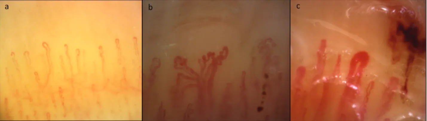
recorded in 26.2% of patients with AS. Interestingly, giant
and ramified capillaries were found in 73.8% of the patients
with SSc-like pattern (Figure 1).
Association between NVC and demographic, clinical, and
serological features of AS. In patients with AS, the presence
of at least 1 NVC abnormality was associated with anti-Jo1
antibodies positivity (p = 0.008) and presence of ILD
(p = 0.02). Interestingly, ramified capillaries were twice as
frequent in patients with ILD as in patients without (53.4%
vs 27.6%, respectively; p = 0.01).
Among the single NVC features, avascular areas, detected
in 17.9% of patients, were significantly associated with
autoimmunity markers (anti-SSA, anti-Jo1), myositis, and
RP (Supplementary Data, available with the online version
of this article).
An SSc-like pattern was associated with anti-Jo1
antibodies (44% vs 21.5%, in patients with Jo1
anti-bodies and other antisynthetase specificities, respectively;
p = 0.002) and also with a longer disease duration (107.1 ±
87.6 vs 84.6 ± 85.9 mos in patients with or without SSc-like
pattern, respectively; p = 0.004). Moreover, multivariate
analysis showed a correlation between disease duration and
SSc-like pattern (OR 2.73, 95% CI 1.04–7.17; p = 0.04, Table
3). There was no correlation between an SSc-like pattern and
the final number of clinical manifestations of the disease
(ILD, arthritis, myositis, mechanic’s hands, fever; all
compar-isons showing p > 0.05), but SSc-like pattern was more
commonly observed in patients with a later appearance of
ILD (63 patients) regarding patients presenting ILD from
disease onset (102 cases) or not presenting ILD (25 subjects;
p = 0.018). A multivariate analysis confirmed the independent
association between anti-Jo1 antibodies, disease duration, the
late occurrence of ILD, and the presence of an SSc-like
pattern (Table 3).
Interestingly, there was no significant correlation between
the presence of SSc-like pattern and RP, and only 47% of
patients with SSc-like pattern had RP.
DISCUSSION
To our knowledge, this is the first multicenter study
examining NVC features systematically in a large population
of patients with AS. Moreover, the study described for the
first time the characteristics and the frequency of SSc-like
pattern in patients with AS, and its correlation with the
clinical features of the disease. The analysis of 190 patients
with AS from participating centers allowed us to establish
that NVC pattern of AS is abnormal regarding people with
primary RP, thus confirming that vasculopathy is a typical
manifestation of this rare condition. The more frequent NVC
finding was ramified capillaries, while SSc-like pattern was
observed in 35.3% of cases. A significant correlation was
found between ILD and ramified capillaries, but not with
SSc-like pattern.
Previously, only a few studies analyzed the relationship
between anti-ARS and NVC features
12,13. A small study
concerning the NVC alterations in 24 patients with idiopathic
inflammatory myopathies (IIM) reported a positivity of
anti-Jo1 antibodies in 8 patients
12. The only abnormality
reported in this small cohort of anti-Jo1–positive patients was
a reduction of the capillary density in comparison to other
patients.
Positivity of anti-ARS was not associated with the
presence of SSc-like pattern in a study on 13 patients with
DM reported by Mugii, et al
10. In contrast, our study showed
Table 1. Demographic, clinical, and serological features of 190 patients withantisynthetase syndrome.
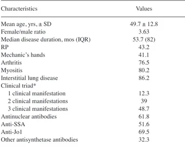
Characteristics Values Mean age, yrs, ± SD 49.7 ± 12.8 Female/male ratio 3.63 Median disease duration, mos (IQR) 53.7 (82) RP 43.2 Mechanic’s hands 41.1 Arthritis 76.5 Myositis 80.2 Interstitial lung disease 86.2 Clinical triad* 1 clinical manifestation 12.3 2 clinical manifestations 39 3 clinical manifestations 48.7 Antinuclear antibodies 61.8 Anti-SSA 51.6 Anti-Jo1 69.5 Other antisynthetase antibodies 32.3
Data are percentages unless otherwise indicated. *Arthritis, myositis, inter-stitial lung disease. IQR: interquartile range; RP: Raynaud phenomenon.
Table 2. Capillaroscopic features of patients with AS and controls.
Variables AS, n = 190 Controls, n = 75 p Frequency (%) Microhemorrhages 23.2 24 ns Ramified capillaries 49.5 6.7 < 0.005 SSc-like pattern 35.3 0 < 0.005 No. per mm Capillaries 8.49 ± 2.27 10.8 ± 0.67 < 0.005 Lowest no. capillaries 6.90 ± 2.63 9.27 ± 0.86 < 0.005 No. giant capillaries 0.18 ± 0.39 0 < 0.005 Severity (no. images per patient with at least a finding/10)
Microhemorrhages 0.7 ± 1.6 0.2 ± 0.3 0.008 Ramified capillaries 2.3 ± 3.1 0.6 ± 0.2 < 0.005 SSc: systemic sclerosis; AS: antisynthetase syndrome.
a significant association between anti-Jo1 antibodies and
SSc-like pattern. Nevertheless, it is possible that the low
number of patients in the subgroup analysis of Mugii, et al
could have been the reason for the lack of a significant
association
10.
Selva-O’Callaghan, et al investigated 53 patients with
IIM
11and reported a positivity for anti-ARS in 16 (12
positive for anti-Jo1 and 4 for other ARS). The comparison
between patients with the 16 anti-ARS and 6 anti-Pm/Scl
antibodies–positive patients did not show statistically
signifi-cant differences in regard to NVC alterations.
Interestingly, in a study examining 27 patients with DM,
an SSc-like pattern was recorded in 4/4 patients with anti-Jo1
antibodies
13.
In our population, NVC abnormalities were detected in
more than 60% of patients with AS, while an SSc-like pattern
was observed in the 35.3% of cases.
NVC findings were differently associated with the main
clinical features of AS. In this regard, NVC alterations were
more frequently observed in patients with ILD compared to
patients without (p = 0.02), and myositis was associated to
avascular areas (p = 0.004), while no specific NVC features
were detected in patients with arthritis. Interestingly, we
observed no differences according to the final number of
major clinical manifestations.
We previously observed a high prevalence of NVC
alter-ations in patients with inflammatory muscle diseases,
observing that NVC major alterations are quite exclusive of
DM, while PM was not different by primary RP
20. Moreover,
we observed differences between SSc pattern and DM pattern
(observed in patients with DM). In DM, giant and ramified
capillaries were seen at disease onset, while in patients with
longer disease duration, DM pattern regressed and only
brushing capillaries were observed
23.
At present, we cannot establish whether patients with AS
are characterized by true SSc-specific patterns
24or DM
pattern
23or by a specific AS pattern. For this reason,
comparison studies of AS with SSc and DM are required to
address this question, and the term SSc-like pattern appears
to be more proper. In contrast to that observed in DM, we
observed a direct correlation between disease duration and
SSc-like pattern in patients with AS, suggesting a different
phenotype of microangiopathy in AS and DM.
Remarkably, patients with SSc-like pattern at NVC seem
to develop ILD late in the course of the disease. Prospective
studies are needed to confirm this finding and to evaluate the
possible value of NVC in the prediction of interstitial lung
involvement. This point is not a secondary issue, because
together with other clinical aspects such as the occurrence of
de novo accompanying findings during followup
18, NVC
could result in an easily obtainable marker providing
additional information on the degree of risk for the clinical
spectrum progression in patients with AS.
Further, clinically relevant RP in AS is only associated
with the presence of avascular areas, but not with SSc-like
pattern. It is also worth noting that SSc-like pattern was found
in 32.1% of patients without RP from our series and that only
47% of patients with SSc-like pattern had RP. This point
highlights the practical value of performing NVC in patients
with connective tissue disease, arthritis, or ILD, regardless
of the presence of RP. In particular, all patients with
rheumatoid arthritis or ILD with SSc-like pattern at NVC
should be investigated for an underlying AS. Even if
Figure 1. Different morphological patterns on nailfold videocapillaroscopy. A. Raynaud phenomenon (homogeneously distributed capillaries with
normal architecture and morphology). B. and C. SSc-like pattern in antisynthetase syndrome: architectural disorganization, giant and ramified capillaries, microhemorrhages, reduction of the number of capillaries per linear mm. SSc: systemic sclerosis.
Table 3. Multivariate analysis. Association with SSc-like pattern.
Variables OR 95% CI p Lower Higher Disease duration ≥ 24 mos 2.731 1.041 7.167 0.041 ILD ≥ second AS manifestation 2.245 1.147 4.395 0.018 Anti-Jo1 antibodies 2.885 1.342 6.205 0.007j1 SSc: systemic sclerosis; ILD: interstitial lung disease; AS: antisynthetase syndrome.
inclusion of patients has not occurred at disease onset,
findings of the investigation can also be of importance
because clinical spectrum time course of this disease is highly
variable, and clinical manifestations may occur even after
years from disease onset
15,16.
The major limitation of this study is its retrospective
design. However, the high number of examined NVC and the
large amount of clinical data made it possible to obtain
clini-cally relevant and novel information. Nevertheless,
longitu-dinal studies are needed and planned by our group to examine
the temporal relationship between NVC findings and the
clinical manifestations of AS in a prospective manner. An
additional limitation could be that currently NVC is not
routinely performed in patients with AS. Therefore, we could
have a challenging bias in our population, with an
overesti-mation of the prevalence of RP, the main indication of NVC.
AS is a complex entity, in which the different clinical
manifestations are variably associated, determining a large
spectrum of disease
16. According to our results,
micro-angiopathy should be defined as a hallmark of disease
together with arthritis, myositis, and ILD, and in association
with the most frequent serum biomarker of the disease
(anti-Jo1), could allow identifying a more defined AS
subtype.
Prospective studies are needed to confirm our data, in
particular the potential predictive role of NVC for the
sub-sequent occurrence of ILD. Further, our results suggest the
need to better evaluate the role of microangiopathy in the
pathogenesis of disease, the association with specific
antibodies, and the possible association with disease clinical
manifestations.
ONLINE SUPPLEMENT
Supplementary material accompanies the online version of this article.
REFERENCES
1. Chatterjee S, Prayson R, Farver C. Antisynthetase syndrome: Not just an inflammatory myopathy. Cleve Clin J Med 2013;80:655–66. 2. Dugar M, Cox S, Limaye V, Blumbergs P, Roberts-Thomson PJ.
Clinical heterogeneity and prognostic features of South Australian patients with anti-synthetase autoantibodies. Intern Med J 2011;41:674–9.
3. Lega JC, Fabien N, Reynaud Q, Durieu I, Durupt S, Dutertre M, et al. The clinical phenotype associated with myositis-specific and associated autoantibodies: a meta-analysis revisiting the so-called antisynthetase syndrome. Autoimmun Rev 2014;13:883-91. 4. Hervier B, Devilliers H, Stanciu R, Meyer A, Uzunhan Y, Masseau
A, et al. Hierarchical cluster and survival analyses of antisynthetase syndrome: phenotype and outcome are correlated with anti-tRNA synthetase antibody specificity. Autoimmun Rev 2012;12:210-7. 5. Cutolo M, Smith V. State of the art on nailfold capillaroscopy: a
reliable diagnostic tool and putative biomarker in rheumatology? Rheumatology 2013;52:1933-40.
6. Cutolo M, Pizzorni C, Secchi ME, Sulli A. Capillaroscopy. Best Pract Res Clin Rheumatol 2008;22:1093–108.
7. van den Hoogen F, Khanna D, Fransen J, Johnson S. Classification criteria for systemic sclerosis: an ACR-EULAR collaborative initiative. Arthritis Rheum 2013;65:2737–47.
8. Smith V, Beeckman S, Herrick AL, Decuman S, Deschepper E, De Keyser F, et al. An EULAR study group pilot study on reliability of simple capillaroscopic definitions to describe capillary morphology in rheumatic diseases. Rheumatology 2016;55:883-90.
9. Cutolo M, Melsens K, Herrick AL, Foeldvari I, Deschepper E, De Keyser F, et al. Reliability of simple capillaroscopic definitions in describing capillary morphology in rheumatic diseases.
Rheumatology 2018;57:757-9.
10. Mugii N, Hasegawa M, Matsushita T, Hamaguchi Y, Horie S, Yahata T, et al. Association between nail-fold capillary findings and disease activity in dermatomyositis. Rheumatology 2011;50:1091-8. 11. Selva-O’Callaghan A, Fonollosa-Pla V, Trallero-Araguás E,
Martínez-Gómez X, Simeon-Aznar CP, Labrador-Horrillo M, et al. Nailfold capillary microscopy in adults with inflammatory myopathy. Semin Arthritis Rheum 2010;39:398-404.
12. Mercer LK, Moore TL, Chinoy H, Murray AK, Vail A, Cooper RG, et al. Quantitative nailfold video capillaroscopy in patients with idiopathic inflammatory myopathy. Rheumatology 2010; 49:1699–705.
13. Shenavandeh S, Zarei Nezhad M. Association of nailfold capillary changes with disease activity, clinical and laboratory findings in patients with dermatomyositis. Med J Islam Repub Iran 2015;29:586–90.
14. Riccieri V, Vasile M, Macri V, Sciarra I, Stefanantoni K, De Luca N, et al. Successful immunosuppressive treatment of dermatomyositis: a nailfold capillaroscopy survey. J Rheumatol 2010;37:443–5. 15. Cavagna L, Castañeda S, Sciré C, Gonzalez-Gay MA; AENEAS
Collaborative Group Members. Antisynthetase syndrome or what else? Different perspectives indicate the need for new classification criteria. Ann Rheum Dis 2018;77:e50.
16. Cavagna L, Nuño L, Scirè CA, Govoni M, Longo FJ, Franceschini F, et al. Clinical spectrum time course in anti Jo-1 positive antisynthetase syndrome: results from an international retrospective multicenter study. Medicine 2015;94:e1144.
17. González-Gay MA, Montecucco C, Selva-O’Callaghan A, Trallero-Araguas E, Molberg O, Andersson H, et al. Timing of onset affects arthritis presentation pattern in antisyntethase syndrome. Clin Exp Rheumatol 2018;36:44-9.
18. Bartoloni E, Gonzalez-Gay MA, Scirè C, Castaneda S, Gerli R, Lopez-Longo FJ, et al. Clinical follow-up predictors of disease pattern change in anti-Jo1 positive anti-synthetase syndrome: results from a multicenter, international and retrospective study.
Autoimmun Rev 2017;16:253-7.
19. Etehad Tavakol M, Fatemi A, Karbalaie A, Emrani Z, Erlandsson BE. Nailfold capillaroscopy in rheumatic diseases: which parameters should be evaluated? Biomed Res Int 2015;2015:1-17. 20. Cutolo M, Sulli A, Smith V. How to perform and interpret
capillaroscopy. Best Pract Res Clin Rheumatol 2013;27:237-48. 21. Manfredi A, Sebastiani M, Cassone G, Pipitone N, Giuggioli D,
Colaci M, et al. Nailfold capillaroscopic changes in dermatomyositis and polymyositis. Clin Rheumatol 2015; 34:279-84.
22. Sulli A, Secchi ME, Pizzorni C, Cutolo M. Scoring the nailfold microvascular changes during the capillaroscopic analysis in systemic sclerosis patients. Ann Rheum Dis 2008;67:885-7. 23. Manfredi A, Sebastiani M, Campomori F, Pipitone N, Giuggioli D,
Colaci M, et al. Nailfold videocapillaroscopy alterations in dermatomyositis and systemic sclerosis: toward identification of a specific pattern. J Rheumatol 2016;43:1575-80.
24. Cutolo M, Sulli A, Pizzorni C, Accardo A. Nailfold videocapillaroscopy assessment of microvascular damage in systemic sclerosis. J Rheumatol 2000;27:155-60.
25. Altman DG. Practical statistic for medical research. London: Chapman and Hall; 1991.