Contents lists available atScienceDirect
NeuroImage
journal homepage:www.elsevier.com/locate/neuroimage
The proactive self-control of actions: Time-course of underlying brain
activities
V. Bianco
a, M. Berchicci
a, R.L. Perri
a, D. Spinelli
a,b, F. Di Russo
a,b,⁎aDept. of Movement, Human and Health Sciences, University of Rome "Foro Italico", Rome, Italy bIRCCS Santa Lucia Foundation, Rome, Italy
A R T I C L E I N F O
Keywords: Self-control Proactive control Decision making ERP PN BPA B S T R A C T
Proactive brain control optimizes upcoming actions and inhibits unwanted responses. In the present event-related potential (ERP) study, participants freely decided in advance whether to respond or not to an upcoming stimulus, then prepared or not the action according to their decision;finally, a stimulus was delivered, and subjects had to respond (or not). During the decision-making stage, a prefrontal negativity raised bilaterally in case no-response was decided, reflecting the first brain signal of proactive inhibition. Simultaneously, slow activity raised over premotor cortices independently from the decision taken, and then raised during the preparation phase only in the case of response decision (as a sort of accelerator). When the decision was not to respond, the prefrontal activity remained sustained (as a sort of brake) and showed a right-lateralized distribution during the preparation phase. Overall, we described the time-course of a proactive accelerating-braking system regulating self-control of actions.
Introduction
Self-control is a key-feature of human cognition and refers to the ability of modifying responses in a deliberate and conscious way (e.g. Baumeister et al., 2007); in particular, self-control includes the ability not to react to upcoming stimuli, and to subdue undesired responses according to decisions taken in advance. For instance, one can decide not to react to an aggressive event in interpersonal interactions or to a child caprice. Thus, self-control is a form of behavioral inhibition and is particularly meaningful in cognitive control and social skills.
The dual-mechanisms of cognitive control theory postulates that individuals can engage in either proactive or reactive modes of cognitive control (Braver et al., 2009). The proactive control represents a future-oriented form of regulation, which relies upon anticipation and prevention of interferences before the presentation of a critical event for the planned behavior. In contrast, reactive control represents a just-in-time form of regulation that is implemented after the event (Cai et al., 2011;Kenemans, 2015). Proactive inhibitory control, which is the focus of the present study, reflects a mechanism that has also been referred to as“to hold the horses” (Frank et al., 2006) or“braking” (Gillies and Willshaw, 1998). Previous studies on the inhibitory control focused on externally driven actions. In particular, it has been investigated with stop-signal tasks by means of behavioral and fMRI recordings (Kleerekooper et al., 2016;Vink et al., 2005;Verbruggen
et al., 2014;Verbruggen and Logan, 2009;Logan and Burkell, 1986). Some paradigms investigated the proactive inhibition measuring the RT slowing down in stop-signal blocks compared with choice-response task blocks without stop signals (Aron, 2011), while others increased the stop-signal probability showing response time slowing (Verbruggen and Logan, 2009), which was strongly related to the increased activation of the striatum (Kleerekooper et al., 2016; Vink et al., 2005). In contrast, the present study investigates brain activities behind internally driven actions. In particular, we aim to describe the cortical mechanisms underlying the decision to act or not to act when the choice between these two options is internally generated.Brass and Haggard (2007) investigated similar mechanisms using functional magnetic resonance imaging (fMRI) and showed that, when a volun-tary action was auto-inhibited at a time close to the subjective decision to move, three cortical regions were more active (dorsal frontal-medial cortex, anterior ventral insula and right superior temporal sulcus) than when the action was executed. The supplementary motor area (SMA) and pre-SMA areas were equally active in both conditions. Authors proposed that, among these areas, the medial prefrontal cortex is the one especially involved in this“last-minute” self-control.
Event-related potential (ERP) studies involving visuo-motor tasks described three main cortical proactive activities. One is the Bereitschaftspotential (BP) or readiness potential, reflecting the pro-gressive cortical excitability of the supplementary and cingulate motor
http://dx.doi.org/10.1016/j.neuroimage.2017.05.043 Received 4 January 2017; Accepted 18 May 2017
⁎Corresponding author at: Dept. of Movement, Human and Health Sciences, University of Rome "Foro Italico", Rome, Italy.
E-mail address:[email protected](F. Di Russo).
Available online 19 May 2017
1053-8119/ © 2017 Elsevier Inc. All rights reserved.
areas in both self-paced (Kornhuber and Deecke, 1965;Vaughan et al., 1968) and externally triggered (Jahanshahi et al., 1995;Berchicci et al., 2016) movements. Other ERP studies using cued paradigms described the contingent negative variation (CNV) reflecting temporal anticipa-tion of acanticipa-tion, expectancy processes related to an informative cue and to motor preparation (seevan Boxtel and Böcker (2004)); in particular, the late phase of CNV is considered equivalent to the BP component (Loveless and Sanford, 1974; Gomez et al., 2003; Di Russo et al., 2016).
A third proactive activity is a slow rising prefrontal negative (pN) component, which was previously reported in Go/No-go tasks (e.g., Berchicci et al., 2012). By combining electroencephalogram (EEG) and fMRI techniques, the source of the pN component was localized in the pars opercularis of the inferior frontal gyrus (iFg; Di Russo et al., 2016). Many studies found that the pN is modulated by several cognitive factors as task complexity, individual response consistency (Perri et al., 2015), and it seems to compensate the age-related cognitive decline (Berchicci et al., 2012. This component has been associated with top-down control (bilateral distribution) and proactive inhibition (especially in the right hemisphere) of an upcoming response (Berchicci et al., 2012, 2013, 2014, 2015, 2016;Di Russo et al., 2016; Perri et al., 2014, 2015, 2016). Our main hypothesis is that the pN component (especially if right distributed) may represent the electro-physiological correlate of the proactive inhibitory control or“braking” activity, while the BP may reflect a sort of action “accelerator”. Since the pN has been reported in externally triggered tasks, it is hard to dissociate inhibitory control from other concomitant processes, like sustained attention and working memory. Rather, in the present study participants had to freely decide to respond or not to an upcoming stimulus. This novel procedure may reduce, at least in part, the contribution of brain processes related to external cues and make less arguable the results’ interpretation. Using this procedure, we sought to evaluate whether the pN component (which was proposed to be an index of proactive inhibitory control) could be recorded also when these external factors (and related processing) are minimized. This would show the pN top-down inhibitory function during voluntary decision and execution processes, and, therefore, its role in self-control. Indeed, the present task does not require neither stimulus discrimina-tion nor the necessity to hold in mind multiple stimulus-response associations. More important, the task was designed to temporally isolate the decision stage from the response-preparation phase, with thefinal goal of unrevealing whether a top-down inhibitory control can be observed also during a purely decisional stage, particularly when the decision is to not act.
Thus, we evaluated the pN over two stages of processing: (a) during a voluntary decision process on whether to make an action or not and (b) during the subsequent response preparation. To this aim, the participants were given a time interval during which they had to decide in advance whether to respond or not to an upcoming stimulus (decision stage); during the following time-interval they had to prepare or not for the action according to their previous decision (preparation stage); afterwards, the stimulus was presented and the participant had to press a response button or refrain to respond according to the previous decision.
Based on previous studies, we cannot have a clear expectation on the ERP activities associated with the decision on whether to act, because literature is lacking. The fMRI study byHaynes et al. (2007) showed that it is possible to predict the intention of the subject based on the brain activity in medial and lateral prefrontal cortex during the decision phase; however, in that study the decision was to add or to subtract numbers, which is quite different from the decision to physically react or not to a future stimulus, as in the present experiment. In theBrass and Haggard (2007)experiment, the volun-tary decision to act overlapped with the preparation and, in case of auto-vetoing, it was unlikely to separate different processing stages. In Filipovic et al. (1999)study, the participant was instructed by a cue on
whether to go, not to go or to delay his/her response; these authors observed that the CNV amplitude was larger for go-trials and smaller or null for no-go trials. However, the decision was not voluntary and the analysis focused on the preparation stage only.
In the present study, the goal is to identify the electrophysiological correlates of deciding to act vs. deciding not to act. Our hypothesis is that the decision whether to respond or not may be associated with changes of bilateral activity within the prefrontal areas. In the subsequent preparation stage, in which participants are about to “reveal” their decision, we expect a prevalent excitatory pattern characterized by enhanced BP amplitude and reduced pN amplitude in case they decided to respond, while the intention not to respond may be associated with a prevalent inhibitory pattern characterized by reduced BP and enhanced pN amplitudes, especially over the right hemisphere.
Materials and methods Subjects
Sixteen healthy participants (10 females, mean age 22.1 years, SD=3) were recruited. They had normal or corrected-to-normal vision and no history of neurological or psychiatric disorders; all were right-handed (Edinburgh right-handedness inventory; Oldfield, 1971). After explaining the procedures to the participants, they provided written informed consent, approved by the Ethical Committee of the Santa Lucia Foundation.
Task, procedure and stimulus
During the experiment, the participant sat comfortably in front of a 24″ CRT monitor at a distance of 114 cm in a sound attenuated, dimly lit room. A board wasfixed on the armchair allowing the participants to easily push a button with their right indexfinger. The fixation point was a yellow filled circle (diameter 0.15°×0.15° of visual angle) on the monitor center.
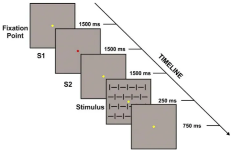
As shown inFig. 1, each trial started with the yellowfixation point and after 1.5 s its color turned to red (called S1) signaling the beginning of the decision period (or S1 interval); during this time the subject had to decide whether to act or not to an upcoming visual stimulus. After 1.5 s, the S1 turned to yellow (called S2) and lasted for 1.5 s; during this phase (S2 interval or preparation interval), the
Fig. 1. Schematic representation and time course of the cues and the stimulus adopted in the present task. Each trial started with the yellowfixation point, after 1500 ms its color turned to red (called S1) signaling the beginning of the decision period (or S1 interval); during this time the subject had to decide whether to act or not to an upcoming visual stimulus. After 1500 ms, the S1 turned to yellow (called S2) and lasted for further 1500 ms; during this phase (S2 interval or preparation interval) the subject had to prepare or not for the motor response according to his/her previous decision. The preparation phase ended with the stimulus onset (250 ms duration).
subject had to prepare or not for the motor response according to his/ her previous decision. The preparation phase ended with the stimulus onset (250 ms duration). The stimulus consisted of one (4°x4°) squared pattern made by vertical and horizontal bars. After stimulus onset, the participant pressed the button as soon as possible, but only if she/he had decided to do so during the S1 interval. The yellowfixation point lasted for other 750 ms after stimulus offset, then turned to red starting a new trial. Participants were instructed to take a balanced number of action choices without having a specific rule of responding (i.e. they could not simply alternate between choices). Participants received informative warning messages at the end of each block if they had made the same decision for more than half trials during that block, and were asked to compensate in the next block. One block consisted of 40 trials and each participant received 10 blocks. A total of 10 blocks allowed us to obtain 400 trials for each subject, of which approximately 200 Response and 200 No-response trials.
Behavioral analysis
The performance speed was assessed using median response time (RT).
Electrophysiological analysis
The EEG signal was recorded using three BrainAmp amplifiers (BrainProducts GmbH, Munich, Germany) with 64 scalp electrodes mounted according to the 10-10 international system initially refer-enced to the left mastoid. Horizontal and vertical electrooculogram (HEOG and VEOG) were additionally recorded with bipolar montages using electrodes at left and right external canthi and below and above the left eye, respectively. Electrode impedances were kept below 5 KΩ. The EEG was digitized at 250 Hz, amplified (band-pass of 0.01–80 Hz including a 50 Hz notch filter) and stored for offline averaging. To reduce high frequency noise, the signal was low passfiltered at 30 Hz (slope 24 dB/octave). The removal of the eye-movement artifacts was performed using the ocular correction based on independent compo-nent analysis (ICA): this method was introduced byJung et al. (2000) and revealed better results when compared to other ocular correction methods (e.g., Hoffmann and Falkenstein, 2008). Then, the artifact rejection was performed to discard epochs contaminated by artifacts or other signals exceeding the amplitude threshold of ± 55μV. The EEG signal was segmented in epochs starting 3500 ms prior the stimulus onset (time 0) and lasting for 5500 ms, with a baseline measured during the initial 500 ms (−3500/−3000 ms). The epochs were aver-aged into two conditions, as follows: Response condition (stimuli followed by response) and No-response condition (stimuli not followed by response).
According to both topographical distribution and previous studies (e.g.,Di Russo et al., 2016), the pN component was measured on Fp1 and Fp2 sites; because of its long-lasting activity, it was calculated as the mean amplitude in six consecutive 500 ms time windows, as follows: −3000/−2500 ms, −2500/−2000 ms, −2000/−1500 ms, −1500/−1000 ms, −1000/−500, −500/0 ms. For the same reason, the BP component was measured as the mean amplitude in the above intervals at Cz site. Given the constant topographical distribution focusing on the vertex, we will refer to the whole pre-stimulus activity at Cz as the BP component, even though the earlier interval (from S1 to S2) can be seen as a CNV-like activity; however, the later interval from S2 to stimulus onset are more properly defined as the BP.
To be more confident in differentiating the decision and the preparation stages, lateralized readiness potentials (LRP) and motor-related amplitude asymmetry in the EEG mu and beta rhythms (mu-and beta-MRAA;de Jong et al., 2006;Poljac and Yeung, 2014) were calculated between C3 and C4 electrodes. LRP was calculated subtract-ing the ERP on C4 from the ERP on C3. The mu-MRAA was calculate extracting the EEG power in the 9–11 Hz range using a complex
demodulation procedure, and then the frequency power was subtracted between C3 and C4. Likewise, the beta-MRAA was calculate in the frequency range 18–26 Hz, and then the same procedures were applied.
ERPs following the stimulus onset were not analyzed because outside the aims of the present study.
Statistical analysis
The pN amplitude was submitted to 6×2×2 repeated measures analysis of variance (RM-ANOVA) with Time window (−3000/−2500, −2500/−2000, −2000/−1500, −1500/−1000, −1000/−500, −500/0), Condition (Response vs. No-response) and Site (Fp1 vs. Fp2) as factors. The BP amplitude was submitted to 6×2 RM-ANOVA with Time window and Condition as factors. Critical alpha was set at p=0.05 and post-hoc comparisons were performed using the Fisher LSD test. Results
Behavioral results
The participants successfully performed the task, as proved by the individual percentage of response and No-response (49.55% Response, 50.45% No-response; t(16)< 1, ns). The median RT was very fast:
202 ms (SD=37.7). Electrophysiological results
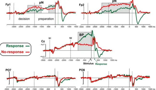
The grand-average ERPs of Response and No-response conditions at the electrodes Fp1, Fp2, Cz, PO7, PO8 are depicted inFig. 2. Top-flat topographical voltage maps of the six 500 ms intervals preceding the stimulus are shown inFig. 3.
The earliest ERP activity (seeFig. 2) was a small P1-N1-P2 complex related to the S1 detection; this activity, peaking over bilateral parietal-occipital sites at 150–350 ms after S1 (−2850/−2650 ms before the stimulus), was detectable also at Cz. Concomitant to this visual activity, a slow rising negativity (CNV/BP) was present over medial central and frontal areas (see Cz inFig. 2) independently from the decision (to respond or not to respond) for the whole S1 interval. At more anterior sites, a prefrontal negativity (pN) started to rise from−2200 ms on both hemispheres, with larger amplitude for No-response than Response condition. The S2 produced a P1-N1-P2 tri-phasic activity, as for the S1; this activity was superimposed on the ongoing slow negativities at Cz. From −1000 ms, the BP component was clearly visible at the vertex and increased in the Response condition only; in the No-response condition, the BP at Cz decreased. In the preparatory phase, the pN component increased in the No-response condition over the right frontopolar site.
ANOVA on the BP amplitude showed significant Time Window effect (F(5,75)=16.71, p < 0.01), indicating larger amplitude in the last
two time windows of the preparation phase (−1000/−500 and −500/0) with respect to previous intervals (all ps < 0.05). A Time Window×Condition significant interaction was found (F(5,75)=8.76, p
< 0.01); post-hoc analysis showed that the amplitude of Response condition was larger than No-response condition in the last two time windows−1000/−500 and −500/0 (all ps < 0.05), while the difference was not significant in the time windows from −3000 to −1000 (all ps > 0.05).
Statistical analysis on pN amplitudes showed a significant Time Window effect (F(5,75)=6.01, p < 0.01). A Site×Condition Interaction
(F(1,15)=6.01, p < 0.05) was found to be significant; post-hoc analysis
indicated that the amplitude of the No-response was larger than Response condition at Fp2 (all ps < 0.05). Further, the Time Window×Site×Condition interaction (F(5,75)=6.98, p < 0.01) was
sig-nificant. Post-hoc analysis showed that in the time windows −2000/ −1500 (decision phase) and −1500/1000 (preparation phase) the
amplitudes of the No-response condition were larger than Response condition in both hemispheres (all ps < 0.05); in the −1000/−500 amplitude did not differ between conditions at Fp1, while it was larger for No-response condition at Fp2; in the last interval (−500/0), the hemispheric difference was also evident, with larger amplitude for No-response compared to Response at Fp2, and smaller amplitude at Fp1 than Fp2 (all ps < 0.05). The time windows showing significant differences between Response and No-response conditions are indi-cated by the gray regions inFig. 2.
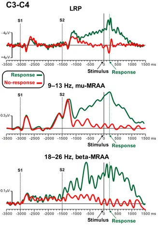
Fig. 4shows the LRP and the mu- and beta-MRAA (C3 minus C4)
for the two conditions (Response vs. No-response) in the same interval of Fig. 2. The LRP during the decision interval did not show any difference between conditions. In contrast, strong lateralized activity (contralateral to the hand used for the response) was present after S2 (the preparation cue) in the Response trials only, while no lateralized activity was detected in the No-response preparation interval. Mu- and beta-MRAA replicated these results. These data indicated that the brain activity during the decision period was very different from that recorded during the preparation interval: only the latter showed a difference between conditions. This support the view that subjects
Fig. 2. Grand averaged ERP waveforms for the−3000/1500 epoch at prefrontal (Fp1 and Fp2), medial central (Cz) and parietal occipital (PO7 and PO8) sites. The Response and the No-response conditions are superimposed. The−3500/−3000 interval was taken as baseline. Based on the instruction given to the subjects, the −3000/−1500 was labeled “decision” stage; the−1500/0 interval was labeled “preparation” stage. Time zero represents the stimulus onset. The pN (prefrontal negativity) and the BP (Bereitschaftspotential) components are labeled in thefigures. Black dotted vertical lines represent S1 and S2; green dotted vertical lines represent response emission in case of Response condition. The gray regions indicate the portions showing significant difference between Response and No-response conditions.
Fig. 3. Topographical maps of the pre-stimulus activity at different time windows during the “decision” and “preparation” stages for the Response (top-panel) and No-response (bottom-panel) conditions. The pN component emerged as a negative activity over anterior sites present only in the No-response condition: it shows a bilateral distribution during the last time window of the“decision” stage and a right lateralization during the final window of the “preparation” stage.
could conform to the instructions differentiating between decision and preparation stages.
Discussion
The main result of the present study is that Response and No-response conditions show different electrophysiological features during both the decision and the preparation stages. Thus, the ERP activities allow to predict whether the subject will respond or not to the upcoming stimulus.
During the decision phase, the prefrontal pN component grows bilaterally when participants decide not to respond, while this activity is smaller when they decide to respond. In contrast, in the same interval the activity recorded over the premotor areas grows indepen-dently from the decision taken, being comparable between response and no response trials. Thus, the bilateral pN is thefirst electrophy-siological sign of the no-response decision, and supports the view that the pN may reflect a form of top-down proactive inhibitory control. On the other hand,findings on the slow negative activity over the vertex (Cz), which was independent from the decision category, can be explained in light of the CNV literature suggesting an interpretation of this activity in cognitive terms, likely a temporal orienting to the task (see Kononowicz and Penney (2016) for review). In other words, presentfindings indicate that any decision about future action (includ-ing not act(includ-ing) is mapped within pre-motor cortices. This data might support the view that alternative intentions (both action and no-action) are present at motor level, but not at prefrontal level where the activity is enhanced only in case of inhibition, as discussed above.
The preparation stage is also characterized by sustained pN activity in case of decision to not respond, but, differently from the previous decision stage, the pN distribution is lateralized on the right hemi-sphere. Thisfinding is consistent with neuroimaging results suggesting that proactive inhibitory control relies on activation of the right inferior
frontal regions (Garavan et al., 1999;Konishi et al., 1998;Rubia et al., 2003;Aron et al., 2003). Especially interesting is the comparison with the Brass and Haggard (2007) results, indicating that the medial prefrontal cortex is involved in self-inhibition, while the pre-motor areas are equally activated in intentional actions, both in the action trials and in the inhibition trials. In partial contrast to these latter data, we clearly observed larger amplitude in premotor areas in case of Response than No-response trials during the preparation phase, although the amplitude over the same areas was similar during the previous decision phase. The observed differences may be due to the different experimental paradigms; in our study, we tried to separate decision and preparation processes, while activities related to these two processing stages overlap in Brass and Haggard (2007) self-paced paradigm. However, present results are in line with previous ERP data (Filipovic et al., 1999), showing that the late CNV (comparable to the BP) was present only when the instruction was to move after the cue presentation. Thus, it seems that thefine temporal analysis typical of ERP can distinguish between preparation to respond and not to respond better than neuroimaging recordings. Further, present find-ings show a prominent inhibitory activity at prefrontal sites (especially on the right hemisphere), likely originating in the iFg (Di Russo et al., 2016).
Further, in the preparation phase, we observed a parallel amplitude increase of both prefrontal (pN) and premotor (BP) activities when the previously taken decision was to respond (see the green trace inFig. 2). The excitability level of premotor regions (BP) seems therefore to reflect a sort of accelerating system. At the same time, the activity within the right prefrontal cortex (pN) seem to reflect a sort of braking system, regulating the action intention in order to release the action only at the appropriate time.
One could argue that the decision and the preparation stages may involve not only proactive inhibitory control process per se, but also "switch", "tracking proceeding response", and "motor preparation" processes. Although this cannot be completely excluded, we should highlight that we instructed the participants of the present study to decide the next action in the initial 1.5 time-frame and to prepare to act (or not) in the successive 1.5 s interval, which was signaled by cues. At the end of each run, all participants reported to be able to follow the instructions and to perform the task according to the request. In addition, motor preparation, revealed by the BP component, was only present in the preparation phase of response trials, indicating that the action execution was actually prepared in that stage. Moreover, no differences between Response and No-Response conditions were found over pre-motor areas (see Cz) and in the LRP, mu- and beta-MRAA during the decision stage providing further support to the ability of the subjects to conform to the task instruction.
Present findings reinforce and extend the notion of proactive inhibitory control in the prefrontal cortex, consistent with results showing its involvement in a more general regulation of behavioral inhibition (Meyer and Bucci, 2016).
Conclusions
In conclusion, the present work contributes to the description of the electro-cortical correlates of proactive action control, necessary when one should take and maintain a decision to react or not to expected events, which is a fundamental expression of the social self-control. Present data suggests a model of self-control in which thefirst brain signal of the decision of not to respond is the bilateral prefrontal negative activity (pN), which supports the view that the pN reflects proactive action inhibition. During the preparation stage, in case the subject decided not to react, only the right iFg increases its activity, further supporting the inhibitory interpretation of this brain wave. In contrast, after choosing to respond, the right iFg activity is clearly reduced and the excitatory premotor activity grows with a high gain.
Overall, we extend the hypothesis of an accelerating-breaking
Fig. 4. LRP, mu- and beta-MRAA during the decision and preparation phases for the Response and No-response conditions.
system during proactive action control, indicating the neural origin and the time-course of this fundamental cognitive and social skill. References
Aron, A.R., Fletcher, P.C., Bullmore, E.T., Sahakian, B.J., Robbins, T.W., 2003. Stop-signal inhibition disrupted by damage to right inferior frontal gyrus in humans. Nat. Neurosci. 6 (2), 115–116.
Aron, A.R., 2011. From reactive to proactive and selective control: developing a richer model for stopping inappropriate responses. Biol. Psychiatry 69 (12), e55–e68. Baumeister, R.F., Vohs, K.D., Tice, D.M., 2007. The strength model of self-control. Curr.
Dir. Psychol. Sci. 16 (6), 351–355.
Berchicci, M., Lucci, G., Pesce, C., Spinelli, D., Di Russo, F., 2012. Prefrontal hyperactivity in older people during motor planning. Neuroimage 62 (3), 1750–1760.
Berchicci, M., Lucci, G., Di Russo, F., 2013. Benefits of physical exercise on the aging brain: the role of the prefrontal cortex. J. Gerontol. Ser. A: Biol. Sci. Med. Sci. 68 (11), 1337–1341.
Berchicci, M., Lucci, G., Perri, R.L., Spinelli, D., Di Russo, F., 2014. Benefits of physical exercise on basic visuo-motor functions across age. Front. Aging Neurosci. 6, 48. Berchicci, M., Lucci, G., Spinelli, D., Di Russo, F., 2015. Stimulus onset predictability modulates proactive action control in a Go/No-Go task. Front. Behav. Neurosci., 9. Berchicci, M., Spinelli, D., Di Russo, F., 2016. New insights into old waves. Matching
stimulus-and response-locked ERPs on the same time-window. Biol. Psychol. 117, 202–215.
Brass, M., Haggard, P., 2007. To do or not to do: the neural signature of self-control. J. Neurosci. 27 (34), 9141–9145.
Braver, T.S., Paxton, J.L., Locke, H.S., Barch, D.M., 2009. Flexible neural mechanisms of cognitive control within human prefrontal cortex. Proc. Natl. Acad. Sci. 106 (18), 7351–7356.
Cai, W., Oldenkamp, C.L., Aron, A.R., 2011. A proactive mechanism for selective suppression of response tendencies. J. Neurosci. 31 (16), 5965–5969. Di Russo, F., Lucci, G., Sulpizio, V., Berchicci, M., Spinelli, D., Pitzalis, S., Galati, G.,
2016. Spatiotemporal brain mapping during preparation, perception, and action. NeuroImage 126, 1–14.
de Jong, R., Gladwin, T.E., M't Hart, B., 2006. Movement-related EEG indices of preparation in task switching and motor control. Brain Res. 1105 (1), 73–82. Filipovic, S.R., Jahanshahi, M., Rothwell, J.C., 1999. Cortical potentials related to decision‐making: comparison of two types of go/no‐go decision. Neuroreport 10 (17), 3583–3587.
Frank, M.J., 2006. Hold your horses: a dynamic computational role for the subthalamic nucleus in decision making. Neural Netw. 19 (8), 1120–1136.
Garavan, H., Ross, T.J., Stein, E.A., 1999. Right hemispheric dominance of inhibitory control: an event-related functional MRI study. Proc. Natl. Acad. Sci. 96 (14), 8301–8306.
Gillies, A.J., Willshaw, D.J., 1998. A massively connected subthalamic nucleus leads to the generation of widespread pulses. Proc. R. Soc. Lond. B: Biol. Sci. 265 (1410), 2101–2109.
Gomez, C.M., Marco, J., Grau, C., 2003. Preparatory visuo-motor cortical network of the contingent negative variation estimated by current density. Neuroimage 20 (1), 216–224.
Haynes, J.D., Sakai, K., Rees, G., Gilbert, S., Frith, C., Passingham, R.E., 2007. Reading hidden intentions in the human brain. Curr. Biol. 17 (4), 323–328.
Hoffmann, S., Falkenstein, M., 2008. The correction of eye blink artefacts in the EEG: a comparison of two prominent methods. PLoS One 3 (8), e3004.
Jahanshahi, M., Jenkins, I.H., Brown, R.G., Marsden, C.D., Passingham, R.E., Brooks, D.J., 1995. Self-initiated versus externally triggered movements. Brain 118 (4), 913–933.
Jung, T.P., Makeig, S., Westerfield, M., Townsend, J., Courchesne, E., Sejnowski, T.J., 2000. Removal of eye activity artifacts from visual event-related potentials in normal and clinical subjects. Clin. Neurophysiol. 111 (10), 1745–1758.
Kenemans, J.L., 2015. specific proactive and generic reactive inhibition. Neurosci. Biobehav. Rev. 56, 115–126.
Kleerekooper, I., van Rooij, S.J., van den Wildenberg, W.P., de Leeuw, M., Kahn, R.S., Vink, M., 2016. The effect of aging on fronto-striatal reactive and proactive inhibitory control. NeuroImage 132, 51–58.
Konishi, S., Nakajima, K., Uchida, I., Sekihara, K., Miyashita, Y., 1998. No‐go dominant brain activity in human inferior prefrontal cortex revealed by functional magnetic resonance imaging. Eur. J. Neurosci. 10 (3), 1209–1213.
Kononowicz, T.W., Penney, T.B., 2016. The contingent negative variation (CNV): timing isn’t everything. Curr. Opin. Behav. Sci. 8, 231–237.
Kornhuber, H.H., Deecke, L., 1965. Hirnpotentialänderungen bei Willkürbewegungen und passiven Bewegungen des Menschen: Bereitschaftspotential und reafferente Potentiale. Pflüg.'S. Arch. Gesamt. Physiol. Mensch. Tiere 284 (1), 1–17. Logan, G.D., Burkell, J., 1986. Dependence and independence in responding to double
stimulation: a comparison of stop, change, and dual-task paradigms. J. Exp. Psychol.: Hum. Percept. Perform. 12 (4), 549–563.
Loveless, N.E., Sanford, A.J., 1974. Slow potential correlates of preparatory set. Biol. Psychol. 1 (4), 303–314.
Meyer, H.C., Bucci, D.J., 2016. Imbalanced activity in the orbitofrontal cortex and nucleus accumbens impairs behavioral inhibition. Curr. Biol. 26 (20), 2834–2839. Oldfield, R.C., 1971. The assessment and analysis of handedness: the Edinburgh
inventory. Neuropsychologia 9 (1), 97–113.
Perri, R.L., Berchicci, M., Spinelli, D., Di Russo, F., 2014. Individual differences in response speed and accuracy are associated to specific brain activities of two interacting systems. Front. Behav. Neurosci. 8, 251.
Perri, R.L., Berchicci, M., Lucci, G., Spinelli, D., Di Russo, F., 2015. The premotor role of the prefrontal cortex in response consistency. Neuropsychology 29 (5), 767. Perri, R.L., Berchicci, M., Lucci, G., Spinelli, D., Di Russo, F., 2016. How the brain
prevents a second error in a perceptual decision-making task. Sci. Rep., 6. Poljac, E., Yeung, N., 2014. Dissociable neural correlates of intention and action
preparation in voluntary task switching. Cereb. Cortex 24 (2), 465–478. Rubia, K., Smith, A.B., Brammer, M.J., Taylor, E., 2003. Right inferior prefrontal cortex
mediates response inhibition while mesial prefrontal cortex is responsible for error detection. Neuroimage 20 (1), 351–358.
van Boxtel, G.J., Böcker, K.B., 2004. Cortical measures of anticipation. J. Psychophysiol. 18 (2/3), 61–76.
Vaughan, H.G., Costa, L.D., Ritter, W., 1968. Topography of the human motor potential. Electroencephalogr. Clin. Neurophysiol. 25 (1), 1–10.
Verbruggen, F., Logan, G.D., 2009. Proactive adjustments of response strategies in the stop-signal paradigm. J. Exp. Psychol.: Hum. Percept. Perform. 35 (3), 835–854. Verbruggen, F., Stevens, T., Chambers, C.D., 2014. Proactive and reactive stopping when
distracted: an attentional account. J. Exp. Psychol.: Hum. Percept. Perform. 40 (4), 1295–1300.
Vink, M., Kahn, R.S., Raemaekers, M., van den Heuvel, M., Boersma, M., Ramsey, N.F., 2005. Function of striatum beyond inhibition and execution of motor responses. Hum. Brain Mapp. 25 (3), 336–344.