Downloaded from https://journals.lww.com/pec-online by BhDMf5ePHKav1zEoum1tQfN4a+kJLhEZgbsIHo4XMi0hCywCX1AWnYQp/IlQrHD32kySThwcl1uRPnK6UuhH3uOWPTtnCEYxib8q6ASGN30= on 12/18/2019 Downloadedfrom https://journals.lww.com/pec-onlineby BhDMf5ePHKav1zEoum1tQfN4a+kJLhEZgbsIHo4XMi0hCywCX1AWnYQp/IlQrHD32kySThwcl1uRPnK6UuhH3uOWPTtnCEYxib8q6ASGN30=on 12/18/2019
Esophageal Retained Lithium Battery in
Children Younger than 6 Years
A Prompt Structurated Multidisciplinary Approach Is Essential
to Reduce Long-Term Consequences
Gabriele Lisi, MD, PhD,* Maria Teresa Illiceto, MD,
† Erminia Francesca Romeo, MD,‡
Giuseppe Lauriti, MD, PhD,* Simona Faraci, MD,
‡ Giuliano Lombardi, MD,†
Luigi Dall'Oglio, MD,
‡ and Pierluigi Lelli Chiesa, MD*
Objectives:Disk battery esophageal retention in children younger than 6 years represents an increasing endoscopic emergency, followed by a rel-evant risk of life-threatening late complications. Surgical removal after a failed endoscopic approach is rarely reported in the literature. We describe our experience in this scenario.
Methods:Two female asymptomatic patients aged 26 and 29 months pre-sented within 4 hours after a witnessed ingestion of a 2-cm, 3-V lithium battery (CR2032) retained in the cervical esophagus. Both patients underwent a prolonged unsuccessful emergent endoscopic removal with a flexible instrument performed by an adult gastroenterologist. Both batteries fused with the esophageal wall were extracted through a longitudinal left cervical esophagotomy combined with minimal resection of necrotic tis-sues and repaired over a 12F feeding tube.
Results:Patients were extubated after 12 and 72 hours, respectively. Con-trast study was performed after 20 and 13 days, respectively, before re-suming oral feeding. At endoscopy, the first patient developed a 3-cm-long severe esophageal stenosis (35th day), followed by an asymptomatic tracheoesophageal fistula (60th day), which was conservatively treated. After spontaneous resolution of the tracheoesophageal fistula, esopha-geal stenosis progressed, partially responsive to esophaesopha-geal stenting. Short esophagectomy is under evaluation. The second patient developed an asymptomatic limited stenosis, not requiring dilatation.
Conclusions:The emergent management of lithium battery ingestion needs a structured timely multidisciplinary approach in the emergency department, an experienced pediatric endoscopist, and a simultaneous engagement of pediatric surgical expertise, even in patients who do not show bleeding, to reduce esophageal exposure time to high-voltage current released by batteries, which represents the main factor conditioning tissue damage and prognosis.
Key Words: disk batteries, ingestion, esophagus, esophageal stenosis (Pediatr Emer Care 2018;00: 00–00)
L
ithium battery (LB) esophageal retention in children younger than 6 years represents an increasing social and endoscopicemergency, followed by a relevant risk of life-threatening complications.1–3 To date, 59 deaths in children younger than 6 years who underwent battery ingestion have been reported worldwide from 1977, mainly related to LB.4The causal
mecha-nism of death in at least 36 children was massive bleeding from aortoesophageal fistula5or fistulae with other major vessels of the mediastinum,6followed by esophageal perforation (10 cases) or tracheoesophageal fistula (TEF; 9 cases). In the same period, 231 cases with severe esophageal or airway injury are reported.7 The severity of injury depends on battery type, size, voltage, loca-tion, and duration of close contact with the mucosa. The most im-portant lesion mechanism consists of the electrical generation of caustic hydroxide ions at the negative pole proportional to the bat-tery voltage. Tanaka et al,8in an animal canine model, demon-strated that sodium hydroxide is produced much more rapidly with LB (3 V) than with other button cells because the amount of alkali produced in tissue is proportional to the electric current produced, and the same amount of current is produced more rap-idly with the higher-voltage lithium cell.
The recent and increased use of the more powerful LB has increased the risk of significant tissue damage, which can oc-cur after just 2 hours, from their lodgment in the esophagus in small children.9
In this dramatic scenario, endoscopic removal of LB retained in the esophagus of small children represents a frequently success-ful endoscopic emergency to be performed as soon as possible, under safety conditions.
The role of surgery after ingestion of LB is mainly reported as an emergent attempt in patients presenting with massive bleeding10–12or for the treatment of complications such as esoph-ageal perforation,13TEF,14,15or esophageal stenosis, whereas sur-gical successful removal of batteries after a failed endoscopic approach is not reported in the literature.
We report 2 cases of surgical approach to LBs retained in the esophagus after a failed prolonged attempt of endoscopic removal, describing postoperative management and outcomes.
METHODS
The medical records of 2 consecutive female patients aged 26 and 29 months, respectively, presented in our department from October 2015 to October 2016 within 4 hours after a witnessed in-gestion of a 2-cm, 3-V LB (CR2032) retained in the cervical esophagus were reviewed. Both patients underwent a prolonged unsuccessful emergent endoscopic removal with a flexible instru-ment, followed by a successful extraction through a longitudinal left cervical esophagotomy combined with minimal resection of necrotic tissues. The postoperative management and outcomes are reported.
From the *Pediatric Surgery Unit—University “G. d'Annunzio” of Chieti-Pescara; †Pediatric Gastroenterology and Endoscopic Service—Hospital “Santo Spirito”, Pescara; and‡Digestive Endoscopy and Surgery Unit—Children's Hospital “Bambino Gesù”, Rome, Italy.
Disclosure: The authors declare no conflict of interest.
Reprints: Lisi Gabriele, MD, PhD, UOC Clinicizzata di Chirurgia Pediatrica, P.O. “Santo Spirito,” Via Fonte Romana, 8, 65124 Pescara, Italy
(e‐mail: [email protected]).
Copyright © 2018 The Author(s). Published by Wolters Kluwer Health, Inc. This is an open-access article distributed under the terms of the Creative Commons Attribution-Non Commercial-No Derivatives License 4.0 (CCBY-NC-ND), where it is permissible to download and share the work provided it is properly cited. The work cannot be changed in any way or used commercially without permission from the journal.
RESULTS Case 1
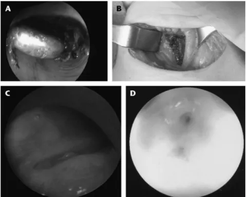
A 26-month-old female patient was sent to our department from a surrounding hospital 2½ hours after a witnessed ingestion of a 2-cm, 3-V LB (CR2032), which was changed from the televi-sion remote controller by her father 4 days ahead. The patient underwent a cervicothoracic x-ray assessment that returned the presence of a large button battery with a“halo sign” retained in the cervical esophagus. Before sending the patient to our hospital, where pediatric endoscopic and surgical expertise is present, the doctors of the accepting emergency department (ED) expected the blood tests to return from the laboratory. A total of 4 hours passed between ingestion of LB and delivery of the baby. In our hospital during the night, with the baby intubated, an emergent en-doscopy with an 8-mm flexible instrument was performed by the adult gastroenterologist on call, in the operating room, in the pres-ence of the pediatric surgeon on duty. An unsuccessful endoscopic removal of the LB using different endoscopic retrieval instruments was attempted for 1 hour, but the battery was literally fused with necrotic esophageal wall on his negative pole (Fig. 1A); thus, any further attempt was considered at risk. At this time, using a left lower cervical approach, a 3-cm-long longitudinal esophagotomy was performed where transmural full-thickness esophageal necrosis was found at the level of the left common carotid artery that was adherent to necrotic tissues but apparently not involved (Fig. 1B). After retrieving the intact battery, the necrotic esophageal wall was partially removed and the controlateral esophageal mucosa was inspected, evidencing a 360-degree mucosal burns. The esophageal wall was repaired over a 12F polyurethane feeding tube and a Penrose drain was left in periesophageal space. The patient was transferred intubated in the pediatric intensive unit, where therapy with proton-pump inhibitors and antibiotic was started. The patient was extubated the afternoon
after surgery, after a contrast-enhanced computed tomographic scan demonstrating the absence of evolution of the vascular involvement. A swallow contrast study was performed after 20 days, before considering to resuming oral feeding, returning the absence of contrast extravasation but evidencing an esophageal stenosis at the level of esophagotomy. After further 2 weeks (35th postoperative day), the endoscopic evaluation confirmed the presence of a 3-cm-long severe esophageal stenosis, but tissues consistence was considered inadequate to perform a dilatation safely. A 12F polyurethane feeding was still left in site for enteral nutrition, which was well tolerated. A second endoscopy (60th postoperative day) with radiologic evaluation of the stenosis returned the suspicion of a TEF, which was confirmed at tracheoscopy (Fig. 1C). Considering the absence of symptoms related to the TEF, a conservative treatment was attempted: the stenosis was not dilated and a 12F feeding tube was left still in site. After 4 weeks, the TEF evolved to a spontaneous resolution, endoscopically documented, whereas the esophageal stenosis worsened, allowing nothing but the passage of an 8F feeding tube under direct vision. At this time, considered the freshly resolved TEF, esophageal dilatation was still considered at risk for TEF recurrence or esophageal rupture and postplaced. After a few days, the girl removed accidentally the probe. An attempt to reinsert the feeding tube under direct endoscopic vision failed, because of the progression of the stenosis that made the esophageal lumen virtual (Fig. 1D). After a limited longitudinal mucosal incision on the posterior esophageal wall with precut endoscopic needle, a progressive esophageal dilatation with Savary-Gilliards dilators was performed that allowed for the positioning of a first custom dynamic stent,16
left in site for 4 months, obtaining an adequate esophageal caliber. After relapse of the esophageal stenosis, a second dynamic stent was positioned, followed by a cycle of successful progressive esophageal dilatation with Savary-Gilliards
FIGURE 1. A, Endoscopic view of the LB in the cervical esophagus adjacent to necrotic tissue of the esophageal wall. B, Through a basal transverse left cervicothomy, the whole-thickness necrotic esophagus adjacent to the left common carotid artery is opened and the DB extracted. C, Tracheoscopic view of the TEF. D, Severe esophageal stenosis with virtual lumen 3 months postoperatively.
dilators until reaching the goal to accommodate a 16F feeding tube. At the time of writing, the patient has a stricture 0.5 cm in length and she is able to assume a creamy diet, integrated with a gastric feeding through the tube. Because of the level of the stricture, just below the pharyngoesophageal junction, further attempts of conservative stricture management have been scheduled, avoiding surgery.
Case 2
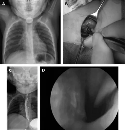
A 24-month-old girl came to our hospital after 2 hours from a witnessed ingestion of a foreign body taken from a basket of used batteries. Thoracoabdominal x-rays evidenced the presence of a lower cervical foreign body with a“halo sign,” compatible with a 2-cm, 3-V LB (CR2032) (Fig. 2A). Within 1 hour, the patient underwent an unsuccessful attempt of endoscopic removal with a flexible instrument, performed by the adult gastroenterologist on call, in the operating room under general anesthesia. At this time, with a left lower cervical approach, a 2-cm-long longitudinal esophagotomy was performed on the esophageal wall, not transmurally necrotic, at the level of left common carotid artery, which was apparently not involved (Fig. 2B). After retrieving the intact battery, the esophageal mucosa was inspected, evidencing a mucosal edema. The esophageal wall was repaired over a 12F polyurethane feeding tube and a Penrose drain was left in periesophageal space. The patient was transferred intubated
in the pediatric intensive unit where therapy with proton-pump inhibitors and antibiotic was started. In the first postoperative day, a contrast-enhanced computed tomographic scan excluded any progression of tissue damage. The patient was extubated after 72 hours. A contrast study, performed 13 days after surgery (Fig. 2C), excluded esophageal perforation or TEF, allowing for a gradual introduction of liquid oral feeding. An endoscopic control was performed at 1 and 4 months postoperatively. returning an asymptomatic limited stenosis, not requiring dilatation (Fig. 2D). One year after surgery, the patient is asymptomatic, at full diet.
DISCUSSION
In most cases, battery ingestion does not result in any or mi-nor sequelae for the health of affected children. However, despite a moderate decrease in the overall rate of ingestion in the last de-cade (from 12 to <10.5 cases per million population), in the same period, a linear increase of major or fatal outcomes has been reg-istered (from 0.4% to 1% of patients).17These data are derived
from the increased use of large-diameter button batteries, espe-cially 20-mm diameter high-voltage LB, that are demonstrated to severely damage the esophageal wall within a very limited time of contact with mucosa (<2 hours from ingestion).
If we look at monocentric11or large epidemiological series,4 the percentage of fatal cases is around 20% to 25% of the entire cohort of patients at risk for severe effects. These data indicate that
FIGURE 2. A, Radiologic anteroposterior view of the DB retained at the cervical level, with the classical peripheral double rim. B, The DB retained in the cervical esophagus during surgical extraction. C, Contrast-enhanced radiographic esophagram at the 14th postoperative day demonstrating a limited esophageal stenosis without evidence of esophageal perforation or TEF. D, Endoscopic view of the esophageal hyperemic mucosa 4 months postoperatively.
an essential effort has to be made to rapidly identify and ade-quately treat the limited number of children with high risk of esophageal, vascular, or airways damage. Children younger than 6 years who ingested high-voltage button batteries of at least 20 mm are particularly at risk, mainly in case of unwitnessed in-gestion with battery permanence in the esophagus lasting more than 2 hours.18,19Our 2 patients presented with many of the pre-viously cited risk factors: small age, high-voltage large-diameter LB, and prolonged contact time with esophageal mucosa.
According with the European Society of Gastrointestinal Endoscopy–European Society for Paediatric Gastroenterology Hepatology and Nutrition guidelines,20 emergent removal (<2 hours) of the retained disk battery (DB) is the goal of the acute phase after the radiologic highlighting of the battery posi-tion. This goal can be accomplished through different methods, such as endoscopic removal, balloon extraction with fluoroscopy, and esophageal bougienage, with the endoscopic approach to be preferred for its safety and completeness of derivable information, being the only one method that, under direct vision, can return the extent of mucosal damage. Whenever possible, the intervention of an endoscopist with adequate pediatric expertise coming from different subspecialties (pediatric surgeon, otolaryngologist, or gastroenterologist, depending on the center policy) should be obtained. Regarding the type of endoscopic instrument to pre-fer, the pediatric literature do not show any significant differences among rigid and flexible endoscopies in terms of the efficacy of
esophageal foreign body retrieval in children,21,22but any of the
articles available in literature refers only to the specific situation of DB esophageal retention.
In our 2 cases, a prolonged attempt of endoscopic removal was performed by experienced adult gastroenterologists on call with flexible instruments that failed to detach the DB from the esophageal mucosa. On the base of subsequent surgical finding of batteries fused with esophageal wall, we could only speculate that, in these specific cases, a rigid endoscope would not have added any advantage in terms of effectiveness; rather, it would have increased the risk of esophageal perforation.
Success rate of endoscopic approach has been reported as high as 98% to 100% of cases of foreign bodies retained in the esophagus of small children,22with very few cases requiring a
surgical removal,21but to the best of our knowledge, this event
has never been described for DB presenting without massive or sentinel bleeding, such as in our 2 cases.
Some authors, on the basis of the high risk of mortality, agree to advocate a contemporary emergent cardiothoracovascular sur-gical approach performed by the general (pediatric) surgeon or cardiovascular (pediatric) surgeon, possibly combined with intra-operative endoscopy, in cases of documented DB ingestion retained in the esophagus presenting at the ED with clinical signs of vascular fistula (sentinel or massive bleeding).23,24We agree not only that, under these life-threatening circumstances, the pres-ence of a surgeon in the management protocol is recommended,
but also that an emergent surgical approach is a priority over the endoscopic removal of the retained DB.
The protocols used by the aforementioned authors differ when patients present at the ED without a sign of vascular fistula, such as in our 2 cases. Brumbaugh et al23do not take into consid-eration a surgical presence during the endoscopic maneuver, un-less for immediate rigid esophagoscopy where significant esophageal edema makes flexible endoscopy battery removal im-possible. Barabino et al,24in case of DB incarcerated in the esoph-ageal wall with endoscopic finding of severe and deep ulceration of the esophageal wall, suggest a surgical cervical or thoracotomic approach combined with the endoscopic removal of the DB, thus preventing uncontrollable fatal bleeding.
In the 2 reported patients from our experience, both present-ing without a sign of bleedpresent-ing, the presence of pediatric surgeons assisting live the endoscopic maneuver made a direct multidisci-plinary evaluation of the mucosal damage possible, suggesting in a shared way not to proceed further with endoscopic attempt and to immediately convert to a surgical successful approach. The latter certainly resulted in a partial resection of necrotic esophageal wall which ultimately contributed, together with the tissue damage induced by the LB, to the following esophageal ste-nosis. However, the decision to proceed surgically was at that point the only effective method to prevent the progression of ne-crosis to the surrounding vascular structures.
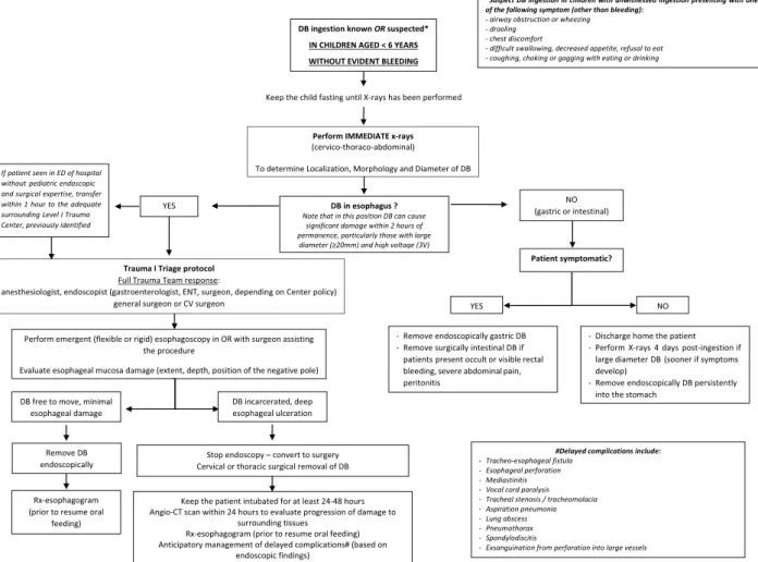
We agree with Barabino et al that, under these circumstances at high risk of severe short-term or midterm complications, a sur-gical presence in the operating room during endoscopy is essential to offer the child the maximal chance of safe management. What is stated is particularly sensible in children at major risk (age <6 years, large-diameter high-voltage DB, battery persistence in the esophagus lasting >2 hours), for whom a specific manage-ment protocol is proposed (Fig. 3).
Epidemiological data and our experience put the spotlight on 2 essential aspects of the urgent management of these cases in the ED:
1. The absolute need to promptly send the patient to the operating room to perform the endoscopic removal attempt, to be carried out as soon as possible. For this purpose, Russell and col-leagues25clearly demonstrated that, in their level I trauma cen-ter, the activation of an immediate full trauma team response (trauma I triage protocol) is useful to reduce the risk of compli-cations in patients at risk. A trauma I triage protocol entails im-mediate notification of the pediatric trauma team, the anesthesia team, the radiology technician, and the operating room charge nurse of the arrival of trauma patients to the ED. This allows for immediate response of the pediatric trauma surgeon and/or endoscopist and immediate confirmation of the presence and site of battery by imaging. Availability of an operating room for potential operative intervention is also secured.
2. The opportunity to guarantee the patient the assistance of a multidisciplinary team that can manage urgently both the endo-scopic priority and any surgical requirement. We are well aware of the limitations imposed by an approach requiring the simul-taneous presence of endoscopic and surgical expertise, particu-larly in trauma centers other than level I. However, the duty to prevent severe damages in selected pediatric cases and the pos-sibility of a surgical procedure after endoscopy should be well known. This knowledge should direct the protocols of ED when facing a child with documented DB ingestion belonging to categories at high risk to rapidly address the patient to the more adequate level I trauma referral center, previously identi-fied, reachable within a reasonable time frame of no more than
1 hour, with which a transfer agreement was found. Any at-tempt at endoscopic or fluoroscopic removal of large DB retained in the cervical esophagus should absolutely be avoided if there is no adequate pediatric surgical expertise in the hospi-tal, also in case without sentinel or massive bleeding.
In conclusion, the emergent management of LB ingestion needs a structured timely multidisciplinary approach beginning from the ED, an endoscopist with pediatric experience, and a si-multaneous engagement of pediatric surgical expertise, to reduce esophageal exposure time to high-voltage current released by bat-teries, which represents the main factor conditioning tissue dam-age and prognosis.
REFERENCES
1. Panella NJ, Kirse DJ, Pranikoff T, et al. Disk battery ingestion: case series with assessment of clinical and financial impact of a preventable disease. Pediatr Emerg Care. 2013;29:165–169.
2. Patel SA, Hillel AD, Perkins J. Battery ingestion leading to bilateral vocal cord paresis. JAMA Otolaryngol Head Neck Surg. 2013;139:304–306. 3. Peters NJ, Mahajan JK, Bawa M, et al. Esophageal perforations due to foreign body impaction in children. J Pediatr Surg. 2015;50:1260–1263. 4. National Capital Poison Center. Fatal button battery ingestions: 59 reported
cases. Available at: http://www.poison.org/battery/fatalcases.asp. Accessed May 7, 2018.
5. Mortensen A, Hansen NF, Schiødt OM. Fatal aortoesophageal fistula caused by button battery ingestion in a 1-year-old child. Am J Emerg Med. 2010;28:984.e5–984.e6.
6. Loots DP, du Toit-Prinsloo L, Saayman G. Disk battery ingestion: a rare cause of perforation of the brachiocephalic artery. Forensic Sci Med Pathol. 2015;11:614–617.
7. National Capital Poison Center. Nonfatal button batteries ingestions with severe esophageal or airways injury: 231 cases. Available at: https://www. poison.org/battery/severecases. Accessed May 7, 2018.
8. Tanaka J, Yamashita M, Yamashita M, et al. Esophageal electrochemical burns due to button type lithium batteries in dogs. Vet Hum Toxicol. 1998; 40:193–196.
9. Jatana KR, Litovitz T, Reilly JS, et al. Pediatric button battery injuries: 2013 task force update. Int J Pediatr Otorhinolaryngol. 2013;77:1392–1399. 10. Hamilton JM, Schraff SA, Notrica DM. Severe injuries from coin cell
battery ingestions: 2 case reports. J Pediatr Surg. 2009;44:644–647. 11. Leinwand K, Brumbaugh DE, Kramer RE. Button battery ingestion in
children: a paradigm for management of severe pediatric foreign body ingestions. Gastrointest Endosc Clin N Am. 2016;26:99–118. 12. Nisse P, Lampin ME, Aubry E, et al. Ingestion d'une pile bouton
compliquée d'une fistule oeso-aortique fatale. Proposition d'un algorithme de prise en charge chez l'enfant de moins de 6 ans. Presse Med. 2016;45: 947–953.
13. Soccorso G, Grossman O, Martinelli M, et al. 20 mm lithium button battery causing an oesophageal perforation in a toddler: lessons in diagnosis and treatment. Arch Dis Child. 2012;97:746–747.
14. Hammond P, Jaffray B, Hamilton L. Tracheoesophageal fistula secondary to disk battery ingestion: a case report of gastric interposition and tracheal patch. J Pediatr Surg. 2007;42:e39–e41.
15. Okuyama H, Kubota A, Oue T, et al. Primary repair of tracheoesophageal fistula secondary to disc battery ingestion: a case report. J Pediatr Surg. 2004;39:243–244.
16. Foschia F, De Angelis P, Torroni F, et al. Custom dynamic stent for esophageal strictures in children. J Pediatr Surg. 2011;46:848–853.
17. National Capital Poison Center. Button batteries ingestions statistics. Available at: https://www.poison.org/battery/stats#2016. Accessed November 5, 2017.
18. Litovitz T, Whitaker N, Clark L, et al. Emerging battery-ingestion hazard: clinical implications. Pediatrics. 2010;125:1168–1177.
19. Eliason MJ, Melzer JM, Winters JR, et al. Identifying predictive factors for long-term complications following button battery impactions: a case series and literature review. Int J Pediatr Otorhinolaryngol. 2016;87: 198–202.
20. Thomson M, Tringali A, Dumonceau JM, et al. Paediatric gastrointestinal endoscopy: European Society for Paediatric Gastroenterology Hepatology and Nutrition and European Society of Gastrointestinal Endoscopy Guidelines. J Pediatr Gastroenterol Nutr. 2017;64:133–153.
21. Popel J, El-Hakim H, El-Matary W. Esophageal foreign body extraction in children: flexible versus rigid endoscopy. Surg Endosc. 2011;25:919–922. 22. Russell R, Lucas A, Johnson J, et al. Extraction of esophageal foreign
bodies in children: rigid versus flexible endoscopy. Pediatr Surg Int. 2014; 30:417–422.
23. Brumbaugh DE, Colson SB, Sandoval JA, et al. Management of button battery-induced hemorrhage in children. J Pediatr Gastroenterol Nutr. 2011;52:585–589.
24. Barabino AV, Gandullia P, Vignola S, et al. Lithium battery lodged in the oesophagus: a report of three paediatric cases. Dig Liver Dis. 2015;47: 984–986.
25. Russell RT, Griffin RL, Weinstein E, et al. Esophageal button battery ingestions: decreasing time to operative intervention by level I trauma activation. J Pediatr Surg. 2014;49:1360–1362.