Basic Science Investigations
Respiration 2015;89:329–342 DOI: 10.1159/000375168
Phospho-p38 MAPK Expression in
COPD Patients and Asthmatics and in
Challenged Bronchial Epithelium
Davide Vallese
a
Fabio L.M. Ricciardolo
b
Isabella Gnemmi
a
Paolo Casolari
c
Paola Brun
d
Valentina Sorbello
b
Armando Capelli
a
Francesco Cappello
e, f
Giorgio Narciso Cavallesco
g
Alberto Papi
c
Kian Fan Chung
h
Bruno Balbi
a
Ian M. Adcock
h
Gaetano Caramori
c
Antonino Di Stefano
a
a
Divisione di Pneumologia e Laboratorio di Citoimmunopatologia dell’Apparato Cardiorespiratorio, Fondazione Salvatore Maugeri, IRCCS, Veruno , b
Divisione di Pneumologia, Ospedale San Luigi, Università di Torino, Orbassano ,
c
Centro Interdipartimentale per lo Studio delle Malattie Infiammatorie delle Vie Aeree e Patologie Fumo-Correlate, Sezione di Medicina Interna e Cardiorespiratoria, Dipartimento di Scienze Mediche, and d
Dipartimento di Medicina Molecolare, Università di Padova, Padova , and e
Sezione di Anatomia Umana, Dipartimento di Biomedicina Sperimentale e Neuroscienze Cliniche, Università di Palermo, and f
Euro- Mediterranean Institute of Science and
Technology, Palermo , g
Section of General and Thoracic Surgery, Department of Morphology, Experimental Medicine and Surgery, Sant’Anna Hospital, University of Ferrara, Ferrara, Italy; h
National Heart and Lung Institute, Imperial College London, London , UK
patients with mild/moderate (n = 17), severe/very severe (n = 16) stable COPD, control smokers (n = 16), control non-smokers (n = 9), in mild asthma (n = 9) and in peripheral air-ways from COPD patients (n = 15) and control smokers (n = 15). Interleukin (IL)-8 and MAPK mRNA was measured in stimulated 16HBE cells. Results: No significant differences in p-p38 MAPK, p-JNK or p-ERK1/2 expression were seen in bronchial biopsies and peripheral airways between COPD and control subjects. Asthmatics showed increased submu-cosal p-p38 MAPK expression compared to COPD patients (p < 0.003) and control non-smokers (p < 0.05). Hydrogen
peroxide (H 2 O 2 ), cytomix (tumour necrosis factor-α + IL-1β +
interferon-γ) and lipopolysaccharide (LPS) upregulated IL-8 mRNA at 1 or 2 h. p38 MAPKα mRNA was significantly
in-creased after H 2 O 2 and LPS treatment. JNK1 and ERK1 mRNA
were unchanged after H 2 O 2 , cytomix or LPS treatments.
Con-clusion: p-p38 MAPK expression is similar in stable COPD
and control subjects but increased in the bronchi of mild Key Words
Mitogen-activated protein kinases · p65 · Pathology of chronic obstructive pulmonary disease · Chronic obstructive pulmonary disease phenotypes · Asthma phenotypes
Abstract
Background: The role of mitogen-activated protein kinases (MAPK) in regulating the inflammatory response in the air-ways of patients with chronic obstructive pulmonary disease (COPD) and asthmatic patients is unclear. Objectives: To in-vestigate the expression of activated MAPK in lungs of COPD patients and in bronchial biopsies of asthmatic patients and to study MAPK expression in bronchial epithelial cells in re-sponse to oxidative and inflammatory stimuli. Methods: Im-munohistochemical expression of phospho (p)-p38 MAPK, p-JNK1 and p-ERK1/2 was measured in bronchial mucosa in
Received: July 8, 2014
Accepted after revision: January 2, 2015 Published online: March 14, 2015
Antonino Di Stefano, PhD © 2015 S. Karger AG, Basel
asthmatics compared to stable COPD patients. p38 MAPK mRNA is increased after bronchial epithelial challenges in vi-tro. These data together suggest a potential role for this MAPK in Th2 inflammation and possibly during COPD
exac-erbations. © 2015 S. Karger AG, Basel
Introduction
The mitogen-activated protein kinase (MAPK) family
includes 3 distinct stress-activated protein kinase
path-ways: p38, c-Jun N-terminal kinase (JNK) and
extracel-lular regulating kinase (ERK) [1] . The ERK pathway is
predominantly activated by mitogenic and proliferative
stimuli, whereas the JNK and p38 MAPK respond to
en-vironmental stresses [1] . A potential role of activated p38
MAPK has been reported for neutrophil and eosinophil
lung cell migration, cytokine release from inflammatory
cells and airway smooth muscle, release of matrix
metal-loproteinases, Th1 cell differentiation and interferon
(IFN)-γ production and induction of corticosteroid
in-sensitivity [2] . MAPK and the pro-inflammatory
tran-scription factor nuclear factor-κB (NF-κB) are activated
in airway epithelial cells and macrophages exposed to
dif-ferent stimuli such as lipopolysaccharide (LPS), cigarette
smoke extracts and oxidants [3–8] . The production of
mucin by airway epithelial cells is induced by oxidants
and is accompanied by p38 MAPK phosphorylation and
activation [6] . Cigarette smoke exposure of airway
epi-thelial cells resulted in upregulation of interleukin (IL)-6
and IL-8 mediated by p38 MAPK and NF-κB [9] .
Rhino-virus exposure of human macrophages induces p38
MAPK activation and release of CCL1 (monocyte
che-moattractant protein-1) [10] . Increased phospho (p)-p38
MAPK immunopositivity was reported in lung alveolar
septa of chronic obstructive pulmonary disease (COPD)
patients when compared to control smokers and
non-smokers [11] , whilst p-p38 MAPK expression in
lympho-cytes within the submucosa of peripheral airways was
similar in COPD patients, control smokers and
non-smokers [12] . In bronchial biopsies of asthmatics,
epithe-lial staining for p-p38 MAPK and p-ERK1/2 was
in-creased compared to healthy controls, while healthy
sub-jects showed the highest p-JNK intensity [13] . MAPK
expression and activation have not been studied to date
in bronchial biopsies of COPD patients. Due to the role
of p38 MAPK in inducing inflammation, p38 MAPK
in-hibitors have recently been studied, particularly in severe
asthmatics and COPD patients [2] . The results obtained
in these clinical trials are not particularly encouraging
[14–16] .
In COPD, the airflow limitation is usually progressive
and associated with an abnormal inflammatory response
of the lungs to noxious particles or gases [17, 18] .
Previ-ous studies have emphasized the potential role of p38
MAPK in the pathogenesis of COPD and asthma [2] .
However, the mechanisms of inducing p38 MAPK
up-regulation and its participation in the inflammatory state
in the bronchi of COPD patients are not yet fully clarified.
The present study aimed: (1) to quantify by
immuno-histochemistry the activated MAPK expression in the
bronchial biopsies and peripheral airways of patients with
COPD and to compare these findings with control
smok-ers, non-smoking subjects and a group of asthmatics, and
(2) to investigate MAPK mRNA expression in vitro in
bronchial epithelial cells in response to oxidative (H
2O
2)
and inflammatory (LPS, cytomix) stimuli which are
im-plicated in COPD and asthma.
Methods
Subjects
All COPD and healthy control subjects who underwent bron-choscopy and bronchial biopsy collection were recruited from the Respiratory Medicine Unit of the Fondazione Salvatore Maugeri (Veruno, Italy). Asthmatics were recruited from the Division of Pneumology, Ospedale San Luigi, Orbassano, University of Tori-no, and the severity of asthma was classified according to the GINA and ATS criteria [19, 20] . In COPD patients, the severity of the airflow obstruction was staged using current GOLD criteria (www. goldcopd.com). All former smokers had stopped smoking for at least 1 year. COPD and chronic bronchitis were defined, according to international guidelines, as follows: COPD, presence of a post-bronchodilator forced expiratory volume in 1 s (FEV 1 )/forced vital
capacity (FVC) ratio <70%; levels of shortness of breath, chronic cough, sputum and numbers of exacerbations per year were also taken into account as suggested by new GOLD criteria; chronic bronchitis, presence of cough and sputum production for at least 3 months in each of 2 consecutive years [18] . All COPD patients were stable with no previous exacerbation in the 6 months before bronchoscopy. None of the COPD patients was treated with the-ophylline, antibiotics, antioxidants, mucolytics and/or glucocorti-coids in the month prior to the bronchial biopsy. The peripheral lung tissues were collected at the University Hospital of Ferrara, during lung resection for a solitary peripheral neoplasm, and all subjects were not under regular treatment with glucocorticoids and/or bronchodilators. Asthmatics were under therapy following GINA criteria [19] . The study conformed to the Declaration of Helsinki and was approved by the ethics committees of the Fon-dazione Salvatore Maugeri, Veruno (Novara), San Luigi Hospital, Orbassano (Torino), and the University Hospital of Ferrara, Italy; written informed consent was obtained from each participant, and bronchial biopsies were performed according to the guidelines of the local ethics committee.
Lung Function Tests and Volumes
Pulmonary function tests were performed as previously de-scribed [21] according to guideline recommendations [22] . Pul-monary function tests included measurements of FEV 1 and FEV 1 /
FVC under baseline conditions in all the subjects examined (6200 Autobox Pulmonary Function Laboratory; Sensormedics Corp., Yorba Linda, Calif., USA). In order to assess the reversibility of airflow obstruction and postbronchodilator functional values, the FEV 1 and FEV 1 /FVC percent measurements in the groups of
sub-jects with FEV 1 /FVC ≤ 70% before bronchodilator use was
repeat-ed 20 min after the inhalation of 0.4 mg of salbutamol.
Fibre-Optic Bronchoscopy, Collection and Processing of Bronchial Biopsies
Subjects were at the bronchoscopy suite at 8.30 a.m. after hav-ing fasted from midnight and were pretreated with atropine (0.6 mg i.v.) and midazolam (5–10 mg i.v.). Oxygen (3 l/min) was ad-ministered via nasal prongs throughout the procedure, and oxygen saturation was monitored with a digital oximeter. Using local an-aesthesia with lidocaine (4%) to the upper airways and larynx, a fibre-optic bronchoscope (Olympus BF10 Key-Med, Southend, UK) was passed through the nasal passages into the trachea. Fur-ther lidocaine (2%) was sprayed into the lower airways, and 4 bron-chial biopsy specimens were taken from segmental and subseg-mental airways of the right lower and upper lobes using size 19 cupped forceps. Bronchial biopsies for immunohistochemistry were gently extracted from the forceps and processed for light mi-croscopy as previously described [21] . At least 2 samples were em-bedded in Tissue Tek II OCT (Miles Scientific, Naperville, Ill., USA), frozen within 15 min in isopentane precooled in liquid ni-trogen and stored at –80 ° C. The best frozen sample was then ori-ented, and 6-μm-thick cryostat sections were cut for immunohis-tochemical light microscopy analysis and processed as described below.
Collection and Processing of the Peripheral Lung Tissue
Thirty subjects undergoing lung resection surgery for a solitary peripheral neoplasm were recruited. Fifteen were smokers with normal lung function, and 15 subjects were smokers with COPD (table 3). All former smokers had stopped smoking for more than 1 year. No subject underwent pre-operative chemotherapy and/or radiotherapy and had been treated with bronchodilators, theoph-ylline, antibiotics, antioxidants and/or glucocorticoids in the month prior to surgery. Lung tissue processing was performed as previously described [23, 24] . Two to 4 randomly selected tissue
blocks were taken from the subpleural parenchyma of the lobe ob-tained at surgery, avoiding areas grossly invaded by tumour. Sam-ples were fixed in 4% formaldehyde in phosphate-buffered saline (PBS) at pH 7.2 and, after dehydration, embedded in paraffin wax. Serial sections 4 μm thick were first cut and stained with haema-toxylin-eosin in order to visualize the morphology and to exclude the presence of microscopically evident tumour infiltration. Tissue specimens were then cut for immunohistochemical analysis and were placed on charged slides as previously reported [24] .
Immunohistochemistry on OCT-Embedded Bronchial Biopsies
One cryostat section from each biopsy was stained applying immunohistochemical methods with a panel of antibodies specif-ic for inflammatory cells (CD4+, CD8+, CD68+, neutrophil elas-tase+) or p-MAPK ( table 1 ). Briefly, after blocking non-specific binding sites with serum derived from the same animal species as the secondary antibody, primary antibody was applied at optimal dilutions in Tris-buffered saline (0.15 M saline containing 0.05 M Tris-hydrochloric acid at pH 7.6) and incubated for 1 h at room temperature in a humid chamber. Antibody binding was demon-strated with secondary anti-mouse (Vector, BA 2000) or anti-rab-bit (Vector, BA 1000) antibodies followed by ABC kit AP AK5000 Vectastain and fast red substrate (red colour) or ABC kit HRP Elite, PK6100, Vectastain and diaminobenzidine substrate (brown colour). Human tonsils or nasal polyps were used as positive con-trols. For the negative control, normal mouse or rabbit non-spe-cific immunoglobulins (Santa Cruz Biotechnology, Santa Cruz, Calif., USA) were used at the same protein concentration as the primary antibody.
Immunohistochemistry in Human Peripheral Lung Tissue
Immunostaining of paraffin-embedded peripheral lung tissue was performed as previously described [23] . After deparaffinization and rehydration to expose the immunoreactive epitopes, the sec-tions to be stained, immersed in retrieval solution citrate, pH 6.0, or EDTA, pH 8.0, were incubated in a microwave oven (model NN S200W; Panasonic, Milano, Italy) on high power for 40 min. Endog-enous peroxidase activity was blocked by incubating slides in 3% H 2 O 2 in PBS followed by washing in PBS. Cell membranes were
permeabilized adding 0.1% saponin to the PBS. Non-specific label-ling was blocked by coating with blocking serum (5% normal goat serum) for 20 min at room temperature. After washing in PBS the sections were treated with the following primary antibodies: rabbit monoclonal p-p38 MAPK (p-Thr180/p-Tyr182; code 1229-1) and rabbit p-ERK1/p-ERK2 (p-Thr202/p-Tyr204; code 1229-1), both Table 1. Primary antibodies and immunohistochemical conditions used for identification of MAPK and inflammatory cells
Primary antibody Origin ID Secondary antibody Dilution Positive control
p-p38 Santa Cruz SC-17852-R Rabbit 1:150 Nasal polyp, human tonsil
p-JNK1 Abcam Ab-18680 Rabbit 1:150 Nasal polyp, human tonsil
p-ERK1/2 Epitomics 1481-1 Rabbit 1:100 Nasal polyp, human tonsil
CD4 Dako M716 Mouse 1:100 Nasal polyp, human tonsil
CD8 Dako M7103 Mouse 1:200 Nasal polyp, human tonsil
CD68 Dako M814 Mouse 1:200 Nasal polyp, human tonsil
obtained from Epitomics, and mouse monoclonal anti-p-SAPK/ JNK (Thr183/Tyr185; www.scbt.com; code sc6254) at the dilution of 1.25, 1: 50 and 1: 300, respectively. Sometimes we have used differ-ent primary antibodies for the immunohistochemical and the West-ern blotting (see below) studies because the cell signalling technol-ogy does not provide the concentration of their primary antibody, and this does not allow the use of appropriate negative controls (non-specific IgG at the same concentration of the primary anti-body) for immunohistochemical studies. For the negative control slides normal rabbit or mouse non-specific immunoglobulins (San-ta Cruz Biotechnology) were used at the same protein concentration as the primary antibody. Control slides were included in each stain-ing run usstain-ing human normal tonsils (kindly provided by Prof. Ste-fano Pelucchi, ENT Section at the University of Ferrara, Italy) as a positive control for all the immunostaining performed. After re-peated washing steps with PBS, the sections were subsequently in-cubated with goat anti-rabbit or horse anti-mouse biotinylated an-tibody (Vector ABC Kit, Vector Laboratories; www.vectorlabs.com) for 30 min at room temperature. After further washing, the sections were subsequently incubated with ABC reagent (Vector ABC kit, Vector Laboratories) for 30 min at room temperature. Slides were then incubated with chromogen-fast diaminobenzidine as chromo-genic substance, after which they were counterstained in haema-toxylin and mounted on permanent mounting medium.
Scoring System for Immunohistochemistry in the Bronchial Biopsies
Morphometric measurements were performed with a light mi-croscope (Leitz Biomed, Leica, Cambridge, UK) connected to a video recorder linked to a computerized image system (Quanti-met 500 Image Processing and Analysis System, Software Qwin V0200B, Leica). Light-microscopic analysis was performed at a magnification of ×630.
The immunostaining for all antigens studied was scored (range: from 0 = absence of immunostaining to 3 = extensive intense im-munostaining) in the intact (columnar and basal epithelial cells) bronchial epithelium, as previously described [21] . The final result was expressed as the average of all scored fields performed in each biopsy. A mean ± SD of 0.700 ± 0.260 mm of epithelium was ana-lysed in COPD patients and control subjects. Immunostained cells in bronchial biopsy lamina propria were quantified 100 μm be-neath the epithelial basement membrane in several non-overlap-ping high-power fields until the whole specimen was examined. The final result, expressed as the number of positive cells per square millimetre, was calculated as the average of all the cellular counts performed in each biopsy.
Scoring System for Immunohistochemistry in the Peripheral Lung Tissue
Staining analysis was performed as previously described [24] . Staining data were interpreted blinded with no prior knowledge of the clinicopathological parameters. A bronchiole was taken to be an airway with no cartilage and glands in its wall. To count the number of positive cells on the sections stained for p-p38 MAPK, p-JNK and p-ERK, the area of bronchiolar epithelium to be studied was selected randomly. Cells with nuclear immunostaining were counted on each of 10 consecutive, non-overlapping, high-power fields (about 300 cells) with 1 count on each of 3, when available, bronchioles for each section stained. Results were expressed as per-centages of total bronchiolar epithelial cells counted.
To quantify kinase expression in alveolar macrophages, at least 20 high-power fields of lung parenchyma were randomly selected for each section, and at least 100 macrophages inside alveoli were evaluated. Alveolar macrophages were defined as mononuclear cells with well-represented cytoplasm present in the alveolar spac-es and not attached to the alveolar walls using a previously vali-dated method [25] . Results were expressed as percentages of total alveolar macrophages counted.
Western Blot Analysis for p-p38 MAPK, p-JNK and p-ERK in the Peripheral Lung
Whole cell protein extraction from peripheral lung parenchy-ma, gel electrophoresis and nitrocellulose filter transfer were per-formed as previously described [21] . After blocking for 45 min at room temperature in Tris-buffered saline, 0.05% Tween-20 and 5% non-fat dry milk, filters were incubated with rabbit anti-p-p38 MAPK (Cell Signalling, monoclonal antibody No. 9215) or rabbit anti-p-JNK1 (Abcam, Ab-18680) or rabbit anti-p-pERK1/2 (Epitomics, 1481-1) for 1 h at room temperature in Tris-buffered saline, 0.05% Tween-20 and 5% non-fat dry milk at a dilution of 1: 500 to 1: 1,000 (0.1–0.2 mg/ml). HeLa cells were used as positive controls. After washing, filters were incubated with goat anti-rabbit (Dako, UK) antibody conjugated to horseradish peroxi-dase at a dilution of 1: 4,000. Visualization was performed using enhanced chemiluminescence as recommended by the manufac-turer (Amersham Pharmacia Biotech). Anti-human actin anti-body (Santa Cruz Biotechnology) was used as an internal control. Bands were quantified using densitometry with Grab-It and VisionWorks LS software (UVP, Cambridge, UK) and expressed as a ratio with the corresponding actin optical density value of the same line.
Cell Culture and Treatments
We used the SV40 large T antigen-transformed 16HBE cell line that retains the differentiated morphology and function of normal human bronchial epithelial cells [26] . For experiments 16HBE cells were passaged using Dulbecco’s modified minimum essential me-dium (DMEM), supplemented with 10% v/v fetal bovine serum (FBS), 50 IU/ml penicillin, 50 μg/ml streptomycin, 1× non-essen-tial amino acids, 1 m M sodium pyruvate and 2 m M glutamine (37 ° C, 5% CO 2 ). When cells were at 60–70% confluence, the com-plete medium was replaced with DMEM without FBS for starva-tion time (24 h), followed by DMEM plus 1% FBS in the absence or presence of H 2 O 2 (100 μ M ), cytomix (TNF-α 10 ng/ml + IL-1β
1 ng/ml + IFN-γ 10 ng/ml; R&D System), LPS from Pseudomonas
aeruginosa (Sigma, L9143; 10 μg/ml), at 1, 2 and 4 h. Passage
num-bers of cells used in this study ranged from 22 to 24. All experi-ments were performed at least in quadruplicate for 4 independent experiments for each type of treatment (H 2 O 2 , cytomix, LPS) and
each time of exposure (1, 2, 4 h).
Extraction and Quantification of RNA and qRT-PCR from 16HBE
Total cellular RNA from exposed and non-exposed cultures was purified and isolated using an RNAspin Mini RNA Isolation kit (GE Healthcare) following the manufacturer’s instructions. To-tal RNA was resuspended in 100 μl nuclease-free water. RNA con-centration was determined using a UV/visible spectrophotometer (λ 260/280 nm, Eppendorf BioPhotometer plus) and stored at –80 ° C.
The expression of genes of interest was measured using Syber green (Qiagen, UK) for qPCR in a Corbett Rotor Gene 6 (Corbett, Cambridge, UK) system. One-step real-time PCR was carried out by amplifying mRNA using the QuantiFast TM Syber green
RT-PCR kit (Qiagen, Italy) according to the manufacturer’s instruc-tions and the gene specific primers (Qiagen, Italy). We detected the expression of CXCL8 (IL-8; cat. QT00000322, Qiagen), p38α (cat. QT00079345), JNK1 (cat. QT00091056), ERK1 (cat. QT00065933) and NF-κB p65 (cat. QT01007370) mRNA after each stimulation. We performed independent experiments and quantitative PCR measurements in quadruplicate for each type of treatment (H 2 O 2 , cytomix, LPS) and each 16HBE time of
expo-sure (1, 2, 4 h). A single qPCR determination was performed for each type of treatment and time of exposure. Briefly, the PCR re-action mix, prepared in a total volume of 25 μl, was run on the Rotor Gene Q (Qiagen, Italy), and the following PCR run protocol was used: 55 ° C for 10 min (reverse transcription); 95 ° C for 5 min (PCR initial activation step); 40 amplification cycles of 95 ° C for 5 s (denaturation) and 60 ° C for 10 s (combined annealing/exten-sion), followed by melting curve analysis to ensure the specificity of PCR amplification. Glyceraldehyde-3-phosphate dehydroge-nase (GAPDH; QT01192646, Qiagen) was used as the reference gene for every target gene per sample, and the data were normal-ized against the respective GAPDH signalling. Cycle threshold values were determined using the Rotor Gene Q software (Rotor-Gene Q Series Software 2.0.2). The expression levels of all genes studied were normalized to GAPDH levels in each sample to de-termine the expression between treated and non-treated cells us-ing the 2 –ΔC t method [27] .
Statistical Analysis
Group data were expressed as mean (with standard deviation) for functional data and median (with range) or interquartile range for morphological data. Differences between groups were analysed using analysis of variance (ANOVA) for functional data. The ANOVA test was followed by the unpaired t test for comparison between groups. The Kruskal-Wallis test applied for morphologi-cal data was followed by the Mann-Whitney U test for comparison between groups. In vitro data were expressed as means ± standard
deviation and analysed by the t test. Correlation coefficients were calculated using the Spearman rank method. Probability values of p < 0.05 were considered significant. Data analysis was performed using the Stat View SE Graphics program (Abacus Concepts Inc., Berkeley, Calif., USA).
Results
Clinical Characteristics of Subjects Studied by
Immunohistochemistry
We obtained and studied bronchial biopsies from 58
subjects: 33 with stable COPD, 16 were current or
ex-smokers with normal lung function, and 9 were
non-smokers with normal lung function ( table 2 ). COPD
pa-tients were divided into 2 groups: mild/moderate (GOLD
stage I–II, n = 17) and severe/very severe (GOLD stage
III–IV, n = 16; www.goldcopd.com). Subjects in all 4
groups were age-matched. The smoking history was
sim-ilar in the 3 smoking groups. Values of FEV
1(%
predict-ed) and FEV
1/FVC (%) were significantly different in the
groups with mild/moderate and severe/very severe COPD
compared to both control groups (healthy smokers and
healthy non-smokers). Severe/very severe COPD patients
also differed significantly from mild/moderate COPD
pa-tients (for overall groups, ANOVA test: p < 0.0001 for
FEV
1% predicted and FEV
1/FVC % values). Thirty-six
percent (n = 12) of the total COPD patients and 25% (n =
4) of healthy smokers with normal lung function also had
symptoms of chronic bronchitis. There was no significant
difference when COPD patients and healthy smokers
were compared for the presence of chronic bronchitis.
For comparative purposes related to the expression of
Table 2. Clinical characteristics of COPD, asthmatics and control subjects studied by bronchial biopsy analysis
Groups n Age, years M/F Pack-years Ex/current smokers FEV1 before BD, % pred. FEV1 after BD, % pred. FEV1/ FVC, % Control non-smokers 9 64±10 8/1 0 0 112±15 n.d. 89±12
Control smokers with normal lung function 16 60±9 11/5 43±29 4/12 100±13 n.d. 81±6
COPD stages I and II (mild/moderate) 17 71±8c 14/3 50±28 6/11 63±11a 67±13 57±9a
COPD stages III and IV (severe/very severe) 16 70±16c 10/6 59±39 12/4 36±8a, b 43±9 43±11a, b
Asthmatics 9 66±8 5/4 0 – 85±17 97±17 81±10
Patients were classified according to GOLD (http://www-goldcopd.com) levels of severity for COPD into mild (stage I), moderate (stage II), severe (stage III) and very severe (stage IV). Mild asthmatics were classified according to the GINA and ATS criteria. Data are means ± SD. For COPD and asthmatic patients FEV1/FVC (%) are postbronchodilator values. M = Male; F = female; BD = bronchodilator; n.d. = not determined. Statistics (ANOVA): a p < 0.0001, significantly different from control smokers with normal lung function and control never-smokers; b p < 0.0001, significantly different from mild/moderate COPD; c p < 0.05, significantly different from control smokers with normal lung function.
MAPK in the bronchi of COPD patients and asthmatics,
we also studied bronchial biopsies from 9 stable
age-matched mildly asthmatic patients. Clinical
characteris-tics of asthmacharacteris-tics are included in table 2 . Asthmacharacteris-tics were
using short-acting bronchodilators on demand. Two out
of 9 asthmatics were using inhaled corticosteroids at low
doses.
We studied peripheral lung specimens from 15 stable
COPD patients and 15 control smokers with normal lung
function matched for their age and smoking history
( table 3 ).
Measurement of Inflammatory Cells in the Bronchial
Submucosal Biopsies of COPD Patients
These data, obtained from stable COPD patients by
immunohistochemistry, confirm previously reported
higher numbers of neutrophils and CD8+ cells in severe/
very severe COPD ( table 4 ) [28] . COPD patients with
chronic bronchitis had a similar number of neutrophils
when compared with COPD patients without chronic
bronchitis [21, 28] .
p-MAPK Immunohistochemistry in Bronchial Biopsies
and Peripheral Airways
Immunohistochemistry in the Bronchial and
Bronchiolar Epithelium and Alveolar Macrophages
In COPD and control subjects the most frequently
ex-pressed activated MAPK in bronchial epithelium was
p-p38 MAPK. p-JNK1 was occasionally expressed in
bronchial epithelium whereas p-ERK1/2 was virtually
ab-sent. Scored values for p-p38 MAPK and p-JNK1 did not
show any significant differences between groups ( table 4 ).
No significant differences were observed for p-p38 MAPK
between asthmatics (median 0.75, range 0.25–1.5) and
Table 4. Immunohistochemical quantification of inflammatory cells and MAPK related to inflammatory response in bronchial mucosa Control
non-smokers
Control smokers Mild/moderate
COPD Severe/very severe COPD Kruskal-Wallis p value Epithelium (score 0–3) p-p38 0.75 (0.25–1) 0.75 (0.25–1.25) 0.5 (0.25–1.25) 0.5 (0.25–1.5) 0.618 p-JNK1 0.12 (0–0.25) 0.1 (0–1) 0.12 (0–1) 0.25 (0–1) 0.521 p-ERK1/2 0 (0–0) 0 (0–0) 0 (0–0) 0 (0–0) n.a. Submucosa, cells/mm2 p-p38 89 (27–118) 105 (32–155) 52 (20–106) 49 (27–161) 0.223 p-JNK1 5 (0–20) 7 (0–45) 17 (0–64) 11 (3–51) 0.262 p-ERK1/2 0 (0–6) 0 (0–21) 0 (0–13) 0 (0–2) 0.212 CD4 164 (101–212) 246 (37–500) 258 (107–731) 252 (66–470) 0.206 CD8 147 (76–301) 179 (86–657) 195 (86–523) 244 (111–355)a 0.365 CD68 284 (128–516) 275 (97–904) 367 (158–759) 340 (204–1,054) 0.671 Neutrophil elastase 93 (58–166) 97 (45–308) 94 (28–512) 151 (47–470)a, b 0.045
n.a. = Not applicable. Data expressed as medians (ranges). Statistics: the Kruskal-Wallis test was used for multiple comparisons fol-lowed by the Mann-Whitney U test for comparison between groups; a p < 0.05, significantly different from control non-smokers; b p <
0.05, significantly different from control smokers with normal lung function; the exact p values for comparison between groups are given in the Results section.
Table 3. Characteristics of subjects for the immunohistochemistry and Western blotting studies on the peripheral lung tissue
Subjects n Age, years M/F Ex/current
smokers
Pack-years Chronic
bronchitis (yes/no)
FEV1, % pred. FEV1/FVC, %
Control smokers 15 66±7 12/3 8/7 40±34 7/8 100.3±15.6 77±3.7
COPD patients 15 69±7 13/2 8/7 42±22 7/8 72.4±20* 58±9.7*
Data are expressed as means ± SD. M = Male; F = female. For COPD patients and smokers with normal lung function, FEV1 and
FEV1/FVC are postbronchodilator values. Statistics (ANOVA): * p < 0.001, significantly different from control smokers with normal
control non-smokers (p = 0.958), severe/very severe
COPD (p = 0.385), mild/moderate COPD (p = 0.247) or
all COPD patients (p = 0.249; fig. 1 ). Similarly, no
sig-nificant differences were observed for p-JNK1 between
asthmatics (median 0.125, range 0–0.25) and control
non-smokers (p = 0.957), severe/very severe COPD (p =
0.270), mild/moderate COPD (p = 0.345) or all COPD
patients (p = 0.256). No significant differences were
ob-served for p-ERK1/2 between asthmatics (median 0,
range 0–0) and control non-smokers (p > 0.999), severe/
very severe COPD (p > 0.999), mild/moderate COPD
(p > 0.999) or all COPD patients either (p > 0.999).
Immunohistochemistry in the Bronchial Submucosal
Biopsies
In COPD and control subjects the most frequently
ex-pressed MAPK in bronchial submucosa was, as for
epi-thelium, p-p38 MAPK. It was mainly expressed by
mono-nuclear cells and occasionally by endothelial cells. p-JNK1
was poorly expressed in all groups studied, and p-ERK1/2
was only occasionally found. In COPD patients and
con-trol subjects no significant differences between groups
were observed for all p-MAPK studied ( table 4 ; fig. 2 ).
Interestingly, asthmatics showed higher levels of p-p38
MAPK protein (median 125, range 54–387) when
com-pared to control non-smokers (p = 0.040), severe/very
severe COPD (p = 0.013), mild/moderate COPD (p =
0.0018) or all COPD patients (p = 0.0016; fig. 1 ). No
sig-nificant differences were observed for p-JNK1/mm
2counted in asthmatics (median 13, range 5–161) when
compared to control non-smokers (p = 0.112), severe/
very severe COPD (p = 0.654), mild/moderate COPD
(p = 0.571) or all COPD patients (p = 0.570). No
signifi-cant differences were observed for p-ERK1/2/mm
2
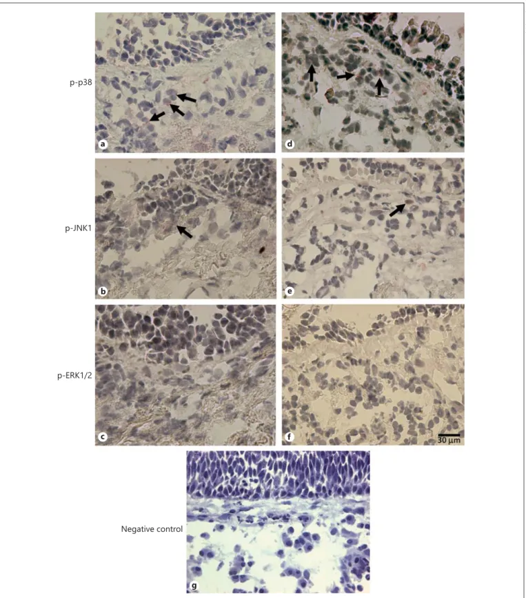
Fig. 1. Photomicrographs showing the bronchial mucosa from an asthmatic subject ( a ) and a patient with COPD ( b ) immunostained for identification of p-p38+ cells (arrows). Results are representa-tive of those from 33 (mild/moderate and severe/very severe) COPD patients and 9 mild asthmatics. Column bar graphs indicate
median (and interquartile range) values of p-p38 scored in the ep-ithelium ( c ) and p-p38+ cells quantified in the submucosa ( d ) of all COPD patients (n = 33) and asthmatics (n = 9) studied. p values are based on the Mann-Whitney test for comparison between groups.
Color version available online
0 0.2 0.4 0.6 0.8 1.0 1.2 p-p38 epithelial scor e COPD Asthmatics p = n.s. c 0 50 100 150 200 p-p38 (cells/mm 2) COPD Asthmatics p = 0.0018 d a b
Fig. 2. Photomicrographs showing the bronchial mucosa from control non-smokers ( a–c) and patients with mild/moderate COPD ( d–f ) immunostained (arrows) for identification of p-p38 ( a , d ), p-JNK1 ( b , e ) and p-ERK1/2 ( c , f ) in the bronchial
epithe-lium and submucosa. Results are representative of those from 9 non-smokers and 33 COPD patients. g Negative control immu-nostaining, performed in a nasal polyp section, including an irrel-evant rabbit primary antibody.
Color version available online
Negative control p-ERK1/2 p-JNK1 p-p38 a b c d e f g
counted between asthmatics (median 5, range 0–17)
and control non-smokers (p = 0.216), mild/moderate
COPD (p = 0.608) or all COPD patients (p = 0.156). A
slight but significant increase was observed in
asthmat-ics in comparison with severe/very severe COPD (p =
0.034).
Immunohistochemistry in the Peripheral Lung
Tissue
In the peripheral airways no significant differences
were observed for percentages of p-p38 MAPK
immu-nostained bronchiolar epithelial cells between COPD
(median 68, range 10–100) and control smokers (70,
range 40–100; p = 0.9181). Percentages of p-p38 MAPK+
alveolar macrophages in COPD (median 87, range 60–
100) versus control smokers (94, range 82–100; p =
0.5265) were not significantly different either ( fig. 3 ).
No significant differences were observed for
percent-ages of p-JNK immunostained bronchiolar epithelial cells
between COPD (median 38, range 2–64) and control
smokers (36, range 5–58; p = 0.5439). Percentages of
p-JNK+ alveolar macrophages in COPD (median 88, range
60–100) versus control smokers (90, range 40–100; p =
0.3216) were not significantly different ( fig. 4 ).
No significant differences were observed for
percent-ages of p-ERK1/2-immunostained bronchiolar
epithe-lial cells between COPD (median 40, range 0–90) and
control smokers (37, range 0–70; p = 0.5495).
Fig. 3. a Western blot analysis of activated p-p38 MAPK in snap-frozen peripheral lung lysates of stable COPD (n = 9) and age-matched control smokers with normal lung function (n = 7) with actin as the control for loading. b Graphical analysis of the densi-tometric ratio (arbitrary units, AU) of p-p38 MAPK/actin. c
Im-munohistochemical images of p-p38 MAPK immunostaining in bronchiolar epithelial cells and alveolar macrophages representa-tive of 15 COPD and 15 control smokers. Magnification ×400. Bar = 50 μm. d , e Graphical presentation of the percentage of epi-thelial and macrophage staining.
Color version available online
0.50 0.75 1.00 1.25 1.50 Smokers COPD p = 1.0000 Smokers COPD 0 50 100 150 p = 0.9181 p-p38 MAPK+ epithelial cells (%) Smokers COPD 0 60 70 110 100 90 80 p = 0.5265 p-p38 MAPK+ macr ophages (%) p-p38 MAPK Actin p-p38 MAPK Actin S1 C1 S2 S3 C2 C3 C4 C5 S4 S5 S6 S7 C6 C7 C8 C9
Bronchiolar epithelium ×400 Alveolar macrophages ×400
p-p38 MAPK/actin ratio (A U) a b c e d Smoker Smoker COPD COPD
ages of p-ERK1/2+ alveolar macrophages in COPD
(me-dian 75, range 25–100) versus control smokers (72,
range 8–100; p = 0.8682) were not significantly different
either ( fig. 4 ).
Western Blotting for p-p38 MAPK, p-JNK and
p-ERK1/2 in Peripheral Lung Tissue
No significant differences were observed for the p-p38
MAPK/actin ratio between COPD (median 1.02, range
0.2–1.40) and control smokers (1.0, range 0.65–1.30; p =
1.0000; fig. 3 ), the p-JNK/actin ratio between COPD
(me-dian 0.10, range 0.05–0.13) and control smokers (0.10,
range 0.06–0.12; p = 0.4079; fig. 4 ) and the p-ERK1/2/
actin ratio between COPD (median 0.10, range 0.05–
0.13) and control smokers (0.10, range 0.06–0.12; p =
0.4079; fig. 4 ).
Correlations between p-MAPK Cell Counts, Clinical
Parameters and Inflammatory Cells in the Bronchial
Biopsies
In all smokers and in patients with COPD alone we did
not observe any significant correlation between numbers
of p-p38+, p-JNK1+ and p-ERK1/2+ cells per square
mil-limetre and numbers of cigarettes smoked (pack-years),
functional (spirometry values and clinical parameters)
data or structural (inflammatory cells) data.
MAPK, NF-κB p65 and IL-8 mRNA Expression in
16HBE Cells Induced by Oxidative and Inflammatory
Stimuli in vitro
Human bronchial epithelial (16HBE) cells were
stim-ulated with H
2O
2(100 μ
M), cytomix (TNF-α, 10 ng/ml,
IL-1β, 1 ng/ml, and IFN-γ, 10 ng/ml) and LPS (10 μg/
ml), and the expression of IL-8 ( fig. 5 a), p38 MAPKα
( fig. 5 b), JNK1 ( fig. 5 c), ERK1 ( fig. 5 d) and NF-κB p65
subunit ( fig. 5 e) mRNA was quantified by qRT-PCR.
IL-8 mRNA was significantly increased 2 h after H
2O
2(p < 0.0001), 1 and 2 h after cytomix (p = 0.001 and p =
0.017, respectively), and 2 h after LPS (p = 0.043; fig. 5 a).
p38 MAPKα mRNA was significantly increased 2 h after
H
2O
2(p = 0.030) and 1 h after LPS (p = 0.010) but it did
not change after cytomix treatment (
fig. 5
b). JNK1
mRNA was not significantly changed by any of the
treat-ments used ( fig. 5 c). ERK1 mRNA was not significantly
increased after H
2O
2, cytomix or LPS treatments ( fig. 5 d).
NF-κB p65 subunit mRNA was significantly increased
0.2 0.6 1.0 1.4 p-ERK1/2/actin ratio (A U) Smokers COPD c p = 0.4945 0 0.05 0.10 0.15 p-JNK (G-7)/actin ratio (A U ) Smokers COPD f p = 0.4079 0 20 40 60 80 p-JNK+ epithelial cells (%) Smokers COPD d p = 0.5439 0 50 100 150 p-JNK+ macr ophages (%) Smokers COPD e p = 0.3216 0 25 50 75 100 p-ERK1/ERK2+ macr ophages (%) Smokers COPD b p = 0.8682 0 10 20 30 40 50 60 70 80 90
p-ERK1/ERK2+ epithelial cells (%)
Smokers COPD
a
p = 0.5495
Fig. 4. Graphical presentation of the percentage of p-ERK1/2-im-munostained bronchiolar epithelial cells ( a ), percentage of
p-ERK1/2-immunostained positive macrophages ( b) and
p-ERK1/2/actin ratio from peripheral lung lysates ( c ), percentage of p-JNK-immunostained bronchiolar epithelial cells ( d ), percent-age of p-JNK-immunostained positive macrophpercent-ages ( e ) and
p-JNK/actin ratio from peripheral lung lysates ( f ) in stable COPD and control smokers. AU = Arbitrary units. Data are representa-tive of 15 stable COPD and 15 control smokers for immunohisto-chemical data and 9 stable COPD and 7 control smokers for West-ern blot analysis. The Mann-Whitney test was used for compari-son between groups.
0 0.01 0.02 0.03 0.04 1 h 2 h 0 0.01 0.02 0.03 0.04 0.07 0.06 0.05 1 h 2 h 0.E+00 1.E-03 2.E-03 1 h 2 h 0 0.01 0.02 0.03 0.04 0.06 0.05 1 h 2 h 0 0.20 0.40 0.60 0.80 1.40 1.20 1.00 1 h 2 h
*
*
Cytomix 0 0.10 0.20 0.30 0.40 1 h 2 h 0 0.1 0.2 0.3 0.4 1 h 2 h 0.E+00 1.E-02 2.E-02 1 h 2 h 0 0.05 0.10 0.15 0.20 1 h 2 h*
0 0.05 0.10 0.15 0.20 0.30 0.25 2 h 1 h*
NT TR LPS Re lativ e IL-8 mRNA 0 0.05 0.10 0.15 0.20 0.25 1 h 2 h*
Re lativ e ERK1 mRNA 0 0.01 0.02 0.03 0.04 0.05 1 h 2 h Re lativ e JNK1 mRNA 0.E+00 5.E-04 1.E-03 1 h 2 h Re lativ e p65 mRNA 0 0.01 0.02 0.03 0.04 1 h 2 h*
Re lativ e p38 į mRNA 0 0.01 0.02 0.03 0.04 0.05 1 h 2 h*
H2O2 a b c d eFig. 5. In vitro expression of CXCL8 (IL-8; a ), p38α ( b ), JNK1 ( c ), ERK1 ( d ) and NF-κB p65 subunit ( e ) mRNA in 16HBE cells treat-ed with H 2 O 2 (100 μ M ), cytomix (TNF-α, 10 ng/ml, IL-1β, 1 ng/ml,
and IFN-γ, 10 ng/ml) and LPS from P. aeruginosa . All treatments upregulated IL-8 mRNA expression 1 or 2 h after treatment ( a ). H 2 O 2 stimulation upregulated p38 ( b ) and p65 ( e ) mRNA 2 h after
stimuli. LPS upregulated p-38 ( b ) mRNA 1 h after stimulus. All experiments were performed in quadruplicate. Data are expressed as means ± standard deviation. Statistical analysis: t test ( * p < 0.05) for comparison between treated (TR) and non-treated (NT) cells. The exact p values for comparison between groups are given in the Results section.
2 h after H
2O
2(p = 0.032) but it did not reach statistical
significance 2 h after cytomix (p = 0.086) and LPS (p =
0.093; fig. 5 e).
Discussion
This study shows similar immune expression of p-p38
MAPK, p-JNK1 and p-ERK1/2 in the bronchial
epitheli-um and submucosa of patients with mild/moderate and
severe/very severe stable COPD when compared to
con-trol smokers and concon-trol non-smokers. Activated MAPK
were also similarly expressed in the bronchiolar
epithe-lium of stable COPD patients compared to control
smok-ers with normal lung function. p-p38 MAPK
immu-nopositivity in the bronchial submucosa of asthmatics
was significantly increased when compared to stable
COPD and control non-smokers. Interestingly,
bronchi-al epithelibronchi-al cells (16HBE) exposed in vitro to H
2O
2,
cyto-mix and LPS showed increased levels of IL-8 mRNA
pro-duction which were accompanied by a parallel increase in
p38 MAPK mRNA after H
2O
2and LPS stimulation but
not by a parallel increase in JNK1 and ERK1 mRNA. In
patients with COPD, p-p38 MAPK immunopositivity
was increased in the small airway epithelium [12] when
compared to control non-smokers [12] and in alveolar
septa [11] when compared to control smokers and
non-smokers [11] . p-p38 MAPK immunopositivity in
lym-phocytes populating the submucosa of peripheral airways
of COPD patients was similar to that found in control
smokers and non-smokers [12] . These last data are in part
in agreement with our present observations of no
chang-es of p-p38 MAPK immunopositivity in the bronchial
bi-opsy submucosa of stable COPD patients compared to
control smokers and non-smokers, peripheral airway
bronchiolar epithelium of stable COPD patients
com-pared to control smokers, and with our observation of the
absence of significant changes in total p-p38 MAPK
pro-tein, measured by Western blot, using lung tissue, when
comparing stable COPD patients and control smokers
with normal lung function. In fact, p-p38 MAPK
immu-nopositivity in our bronchial biopsy specimens was
main-ly observed in mononuclear cells and occasionalmain-ly in
en-dothelial cells, and in agreement with Gaffey et al. [12] ,
the mononuclear cell component expressing p-p38
MAPK immunopositivity was similar in COPD patients
and control subjects. At variance with previously
report-ed data in peripheral airways [12] of COPD patients and
in large airways from asthmatics [13] , we did not find a
significant increase in this MAPK in bronchial biopsy
ep-ithelium in mild/moderate and severe/very severe COPD
or in peripheral tissue. This may be due, in part, to the
presence and influence of a different inflammatory state
of the airways when bronchial biopsies of asthmatics are
compared to COPD patients. It is conceivable that a Th2
prevalent inflammatory state, such as in asthma, could
better activate MAPK, including p-p38 MAPK. To better
understand possible differences between asthma and
COPD, we directly compared immune expression of
ac-tivated MAPK in bronchial biopsies from COPD and
asthmatic subjects, matched for age and sex, and we
ob-served a significant increase in p-p38 MAPK in
bronchi-al submucosa of mild asthmatics compared to stable
COPD patients. Asthmatics also differed significantly
from control non-smokers, confirming, in part,
previous-ly reported data [13] . In our asthmatics we found the most
significant difference in the submucosal area rather than
in epithelium, as previously reported [13] . This indicates
that increased p-p38 MAPK levels in our asthmatics are
mainly due to an increased infiltration of inflammatory
cells. A Th2-type prevalent inflammation, such as in
asth-ma, may also favour the highest p-p38 MAPK expression
observed in the bronchi of asthmatics [29] .
Inhalation of LPS in human volunteers induced
activa-tion of bronchial epithelium by increased expression of
p38 MAPK and IL-8 [30] . P. aeruginosa -challenged
hu-man bronchial epithelial cells showed increased
phos-phorylation of p38 MAPK and IL-8 gene expression
which was reduced by the use of p38 inhibitors [31] .
Fla-gellin from P. aeruginosa increased the expression of IL-8
in BEAS-2B cells compared to untreated cells, and
addi-tion of p38 MAPK inhibitors reduced IL-8 expression
[32] . IL-8 mRNA and protein expression was also
in-creased after LPS stimulation of bronchial epithelial cells
[33] . Poly(I:C) costimulation further increased IL-8
pro-duction which was reversed by dexamethasone and a p38
MAPK inhibitor [33] .
Hydrogen peroxide increased Wnt-4 and IL-8 gene
ex-pression in BEAS-2B cells [34] . Wnt-4-stimulated 16HBE
cells significantly increased IL-8 secretion and p38 MAPK
activation [35] . A lower efficacy of corticosteroids is
re-ported in human bronchial epithelial cells in asthma and
COPD after oxidative stress challenge [2, 36] . In
paediat-ric bronchial epithelial cells, Th2 cytokine challenge in
the presence of rhinovirus-16 infection augmented IL-8
release
[29]
. Housedust mite-induced IL-8 release is
blocked by an ERK inhibitor in human lung epithelial
cells [37] . Our in vitro experiments show that 16HBE cells
stimulated with H
2O
2, cytomix and LPS upregulated IL-8
report-ed observations [30–37] . This was associatreport-ed with
upreg-ulation of p38α MAPK but not JNK1 and ERK1 mRNA
after H
2O
2and LPS exposure. Interestingly, H
2O
2also
in-creased NF-κB p65 mRNA. These data suggest that in
bronchial epithelial cells the p38 MAPK pathway may be
more relevant after LPS and oxidative stimulation.
Our bronchial biopsy data, showing a significant
in-crease in p-p38 MAPK immune positivity in mild
asth-matics and its prevalence in stable COPD patients, taken
together with our in vitro observations of increased p-p38
MAPKα mRNA after bronchial epithelial stimulation
suggests that p-p38 MAPK activation may play a
signifi-cant role in inducing bronchial inflammation in these
diseases.
Recently, MacNee et al. [38] described an
improve-ment in FEV
1after 6 weeks of p38 MAPK inhibitor
treat-ment in moderate-to-severe COPD patients. In contrast,
a larger study, performed in moderate-to-severe stable
COPD patients, treated with the highly selective oral p38
MAPK inhibitor iosmapimod for 6 months, showed no
significant changes in exercise tolerance or lung function
[16] . This highlights the need to better define the clinical
phenotype of patients in order to identify a likely
‘re-sponder’ population of COPD patients [14, 15] . Our
pres-ent results may contribute to this scope since p-p38
MAPK was certainly the most expressed MAPK in our
bronchial specimens from asthmatics and COPD
pa-tients, and specific oxidative and inflammatory
challeng-es, performed in 16HBE cells, showed a significant
in-crease in p-p38 MAPKα mRNA after challenges. Since
mononuclear cell infiltration was reported as the
promi-nent cellular type in bronchial biopsies of mild/moderate
but not of severe/very severe COPD patients [39] , and
p-p38 MAPK expression, reported in the submucosa of
bronchial biopsies, is mainly due to immunostained
mononuclear cells, we can argue, therefore, that selected
populations including mild-to-moderate COPD patients
with a more inflamed airway (cut-off values for
mono-nuclear cell inflammation should be defined) and COPD
patients during an exacerbation or those who frequently
exacerbate, or asthmatic patients, could better respond to
p38 MAPK inhibitors. The potential for combination
therapies, particularly in exacerbated diseased patients,
together with a deeper analysis of molecular events and
possible activation of redundant inflammatory pathways,
developing after p38 MAPK blockade, needs to be further
studied.
Acknowledgements
This work was supported by the Fondazione Salvatore Maugeri, IRCCS, Ricerca Corrente.
I.M.A. is supported by the MRC-ABPI COPD-MAP consor-tium (G1001367/1) and the Welcome Trust (093080/Z/10/Z). Re-search at the National Heart and Lung Institute is also supported by the NIHR Respiratory Disease Biomedical Research Unit at the Royal Brompton NHS Foundation Trust and Imperial College London.
References
1 Johnson GL, Lapadat R: Mitogen-activated protein kinase pathways mediated by ERK, JNK, and p38 protein kinases. Science 2002; 298: 1911–1912.
2 Chung KF: p38 mitogen-activated protein ki-nase pathways in asthma and COPD. Chest 2011; 139: 1470–1479.
3 Birrell MA, Wong S, Catley MC, Belvisi MG: Impact of tobacco-smoke on key signaling pathways in the innate immune response in lung macrophages. J Cell Physiol 2008; 214: 27–37.
4 Bhavsar P, Hew M, Khorasani N, Torrego A, Barnes PJ, Adcock I, Chung KF: Relative corticosteroid insensitivity of alveolar mac-rophages in severe asthma compared with non-severe asthma. Thorax 2008; 63: 784– 790.
5 Woo CH, Lim JH, Kim JH: Lipopolysaccha-ride induces matrix metalloproteinase-9 ex-pression via a mitochondrial reactive oxygen species-p38 kinase-activator protein-1
path-way in Raw 264.7 cells. J Immunol 2004; 173: 6973–6980.
6 Jang MK, Kim SH, Lee KY, Kim TB, Moon KA, Park CS, Bae YJ, Zhu Z, Moon HB, Cho YS: The tyrosine phosphatase, SHP-1, is in-volved in bronchial mucin production during oxidative stress. Biochem Biophys Res Com-mun 2010; 393: 137–143.
7 Heit B, Tavener S, Raharjo E, Kubes P: An in-tracellular signaling hierarchy determines di-rection of migration in opposing chemotactic gradients. J Cell Biol 2002; 159: 91–102. 8 Heuertz RM, Tricomi SM, Ezekiel UR,
Web-ster RO: C-reactive protein inhibits chemo-tactic peptide-induced p38 mitogen-activated protein kinase activity and human neutrophil movement. J Biol Chem 1999; 274: 17968– 17974.
9 Beisswenger C, Platz J, Seifart C, Vogelmeier C, Bals R: Exposure of differentiated airway epithelial cells to volatile smoke in vitro. Res-piration 2004; 71: 402–409.
10 Hall DJ, Bates ME, Guar L, Cronan M, Korpi N, Bertics PJ: The role of p38 MAPK in rhino-virus-induced monocyte chemoattractant protein-1 production by monocytic-lineage cells. J Immunol 2005; 174: 8056–8063. 11 Renda T, Baraldo S, Pelaia G, Bazzan E,
Tu-rato G, Papi A, Maestrelli P, Maselli R, Va-trella A, Fabbri LM, Zuin R, Marsico SA, Saet-ta M: Increased activation of p38 MAPK in COPD. Eur Respir J 2008; 31: 62–69.
12 Gaffey K, Reynolds S, Plumb J, Kaur M, Singh D: Increased phosphorylated p38 mitogen-activated protein kinase in COPD lungs. Eur Respir J 2013; 42: 28–41.
13 Liu W, Liang Q, Balzar S, Wenzel S, Gorska M, Alam R: Cell-specific activation profile of extracellular signal-regulated kinase 1/2, Jun N-terminal kinase, and p38 mitogen-activat-ed protein kinases in asthmatic airways. J Al-lergy Clin Immunol 2008; 121: 893–902. 14 Singh D: p38 inhibition in COPD: cautious
15 Calverley PM: New treatments for COPD: many miles still to go. Lancet Respir Med 2014; 2: 6–7.
16 Watz H, Barnacle H, Hartley BF, Chan R: Ef-ficacy and safety of the p38 MAPK inhibitor losmapimod for patients with chronic ob-structive pulmonary disease: a randomised, double-blind, placebo-controlled trial. Lancet Respir Med 2014; 2: 63–72.
17 Global Initiative for Chronic Obstructive Lung Disease (GOLD): global strategy for the diagnosis, management and prevention of chronic obstructive pulmonary disease. NHLBI/WHO workshop report. NIH Publi-cation No 2701A. Last update 2011. http:// www.goldcopd.com/ (accessed May 28, 2014). 18 Vestbo J, Hurd SS, Agusti AG, Jones PW, Vo-gelmeier C, Anzueto A, et al: Global strategy for the diagnosis, management and preven-tion of chronic obstructive pulmonary dis-ease, GOLD executive summary. Am J Respir Crit Care Med 2013; 187: 347–365.
19 Global Strategy for Asthma Management and Prevention: Global Initiative for Asthma (GINA). 2010. www.ginasthma.org.
20 Wenzel SE, Fahy JV, Irvin CG, Peters SP, Spector S, Szefler SJ: Proceedings of the ATS Workshop on Refractory Asthma: current understanding, recommendations and unan-swered questions. Am J Respir Crit Care Med 2000; 162: 2341–2351.
21 Di Stefano A, Caramori G, Gnemmi I, Con-toli M, Bristot L, Capelli A, Ricciardolo FL, Magno F, D’Anna SE, Zanini A, Carbone M, Sabatini F, Usai C, Brun P, Chung KF, Barnes PJ, Papi A, Adcock IM, Balbi B: Association of increased CCL5 and CXCL7 chemokine ex-pression with neutrophil activation in severe stable COPD. Thorax 2009; 64: 968–975. 22 Quanjer PH, Tammeling GJ, Cotes JE,
Peder-sen OF, Peslin R, Yernault JC: Lung volumes and forced ventilatory flows. Report Working Party Standardization of Lung Function Tests, European Community for Steel and Coal. Official Statement of the European Re-spiratory Society. Eur Respir J Suppl 1993; 16: 5–40.
23 Marwick JA, Caramori G, Stevenson CS, Ca-solari P, Jazrawi E, Barnes PJ, Ito K, Adcock IM, Kirkham P, Papi A: Inhibition of PI-3Kδ restores steroid responsiveness in smoking-induced steroid insensitivity. Am J Respir Crit Care Med 2009; 179: 542–548.
24 Kirkham PA, Caramori G, Casolari P, Papi AA, Edwards M, Shamji B, Triantaphyllopou-los K, Hussain F, Pinart M, Khan Y, Heine-mann L, Stevens L, Yeadon M, Barnes PJ, Chung KF, Adcock IM: Oxidative stress-in-duced antibodies to carbonyl-modified pro-tein correlate with severity of chronic ob-structive pulmonary disease. Am J Respir Crit Care Med 2011; 184: 796–802.
25 Maestrelli P, El Messlemani AH, De Fina O, Nowicki Y, Saetta M, Mapp C, Fabbri LM: In-creased expression of heme oxygenase (HO)-1 in alveolar spaces and HO-2 in alveolar walls of smokers. Am J Respir Crit Care Med 2001; 164: 1508–1513.
26 Cozens AL, Yezzi MJ, Kunzelmann K, Ohrui T, Chin L, Eng K, Finkbeiner WE, Widdi-combe JH, Gruenert DC: CFTR expression and chloride secretion in polarized immortal human bronchial epithelial cells. Am J Respir Cell Mol Biol 1994; 10: 38–47.
27 Livak KJ, Schmittgen TD: Analysis of relative gene expression data using real-time quanti-tative PCR and the 2(–delta delta C(T)) meth-od. Methods 2001; 25: 402–408.
28 Di Stefano A, Caramori G, Barczyk A, Vicari C, Brun P, Zanini A, Cappello F, Garofano E, Padovani A, Contoli M, Casolari P, Durham AL, Chung KF, Barnes PJ, Papi A, Adcock I, Balbi B: Innate immunity but not NLRP3 in-flammasome activation correlates with sever-ity of stable COPD. Thorax 2014; 69: 516–524. 29 Cakebread JA, Haitchi HM, Xu Y, Holgate ST, Roberts G, Davies DE: Rhinovirus-16 in-duced release of IP-10 and IL-8 is augmented by Th2 cytokines in a pediatric bronchial epi-thelial cell model. PLoS One 2014; 9:e94010. 30 Roos-Engstrand E, Wallin A, Bucht A,
Poura-zar J, Sandström T, Blomberg A: Increased ex-pression of p38 MAPK in human bronchial epithelium after lipopolysaccharide exposure. Eur Respir J 2005; 25: 797–803.
31 Bezzerri V, Borgatti M, Finotti A, Tamanini A, Gambari R, Cabrini G: Mapping the tran-scriptional machinery of the IL-8 gene in hu-man bronchial epithelial cells. J Immunol 2011; 187: 6069–6081.
32 Yang JJ, Wang DD, Sun TY: Flagellin of
Pseu-domonas aeruginosa induces transforming
growth factor beta 1 expression in normal bronchial epithelial cells through mitogen ac-tivated protein kinase cascades. Chin Med J (Engl) 2011; 124: 599–605.
33 Zhang JX, Xu WJ, Han YP, Bai C, Li Q: Effects on chemotactic factor expression in bronchi-al epithelibronchi-al cells by co-stimulation of poly(I:C) and lipopolysaccharide and the un-derlying mechanism (in Chinese). Xi Bao Yu Fen Zi Mian Yi Xue Za Zhi 2012; 28: 1046– 1050.
34 Durham AL, McLaren A, Hayes BP, Caramo-ri G, Clayton CL, Barnes PJ, Chung KF, Ad-cock IM: Regulation of Wnt4 in chronic ob-structive pulmonary disease. FASEB J 2013; 27: 2367–2381.
35 Heijink IH, de Bruin HG, van den Berge M, Bennink LJ, Brandenburg SM, Gosens R, van Oosterhout AJ, Postma DS: Role of aberrant WNT signalling in the airway epithelial re-sponse to cigarette smoke in chronic obstruc-tive pulmonary disease. Thorax 2013; 68: 709– 716.
36 Heijink I, van Oosterhout A, Kliphuis N, Jonker M, Hoffmann R, Telenga E, Klooster K, Slebos DJ, ten Hacken N, Postma D, van den Berge M: Oxidant-induced corticosteroid unresponsiveness in human bronchial epithe-lial cells. Thorax 2014; 69: 5–13.
37 Sohn MH, Lee KE, Kim KW, Kim ES, Park JY, Kim KE: Calcium-calmodulin mediates house dust mite-induced ERK activation and IL-8 production in human respiratory epithe-lial cells. Respiration 2007; 74: 447–453. 38 MacNee W, Allan RJ, Jones I, De Salvo MC,
Tan LF: Efficacy and safety of the oral p38 in-hibitor PH-797804 in chronic obstructive pulmonary disease: a randomised clinical tri-al. Thorax 2013; 68: 738–745.
39 Di Stefano A, Caramori G, Ricciardolo FL, Capelli A, Adcock IM, Donner CF: Cellular and molecular mechanisms in chronic ob-structive pulmonary disease: an overview. Clin Exp Allergy 2004; 34: 1156–1167.