Faculty of Pharmacy and Medicine
PhD Thesis in Pharmaceutical Sciences
XXXIII cycle
Development of novel inhibitors targeting the
tumour-related carbonic anhydrase isoforms and of small molecule
ligands targeting the human tyrosinase and tyrosinase
related protein 1
PhD student Tutor
Table of contents
Section I. ... 6
Development of novel inhibitors targeting the tumour-related carbonic
anhydrase isoforms IX and XII ... 6
1. Introduction to the human carbonic anhydrases ... 7
1.1. Structural features and functions of human carbonic anhydrases ... 7
1.2. Carbonic anhydrase IX and XII: the tumour-related isoforms ... 10
1.3. Carbonic anhydrases: activators and inhibitors ... 14
1.4. The aim of the project ... 20
2. Design, synthesis and biological activity of selective carbonic anhydrase
inhibitors based on 2-(benzylsulfinyl)benzoic acid scaffold ... 22
2.1. Design of novel inhibitors based on 2-(benzylsulfinyl)benzoic acid scaffold ... 22
2.2. Chemistry ... 23
2.3. Results and discussion ... 24
2.4. Conclusions ... 27
2.5. Experimental section ... 27
2.5.1. General ... 27
2.5.2. Synthesis and characterization data of derivatives 1-15 ... 28
2.5.3. Enzyme inhibition assays ... 38
3. Novel insights on saccharin- and acesulfame-based carbonic anhydrase
inhibitors: design, synthesis, modelling investigations and biological activity
evaluation ... 41
3.1. Design of novel inhibitors based on the saccharin and acesulfame scaffold ... 41
3.2. Chemistry ... 45
3.3. Results and discussion ... 49
3.3.2. Inhibitory activity of acesulfame-based derivatives... 57
3.3.3. Modelling studies ... 59
3.4. Conclusions ... 63
3.5. Experimental section ... 63
3.5.1. General ... 63
3.5.2. Synthesis and characterization data of saccharin-based derivatives ... 65
3.5.3. Synthesis and characterization data of acesulfame-based derivatives ... 84
3.5.4. Enzyme inhibition assays ... 94
3.5.5. Molecular modelling studies ... 95
Section II. ... 98
Development of novel ligands targeting human tyrosinase and tyrosinase
related protein 1 ... 98
1. Introduction to human tyrosinase and tyrosinase related proteins ... 99
1.1. Structural features and functions of human tyrosinase and tyrosinase related proteins 99 1.2. Tyrosinase and tyrosinase related protein 1: activators and inhibitors ... 103
1.3. Tyrosinase and tyrosinase related protein 1: melanoma specific antigens ... 107
1.4. The aim of the project ... 109
2. Development of small molecule ligands from DNA encoded chemical
libraries ... 111
2.1. Design of small molecule ligands from DNA encoded chemical libraries ... 111
2.2. Chemistry ... 113
2.3. Biological evaluation ... 115
2.4. Results and discussion ... 116
2.5. Conclusions ... 117
2.6. Experimental section ... 118
2.6.1 General ... 118
2.6.3. Biological evaluation... 124
3. Development of multimeric ligands of Thiamidol™... 125
3.1. Design of multimeric ligands of Thiamidol™ ... 125
3.2. Chemistry ... 130
3.3. Biological evaluation ... 136
3.4. Results and discussion ... 136
3.5. Conclusions ... 139
3.6. Experimental section ... 139
3.6.1. General ... 139
3.6.2. Synthesis and characterization data... 140
3.6.3. Biological evaluation... 152
Bibliography ... 154
During my PhD studies in the field of medicinal chemistry, I dealt with two different targets thus, this Thesis consists of two Sections.
The first one is focused on my main project conducted in the Research Group of Professor Daniela Secci: the development of novel and selective inhibitors targeting the tumour-related carbonic anhydrases isoforms IX and XII.
The second one is focused on the project performed during my stay abroad in the Research group of Professor Dario Neri at the Eidgenössische Technische Hochschule Zürich (ETH Zurich): the development of small molecule ligands targeting the human tyrosinase and tyrosinase-related protein 1.
Section I.
Development of novel inhibitors targeting the
tumour-related carbonic anhydrase isoforms IX and XII
7
1. Introduction to the human carbonic anhydrases
1.1. Structural features and functions of human carbonic anhydrases
The carbonic anhydrases (CAs, EC 4.2.1.1) are a superfamily of metalloenzymes that catalyse the reversible hydration of carbon dioxide (CO2) into bicarbonate (HCO3-) and protons (H+) (eq. 1) [1].
This reaction is too slow in most tissues and organisms at physiological pH [2] and since CO2, HCO3- and H+ are involved in several important physiological processes related to metabolism (photosynthesis, carboxylation reaction, etc.), pH regulation, secretion of electrolytes, etc. throughout the tree of life [3–5], eight distinct genetic families, α-, β-, γ-, δ-, η-, ζ, θ and ι have been identified [6–13]. These CA families possess a similar active site containing a central divalent ion M(II) coordinated to three conserved amino acid residues and to a hydroxide nucleophilic species. The metal ions are Zn(II) [5], Cd(II) in ζ-CAs [14], Fe(II) in γ-CAs, at least in anaerobic conditions [15,16], Co(II) in the class [7] and Mn(II) in ι [13]. The apoenzymes are devoid of CA catalytic activity [5,17,18].
The human CAs (hCAs), belonging to the -family, are involved in several functions such as respiration and transport of CO2/HCO3-, pH and CO2 homeostasis, electrolyte secretion in tissues and organs, biosynthetic reactions, bone resorption and calcification [5,19].
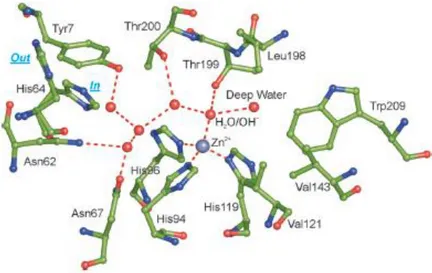
Fifteen human isoforms (hCA I-XIV) have been identified to date and the analysis of their crystal structures revealed high structural and sequence similarity for the catalytic domains [20– 22]. It is cone shaped, composed of a 10-stranded sheet with seven helices located at the surface and divided into a half halve hydrophobic and half halve hydrophilic that lead to a “bipolar” active site. This unique architecture is related to the diverse chemical nature of the CO2 and HCO3- and H+: the hydrophobic part serves as a trap for the CO2 and the hydrophilic part enables the binding of the hydrophilic components, HCO3- and H+, and their release from the active site. The Zn(II) metal ion, located at the bottom of this large cavity (approximately 12 Å wide and 13 Å deep), is coordinated to three conserved histidine (His) residues (His94, His96 and His119 in hCA II active site taken as representative of hCA isoforms) and to a H2O or hydroxide ion that determines the inactive and active form of the enzyme, respectively. The H2O/hydroxide ion is involved in hydrogen bonds with other two molecule of H2O, the first,
8 called deep-H2O, located in the hydrophobic part, and the second H2O located at the entrance of the active site, and with the hydroxyl moiety of the conserved threonine residue (Thr199). This residue is bound in turn to the carboxylate moiety of a conserved glutamic acid residue (Glu106). This pair of amino acids, called gate-keeper residues, trigger the entire process increasing the nucleophilicity of the hydroxide ion and orienting the CO2 in a favourable position for the nucleophilic attack (Figure 1.1) [5,23–27].
Figure 1.1. The active site of hCA II, taken as representative hCA isoform, describing the network of
interactions in the active site. The H2O molecules are indicated as red spheres and the side chain of the His64 is
represented in the “in” and “out” conformation [26].
The hydration of CO2 in HCO3- and H+ occurs in a two-step ping pong catalytic mechanism. The first step in the hydration reaction involves the nucleophilic attack of the Zn2+-OH- on CO2, bound in the hydrophobic pocket, leading to the formation of the HCO3- coordinated to the Zn2+ metal ion. This bond is rather weak so HCO3- is replaced by an incoming H2O molecule yielding the acidic and inactive form of the enzyme, E-Zn2+-OH2 (eq. 2).
The second and rate-limiting step involves the restoration of the active form proceeding through a proton transfer reaction from the active site to the buffer, assisted by an ordered H2O molecule network and by a proton donor/acceptor, the conserved histidine residue (His64), located at the rim of the active site [5,27–29]. Interestingly, His64 has been found in an “in” conformation, pointing inside the active site, and an “out” conformation, pointing outside the active site,
9 suggesting that it is able to accept the proton from the H2O molecule network then turn and release it into the environment, thus restoring the active form of the enzyme, E-Zn2+-OH- (eq. 3) [29,30]. Compounds able to interact with this residue have been shown to act as activators (see 1.3.). Conversely, the absence or the substitution of this proton transfer residue, as in hCA III, impairs the activity of about 500-fold [31,32].
This unique ping-pong mechanism (eq.2 and eq.3) yields some of these isoforms among the most efficient enzymes known in nature, possessing kcat/KM values close to the limit of the diffusion-controlled processes [33].
Though sharing a conserved active site architecture, these isozymes differ from each other for their cellular localization, tissue and organ distribution, oligomeric arrangement and kinetic properties [23] . Five of these isoforms are cytosolic (hCA I-III, hCA VII and hCA XIII), two are mitochondrial (CA VA and VB), four are membrane bound (hCA IV, hCA IX, hCA XII, hCA XIV and hCA XV) and one is secreted (hCA VI) [34]. The three isoforms VIII, X and XI, defined as CA-related proteins (CARPs), are non-catalytic due to the absence of one or more Zn-binding His residues in the active site [35]. Commonly, hCAs are monomers apart from the CA IX and CA XII that are homodimers stabilised through disulphide bond (Figure 1.2) [34,36].
10 The most studied and investigated isoforms are the hCA I, hCA II, known as the ubiquitous ones for their huge distribution in tissues and organs [37] and the hCA IX and hCA XII, known as tumour-related isoforms for their up regulation during tumorigenesis [38,39], extensively described further in this Chapter.
The highest accumulation of hCA I and hCA II is found in erythrocytes for their role in maintaining and regulating the blood pH [40]. Though hCA I is up to 5 or 6 times more dominant, hCA II is the most efficient being defined as the “rapid” isoform [20,26]. It is located almost in every tissue including the gastrointestinal tract, lungs, bone marrow, eyes, kidney, liver, brain, salivary gland and in breast tissue, and covers several important physiological functions. Thus, its dysregulation leads to several pathological disorders such as glaucoma, edema, and epilepsy [26,41] and pathologies such as acute mountain sickness (AMS), atherosclerosis and osteoporosis [42].
The remaining isoforms are more tissue specific. hCA III is found in muscle tissue and adipocytes and it has been suggested to act as antioxidant agent because of the presence of reactive sulfhydryl groups on its surface, that are able to bind to glutathione [43,44]. hCA IV is found in the heart, brain, capillary of the eye, and erythrocytes and it is a possible drug target for several pathologies, including glaucoma, similarly to hCA II [45]. The mitochondrial isoforms, hCA VA and hCA VB, are implicated in metabolism at different stages [41,46]. hCA VI, the secreted isoform, is implicated in cariogenic disorders [47]. hCA VII is involved in neuronal excitation, determining epileptiform conditions, similarly to hCA II and XI [48]. Albeit devoid of catalytic activity, hCA VIII seems to be involved in neurodegenerative diseases and in tumours, being upregulated in colorectal and lung cancer [49]. The physio and pathological role of the other non-catalytic isoforms, hCA X and hCA XI is still under investigation. hCA XIII is distributed in the thymus, kidney, submandibular gland, small intestine, and in reproductive organs, standing for its involvement in sperm motility processes [50,51]. hCA XIV is involved in epileptogenic conditions and is localized in the apical and basal membranes of the retinal pigment epithelium suggesting specific and unique functions for the acid-base balance in the retina [49].
1.2. Carbonic anhydrase IX and XII: the tumour-related isoforms
The tumour-related isoforms hCA IX and hCA XII possess several peculiarities in their structure, localization and implication in tumorigenesis [36,38,39], thus capturing an increased interest over the last years.
11 hCA IX is a homodimer possessing two catalytic domains and associated to form a dimer through an intermolecular disulphide bond. Each monomer consists of four domains: a catalytic domain, which faces the extracellular environment for efficient CO2 hydration and sharing 30-40 % of sequence identity with the other isoforms, a single-pass helical trans-membrane region, a C-terminal short intracellular tail and N-terminal proteoglycan domain (PG), which is a unique feature of hCA IX [36,52]. It has been suggested that each of these constituents could be involved in specific functions in tumorigenesis:
• the catalytic domain is involved in the pH regulation of the tumour microenvironment, thus enabling tumour growth and proliferation;
• the PG domain, located at the entrance of the active site clefts, could support the CO2 hydration at more acidic pH (pKa around 6.49), as the one found in solid tumours micro environment, and could foster cell adhesion and intercellular communications;
• the intracellular tail, containing several phosphorylation sites, could participate in signal transduction, contributing to the activation of a number of cancer related signalling cascades, such as the HIF-1 and the PI3/Akt kinase pathway. Interestingly, it has been shown that mutations of this tail could cause the suppression of cell adhesion and of the extracellular acidification capabilities of hCA IX [36,53,54].
The expression of hCA IX is limited to the stomach and intestine [55] but it is ectopically expressed in a large number of solid tumours including glioblastoma, colorectal, breast cancer and in the clear cell renal carcinoma (ccRCC) [56,57]. It has been related to the activation of the transcription factor HIF-1 that occurs via hypoxia, to peculiarities of solid tumours, and to mutations of Von Hippel Lindau (VHL) tumour suppressor, that mimics hypoxia, respectively [57–63]. hCA IX upregulation is usually related to chemotherapeutic resistance, poor prognosis, and poor clinical outcome [62,64–67].
hCA XII is a dimeric transmembrane isoform with two external catalytic domains (pKa around 7.1) and devoid of the PG domain [68,69]. Similarly to hCA IX, hCA XII is involved in the pH regulation in solid tumours, although there are some differences [69–71]: its dependence on the HIF-1 is not completely understood as its activation in breast cancer occurs via the estrogen receptor (ER) pathway and the hypoxia response element (HRE) on its gene has not been identified; it is related to less aggressive tumour phenotypes; it is more distributed in normal tissues and organs and its catalytic activity is medium high [69–72]. Recent investigations showed that the silencing of the CA9 gene can increase the expression of hCA XII thus defining a potential role of cooperation: the in vivo knockdown of CA9 gene alone led to a tumour
12 regression of 40% and a consequent upregulation of hCA XII, whereas the invalidation of both led to a tumour regression of 85% [73].
The rapid proliferation of tumour tissues resulted in increased distance to the blood vessels leading to inadequate oxygen levels that activate and stabilize the hypoxia-inducible factor 1 (HIF-1) [74,75]. This protein is a heterodimer that consists of the HIF-1α subunit, defined as oxygen sensor as its concentration depends on the concentration of oxygen and the HIF-1 subunit, constitutively located inside the nucleus. Under normoxia, the HIF-1α undergoes proline hydroxylation through the oxygen-dependent prolyl-4-hydroxylase domain (PHD), and thus it is recognized and tagged by the VHL tumour suppressor for ubiquitin proteasome degradation. Under hypoxia, or in case of mutations of the VHL tumour suppressor, the 1α cannot be hydroxylated, and thus it moves to the nucleus and heterodimerizes to the HIF-1 This leads to the formation of the active transcription factor HIF-1 that, binding to the HRE in the promoter-enhancer regions, activates the transcription of genes involved in oxygen and glucose supplies [54,57]. These genes include the glucose transporters (GLUT1 and GLUT3), glycolytic proteins, the vascular endothelial growth factor (VEGF), and specific pumps, transporters and proteins, including hCA IX and hCA XII (Figure 1.3) [74,76–78].
The absence of oxygen forces these cells to switch their metabolism from aerobic (glycolysis and oxidative phosphorylation of pyruvate) to anaerobic (glycolysis, lactic acid fermentation), the so-called Warburg effect [79,80], that leads to an increase of acid side products from the lactic fermentation. Interestingly, anaerobic metabolism is favoured also if adequate levels of oxygen are restored, because it is faster and because it produces fundamental precursors for the rapid replication of tumour cells (nucleotides, amino acids and lipids) [81,82]. The accumulation of acid side products in the cytoplasm leads to a decrease in the intracellular pH that is promptly balanced through a concerted interplay of pumps, transporters, and proteins. These include hCA IX and hCA XII that serve the fundamental role of setting the novel pH set points: weakly alkaline inside (pHi = 7.2-7.4) and more acidic outside (pHe = 6.8) [82–85]. Conversely, in normal cells it is the extracellular space that maintains a slightly more basic environment (pHe > 7.3) than the intracellular environment (pHi = 7.2) [82,83].
13
Figure 1.3. Mechanism of hypoxia-induced gene expression mediated by the HIF-1 transcription factor [54] and
the related transcribed proteins including hCA IX and hCA XII [78].
Particularly, although they have a significant anaerobic metabolism, a lot of tumours produce a large amount of CO2 [86,87]. Its prompt removal is fundamental for the regulation of pH and for eliminating a potential detrimental product from a poorly perfused but metabolically active tissue (mismatch among metabolic demand and the capacity to remove metabolic waste products) [84].
hCA IX converts CO2, which is produced in the cytosol via hCA II, to H+ and HCO3- (Figure
1.3) [83,84,88,89]:
• H+ accumulation leads to a decrease of the pHe, which favours: extracellular matrix (ECM) degradation, which results in tumour dissemination and invasion; the suppression of immune surveillance, which inhibits T-cells activation; the evasion from apoptosis [54], and the multidrug resistance. At lower pHe the antitumor basic drugs undergo ionization and thus the penetration of these compounds is prevented in the so-called ion trapping process [83,90–92]. Furthermore, this slightly acidic pH results suitable for the hCA IX catalytic activity [36,54].
• HCO3- anions are recycled inside the cells through transporters, which are closely associated to hCA IX (this coupled system is defined metabolon) and upregulated by the HIF-1, for the internal titration of H+ formed as tumour side products. This leads to the restoration of a slight alkaline pH that stimulates glycolysis (through the expression
14 of glycolytic enzymes) and lactate hydrogenase (LDH) activity and inhibits gluconeogenesis and apoptosis [39,86,93].
Slight variations, as small as 0.1 pH units, in the intra and extracellular pH, could be detrimental for solid tumours [94]. It has been suggested that hCA IX could fit the role of a pH-stat, continuously sensing the pH in order to keep its value constant [84,87]. Its catalytic activity responds to each single perturbation of the tumour microenvironment: at pH values above 6.8, the rate of hCA IX hydration reaction is higher than dehydration, at pH values below 6.8 its rate of dehydration reaction exceeds the rate of hydration. Thus, it has been proposed that hCA IX does not necessarily decrease pHe and produce HCO3- to be transported inside, but may also play a role in raising the pHe to sustainable acidic values for cell survival [84,89]. This equilibrium could depend on the emission of CO2 over lactic acid, on the substrate availabilities and on the area and the stage of tumours [84,89].
1.3. Carbonic anhydrases: activators and inhibitors
As they cover so many important physiological functions and are widely distributed in diverse tissues and organs, the up- or down-regulation of hCA isoforms has been related to several pathological processes [23,37], as previously reported.
The hCA activators (hCAAs), that are emerging as potential therapeutic agents, have been proposed in the treatment of Alzheimer’s disease, aging and memory consolidation [95]. They could facilitate synaptic transmission in brain cells acting or supporting the His64 during the shuttling of protons from the active site, that constitutes the rate limiting step of the entire process. This unique mechanism of action has been confirmed from the X-ray crystal structure of several compounds bound to hCA II: they are located in the same region but on the opposite side of His64, thus participating in favourable interactions between the amino acid residues and the H2O molecules network [5,96].
Conversely, the hCA inhibitors (hCAIs) have been used in clinical practice and approved as diuretics [97], antiglaucoma [98], antiepileptic [48] and anti-obesity agents [46] and thousands of compounds targeting the tumour-related isoforms hCA IX and hCA XII are in preclinical development [72], [23].
Commonly, hCAIs are divided in four classes based on their mechanism of action plus a fifth class of compounds with an unknown mechanism of action [99].
The first class of hCAIs are the zinc binders [99] and includes sulphonamides and their bio-isosteres (sulfamides and sulfamates) [23,100], the metal complexing anions [101], the
15 dithiocarbamates [102], the xanthates [103], the ureates [104], and the hydroxamates [105] (zinc binding group, ZBG). They act by binding the Zn(II) metal ion preventing its bond to the H2O/hydroxide ion in the active site. Particularly, the sulphonamides (-SO2NH2) [99] the most used in clinical practice (Figure 1.4) [27,106], bind to the Zn(II) metal ion in their deprotonated form: the –NH2 is coordinated to the zinc and it is involved in an hydrogen bond to the Thr199. This binding is strengthened by a second hydrogen bond among the oxygen of the sulphonamide and the -NH of the Thr199 (Figure 1.5) [99].
Figure 1.4. Chemical structure of some of the clinical available hCAIs bearing the primary sulphonamide [23].
Figure 1.5. Schematic representation of the hCAIs anchoring to the central zinc ion [99].
The other classes of hCAIs include inhibitors anchoring to the zinc coordinated H2O/hydroxide, occluding the active site and binding out of the active site [99].
16 Phenols, certain carboxylates and esters, the polyamines and the sulfocoumarins (hydrolysed to sulfonic acid) (anchoring group, AG) act by anchoring the H2O/hydroxide ion avoiding the formation of the pattern of hydrogen bonds in the active site, fundamental in the initial stages of the entire process (Figure 1.6) [99]. The phenol and the spermine are the first inhibitors that have been reported to show this mechanism of action [107,108].
Figure 1.6. Schematic representation of hCAIs anchoring to the zinc-coordinated water/hydroxide ion [99].
Coumarins, substituted coumarins, 5- and 6-membered lactones and thiolactones or quinolines (stick group, SG) act as prodrugs activated in situ from the esterase activity of hCAs that release the cis or trans acid isomers [99,109,110]. Due to their steric hindrance, compounds containing these moieties occlude the active site entrance (Figure 1.7) [99], the most variable region among the isoforms [34].
17 Although compounds containing carboxylic acids are commonly zinc or H2O/hydroxide ion binders, a recent investigation revealed that the 2-(benzylsulfonyl)-benzoic acid possess a unique mechanism of action since it binds out of the active site, representing the fourth mechanism of action to have been identified (Figure 1.8).
Figure 1.8. Schematic representation of the 2-(benzylsulfonyl)-benzoic acid binding out of the active site [99].
The electron density analysis, obtained from the adduct among the 2-(benzylsulfonyl)-benzoic acid and the hCA II, suggested that its carboxylate group is involved in a hydrogen bond with two H2O molecules, one of these bound to the His64, the proton shuttle residue. This favourable pathway of interactions freezes the His64 in its “out” conformation thus preventing and inhibiting the restoration of the catalytic process. Interestingly, 2-(benzylsulfonyl)-benzoic acid possesses a significant inhibitory activity only against the hCA II and hCA IX (IC50 = 0.15 M and IC50 = 1.29 M, respectively) that could be due to the diverse amino acid residues delimiting this pocket [111] (Figure 1.9).
18
Figure 1.9. Representative inhibitors of each of the four classes of hCAIs.
These four mechanisms of action have been established and proven trough kinetic enzymatic activity studies, mass spectroscopy and X-ray crystal structures, but there is still a large number of compounds that possess unknown mechanisms of action [99].
Saccharin and acesulfame derivatives, obtained from the derivatization of the secondary sulphonamide and sulfamate respectively, belong to this class and possess potent and selective hCA IX and XII inhibitory activity [57,99], though apparently devoid of a binding group able to interact in the active site, as proved by several papers in the field [112–115].
The main challenge for the development of selective hCAIs remains the high homology and similarity of the active site architecture among the fifteen isoforms [23]: the same compounds approved in clinical practice as diuretic, antiglaucoma and anticonvulsant agents are often associated with a range of side effects due to their off-target interactions [23].
Several strategies have been proposed and used to overcome this issue, such as the tail approach [18,26,62], leading to isoform selective compounds [82]. The practice consists in the insertion of a tail that, pending from the central core of the inhibitors [18,99], is able to interact with a more variable region far from the active site [34].
This approach turned out to be useful in the design of a class of ureidobenzensulfonamides [116] including SLC-0111 [99], ((4-(4-fluorophenylureido)-benzenesulfonamide)), that successfully completed phase I clinical trials for the treatment of solid tumours overexpressing hCA IX and it has been scheduled for phase II clinical trials (Ki = 45 nM on hCA IX and Ki =
19 4.5 nM on hCA XII) [68,117,118]. X-ray crystal studies suggested that this isoform’s selective profile is due to the active site entrance conformation, which is more occlusive for hCA I and hCA II due to the presence of bulky residues, and more affordable for hCA IX and hCA XII [117].
The other compound advancing in phase II clinical trials is E-7070 ((N-(3-chloro-7-indolyl)-1,4-benzenedisulfonamide)), also known as Indisulam [68,119]: its success in the treatment of solid tumours could be related to its double activity: inhibition of hCA IX and XII trough the sulphonamide moiety and inhibition of cyclin-dependent kinases (CDKs) that regulate cell cycle progression [68] (Figure 1.6).
20
1.4. The aim of the project
hCA IX and hCA XII can be considered as suitable targets for the treatment of solid tumours due to their targetable extracellular domain [36,54], their limited expression in healthy tissues and their implication in tumorigenesis [38,39]. Though a large number of inhibitors have been developed to date, only two of the sulphonamide-based inhibitors, SLC-0111 and E7070/Indisulam, are currently in clinical trials [68], highlighting the urgent need of novel and selective inhibitors targeting these tumour related isoforms.
The aim of this project was the design, synthesis and SAR analysis of two libraries of compounds based on the 2-(benzylsulfinyl)benzoic acid scaffold and on the saccharin and acesulfame nucleus. The biological evaluation and the modelling studies were performed in the Research Group of Professor Claudiu T. Supuran,
These scaffolds possess peculiar features, resulting in attractive lead compounds for the development of potent and selective inhibitors:
• 2-(benzylsulfinyl)benzoic acid acts in a pocket out of the active site consisting of the highest variability in the amino acid sequence [111] ;
• saccharin is affine for hCA IX in the nanomolar range and is more than 50-fold selective over hCA II [120,121],
• acesulfame acts exclusively on hCA IX, not affecting hCA I, hCA II and hCA XII [122].
21
This Chapter is part of the paper “Design, synthesis and biological activity of selective hCAs inhibitors based on 2-(benzylsulfinyl)benzoic acid scaffold” by G. Rotondi, P. Guglielmi, S. Carradori, D. Secci, C. De Monte, B. De Filippis, C. Maccallini, R. Amoroso, R. Cirilli, A. Akdemir, A. Angeli, C.T. Supuran published in J. Enzyme Inhib. Med. Chem. 34 (2019) 1400-1413 [123].
22
2. Design, synthesis and biological activity of selective
carbonic anhydrase inhibitors based on
2-(benzylsulfinyl)benzoic acid scaffold
2.1. Design of novel inhibitors based on 2-(benzylsulfinyl)benzoic acid
scaffold
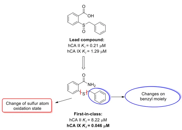
A recent investigation found that the 2-(benzylsulfinyl)benzoic acid displayed an atypical mechanism of action involving the occupancy of a pocket near the entrance of the active site through interactions that froze the His64 residue in the “out” conformation [111]. This amino acid, that can be found in two conformations called “in” and “out”, acts as a proton shuttle residue transferring a proton from the Zn-coordinated H2O molecule to the environment, in order to reconstitute the catalytic active form of the isoform. Interfering in this process led to the blocking of or a strong reduction in enzymatic activity, thus leading to enzyme inhibition. Interestingly, this lead compound resulted as inactive against hCA I and XII (Ki hCA I/XII >10 µM), but showed inhibitory activity in the low micromolar range towards hCA II and IX (Ki hCA II = 0.15 µM, Ki hCA IX = 1.29 µM).
Previous studies by our research group [123], which aimed at evaluating the impact of the substitution of the carboxylate, the benzyl moiety, and the diverse sulphur oxidation state on the activity and selectivity of the (benzylsulfinyl)benzoic acid, led to the identification of 2-(benzylthio)benzamide as the first-in-class derivative. This derivative manifests nanomolar activity against hCA IX (Ki = 46 nM) but also micromolar activity against hCA II (Ki = 8.22
M).
Based on these findings, I attempted the insertion of unsaturated alkyl chains of increasing lengths on the 2-thiobenzamide, in order to evaluate if the presence of groups able to give hydrophobic interactions, could improve the activity, with particular attention to the selectivity against hCA IX (and presumably hCA XII), that possesses a more hydrophobic and affordable active site compared to the off-target isoforms, hCA I and hCA II. The role of the sulphur atom was also investigated, to evaluate if its oxidation to sulfinyl (sulfoxides) and sulphonyl (sulfones) derivatives could affect the inhibitory activity (Figure 2.1).
23
Figure 2.1. Chemical modifications on the first-in-class compound identified from a large library based on the
2-(benzylsulfinyl)benzoic acid scaffold.
2.2. Chemistry
The synthetic procedure employed to obtain derivatives 1-15 is reported in Scheme 2.1. Methyl 2-mercaptobenzoate reacted in the presence of bromoalkanes of increasing length and potassium carbonate K2CO3, in N,N-dimethylformamide (DMF) at reflux, to obtain intermediates E2-E5 (E1, methyl 2-(methylthio)benzoate is commercially available). These intermediates were hydrolysed with an aqueous solution of sodium hydroxide (NaOH) 2 N added in a mixture of equal amounts of water and 1,4-dioxane (50:50, v:v). After the synthesis completion, the reactions were quenched with hydrochloric acid (HCl), yielding the intermediates A1-A5. The activation of the carboxylic acid group for the amide synthesis was obtained using the mixed anhydride approach [124]. In presence of an excess of triethylamine (Et3N), ethyl chloroformate was used in tetrahydrofuran (THF) under nitrogen atmosphere (N2). To check for the reaction with thin layer chromatography (TLC), the disappearance of the acidic intermediate was controlled until the completion of anhydride formation. Finally, ammonium chloride (NH4Cl) was added. The presence of the triethylamine excess caused the “de-blocking” of NH3 from NH4Cl, giving the final products (1, 4, 7, 10, 13).
24 The so-obtained compounds (1 and 15) were treated in an oxidative reaction using meta-chloroperbenzoic acid (mCPBA). Although many routes to afford sulphur oxidation have been published [125], I chose this approach in order to obtain the two oxidation products, the sulfoxide and the sulfone, in the same reaction. In fact, by controlling the amount of oxidant added during the reaction, I was able to obtain both the species resolved chromatographically.
Scheme 2.1. Synthetic procedure of derivatives 1-15.
2.3. Results and discussion
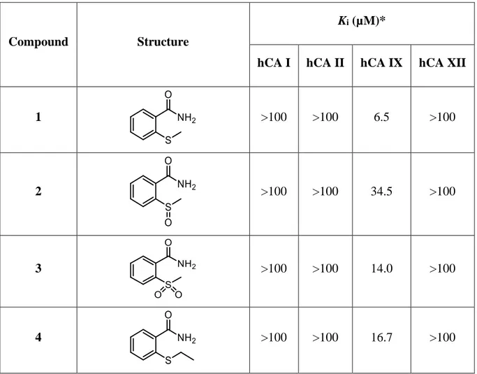
The synthesized compounds were tested to evaluate their inhibitory activity against the ubiquitous off-target isoforms, hCA I and II, and the cancer-related ones, hCA IX and XII, using a stopped-flow, CO2 hydrase assay method [126].
The hCA inhibition data, reported as Ki values, are summarized in Table 2.1.
The effect and importance of the phenyl moiety were evaluated on the 2-(benzylthio)benzamide and its oxidised forms, using linear alkyl chains of increasing length (1-15) (from the methyl to pentyl moieties).
Interestingly, derivatives 1-15 exhibited their effect exclusively against hCA IX, not affecting hCA I, hCA II and hCA XII. This could be due to the hydrophobic hCA IX active site, that could favour interactions among the alkyl chains of these compounds and the hydrophobic
25 amino acids of its cavity. Only derivative 7, bearing a propyl chain bound to the sulphur atom, exhibited a certain affinity against hCA II (Ki hCA II = 90.9 µM), although this was weak. Compound 9, the sulfone analogue of sulphide 7, was the best inhibitor inhibiting the hCA IX with a Ki value of 2.3 µM. Good results were found also for derivative 10 and 13, containing the sulphur atom and bearing a butyl or pentyl chain, respectively (10, Ki hCA IX = 2.5 µM;
13, Ki hCA IX = 2.7 µM), possibly assuming the binding pose of the benzyl group. Apart from the triad bearing the propyl chain (7-9) that possess a reversed trend, the compounds containing the sulfhydryl group displayed better inhibitory activity than the related sulfoxide and sulfone analogues (Table 2.1). A similar result was found for the 2-(benzylthio)benzamide. The reported compounds resulted less active than their lead compound, suggesting a better outcome for the benzyl group.
Table 2.1. Inhibition data of selected human CA isoforms (hCA I, II, IX and XII) with the
proposed derivatives 1-15 and the standard sulphonamide inhibitor acetazolamide (AAZ) by a stopped flow CO2 hydrase assay [126].
Compound Structure
Ki (µM)*
hCA I hCA II hCA IX hCA XII
1 >100 >100 6.5 >100
2 >100 >100 34.5 >100
3 >100 >100 14.0 >100
26 5 >100 >100 40.9 >100 6 >100 >100 22.7 >100 7 >100 90.9 >100 >100 8 >100 >100 38.2 >100 9 >100 >100 2.3 >100 10 >100 >100 2.5 >100 11 >100 >100 36.4 >100 12 >100 >100 13.5 >100 13 >100 >100 2.7 >100
27
14 >100 >100 27.3 >100
15 >100 >100 8.7 >100
AAZ 0.25 0.012 0.025 0.006
2.4. Conclusions
The chemical substitutions of the benzyl moiety of 2-(benzylthio)benzamide were explored. This compound resulted as the first-in class of a large library of compounds obtained from modifications on the 2-(benzylsulfinyl)benzoic acid scaffold, an innovative and atypical inhibitor, recently discovered.
Particularly, the chemical modifications attempted on the 2-(benzylthio)benzamide and its oxidised derivatives involved the substitution of the benzyl group using alkyl chains of increasing lengths that led to 15 derivatives tested against the off-target isoforms, hCA I and II, and the cancer-related ones, hCA IX and XII. Interestingly, these compounds appeared to be effective exclusively against hCA IX, albeit less active than their lead compound, thus suggesting that the replacement of the benzyl ring impaired the activity but increased the selectivity against hCA IX.
2.5. Experimental section
2.5.1. GeneralSolvents were used as supplied without further purification. If solvent mixtures are described, their ratio is expressed as volume:volume (v:v). Starting materials and other chemicals were purchased by Sigma-Aldrich (Italy) and used in the synthesis and in the biological assays without further purification. All synthesized compounds were fully characterized by analytical and spectral data. Analytical thin-layer chromatography (TLC) was carried out on Sigma-Aldrich® silica gel on TLC aluminium foils with fluorescent indicator 254 nm. Visualization was carried out under UV irradiation (254 and 365 nm). Silica for column chromatography were purchased from Sigma-Aldrich (Milan, Italy) (high purity grade, pore size 60 Å, 230-400
28 mesh particle size). 1H NMR spectra were recorded on a Bruker AV400 (1H: 400 MHz, 13C: 101 MHz) using the solvents (CDCl3, DMSO-d6 and D2O) at room temperature. The final concentration of the samples was of ~5 mg/mL for 1H-NMR acquisition and ~25 mg/mL for the recording of the 13C-NMR ones. Chemical shifts are quoted in ppm, based on appearance rather than interpretation, and are referenced to the residual non deuterated solvent peak. Missing signals must be attributed to overlapping peaks Chemical shifts are presented as δ units (parts per millions) using the solvent signal as the internal standard. 1H spectra are described as reported below: δH (spectrometer frequency, solvent): chemical shift/ppm (multiplicity, J-coupling constant(s), number of protons, assignment). 13C spectra are described as reported below: δC (spectrometer frequency, solvent): chemical shift/ppm (assignment). Coupling constants J are valued in Hertz (Hz) using these abbreviations to indicate the splitting: s – singlet; d – doublet; t – triplet; q – quartet; m – multiplet. If needed, it is also reported the abbreviation br – to indicate the broad shape of the specific peak. All melting points were measured on a Stuart® melting point apparatus SMP1 and are uncorrected (temperatures are reported in °C). If given, systematic compound names are those generated by ChemBioDraw Ultra® 12.0 following IUPAC conventions. Silica for column chromatography were purchased from Sigma-Aldrich (Milan, Italy) (high purity grade, pore size 60 Å, 230-400 mesh particle size).
2.5.2. Synthesis and characterization data of derivatives 1-15
2-(Methylthio)benzoic acid (A1): lithium hydroxide (1.2 equiv.), dissolved in 3 mL of water,
was added dropwise to a stirring solution of methyl 2-(methylthio)benzoate (1 equiv.) in H2O:methanol (50 mL, 50:50 v:v). The reaction was stirred at 70 °C for 2 h, concentrated in
vacuo to remove methanol and quenched with 2N HCl. The precipitate was collected by
filtration and washed with n-hexane (2 x 10 mL) to give the title compound as a white solid (89% yield); mp 171-173 °C. 1H NMR (400 MHz, DMSO-d6): δ 2.39 (s, 3H, CH3), 7.19-7.22 (m, 1H, Ar), 7.35 (d, J = 8.1 Hz, 1H, Ar), 7.52-7.57 (m, 1H, Ar), 7.90-7.91 (m, 1H, Ar); 13C NMR (101 MHz, DMSO-d6): δ 15.2 (CH3), 123.9 (Ar), 125.0 (Ar), 127.8 (Ar), 131.4 (Ar), 133.0 (Ar), 142.9 (Ar), 167.9 (COOH).
29
2-(Methylthio)benzamide (1): triethylamine (3 equiv.) and ethyl chloroformate (1.2 equiv.)
were added to a stirring solution of 2-(methylthio)benzoic acid (1 equiv.) in 10 mL of tetrahydrofuran. After monitoring the anhydride intermediate formation with silica TLC plates, ammonium chloride (1.4 equiv.) was added. The reaction was stirred under nitrogen atmosphere for 16 h at 50 °C, concentrated in vacuo and the resulting suspension extracted with ethyl acetate (3 × 20 mL). The organics were reunited, dried over sodium sulphate and concentrated under reduced pressure to give a crude product purified by column chromatography on silica gel (ethyl acetate:cyclohexane, 2:1). The title compound was a white solid (70% yield); mp 145-147 °C. 1H NMR (400 MHz, DMSO-d6): δ 2.39 (s, 3H, CH3), 7.15-7.19 (m, 1H, Ar), 7.32-7.47 (m, 3H Ar + 1H NH2, D2O exch.), 7.76 (br s, 1H, NH2, D2O exch.); 13C NMR (101 MHz, DMSO-d6):
δ 15.7 (CH3), 124.3 (Ar), 125.8 (Ar), 128.0 (Ar), 130.7 (Ar), 135.2 (Ar), 138.6 (Ar), 169.9 (CONH2).
2-(Methylsulfinyl)benzamide (2) and 2-(methylsulfonyl)benzamide (3): 3-chloroperbenzoic
acid (1 equiv.), dissolved in 5 mL of methanol, was added dropwise to a stirring solution of 2-(methylthio)benzamide (1) (1 equiv.) in 25 mL of methanol. After 2 h, another aliquot of 3-chloroperbenzoic acid (1 equiv. in 5 mL of methanol) was added and the reaction stirred for further 24 h. The mixture was concentrated in vacuo and purified by column chromatography on silica gel (ethyl acetate:cyclohexane, 5:1) to give separately the title compounds 2 and 3.
2: white solid, 34% yield, mp 175-177 °C. 1H NMR (400 MHz, DMSO-d6): δ 2.76 (s, 3H, CH3), 7.62 (t, J = 7.4 Hz, 1H, Ar), 7.70 (br s, 1H, NH2, D2O exch.), 7.81 (t, J = 7.5 Hz, 1H, Ar), 7.89 (d, J = 7.6 Hz, 1H, Ar), 8.12 (d, J = 7.8 Hz, 1H, Ar), 8.26 (b rs, 1H, NH2, D2O exch.); 13C NMR (101 MHz, DMSO-d6): δ 45.2 (CH3), 124.0 (Ar), 128.3 (Ar), 130.6 (Ar), 131.9 (Ar), 132.5 (Ar), 149.3 (Ar), 168.1 (CONH2).
3: white solid, 30% yield, mp 165-167 °C. 1H NMR (400 MHz, DMSO-d6): δ 3.38 (s, 3H, CH3),
7.56-7.58 (m, 1H, Ar), 7.65 (br s, 1H, NH2, D2O exch.), 7.66-7.70 (m, 1H, Ar), 7.75-7.79 (m, 1H, Ar), 7.96-7.98 (m, 1H, Ar), 8.07 (br s, 1H, NH2, D2O exch.); 13C NMR (101 MHz,
DMSO-30
d6): δ 45.5 (CH3), 129.1 (Ar), 129.6 (Ar), 130.2 (Ar), 134.1 (Ar), 138.3 (Ar), 138.4 (Ar), 170.0 (CONH2).
Methyl 2-(ethylthio)benzoate (E2): anhydrous potassium carbonate (1.1 equiv.) was added to
a stirring solution of methyl thiosalicylate (1 equiv.) in 10 mL of N,N-dimethylformamide. After 1 h, ethyl bromide was added dropwise and the reaction stirred under nitrogen atmosphere at 70 °C for 5 h. The mixture was poured on ice and the resulting suspension extracted with dichloromethane (3 × 20 mL). The organics were reunited, dried over sodium sulphate and concentrated in vacuo to give a crude product purified by column chromatography on silica gel (cyclohexane:ethyl acetate, 7:1). The title compound was a colourless oil (90% yield). 1H NMR (400 MHz, CDCl3): δ 1.41 (t, J = 7.4 Hz, 3H, CH3), 2.95-3.01 (m, 2H, CH2), 3.93 (s, 3H, CH3), 7.15-7.19 (m, 1H, Ar), 7.33 (d, J = 8.0 Hz, 1H, Ar), 7.44-7.48 (m, 1H, Ar), 7.97-7.99 (m, 1H, Ar); 13C NMR (101 MHz, CDCl3): δ 13.2 (CH3), 26.0 (CH2), 52.1 (CH3), 123.7 (Ar), 125.4 (Ar), 127.4 (Ar), 131.3 (Ar), 132.3 (Ar), 142.1 (Ar), 167.0 (C=O).
2-(Ethylthio)benzoic acid (A2): lithium hydroxide (1.2 equiv.), dissolved in 10 mL of water,
was added dropwise to a stirring solution of methyl 2-(ethylthio)benzoate (E2) (1 equiv.) in H2O/methanol (50 mL, 50:50 v:v). The reaction was stirred at 70 °C for 2 h, concentrated in
vacuo to remove methanol and quenched with 2N HCl. The precipitate was collected by
filtration and washed with n-hexane to give the title compound as a white solid (92% yield); mp 141-143 °C. 1H NMR (400 MHz, DMSO-d6): δ 1.43 (t, J = 7.4 Hz, 3H, CH3), 2.98-3.03 (m, 2H, CH2), 7.20-7.24 (m, 1H, Ar), 7.38 (d, J = 7.9 Hz, 1H, Ar), 7.50-7.54 (m, 1H, Ar), 8.15-8.18 (m, 1H, Ar), 11.46 (br s, 1H, COOH, D2O exch.).
31
2-(Ethylthio)benzamide (4): triethylamine (3 equiv.) and ethyl chloroformate (1.2 equiv.)
were added to a stirring solution of 2-(ethylthio)benzoic acid (A2) (1 equiv.) in 10 mL of tetrahydrofuran. After monitoring the anhydride intermediate formation with silica TLC plates, ammonium chloride (1.4 equiv.) was added. The reaction was stirred under nitrogen atmosphere for 16 h at 50 °C and concentrated in vacuo. Water (70 mL) was added and the resulting suspension extracted with ethyl acetate (3 × 20 mL). The organics were reunited, dried over sodium sulphate and concentrated under reduced pressure to give a crude product purified by column chromatography on silica gel (ethyl acetate:cyclohexane, 2:1). The title compound was a white solid (65% yield); mp 122-124 °C. 1H NMR (400 MHz, CDCl3): δ 1.32 (t, J = 7.4 Hz, 3H, CH3), 2.95-3.00 (m, 2H, CH2), 6.00 (br s, 1H, NH2, D2O exch.), 6.92 (br s, 1H, NH2, D2O exch.), 7.27-7.32 (m, 1H, Ar), 7.39-7.47 (m, 2H, Ar), 7.78-7.80 (m, 1H, Ar); 13C NMR (101 MHz, CDCl3): δ 14.0 (CH3), 28.7 (CH2), 126.4 (Ar), 129.7 (Ar), 130.8 (Ar), 131.0 (Ar), 134.7 (Ar), 135.3 (Ar), 169.8 (CONH2).
2-(Ethylsulfinyl)benzamide (5) and 2-(ethylsulfonyl)benzamide (6): 3-chloroperbenzoic
acid (1 equiv.), dissolved in 5 mL of methanol, was added dropwise to a stirring solution of 2-(ethylthio)benzoic acid (4) (1 equiv.) in 25 mL of methanol. After 2 h, another aliquot of 3-chloroperbenzoic acid (1 equiv. in 5 mL of methanol) was added and the reaction stirred for further 24 h. The mixture was concentrated in vacuo and purified by column chromatography on silica gel (ethyl acetate:cyclohexane, 5:1) to give separately the title compounds 5 and 6.
5: white solid, 48% yield, mp 155-157 °C. 1H NMR (400 MHz, DMSO-d6): δ 1.09 (t, J = 7.4 Hz, 3H, CH3), 2.64-2.73 (m, 1H, CH2), 3.13-3.22 (m, 1H, CH2), 7.60-7.64 (m, 1H, Ar), 7.68 (br s, 1H, NH2, D2O exch.), 7.76-7.80 (m, 1H, Ar), 7.89-7.91 (m, 1H, Ar), 8.01-8.03 (m, 1H, Ar), 8.25 (br s, 1H, NH2, D2O exch.); 13C NMR (101 MHz, CDCl3): δ 6.7 (CH3), 50.0 (CH2), 125.1 (Ar), 128.5 (Ar), 130.5 (Ar), 131.9 (Ar), 132.1 (Ar), 146.4 (Ar), 168.1 (CONH2).
6: white solid, 29% yield, mp 141-143 °C. 1H NMR (400 MHz, CDCl3): δ 1.31 (t, J = 7.5, 3H, CH3), 3.51-3.57 (m, 2H, CH2), 5.98 (br s, 1H, NH2, D2O exch.), 6.29 (br s, 1H, NH2, D2O
32 exch.),7.60-7.64 (m, 2H, Ar), 7.68-7.72 (m, 1H, Ar), 8.02-8.04 (m, 1H, Ar); 13C NMR (101 MHz, CDCl3): δ 7.1 (CH3), 51.1 (CH2), 128.6 (Ar), 130.2 (Ar), 130.8 (Ar), 133.7 (Ar), 136.2 (Ar), 136.8 (Ar), 170.2 (CONH2).
Methyl 2-(propylthio)benzoate (E3): anhydrous potassium carbonate (1.1 equiv.) was added
to a stirring solution of methyl thiosalicylate (1 equiv.) in 10 mL of N,N-dimethylformamide. After 1 h, 1-bromopropane was added dropwise and the reaction stirred under nitrogen atmosphere at 70 °C for 5 h. The mixture was poured on ice and the resulting suspension was extracted with dichloromethane (3 × 20 mL). The organics were reunited, dried over sodium sulphate and concentrated in vacuo to give a crude product purified by column chromatography on silica gel (cyclohexane:ethyl acetate, 5:1). The title compound was a colourless oil (93% yield). 1H NMR (400 MHz, CDCl3): δ 1.10 (t, J = 7.4 Hz, 3H, CH3), 1.75-1.81 (m, 2H, CH2), 2.90-2.94 (m, 2H, CH2), 3.93 (s, 3H, CH3), 7.13-7.17 (m, 1H, Ar), 7.33 (d, J = 8.0 Hz, 1H, Ar), 7.42-7.47 (m, 1H, Ar), 7.95-7.98 (m, 1H, Ar); 13C NMR (101 MHz, CDCl3): δ 13.8 (CH3), 21.7 (CH2), 34.1 (CH2), 52.0 (CH3), 123.6 (Ar), 125.6 (Ar), 127.6 (Ar), 131.3 (Ar), 132.2 (Ar), 142.2 (Ar), 167.0 (C=O).
2-(Propylthio)benzoic acid (A3): lithium hydroxide (1.2 equiv.), dissolved in 10 mL of water,
was added dropwise to a stirring solution of methyl 2-(propylthio)benzoate (E3) (1 equiv.) in H2O/methanol (50 mL, 50:50 v:v). The reaction was stirred at 70 °C for 2 h, concentrated in
vacuo to remove methanol and quenched with 2N HCl. The precipitate was collected by
filtration and washed with n-hexane to give the title compound as a white solid (95% yield); mp 125-127 °C. 1H NMR (400 MHz, CDCl3): δ 1.12 (t, J = 7.4 Hz, 3H, CH3), 1.76-1.85 (m, 2H, CH2), 2.93-2.97 (m, 2H, CH2), 7.20-7.24 (m, 1H, Ar), 7.38 (d, J = 7.8 Hz, 1H, Ar), 7.49-7.53 (m, 1H, Ar), 8.15-8.17 (m, 1H, Ar), 11.94 (br s, 1H, COOH, D2O exch.).
33
2-(Propylthio)benzamide (7): triethylamine (3 equiv.) and ethyl chloroformate (1.2 equiv.)
were added to a stirring solution of 2-(propylthio)benzoic acid (A3) (1 equiv.) in 10 mL of tetrahydrofuran. After monitoring the anhydride intermediate formation with silica TLC plates, ammonium chloride (1.4 equiv.) was added. The reaction was stirred under nitrogen atmosphere for 16 h at 50 °C and concentrated in vacuo. Water (70 mL) was added and the resulting suspension extracted with ethyl acetate (3 × 20 mL). The organics were reunited, dried over sodium sulphate and concentrated under reduced pressure to give a crude product purified by column chromatography on silica gel (ehyl acetate:cyclohexane, 1:1). The title compound was a white solid (68% yield); mp 122-124 °C. 1H NMR (400 MHz, CDCl3): δ 0.94 (t, J = 7.3 Hz, 3H, CH3), 1.54-1.63 (m, 2H, CH2), 2.83 (t, J = 7.3 Hz, 2H, CH2), 6.47 (br s, 1H, NH2, D2O exch.), 6.98 (br s, 1H, NH2, D2O exch.), 7.17-7.21 (m, 1H, Ar), 7.29-7.33 (m, 1H, Ar), 7.36 (d,
J = 7.2 Hz, 1H, Ar), 7.69 (d, J = 7.6 Hz, 1H, Ar); 13C NMR (101 MHz, CDCl3): δ 13.5 (CH3), 22.3 (CH2), 36.7 (CH2), 126.3 (Ar), 129.7 (Ar), 130.8 (Ar), 131.1 (Ar), 135.0 (Ar), 135.1 (Ar), 170.2 (CONH2).
2-(propylsulfinyl)benzamide (8) and 2-(propylsulfonyl)benzamide (9): a solution of
3-chloroperbenzoic acid (1 equiv.), dissolved in 5 mL of methanol, was added dropwise to a stirring solution of 2-(propylthio)benzamide (7) (1 equiv.) in 25 mL of methanol. After 2 h, another aliquot of 3-chloroperbenzoic acid (1 equiv. in 5 mL of methanol) was added and the reaction was stirred for further 24 h. The mixture was concentrated in vacuo and purified by column chromatography on silica gel (ethyl acetate:cyclohexane, 3:1) to give separately the title compounds 8 and 9.
8: white solid, 35% yield, mp 162-164 °C. 1H NMR (400 MHz, DMSO-d6): δ 0.97 (t, J = 7.4 Hz, 3H, CH3), 1.51-1.63 (m, 1H, CH2), 1.72-1.84 (m, 1H, CH2), 2.53-2.60 (m, 1H, CH2), 3.11-3.18 (m, 1H, CH2), 7.59-7.63 (m, 1H, Ar), 7.68 (br s, 1H, NH2, D2O exch.), 7.76-7.80 (m, 1H, Ar), 7.89-7.91 (m, 1H, Ar), 8.04-8.07 (m, 1H, Ar), 8.25 (br s, 1H, NH2, D2O exch.); 13C NMR
34 (101 MHz, DMSO-d6): δ 13.3 (CH3), 16.5 (CH2), 59.3 (CH2), 124.7 (Ar), 128.4 (Ar), 130.4 (Ar), 132.0 (Ar), 132.2 (Ar), 147.3 (Ar), 168.1 (CONH2).
9: white solid, 40% yield, mp 122-124 °C. 1H NMR (400 MHz, CDCl3): δ 1.04 (t, J = 7.4 Hz, 3H, CH3), 1.76-1.81 (m, 2H, CH2), 3.47-3.51 (m, 2H, CH2), 5.96 (br s, 1H, NH2, D2O exch.), 6.28 (br s, 1H, NH2, D2O exch.), 7.60-7.64 (m, 2H, Ar), 7.67-7.71 (m, 1H, Ar), 8.02-8.05 (m, 1H, Ar); 13C NMR (101 MHz, CDCl3): δ 13.0 (CH3), 16.2 (CH2), 58.4 (CH2), 128.6 (Ar), 130.2 (Ar), 130.5 (Ar), 133.7 (Ar), 136.7 (Ar), 136.9 (Ar), 170.2 (CONH2).
Methyl 2-(butylthio)benzoate (E4): anhydrous potassium carbonate (1.1 equiv.) was added to
a stirring solution of methyl thiosalicylate (1 equiv.) in 5 mL of N,N-dimethylformamide. After 1 h, 1-bromobutane was added dropwise and the reaction stirred under nitrogen atmosphere at 70 °C for 5 h. The mixture was poured on ice and the resulting suspension extracted with dichloromethane (3 × 20 mL). The organics were reunited, dried over sodium sulphate and concentrated in vacuo to give a crude product purified by column chromatography on silica gel (cyclohexane:ethyl acetate, 6:1). The title compound was a colourless oil (89% yield). 1H NMR (400 MHz, CDCl3): δ 0.96 (t, J = 7.3 Hz, 3H, CH3), 1.47-1.56 (m, 2H, CH2), 1.69-1.76 (m, 2H, CH2), 2.91-2.95 (m, 2H, CH2), 3.92 (s, 3H, CH3), 7.12-7.16 (m, 1H, Ar), 7.32 (d, J = 7.8 Hz, 1H, Ar), 7.41-7.46 (m, 1H, Ar), 7.95-7.97 (m, 1H, Ar); 13C NMR (101 MHz, CDCl3): δ 13.7 (CH3), 22.3 (CH2), 30.2 (CH2), 31.7 (CH2), 52.0 (CH3), 123.6 (Ar), 125.5 (Ar), 127.5 (Ar), 131.3 (Ar), 132.2 (Ar), 142.3 (Ar), 166.9 (C=O).
2-(Butylthio)benzoic acid (A4): lithium hydroxide (1.2 equiv.), dissolved in 10 mL of water,
was added dropwise to a stirring solution of methyl 2-(butylthio)benzoate (E4) (1 equiv.) in H2O/methanol (50 mL, 50:50 v:v). The reaction was stirred at 70 °C for 2 h, concentrated in
vacuo to remove methanol and quenched with 2N HCl. The precipitate was collected by
filtration and washed with n-hexane (2 x 10 mL) to give the title compound as a white solid (83% yield); mp 100-102 °C. 1H NMR (400 MHz, CDCl3): δ 0.99 (t, J = 7.3 Hz, 3H, CH3), 1.50-1.59 (m, 2H, CH2), 1.72-1.80 (m, 2H, CH2), 2.95-2.98 (m, 2H, CH2), 7.21 (t, J = 7.5 Hz,
35 1H, Ar), 7.37 (d, J = 8.1 Hz, 1H, Ar), 7.49-7.53 (m, 1H, Ar), 8.14-8.17 (m, 1H, Ar), 10.39 (br s, 1H, COOH, D2O exch.); 13C NMR (101 MHz, CDCl3): δ 13.7 (CH3), 22.3 (CH2), 30.2 (CH2), 31.9 (CH2), 123.8 (Ar), 125.7 (Ar), 126.3 (Ar), 132.6 (Ar), 133.1 (Ar), 143.3 (Ar), 171.6 (C=O).
2-(Butylthio)benzamide (10): triethylamine (3 equiv.) and ethyl chloroformate (1.2 equiv.)
were added to a stirring solution of 2-(butylthio)benzoic acid (A4) (1 equiv.) in 10 mL of tetrahydrofuran. After monitoring the anhydride intermediate formation with silica TLC plates, ammonium chloride (1.4 equiv.) was added. The reaction was stirred under nitrogen atmosphere for 16 h at 50 °C and concentrated in vacuo. Water (70 mL) was added and the resulting suspension extracted with ethyl acetate (3 × 30 mL). The organics were reunited, dried over sodium sulphate and concentrated under reduced pressure to give a crude product purified by column chromatography on silica gel (ethyl acetate:cyclohexane, 1:1). The title compound was a white solid (68% yield); mp 100-102 °C. 1H NMR (400 MHz, CDCl3): δ 0.92 (t, J = 7.3 Hz, 3H, CH3), 1.41-1.50 (m, 2H, CH2), 1.59-1.57 (m, 2H, CH2), 2.93 (t, J = 7.3 Hz, 2H, CH2), 6.58 (br s, 1H, NH2, D2O exch.), 7.06 (br s, 1H, NH2, D2O exch.), 7.25-7.27 (m, 1H, Ar), 7.37-7.45 (m, 2H, Ar), 7.75-7.78 (m, 1H, Ar); 13C NMR (101 MHz, CDCl3): δ 13.6 (CH3), 22.0 (CH2), 30.9 (CH2), 34.4 (CH2), 126.2 (Ar), 129.7 (Ar), 130.6 (Ar), 131.1 (Ar), 134.9 (Ar), 135.3 (Ar), 170.2 (CONH2).
2-(Butylsulfinyl)benzamide (11) and 2-(butylsulfonyl)benzamide (12): a solution of
3-chloroperbenzoic acid (1 equiv.), dissolved in 5 mL of methanol, was added dropwise to a stirring solution of 2-(propylthio)benzamide (10) (1 equiv.) in 25 mL of methanol. After 2 h, another aliquot of 3-chloroperbenzoic acid (1 equiv. in 5 mL of methanol) was added and the reaction stirred for further 24 h. The mixture was concentrated in vacuo and purified by column chromatography on silica gel (ethyl acetate:cyclohexane, 3:1) to give separately the title compounds 11 and 12.
36
11: white solid, 44% yield, mp 130-132 °C. 1H NMR (400 MHz, DMSO-d6): δ 0.88 (t, J = 7.3 Hz, 3H, CH3), 1.33-1.43 (m, 2H, CH2), 1.44-1.55 (m, 1H, CH2), 1.72-1.79 (m, 1H, CH2), 2.55-2.61 (m, 1H, CH2), 3.15-3.22 (m, 1H, CH2), 7.59-7.63 (m, 1H, Ar), 7.68 (br s, 1H, NH2, D2O exch.), 7.76-7.80 (m, 1H, Ar), 7.88-7.90 (m, 1H, Ar), 8.04-8.06 (m, 1H, Ar), 8.25 (br s, 1H, NH2, D2O exch.).
12: white solid, 40% yield, mp 129-131 °C. 1H NMR (400 MHz, CDCl3): δ 0.92 (t, J = 7.3 Hz, 3H, CH3), 1.40-1.49 (m, 2H, CH2), 1.69-1.77 (m, 2H, CH2), 3.49-3.53 (m, 2H, CH2), 5.93 (br s, 1H, NH2, D2O exch.), 6.26 (br s, 1H, NH2, D2O exch.), 7.60-7.64 (m, 2H, Ar), 7.67-7.71 (m, 1H, Ar), 8.03-8.05 (m, 1H, Ar).
Methyl 2-(pentylthio)benzoate (E5): anhydrous potassium carbonate (1.1 equiv.) was added
to a stirring solution of methyl thiosalicylate (1 equiv.) in 5 mL of N,N-dimethylformamide. After 1 h, 1-bromopentane was added dropwise and the reaction stirred under nitrogen atmosphere at 70 °C for 5 h. The mixture was poured on ice and the resulting suspension extracted with dichloromethane (3 × 20 mL). The organics were reunited, dried over sodium sulphate and concentrated in vacuo to give a crude product purified by column chromatography on silica gel (cyclohexane:ethyl acetate, 7:1). The title compound was a colourless oil (85% yield). 1H NMR (400 MHz, DMSO-d6): δ 0.86 (t, J = 7.2 Hz, 3H, CH3), 1.27-1.33 (m, 2H, CH2), 1.35-1.43 (m, 2H, CH2), 1.56-1.64 (m, 2H, CH2), 2.92 (t, J = 7.4 Hz, 2H, CH2), 3.82 (s, 3H, CH3), 7.20-7.24 (m, 1H, Ar), 7.42 (d, J = 7.8 Hz, 1H, Ar), 7.51-7.56 (m, 1H, Ar), 7.84-7.86 (m, 1H, Ar); 13C NMR (101 MHz, DMSO-d6): δ 14.3 (CH3), 22.2 (CH2), 28.0 (CH2), 31.1 (CH2), 31.4 (CH2), 52.5 (CH3), 124.4 (Ar), 126.4 (Ar), 128.0 (Ar), 131.1 (Ar), 133.0 (Ar), 141.3 (Ar), 166.6 (C=O).
2-(Pentylthio)benzoic acid (A5): lithium hydroxide (1.2 equiv.), dissolved in 10 mL of water,
was added dropwise to a stirring solution of methyl 2-(butylthio)benzoate (E5) (1 equiv.) in H2O/methanol (70 mL, 50:50 v:v). The reaction was stirred at 70 °C for 2 h, concentrated in
37 filtration and washed with n-hexane (2 x 10 mL) to give the title compound as a white solid (85% yield); mp 104-107 °C. 1H NMR (400 MHz, CDCl3): δ 0.94 (t, J = 7.2 Hz, 3H, CH3), 1.35-1.44 (m, 2H, CH2), 1.47-1.54 (m, 2H, CH2), 1.74-1.81 (m, 2H, CH2), 2.94-2.98 (m, 2H, CH2), 7.21 (t, J = 7.5 Hz, 1H, Ar), 7.37 (d, J = 8.1 Hz, 1H, Ar), 7.49-7.53 (m, 1H, Ar), 8.14-8.17 (m, 1H, Ar), 9.58 (br s, 1H, COOH, D2O exch.); 13C NMR (101 MHz, CDCl3): δ 13.9 (CH3), 22.3 (CH2), 27.9 (CH2), 31.4 (CH2), 32.2 (CH2), 123.8 (Ar), 125.7 (Ar), 126.3 (Ar), 132.6 (Ar), 133.1 (Ar), 142.2 (Ar), 171.6 (COOH).
2-(Pentylthio)benzamide (13): triethylamine (3 equiv.) and ethyl chloroformate (1.2 equiv.)
were added to a stirring solution of 2-(pentylthio)benzoic acid (A5) (1 equiv.) in 10 mL of tetrahydrofuran. After monitoring the anhydride intermediate formation with silica TLC plates, ammonium chloride (1.4 equiv.) was added. The reaction was stirred under nitrogen atmosphere for 16 h at 50 °C and concentrated in vacuo. Water (70 mL) was added and the resulting suspension extracted with ethyl acetate (3 × 30 mL). The organics were reunited, dried over sodium sulphate and concentrated under reduced pressure to give a crude product purified by column chromatography on silica gel (n-hexane:ethyl acetate, 3:1). The title compound was a white solid (60% yield); mp 95-97 °C. 1H NMR (400 MHz, CDCl3): δ 0.91 (t, J = 7.2 Hz, 3H, CH3), 1.27-1.37 (m, 2H, CH2), 1.39-1.46 (m, 2H, CH2), 1.62-1.70 (m, 2H, CH2), 2.94 (t, J = 7.4 Hz, 2H, CH2), 5.87 (br s, 1H, NH2, D2O exch.), 6.95 (br s, 1H, NH2, D2O exch.), 7.27-7.31 (m, 1H, Ar), 7.39-7.47 (m, 2H, Ar), 7.79-7.81 (m, 1H, Ar); 13C NMR (101 MHz, CDCl3): δ 13.9 (CH3), 22.2 (CH2), 28.6 (CH2), 31.0 (CH2), 34.8 (CH2), 126.3 (Ar), 129.8 (Ar), 130.8 (Ar), 131.0 (Ar), 135.1 (Ar), 135.2 (Ar), 169.7 (CONH2).
2-(pentylsulfinyl)benzamide (14) and 2-(pentylsulfonyl)benzamide (15): 3-chloroperbenzoic acid (1 equiv.), dissolved in 5 mL of methanol, was added dropwise to a stirring solution of 2-(propylthio)benzamide (13) (1 equiv.) in 25 mL of methanol. After 2 h, another aliquot of 3-chloroperbenzoic acid (1 equiv. in 5 mL of methanol) was added and the
38 reaction was stirred for further 24 h. The mixture was concentrated in vacuo and purified by column chromatography on silica gel (cyclohexane:ethyl acetate, 1:1) to give separately the title compounds 14 and 15.
14: white solid, 39% yield, mp 126-128 °C. 1H NMR (400 MHz, DMSO-d6): δ 0.88 (t, J = 8 Hz, 3H, CH3), 1.26-1.41 (m, 4H, 2 x CH2), 1.50-1.56 (m, 1H, CH2), 1.74-1.79 (m, 1H, CH2), 2.53-2.60 (m, 1H, CH2), 3.14-3.21 (m, 1H, CH2), 7.59-7.63 (m, 1H, Ar), 7.69 (br s, 1H, NH2, D2O exch.), 7.76-7.80 (m, 1H, CH2), 7.88-7.90 (m, 1H, Ar), 8.04-8.06 (m, 1H, Ar), 8.25 (br s, 1H, NH2, D2O exch.).
15: white solid, 37% yield, mp 95-97 °C. 1H NMR (400 MHz, CDCl3): δ 0.89 (t, J = 7.1 Hz, 3H, CH3), 1.28-1.43 (m, 2H, CH2), 1.61-1.63 (m, 2H, CH2), 1.72-1.80 (m, 2H, CH2), 3.48-3.52 (m, 2H, CH2), 5.91 (br s, 1H, NH2, D2O exch ), 6.24 (br s, 1H, NH2, D2O exch.), 7.61-7.64 (m, 2H, Ar), 7.68-7.72 (m, 1H, Ar), 8.04-8.07 (m, 1H, Ar); 13C NMR (101 MHz, CDCl3): δ 13.7 (CH3), 22.0 (CH2), 22.1 (CH2), 30.3 (CH2), 56.7 (CH2), 128.6 (Ar), 130.2 (Ar), 130.5 (Ar), 133.6 (Ar), 136.7 (Ar), 136.9 (Ar), 170.2 (CONH2).
2.5.3. Enzyme inhibition assays
An Applied Photophysics stopped-flow instrument was used for assaying the CA catalysed CO2 hydration activity [126]. Phenol red (0.2 mM) was used as an indicator, working at the absorbance maximum of 557 nm, with 20 mM Hepes (pH 7.5, for α-CAs) as buffer and 20 mM NaClO4 (for maintaining constant the ionic strength), following the initial rates of the CA-catalysed CO2 hydration reaction for a period of 10-100 s. The CO2 concentrations ranged from 1.7 to 17 mM for the determination of the kinetic parameters and inhibition constants. In particular, CO2 was bubbled in distilled deionized water for 30 min till saturation. A CO2 kit (Sigma, Milan, Italy) was used to measure the concentration in serially diluted solutions from the saturated one at the same temperature. For each inhibitor at least six traces of the initial 5-10% of the reaction were used for determining the initial velocity. The uncatalyzed rates were determined in the same manner and subtracted from the total observed rates. Stock solutions of the inhibitor (1 µM) were prepared in distilled-deionized water and dilutions up to 0.1 nM were conducted thereafter with the assay buffer procedure. Inhibitor and enzyme solutions were preincubated together for 15 min at room temperature prior to the assay buffer, to allow for the formation of the E-I complex or to allow for active site mediated hydrolysis of the inhibitor. The inhibition constants were obtained by non-linear least-squares methods using PRISM 3 and the Cheng-Prusoff equation[127], and represented the average from at least three different
39 determinations. All recombinant CA isoforms were obtained in-house as previously reported [128,129].
40
This Chapter refers to the paper “Novel insights on saccharin- and acesulfame-based carbonic anhydrase inhibitors: design, synthesis, modelling investigations and biological activity” by P. Guglielmi, G. Rotondi, D. Secci, A. Angeli, P. Chimenti, A. Nocentini, A. Bonardi, P. Gratteri, S. Carradori, C. T. Supuran published in J. Enzyme Inhib. Med. Chem. 35 (2020) 1891-1905 [130].
41
3. Novel insights on saccharin- and acesulfame-based
carbonic anhydrase inhibitors: design, synthesis,
modelling investigations and biological activity
evaluation
3.1. Design of novel inhibitors based on the saccharin and acesulfame scaffold
In the past few years many compounds devoid of the primary sulphonamide group attribute have been discovered and evaluated as effective hCAs inhibitors [26,99,131,132]
As previously described, among the scaffolds frequently used for the development of non-classical inhibitors of hCAs, saccharin and acesulfame have gained great importance. Particularly, since the discovery of its inhibitory activity against hCAs, saccharin has caught the attention as a valuable hit compound for the design of novel molecules acting towards hCAs [121]. Several research groups, including ours, have developed numerous hCAs inhibitors based on this promising scaffold [133–137].
The approaches that have been used to modify this nucleus are based on the derivatization of the benzene moiety to retain the cyclic sulphonamide secondary group, or on its derivatization to obtain N-substituted saccharins. Our research group has mostly focused on the second procedure synthesizing a large library of N-alkyl, N-benzyl or N-benzoyl methylene derivatives [112,114,138].
As part of the research group’s efforts to develop novel inhibitors of carbonic anhydrase, I report a large library of derivatives aimed at further explore the saccharin scaffold, taking advantage of a variety of design strategies.
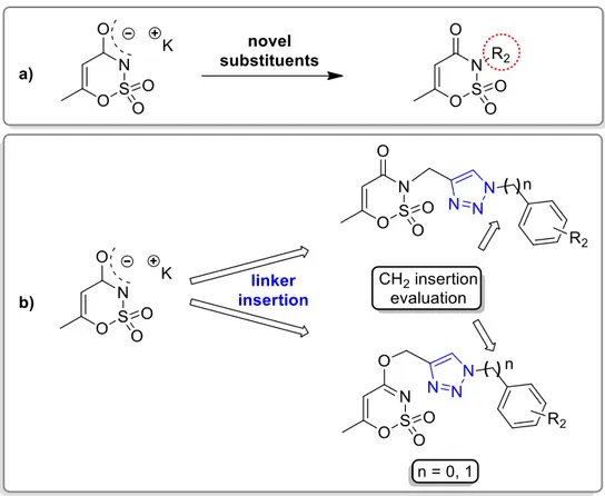
The first strategy I used (Figure 3.1) relied on the replacement of the saccharin nitrogen with an unsaturated and branched alkyl chain, benzyl or benzoyl methylene moieties, as already proposed by our research group in the past years. These substituents could represent a corollary for the robust structure-activity relationships already published [112,114,138].
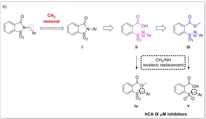
42 The second strategy I used (Figure 3.2) was based on the removal of the methylene bridge included in the benzyl substituted derivatives. I tried to evaluate, on one side, the effects resulting from the loss of conformational freedom and, on the other, the consequences of the direct binding of the sulphonamide nitrogen atom to the phenyl ring. The removal of the methylene linker affected the electronic distribution, due to the onset of conjugation between the electron pair of nitrogen with the π-electrons of the phenyl ring, previously prevented by the presence of the CH2 group.
Furthermore, the synthesis of the saccharin derivatives, consisting of a multistep approach, was studied ad hoc to obtain potential inhibitors for hCAs in each step (Figure 3.2, ii and iii). The acid intermediates ii possess two functional groups suitable for anchoring the zinc ion or the relative anchored water in the catalytic active site (-SO2NH- and COOH, respectively); moreover, they reflect the structure of the opened saccharins, reported by Ivanova et al, that exhibited improved selectivity than their parent closed analogues against the tumour-related isoforms of hCA [137]. These acid derivatives were obtained through the hydrolysis of the ester precursors (Figure 3.2, ii) that could themselves be potential inhibitors of the hCAs since they retain the secondary sulphonamide group. Moreover, considering the esterase activity of hCAs [139], these molecules, if not active as esters, could undergo the enzyme-mediated hydrolysis in the active site, acting as putative prodrugs of the acid analogues. The derivatives belonging to the sub-groups ii and iii can also be considered the isosteres of the series of compounds partly presented in the second Chapter of this Section [123]. These inhibitors, even if devoid of a ZBG, resulted selectively active against hCA IX in the µM range, suggesting that the replacement of -CH2 by -NH could improve the activity against hCAs.
43
Figure 3.2. The second design strategy proposed for the saccharin scaffold: design and retrosynthetic approach.
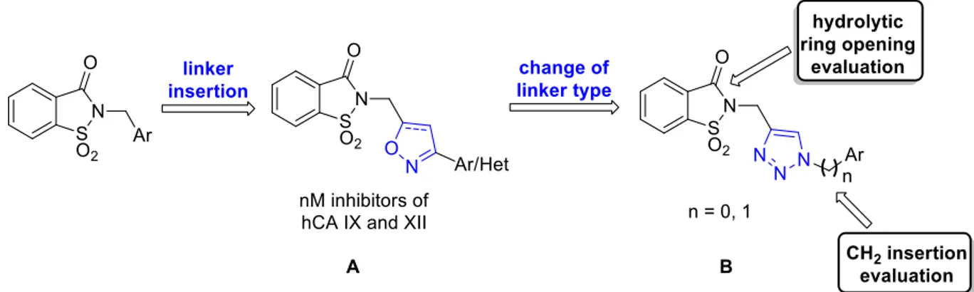
The third design strategy I used was inspired by the findings of our recent paper [115] regarding a series of saccharin/isoxazole and saccharin/isooxazoline derivatives (Figure 3.3) [115]. These compounds, characterized by an isoxazole or isooxazoline linker that disconnected the methylene moiety from the phenyl ring, resulted in a strong affinity for hCA IX and hCA XII and selectivity over hCA I and hCA II (Figure 3.3, A). Thus, based on these promising results, I proposed the replacement of the isoxazole “linker” with the triazole one, that resulted advantageous for some hCAs inhibitors (Figure 3.3, B) [140–143]. The triazole ring is an aromatic five-membered heterocyclic ring, similarly to the isoxazole one, although it contains only the nitrogen heteroatom instead of the oxygen/nitrogen ones of the isoxazole core. Moreover, just as the isoxazole, it can establish stacking interactions in the lipophilic side of the active site, and it is easy to insert through the “click” reaction azide-alkyne cycloaddition. For some of these derivatives, I also evaluated the hydrolytic ring opening and/or the introduction of an additional methylene group between the N1 of the triazole and the phenyl ring bound to it. The ring opening can lead to the same improvements discussed above, and the insertion of the methylene group could increase the “flexibility” of the tail, interrupting the electronic conjugation (Figure 3.3).
![Figure 1.3. Mechanism of hypoxia-induced gene expression mediated by the HIF-1 transcription factor [54] and](https://thumb-eu.123doks.com/thumbv2/123dokorg/2885519.10792/13.892.126.798.104.462/figure-mechanism-hypoxia-induced-expression-mediated-transcription-factor.webp)
![Figure 1.4. Chemical structure of some of the clinical available hCAIs bearing the primary sulphonamide [23]](https://thumb-eu.123doks.com/thumbv2/123dokorg/2885519.10792/15.892.157.777.355.668/figure-chemical-structure-clinical-available-bearing-primary-sulphonamide.webp)
![Figure 1.7. Schematic representation of hCAIs occluding the entrance of the active site [99]](https://thumb-eu.123doks.com/thumbv2/123dokorg/2885519.10792/16.892.294.646.830.1091/figure-schematic-representation-hcais-occluding-entrance-active-site.webp)