F
F
U
U
N
N
G
G
A
A
L
L
S
S
E
E
L
L
F
F
-
-
A
A
S
S
S
S
E
E
M
M
B
B
L
L
I
I
N
N
G
G
P
P
R
R
O
O
T
T
E
E
I
I
N
N
L
L
A
A
Y
Y
E
E
R
R
S
S
:
:
N
N
E
E
W
W
B
B
I
I
O
O
T
T
E
E
C
C
H
H
-
-T
T
O
O
O
O
L
L
S
S
F
F
O
O
R
R
B
B
I
I
O
O
/
/
N
N
O
O
N
N
-
-
B
B
I
I
O
O
H
H
Y
Y
B
B
R
R
I
I
D
D
D
D
E
E
V
V
I
I
C
C
E
E
S
S
Ilaria Sorrentino
Dottorato in Biotecnologie – XXXI ciclo
Dottorato in Biotecnologie – XXXI ciclo Università di Napoli Federico II
F
F
U
U
N
N
G
G
A
A
L
L
S
S
E
E
L
L
F
F
-
-
A
A
S
S
S
S
E
E
M
M
B
B
L
L
I
I
N
N
G
G
P
P
R
R
O
O
T
T
E
E
I
I
N
N
L
L
A
A
Y
Y
E
E
R
R
S
S
:
:
N
N
E
E
W
W
B
B
I
I
O
O
T
T
E
E
C
C
H
H
-
-T
T
O
O
O
O
L
L
S
S
F
F
O
O
R
R
B
B
I
I
O
O
/
/
N
N
O
O
N
N
-
-
B
B
I
I
O
O
H
H
Y
Y
B
B
R
R
I
I
D
D
D
D
E
E
V
V
I
I
C
C
E
E
S
S
Ilaria Sorrentino
Dottorando:
Ilaria Sorrentino
Relatore:
Prof. Paola Giardina
Correlatore:
Dr. Alassandra Piscitelli
Alla mia famiglia
Per non dimenticare
Dove tutto ha avuto inizio …
I have not failed
I’ve just found 10,000 ways
that won’t work.
(Thomas A. Edison)
….
Never, never,
never give up.
(Winston Churchill)
INDEX/INDICE RIASSUNTO ... 1 SUMMARIY ... 7 CHAPTER 1 1 INTRODUCTION ... 8 1.1 Amyloid fibrils ... 8
1.2 Nanotechnology and Nanomaterials ... 13
1.3 Applications of Functional Amyloids from Fungi: Surface Modification by Class I Hydrophobins (review) ... 17
1.4 Vmh2: Class I Hydrophobin from Pleurotus ostreatus ... 29
1.5 Aim of this thesis: work description ... 30
1.6 References ... 32
CHAPTER 2 2 LACCASE ... 36
2.1 Introduction ... 36
2.2 Straightforward laccase immobilization by means of a laccase-hydrophobin chimera (submitted research article) ... 38
2.3 Immobilization of chimeric enzymes on graphene surfaces ... 68
2.4 POXC laccase from Pleurotus ostreatus: a high-performance multicopper enzyme for direct Oxygen Reduction Reaction operating in a proton-exchange membrane fuel cell (research article) ... 76
2.5 Conclusions ... 83
2.6 References ... 84
CHAPTER 3 3 THE RECOMBINANT CHIMERIC PROTEINS PRODUCED IN Escherichia coli 87 3.1 Chimeric protein: Antimicrobial Peptide-Hydrophobin ... 87
3.2 Chimeric protein: Antibody-Hydrophobin ... 96
3.3 Conclusions ... 101
3.4 References ... 102
CHAPTER 4 4 CONCLUSIONS ... 105
COMMUNICATIONS and PUBLICATIONS ... 106
1
RIASSUNTO
INTRODUZIONE
La progettazione e la realizzazione di bionanomateriali con proprietà e prestazioni specifiche sono caratteristiche chiave nello sviluppo di bio-dispositivi, in cui uno o più elementi biologici sono integrati con nano/micro piattaforme inorganiche. Le potenziali applicazioni dei dispositivi dipendono fortemente dalla bio-funzionalizzazione delle loro superfici, che ne modifica le caratteristiche fisiche, chimiche e / o biologiche rispetto a quelle originali. I sistemi auto-assemblanti, per la stabilità chimica e meccanica delle loro strutture, sono candidati promettenti per la bio-funzionalizzazione e progettazione di nuovi biomateriali. In particolare, l'autoassemblaggio è la capacità intrinseca di numerose strutture biologiche multimeriche di assemblare dai loro componenti attraverso movimenti casuali di molecole e formazione di legami chimici deboli tra superfici con forme complementari. Eventi cellulari come la formazione di fibrille amiloidi, il riconoscimento antigene-anticorpo, l'assemblaggio della cromatina e l'autoassemblaggio della membrana fosfolipidica sono eccellenti esempi di autoassemblaggio molecolare.
Le fibrille amiloidi
Le proteine β-amiloidi sono una classe di proteine che possono subire una trasformazione strutturale dal loro stato funzionale nativo, in strutture fibrillari insolubili e altamente organizzate. Durante questa trasformazione le fibrille mature, indipendentemente dalla loro sequenza di amminoacidi o dalla loro struttura nativa, adottano una struttura più rigida rispetto ai loro precursori monomerici ed oligomerici mostrando un caratteristico pattern cross-β.
L'organizzazione molecolare unica delle fibrille amiloidi conferisce loro notevoli proprietà meccaniche e integrità strutturale persistente in presenza di alte temperature, proteasi, denaturanti e forze fisiche. Queste strutture fibrillari sono tipicamente associate a malattie neurodegenerative, come il morbo di Alzheimer e il morbo di Parkinson. Tuttavia, più recentemente è stato osservato che molti organismi, come funghi e alcuni batteri, sfruttano la capacità dei polipeptidi di formare amiloidi in una varietà di processi biologici.
Le idrofobine
Nei funghi filamentosi, le proteine amiloidi sono coinvolte in numerosi processi, ad esempio nella formazione di strutture aeree (spore o corpi fruttiferi) e nell'aderenza delle strutture fungine alle superfici idrofobiche durante le interazioni simbiotiche o patogene.
Le idrofobine sono una famiglia di piccole proteine (circa 100 aminoacidi), e rappresentano un esempio di fibrille amiloidi funzionali prodotte a diversi stadi di crescita da funghi filamentosi, in cui sembrano avere un ruolo chiave. Queste proteine possiedono la caratteristica di auto-assemblare spontaneamente alle interfacce idrofiliche-idrofobiche in strati anfipatici che cambiano la bagnabilità delle superfici. Tradizionalmente sono state divise in idrofobine di classe I e II. Quelle di classe I sono prodotte sia da funghi Basidiomiceti che Ascomiceti, invece quelle di classe II sono state isolate solo da Ascomiceti. Le proteine di questa famiglia hanno bassa identità di sequenza, ma sono caratterizzate da un motivo ben conservato di otto cisteine implicate nella formazione di quattro ponti disolfurici (Cys1-Cys6, Cys2-Cys5, Cys3-Cys4, Cys7-Cys8). Questi otto residui conservati stabilizzano il nucleo proteico e definiscono un modello di idrofobicità tipico di queste proteine. Gli spazi
2
inter-cisteina in quelle di classe I sono meno conservati rispetto a quelle di classe II. La differenza sostanziale tra le due classi risiede nella stabilità dei film anfifilici che formano. Le idrofobine di classe I formano film molto stabili e resistenti ai lavaggi con solventi e detergenti, come ad esempio l’etanolo al 60% e il sodio dodecil solfato (SDS) caldo al 2%; dissociabili solo in presenza di acidi forti come l’acido formico o l’acido trifluoroacetico (TFA), condividendo molte proprietà strutturali con le fibrille amiloidi. Al contrario i film formati dalle idrofobine di classe II sono poco stabili, possono essere più facilmente disciolti in detergenti o solventi organici, e mancano di strutture amiloidi. Entrambe le classi di idrofobine, per le loro interessanti proprietà, sono state sfruttate in molte applicazioni biotecnologiche:
Applicazioni Esempi
Applicazioni Industriali
Emulsionanti Biotensioattivi o emulsionanti, per migliorare la solubilità di preparati Applicazioni ad alto valore aggiunto
Nanotecnologia e diagnostica Biosensori, come componenti per lab-on-chip e per il drug delivery Anti-biofilm Rivestimento di dispositivi medici, come cateteri, per ridurre la possibilità di legame da parte di batteri patogeni Purificazioni Purificazione di proteine di interesse, opportunamente fuse alle idrofobine
Queste capacità delle idrofobine possono essere sfruttate come un'interessante alternativa ai metodi convenzionali per la funzionalizzazione di superfici sviluppando dispositivi ibridi. Per l’ottenimento di uno strato bio-ibrido omogeneo possono essere utilizzate tecniche di ingegneria genetica in cui è possibile fondere le caratteristiche delle idrofobine con le proprietà specifiche delle proteine target. Seguendo questo approccio, le proteine fuse ingegnerizzate possono essere utilizzate direttamente per funzionalizzare le superfici con vantaggi in termini di tempo e di sostanze chimiche consumate.
Nel gruppo di ricerca presso cui è stato svolto Il mio progetto di PhD, è da tempo oggetto di studio il fungo basidiomicete Pleurotus ostreatus, ampiamente caratterizzato ed utilizzato come fonte di proteine di interesse industriale. In questo fungo sono stati identificati diversi geni codificanti idrofobine, espressi in differenti stadi del ciclo vitale. In particolare, è stata purificata dal brodo di coltura di questo fungo l’idrofobina di classe I Vmh2, ampiamente caratterizzata sia in forma solubile che aggregata, che è in grado di formare biofilm stabili su superfici solide. Sfruttando le competenze acquisite dal mio gruppo di ricerca, il presente lavoro verte sullo sviluppo di sistemi versatili e semplici di funzionalizzazione di superfici attraverso l'immobilizzazione di proteine chimeriche auto-immobilizzanti e il loro uso in diversi campi, che vanno dalle applicazioni biomediche, quali rivestimento di dispositivi medici per la sua attività anti-biofilm, a quelle industriali come emulsionante, incluso il
biosensing, immobilizzando diversi enzimi nella loro forma attiva. Quindi, sfruttando
tecniche di ingegneria genetica, Vmh2 è stata fusa a proteine target di interesse biotecnologico ed espressa in due diversi microrganismi (Pichia pastoris ed
Escherichia coli). In particolare, tre classi di proteine target, un enzima ossidativo
3
sono state utilizzate per progettare, produrre, caratterizzare diverse proteine chimeriche utilizzate come case studies per la funzionalizzazione di superfici.
• LACCASI
Le laccasi (EC 1.10.3.2) sono enzimi ubiquitari che sono presenti in natura nei funghi (Ascomiceti, Duteromiceti e Basidiomiceti), nelle piante superiori e, in misura minore, nei batteri e negli insetti. Appartengono alla famiglia delle “blue copper” ossidasi, cioè enzimi ossidativi che contengono rame. Questi enzimi ad attività p-difenolossidasica, in particolare, utilizzano ossigeno atmosferico come accettore finale di elettroni e lo convertono in acqua con contemporanea trasformazione del substrato in prodotto.
Le laccasi sono enzimi molto versatili; catalizzano l’ossidazione di diverse specie chimiche (quali para-difenoli, polifenoli, metossifenoli, amminofenoli, diammine, poliammine, arilammine, pesticidi, idrocarburi policiclici aromatici, coloranti). La capacità di questi enzimi di ossidare un ampio spettro di substrati li rende particolarmente interessanti per le loro potenziali applicazioni biotecnologiche. Di fatto, questa classe di enzimi trova attualmente largo uso in numerosi settori industriali che includono la delignificazione della pasta di cellulosa, la decolorazione di tessuti, la detossificazione di acque reflue, la trasformazione di antibiotici e steroidi, l’utilizzo come componenti di detersivi. Le laccasi inoltre, possono essere immobilizzate su supporti appropriati per il loro utilizzo sia come biocatalizzatori che per il biosensing.
Immobilizzazione facile e diretta della proteina chimerica laccasi-idrofobina e sviluppo di una piattaforma analitica per il monitoraggio di composti fenolici
I fenoli sono tra le impurità organiche più abbondanti che penetrano nell'ambiente a seguito del loro uso in un gran numero di processi, dall'industria cartiera alla sintesi di materie plastiche e farmaceutiche. Per monitorare questi composti è quindi necessario un processo rapido e affidabile. Per raggiungere questo obiettivo, è stato sviluppata piattaforma analitica basata sulla proteina chimerica laccasi-idrofobina. L’interesse per questa proteina chimerica consiste nell’avere una proteina bifunzionale in cui si associano le proprietà dell’idrofobina Vmh2 di aderire alle superfici, alle proprietà enzimatiche della laccasi, utilizzando un approccio di fusione genetica. Si ottiene, così, una proteina auto-assemblante, autoadesiva e con attività catalitica. Quindi, la prima proteina che è stata fusa con l'idrofobina è stato l’enzima POXA1b. Tra gli isoenzimi prodotti da P. ostreatus la laccasi POXA1b è stata scelta per le sue peculiari caratteristiche: i) stabilità e attività in un ampio intervallo di pH (3-9) e temperature (25 ° -65 ° C); ii) alto potenziale redox (+0,650 V); iii) alto livello di produzione in ospiti eterologhi.
4
La proteina chimerica POXA1b-Vmh2 è stata espressa in Pichia pastoris e ritrovata secreta nel mezzo di coltura. Il surnatante della coltura grezza è stato direttamente utilizzato per rivestire piastre multi-pozzetto in polistirene senza ulteriori passaggi di purificazione. Vantaggi della fusione rispetto all'enzima libero prodotto nelle stesse condizioni, sono stati riscontrati sia nella percentuale di immobilizzazione che nella stabilità. La piastra multi-pozzetto così funzionalizzata è stata utilizzata per monitorare due composti fenolici, L-dopa ((S) -2-Amino-3- (3,4-diidrossifenil)
propanoico) e acido caffeico (Acido 3- (3,4-diidrossifenil) -2-propenoico), la cui stima
è di interesse per i settori farmaceutico e alimentare. Il metodo si basa sull'uso degli analiti come inibitori in competizione all'ossidazione dell'ABTS mediata dalla laccasi. I principali vantaggi di questa metodologia di monitoraggio sono la facilità di preparazione, l'uso di piccoli volumi di campioni e l'analisi simultanea di più campioni su un'unica piattaforma. I risultati di questi esperimenti sono oggetto di un lavoro scientifico sottomesso alla rivista “Applied Microbiology and Biotechnology” per la pubblicazione.
Immobilizzazione di enzimi chimerici su nanomateriali
I nanomateriali a base di grafene (GBM) sono materiali nanostrutturati particolarmente utili che sono di grandi prospettive in campo biotecnologico e biomedico. I GBM sono estremamente interessanti perché possono combinare proprietà di elevato rapporto superficie / volume, biocompatibilità, stabilità chimica ed elettrochimica e buona conduttività elettrica. Recentemente, è stato messo a punto un processo per la produzione di GBM biofunzionalizzati in miscele di etanolo-acqua, utilizzando ultrasuoni in sinergia con l'idrofobina di classe I Vmh2. Sia il POXA1b che la proteina chimerica POXA1b-Vmh2 sono stati sfruttati per la funzionalizzazione di nanomateriali (NM) attraverso un approccio “one-step”. L'obiettivo principale è stato l’ottenimento di nuovi nanobio-catalizzatori, sfruttando le proprietà caratteristiche di Vmh2, dei NM e di POXA1b. In particolare, la miglior resa di bio-funzionalizzazione (11%) del grafene con PoxA1b-Vmh2 è stata ottenuta con l'aggiunta dell'enzima negli ultimi 10 minuti dell'esfoliazione. I dati ottenuti confermano che la fusione genica migliora la stabilità e l’adesione della proteina rispetto alla controparte libera. Inoltre, durante il periodo trascorso all’estero presso il laboratorio del Dr. Alan Le Goff (Department of Molecular Chemistry, Groupe Biosystèmes Electrochimiques et Analytiques, Université Grenoble Alpes, Francia), il grafene bio-funzionalizzato è stato depositato su GCE (Glassy Carbon Electrode) e utilizzato come elettrodo di lavoro per test cronometrici per la rivelazione di catecolo. Il risultato di questi esperimenti ha dimostrato che questa tecnica potrebbe rappresentare un'alternativa valida per il rilevamento di questo analita in campioni reali come l'acqua di rubinetto.
La laccasi POXC da Pleurotus ostreatus: un enzima ad alte prestazioni per la reazione diretta di riduzione dell'ossigeno in fuel cell
Le MCO (Multicopper Oxidases) sono una valida opzione come bio-catodi nelle
enzymatic biofuel cells (EBFC), una sottoclasse di celle a combustibile in cui gli
enzimi sostituiscono i catalizzatori convenzionali. A tale scopo, è stata studiata l'elettrochimica diretta di POXC, la laccasi canonica e più abbondante da P.
ostreatus. Grazie all'interazione favorevole tra questa laccasi nativa e i nanotubi di
carbonio (CNT) opportunamente modificati in modo covalente con sali di antrachinone e naftoato di diazonio, POXC presenta un ORR (Oxygen Reduction
Reaction) ad alto potenziale / alta corrente in un ampio intervallo di pH (da 2 a 8),
5
(TvLAC), immobilizzata sugli stessi elettrodi nanostrutturati. Per queste prestazioni uniche, questo enzima è stato in grado di operare all'interfaccia dello strato microporoso, aria umidificata ed elettrolita polimerico, come il nafion®, in una fuel cell convenzionale a scambio di protoni H2 / aria, superando i problemi riscontrati fin qui
con altri enzimi della stessa famiglia. I risultati di questi esperimenti sono oggetto di un lavoro scientifico recentemente pubblicato sulla rivista ChemElectroChem.
• PRODUZIONE RICOMBINANTE DI PROTEINE CHIMERICHE IN
Escherichia coli
La produzione ricombinante in E. coli delle proteine chimeriche è stata eseguita utilizzando il protocollo ottimizzato per altri tipi di proteine di fusione riportati da Piscitelli e coautori. In queste proteine chimeriche, Vmh2 è stata fusa con altre due proteine target, un peptide antimicrobico, LL37, e un anticorpo, HN1 ScFv, che simula l'attività dell'anticorpo umano anti-mesotelina, una glicoproteina sovra espressa in diversi tumori umani.
Proteina chimerica peptide antimicrobico-idrofobina
L’interesse crescente verso i peptidi antimicrobici nasce dall’aumentare della resistenza agli antibiotici di molti microrganismi patogeni. Tra questa classe di peptidi, LL37 è stato intensamente studiato poiché è l’unica catelicidina ritrovata nell’uomo. La proteina chimerica LL37-Vmh2 è stata prodotta e purificata con successo; e le sue capacità adesive e di antibiofilm contro Staphylococcus
epidermidis sono state testate su superfici di polistirene. I risultati più importanti
ottenuti sono stati una buona resa di immobilizzazione (circa 40%) e un’efficiente attività anti-biofilm e antimicrobica
Proteina chimerica anticorpo-idrofobina
L'immobilizzazione dell'anticorpo su una superficie solida è stata studiata estesamente per una serie di applicazioni tra cui immuno-dosaggi, biosensori e cromatografia di affinità. Per la maggior parte di queste applicazioni, un aspetto critico è l'orientamento del sito di legame dell'antigene rispetto alla superficie. Con l'obiettivo di superare questo problema, in questa sezione è stata effettuata la produzione ricombinante in E. coli della proteina chimerica in cui l’idrofobina è fusa alla parte funzionale dell'anticorpo anti-mesotelina. La mesotelina è una glicoproteina di superficie cellulare che è fisiologicamente espressa sulla superficie cellulare delle cellule mesoteliali e sovra espressa in diversi tumori umani, tanto che si possono trovare forme solubili di mesotelina nei fluidi di pazienti affetti da questi tumori. Quindi, la misurazione della mesotelina nel sangue può essere utile per la diagnosi, ma anche per la terapia mirata, data la sua limitata espressione nei tessuti normali e l'alta espressione in diversi tumori. Questa proteina chimerica è stata prodotta in E.
coli.
• CONCLUSIONI
Il principale vantaggio della fusione genica consiste nel combinare le proprietà adesive e auto-assemblanti dell'idrofobina Vmh2 con le proprietà di diverse proteine
target di interesse biotecnologico per sviluppare metodi di immobilizzazione semplici,
arrivando ad ottenere superfici ibride con caratteristiche uniche, che dipendono dalla scelta della proteina target. Di seguito sono riportati i principali risultati conseguiti in questo progetto:
6
Vmh2-PoxA1b è stata prodotta e secreta nel brodo di coltura del lievito P.
pastoris. L'enzima fuso immobilizzato è stato utilizzato per sviluppare una
superficie funzionale per rilevare composti fenolici in matrici reali di interesse alimentare e biomedico.
È stata sviluppata una procedura one-step di esfoliazione e bio-funzionalizzazione del grafene con Vmh2-PoxA1b. Il grafene funzionalizzato è stato depositato su GCE, e può rappresentare una valida alternativa per la rivelazione di catecolo in campioni reali con metodi elettrochimici.
La laccasi POXC ha dimostrato di essere di grande interesse come biocatodo per nuove applicazioni bioelettrocatalitiche.
“layers” assemblanti di LL37-Vmh2 sono stati utilizzati per inibire i bio-films microbici per produrre superfici di polistirene funzionalizzate per lo sviluppo di dispositivi medici.
La proteina HN1-Vmh2 è stata prodotta nel citoplama di E. coli e potrà essere utilizzata per l’immobilizzazione su diverse superfici per immunodosaggi (Elisa Kit) and drug delivery.
7
SUMMARY
During recent years the demand of valuable scaffolds to biofunctionalize nanomaterials has been rapidly increasing. Self-assembling proteins, such as amyloid fibrils, are promising candidates for the functionalization of nanomaterials due to their chemical and mechanical stability. These fibrillar structures are typically associated to neurodegenerative diseases. However, it has been recently observed that many organisms, such as fungi and some bacteria, take advantage of the ability of polypeptides to form amyloids. Hydrophobins represent an example of functional amyloid fibrils produced at different growth stages by filamentous fungi. They are a large family of very active surface proteins and can be grouped into two distinct classes based on the stability of the amphipathic layers that they form. In particular, layers formed by class I share many structural properties with amyloid fibrils, solubilized only after harsh acid treatments Immobilization of active proteins on the hydrophobin layer has been proven as a simple approach for the functionalization of different surfaces useful in biotechnological applications. For most of these applications, a critical aspect concerns protein orientation with respect to the surface. To overcome this problem, design and recombinant production of fused engineered proteins combining the adhesive moiety of hydrophobins to target proteins can be a valuable choice. This PhD project has been focused on recombinant production in different hosts (Pichia pastoris and Escherichia coli) of chimera proteins built by selected target proteins (laccase, antimicrobial peptides and antibody) fused to an adhesive self-assembling class I hydrophobin, and on their exploitation in some application fields. The selected class I hydrophobin was Vmh2, extracted from the mycelium of the fungus Pleurotus ostreatus, one of the most hydrophobic hydrophobin able to spontaneously form stable and homogeneous layers on hydrophilic or hydrophobic surfaces, changing their wettability. As a first goal, the hydrophobin Vmh2 was fused to the laccase POXA1b from P. ostreatus, a high redox potential oxidase endowed with a remarkable stability at high temperature and at alkaline pH. The resulting fusion enzyme was secreted into the culture medium by the heterologous yeast P. pastoris and directly used for coating different surfaces, i.e. graphene and polystyrene, without additional purification steps. The immobilized enzyme was exploited to develop innovative optical and electrochemical biosensing platform for the detection of phenolic compounds. Subsequently, two chimeric proteins were recombinantly expressed in E. coli: in the first case, Vmh2 was fused to the antimicrobial peptide LL37, a small cationic amphiphilic peptide belonging to the family of cathelicidins which has an effective activity against a wide range of bacteria and fungi. The LL37-Vmh2 chimeric protein was successfully produced and its adhesive and antibiofilm capabilities, against Staphylococcus epidermidis, tested by coating polystyrene surfaces. The chimera HN1-Vmh2, the fusion between the hydrophobin and the anti-mesothelin ScFv antibody, was produced in the soluble fraction of E. coli whilst its purification and exploiation needs to be optimized.
In conclusion, chimeric proteins obtained by the genetic fusion of the hydrophobin Vmh2 to target proteins represent a proof of concept of versatile and straightforward solutions of surface functionalizations that could be explored in several fields.
Moreover, during the six months stay at the CNRS of Grenoble, the work was focused on carbon nanotubes (CNT) functionalization using the native laccase POXC from P. ostreatus. POXC is a promising biocathode for enzymatic biofuel cells (EBFCs), since it shows efficient ORR (Oxygen Reduction Reaction) performances and it is capable to operate in a conventional H2/air proton-exchange membrane fuel
8
1. INTRODUCTION
The design and the realization of bionanomaterials with specific properties and performances are key features in bio-device development. The potential applications of these bionanomaterials strongly depend on the bio-functionalization of device surfaces 1. Biological molecules fully integrated with micro or nano technological
platforms are useful tools to fabricate hybrid devices, showing physical, chemical and/or biological characteristics different from those originally found on the surface. Self-assembled systems are promising candidates for designing novel biomaterials, due to the chemical and mechanical stability of their structures 2,3. In particular,
self-assembly is the inherent ability of numerous multimeric biological structures to assemble from their component parts through random movements of molecules and formation of weak chemical bonds between surfaces with complementary shapes. This phenomenon is mainly governed by weak noncovalent bonds like electrostatic interactions (ionic bonds), hydrogen bonds, hydrophobic and hydrophilic interactions, water-mediated hydrogen bonds, and van der Waals interactions 4. Although these
forces are weak, their collective interactions can produce molecules that are structurally and chemically stable. For all those characteristics, molecular self-assembly is a powerful phenomenon borrowed from nature by scientists for fabricating novel supramolecular architectures. Cellular events like amyloid fibril formation, antigen−antibody recognition, chromatin assembly, and phospholipid membrane self-assembly are excellent examples of molecular self-assembly.
1.1 Amyloid fibrils
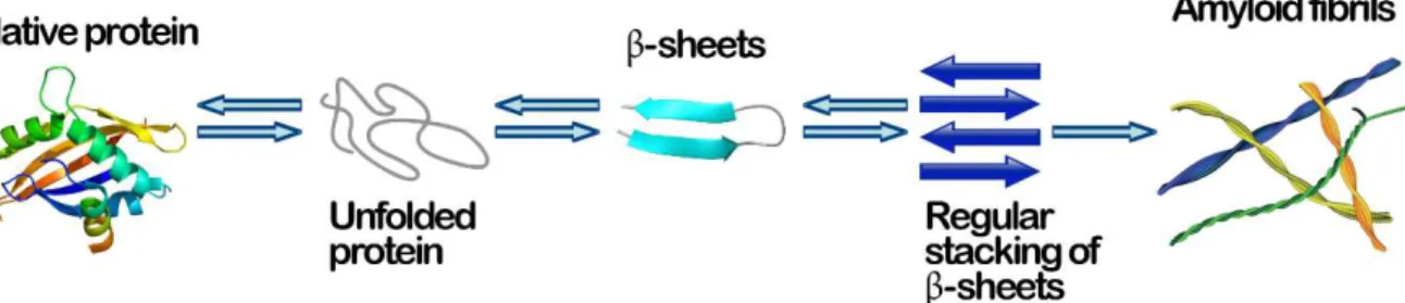
Amyloid proteins are a large class of proteins that may undergo a structural transformation from their native, functional folded state, into highly organized, insoluble fibrillar structures (Fig.1).
Fig. 1– A schematic representation of a possible amyloid fibril self-assembly process
During this transformation, the proteins lose their native form and functionality. Additionally, unlike native protein states, the stability of amyloid fibrils is largely attributed to intermolecular interactions. Mature fibrils adopt a more rigid structure than their monomeric and oligomeric precursors, which can be attributed to the high β-sheet content of the fibrils 5–7. These sheets form a “cross-β” motif, in which
9
between side chains promote hydrogen bonding parallel to the fibril 8,9. Regardless of
their amino acid sequence or native structure, amyloid fibrils display a characteristic cross-β pattern in X-ray diffraction experiments, indicating the orthogonal orientation of individual b-strands with respect to the fibril axis 10,11 (Fig.2).
Fig. 2 – A: atomic force microscopy (AFM) of amyloid fibrils generated by the PSMa1 peptide from S. aureus; B: the typical ‘‘cross-b’’ X-ray diffraction pattern for amyloid fibrils and C: atomic models for secondary, tertiary,
and quaternary structure (adapted from 8)
The unique molecular organization of amyloid fibrils endows them with remarkable mechanical properties as well as persistent structural integrity in the presence of high temperatures, proteases, denaturants, and physical forces 12.
Functional amyloids
These fibrillar structures are typically associated to neurodegenerative diseases such as Alzheimer’s and Parkinson’s diseases. However, more recently it has been observed that they can also fulfill important physiological roles in a variety of biological processes 13,14. Many organisms, such as fungi and some bacteria, take
10
Fig. 3– Functional and structural diversity of amyloid proteins (adapted from 17).
Amyloid fibrils are known to display impressive material properties, including high deformation resistance, elasticity, and persistence under extreme conditions. These unique properties make them excellent candidate materials for the design of functional biomaterials (Fig.4).
11 Hydophobins
In filamentous fungi, amyloids are involved in numerous processes, e.g., in the formation of aerial structures (spores or fruiting bodies) 19 and in the adherence of
fungal structures to hydrophobic surfaces. This adherence can occur on the surface of a host organism, thereby facilitating pathogenesis and playing a role in symbiosis
20. Hydrophobins, a large family of small proteins (about 100 aminoacids), represent
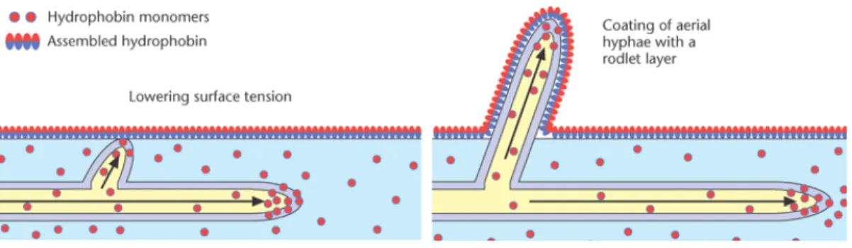
an example of functional amyloid fibrils produced at different growth stages by filamentous fungi (Fig.5). Their biological functions seem to be diverse, these proteins self-assemble at hydrophobic/hydrophilic interfaces into amphipathic layers changing the wettability of surfaces, they are very active surface and play a role as coating/protective agents, in adhesion, surface modification, or other types of functions that require surfactant-like properties 21.
Fig. 5 – Models showing the role of hydrophobins in the emergence of aerial hyphae (adapted from 22)
Hydrophobins are characterized by a β-barrel motif in the 3D structure and the presence of eight conserved cysteine residues that form four disulphide bonds (Cys1–Cys6, Cys2–Cys5, Cys3–Cys4, Cys7–Cys8). These eight conserved residues stabilize the protein core and define a hydrophobicity pattern typical of these proteins.
12
Fig. 6 – A: Amino acid sequence comparison of class I and II hydrophobins. The conserved Cys residues are
highlighted with the conserved disulphide-bonding pattern indicated with brackets. B: Comparative models
between class I (EAS from Neurospora crassa) and class II (HFBI fromTrichoderma reesei) hydrophobin. The
blue circle highlights the hydrophobic domains and the red circle the hydrophilic ones
Hydrophobin family can be divided into two distinct classes based on their structural differences, such as lengths of the inter-cysteine spaces and the stability of the amphipathic monolayers that they form. In particular, class I hydrophobins are produced in both ascomycetes and basidiomycetes species and the inter-cysteine space is more variable; class II hydrophobins are observed only in the ascomycetes and have a conserved length of the inter-cysteine spaces (Fig.6A). Moreover, fibrillar structures formed by class I hydrophobins, share many structural properties with amyloid fibrils. They assemble into insoluble polymeric layers, known as rodlets, extremely stable (resisting to hot 2% sodium dodecyl sulphate) and soluble only after harsh acid treatment (pure formic acid or trifluoroacetic acid (TFA))23. Class II
aggregates are nonfibrillar and less stable, they can be more easily dissolved in detergent or organic solvents (i.e, ethanol or sodium dodecyl sulphate) 24. Both
classes of Hydrophobin for their intriguing properties have been exploited in many biotechnological applications 25, summarized in Table 1.
Table 1 – Biotechnological applications of hydrophobins
Application Example
Industrial applications
Emulsions Personal care product, Food industry, Drug delivery High-value applications
Nanotechnology and diagnostic Biosensors, Electrodes, DNA/protein Microarrays, Immunological assays Tissue engineering Biocompatibility of medical implants and devices Separation technologies Co-purification of hybridized protein of interest
The layers of hydrophobins can be exploited as an attractive platform to immobilize proteins on different surfaces, due to their adhesive abilities. Immobilization of proteins has been widely studied and several methods are used to functionalize surface 26. For example, the adsorption method is the easiest and oldest
13
immobilization techniques 27. The interaction between the target protein and the
surface of the matrix occurs by various weak interactions such as electrostatic, hydrophobic/hydrophilic and van der waals forces. The cross-linking/covalent method is a conventional method for immobilization. It can be achieved by direct attachment between the protein and the material through a covalent linkage 28. In the entrapment
method target proteins are occluded in a permeable membrane (gel, fibre and microcapsule 29) which allows the substrates and the products to pass, but it retains
the target protein inside the network. In affinity binding, the target protein is linked to the matrix through specific interactions 30. In most of these cases, immobilization
leads to the reduction of the biological activity. The loss in activity could be due to changes in the protein conformation after immobilization, structural modification of the protein during immobilization, or changes in the protein microenvironment resulting from the interaction between the support and the protein.
Protein immobilization mediated by hydrophobin layer is a valid alternative to conventional methods and two main approaches have been carried out: deposition on a preformed self-assembled film 31–35, or genetical fusion to the self-assembling
protein 36,37. Protein immobilization by deposition on a preformed self-assembled film
may depend on the characteristics of the adhering protein and has to be optimized varying pH and ionic strength 31. A step forward the obtainment of a homogeneous
biohybrid layer is the exploitation of genetic engineering techniques to combine the hydrophobins features with the specific properties of target proteins. Following this approach, the engineered fused proteins can be straightly used to functionalize surfaces with advantage in terms of time required and chemicals consumed.
Among surfaces functionalized by class I hydrophobins, it is worth underlining the possibility of functionalization of nanomaterials:
1.2 Nanotechnology and Nanomaterials
The definition of a nanomaterial is an aggregate of atoms bonded together with a radius between 1 and 100 nm. (Fig.7)
14
Fig. 7 – Nanoscale
Nano system components are fabricated using top-down approach, that starts with a bulk or thin film material and removes selective regions to fabricate nanostructures, or bottom-up approach, that is based on molecular recognition and self-assembly to fabricate nanostructures out of smaller building blocks (molecules, colloids, and clusters).
When the dimension of a material is reduced from a large size, the properties remain the same at first, then small changes occur, until finally when the size drops below 100 nm, dramatic changes in properties can occur. If only one length of a three-dimensional nanostructure is of Nano dimension, the structure is referred to as a quantum well; if two sides are of nanometer length, the structure is referred to as a quantum wire. A quantum dot has all three dimensions in the nano range. The term
quantum is associated with these three types of nanostructures because the changes
in properties depend from the quantum-mechanical nature of physics in the domain of the ultrasmall. Thus physical, chemical e biological properties of nanomaterials are different from those of bulk matter. For instance, nanomaterials show i) high surface to volume ratio that makes them more chemically reactive; ii) optical properties that depend on parameters such as feature size, shape and surface characteristics; iii) magnetic properties that increase for particles small in size, in fact a paramagnetic bulk matter become a ferromagnetic cluster.
Hence, the advancement of nanomaterial research plays an essential role in the exploration of the biochemical and nanotechnological fields 38.
Graphene
Carbon materials are known to be more environmentally and biologically friendly than inorganic materials, since the carbon is one of the most common elements in our ecosystem. In particular, graphite is a naturally occurring material that has been used in our daily lives for hundreds of years without critical toxicity issues.
15
Graphene is the name given to a two-dimensional sheet of sp2-hybridized carbon. Its
extended honeycomb network is the fundamental building block of other important allotropes; it can be stacked to form 3D graphite, rolled to form 1D nanotubes, and wrapped to form 0D fullerenes 39 (Fig.8).
Fig. 8 – Carbon's allotropic forms
Graphene exhibits unparalleled properties such as high planar surface, superlative mechanical strength, remarkable thermal and electrical conductivity, high absorption of incident white light and highly efficient fluorescence quenching. Due to its extraordinary structure and fascinating properties, graphene is definitely the most studied nanomaterial 40. Consequently, it can be integrated as the core of
cutting-edge technologies and devices related to photonics, electronics, composite materials, environment, energy, biotechnology, biomedicine and biosensors 41. Since
the innovative discovery of the surprising properties of graphene, the industrial and scientific communities have focused their attention on the development of new graphene synthesis methods enabling a variety of options in terms of oxidation grade, number of layers, edge and basal defects, lateral size, quality, and cost for any particular application. According to the literature 42,43, the most relevant methods
for graphene generation are the chemical vapor deposition, mechanical cleavage, wet chemical synthesis, and exfoliation of graphite 44. Generally, liquid-phase
exfoliation of graphite entails the use of ultrasonication as a key method which promotes the generation of laminated material that is subsequently bound to aggregate due to the lack of hydrophilic groups onto the exfoliated material. In fact, re/aggregation is one of the main challenges to address during the exfoliation procedure and the stabilization of solvent-dispersed graphene flakes 45. However,
re/aggregation is typically minimized by using organic solvents with suitable characteristics or surfactant–water solutions. Biological interfacing of graphene has become crucial to improve its biocompatibility and selectivity toward various applications in the biotechnological and biomedical fields 46. Although graphene
16
modification and biofunctionalization are under active research, the in situ production of biofunctionalized graphene has been still little explored. However, fungal biosurfactants have been recently exploited to disperse and stabilize few-layered graphene in ethanol-water mixtures 47.
Carbon Nanotubes (CNT)
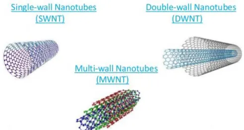
Carbon Nanotubes have sparked much excitement in recent years. These intriguing structures are an example of true nanotechnology: they are less than 100 nanometers in diameter and can be as thin as 1 or 2 nm, while they can be up to 18 centimeters in length 48. Nanotubes have a very broad range of electronic, thermal,
and structural properties that change depending on the different kinds of nanotube (defined by its diameter, length, and chirality, or twist). CNTs are made by rolling up of sheet of graphene into a cylinder. Depending on the number of concentrically rolled-up graphene sheets, they are also classified to single-walled (SWNT), double-walled (DWNT), and multidouble-walled (MWNT) (Fig.9). The structure of SWNT can be conceptualized by rolling-up layer of graphene into a seamless cylinder 49. MWNT
consists of two or more numbers of rolled-up concentric layers of graphene. DWNT is considered as a special type of MWNT where only two concentrically rolled up graphene sheets are present.
Fig. 9 – Basic structures of a single-walled, double-walled and multi-walled CNTs
CNTs have not only unique atomic arrangements but also have unique properties. These extraordinary properties of CNTs qualifies them exciting prospects and variety of applications. Particularly, in the area of microelectronics/nanoelectronics 50, CNTs
and graphene nanoribbons (GNRs) demonstrates wide range of applications such as energy storage devices; energy conversion devices that includes thermoelectric 51
and photovoltaic 52 devices; field emission displays and radiation sources 53;
nanometer semiconductor transistors 54, nanoelectromechanical systems (NEMS) 55,
electrostatic discharge (ESD) protection (Hyperion Catalysis), interconnects 56,57, and
17
1.3 Applications of Functional Amyloids from Fungi: Surface Modification by Class I Hydrophobins
As a general introduction about functionalization of different surfaces with class I hydrophobins, the review published in 2017 on “Biomolecules” is reported below.
biomolecules
Review
Applications of Functional Amyloids from Fungi:
Surface Modification by Class I Hydrophobins
Alessandra Piscitelli1 , Paola Cicatiello1, Alfredo Maria Gravagnuolo1,2, Ilaria Sorrentino1, Cinzia Pezzella1and Paola Giardina1,*
1 Department of Chemical Sciences, Università degli Studi di Napoli Federico II, Complesso Universitario
Monte S. Angelo, Via Cintia 4, 80126 Naples, Italy; [email protected] (A.P.); [email protected] (P.C.); [email protected] (A.M.G.); [email protected] (I.S.); [email protected] (C.P.)
2 Division of Pharmacy and Optometry, Faculty of Biology, Medicine and Health, The University of
Manchester, M13 9PT Manchester, UK
* Correspondence: [email protected]; Tel.: +39-081-674-319
Academic Editors: Margaret Sunde, Matthew Chapman, Daniel Otzen and Sarah Perrett Received: 15 May 2017; Accepted: 22 June 2017; Published: 26 June 2017
Abstract:Class I hydrophobins produced from fungi are amongst the first proteins recognized as functional amyloids. They are amphiphilic proteins involved in the formation of aerial structures such as spores or fruiting bodies. They form chemically robust layers which can only be dissolved in strong acids. These layers adhere to different surfaces, changing their wettability, and allow the binding of other proteins. Herein, the modification of diverse types of surfaces with Class I hydrophobins is reported, highlighting the applications of the coated surfaces. Indeed, these coatings can be exploited in several fields, spanning from biomedical to industrial applications, which include biosensing and textile manufacturing.
Keywords:functionalization; adhesion; biosensors; protein immobilization; biomedical applications; nanomaterials
1. Introduction
In the last decade, several papers have reported that amyloids can fulfill important functional roles in a variety of biological processes of taxonomically distant organisms. Many organisms take advantage of the ability of polypeptides to form amyloids [1,2]. In filamentous fungi, amyloids are involved in numerous processes, e.g., in signal transduction mechanism, in which Nod-like receptors control the induction of programmed cell death [3]; controlling the translation termination by Sup35 [4] and the nitrogen catabolism by Ure2 [5]; and in the formation of aerial structures (spores or fruiting bodies) by amphipathic proteins known as hydrophobins (HFBs) [6]. HFBs self-assemble into an amphipathic membrane at hydrophilic : hydrophobic interfaces fulfilling many other fungal functions, such as the adherence of fungal structures to hydrophobic surfaces, including the surface of a host organism, thereby facilitating pathogenesis and playing a role in symbiosis [7]. Analysis of fungal genomes indicates that HFBs exist as gene families. Different HFBs are expressed at different stages in the fungal life cycle accomplishing specific functions [8].
The HFB family is composed of small proteins (<20 kDa) with high sequence variability, however they share a β-barrel motif in the 3D structure and a pattern of eight cysteine residues forming four disulfide bonds that stabilize the protein core [8]. These proteins are divided into two main classes based on their structural differences, such as the lengths of the inter-cysteine spaces, which determine their different properties. Class I HFBs assemble into insoluble polymeric layers composed of fibrillar structures known as rodlets and have a morphology like amyloid fibrils associated with diseases states [9–11]. These layers are extremely stable (resistant to treatment with hot 2% sodium
Biomolecules 2017, 7, 45 2 of 11
dodecyl sulfate), can only be solubilized with harsh acid treatments (very concentrated formic acid or trifluoroacetic acid) and the soluble forms can polymerize back into rodlets under appropriate conditions [12]. Conversely, the layers formed by class II hydrophobins lack the fibrillary rodlet morphology and can be solubilized with organic solvents and detergents [13].
Only class I HFBs belong to the functional amyloid family, since the structural and morphological similarities between rodlets and amyloid fibrils have been confirmed many times. Indeed, rodlets bind amyloid-specific dyes (Congo Red, Thioflavin T) and their diffraction pattern displays typical reflections of amyloid structures (4.8 and 10–12 Å). [9].
The propensity of class I HFBs to self-assemble and the presence of disorder portions in their soluble forms, has precluded the achievement of crystals suitable for X-ray crystallography. However 3D structures of soluble class I HFBs have been obtained by nuclear magnetic resonance (NMR) studies for the EAS from Neurospora crassa [9], DewA from the fungus Aspergillus nidulans [14], MPG1 from the fungus Magnaporthe oryzae [15], and very recently for SC16 from Schizophyllum commune [16].
Both classes of HFBs have been exploited in many biotechnological applications [17]. Bioinspired coatings based on HFBs can offer novel opportunities for surface modification [13]. Over the last decade the surfaces coated with the layers of HFBs have been employed to immobilize several enzymes of industrial interest, and several materials coated with HFBs and their engineered variants have been proven effective for a wide range of biotechnological applications. Taking into consideration the topic of this special issue, this review is focused on the recent advances in the use of the functional amyloids class I HFBs in surface coating, organizing the reported results based on the typology of the surfaces. 2. Metal and Metalloid Functionalization
Thin films of titanium show unique chemical, optical, and electrical properties [18]. The interest on their fabrication is increasing due to their attractive applications (i.e., microelectronic devices, photonic materials, high-efficiency catalysts, environmental remediation, optical devices, and medical treatments). Santhiya et al. [19] set up a novel method for the aqueous phase deposition of smooth, nanocrystalline TiO2thin films using a self-assembled HFB layer on a silicon substrate. In this report,
the class I HFB used was H*Protein B, an engineered protein based on the class I HFB DewA from A. nidulans. A HFB layer on the silicon surface was prepared and used to deposit highly uniform nanocrystalline TiO2 thin films. Resistance and elasticity properties of the developed films were compatible with implant coatings and other biomedical devices.
Boeuf et al. [20] engineered DewA from A. nidulans by inserting the RGD sequence or the laminin globular domain (LG3 binding motif) at surface-accessible sites of the protein to functionalize the surfaces of orthopedic implants made of titanium. The purified proteins were used to produce surfaces that can enhance the adhesion of the human cells, while the adhesion of Staphylococcus aureus did not increase, thus minimizing the risk of bacterial infection.
Vmh2 from Pleurotus ostreatus was used to functionalize different surfaces, such as silicon, steel and gold. Vmh2 forms a chemically and mechanically stable layer of self-assembled proteins on crystalline silicon. This biomolecular film was exploited as a masking material, since the protein film perfectly protected the coated silicon surface during the standard KOH etching process [21]. The protein-modified silicon surface exhibited also an improvement in wettability and suitability for the immobilization of other proteins, such as BSA or the enzyme laccase, improving its stability [22]. The self-assembled Vmh2 layer also changed the wettability of the Porous silicon (PSi) structures and protected this nanocrystalline material from the basic dissolution process. PSi is a versatile material owing to its peculiar morphological, physical, and chemical properties [23] The major drawback of the “as etched” PSi is its chemical instability, since it is oxidized at room temperature by atmospheric oxygen [24]. The Vmh2 coating added chemical stability to PSi (Figure1), without altering the sensing ability of this optical transducer, which can be a key tool for biomolecular experiments.
Biomolecules 2017, 7, 45ȱ ȱ ȱ ȱ ȱ 3 of 11ȱ ȱ ȱ ȱ ȱ ȱ ȱ ȱ ȱ ȱ ȱ ȱ ȱ ȱ ȱ ȱ ȱ ȱ ȱ ȱ ȱ ȱ ȱ ȱ ȱ ȱ ȱ Ȭ ȱ ȱ ȱ ȱ ȱ Ȭ ȱ ȱ ȱ Ȭ Ȭ ȱ ȱ ȱ Ȭ ȱ ȱ ȱ ȱ ȱ ȱ ȱ ȱ ȱ ȱ ȱ ȱ ȱ ȱ ȱ ȱ ȱ ȱ ȱ ȱ Ȭ ȱ ȱ ȱ ȱ ȱ ȱ ȱ ȱ ȱ ȱ ȱ ȱ ȱ ȱ ȱ ȱ ȱ ȱ ȱ ȱ ȱ ȱ ȱ ȱ ȱ ȱ ȱ Ȭ ȱ Ȭ ȱ ȱ ȱ ȱ ȱ ȱ ȱ ȱ ȱ ȱ ȱ ȱ ȱ ȱ ȱ ȱ ȱ ȱ ȱ ȱ ȱ ȱ ȱ ȱ ȱ ȱ ȱ ȱ ȱ ȱ ȱ ȱ ȱ ȱ ȱ ȱ ȱ ȱ ȱ ȱ ȱ ȱ ȱ ȱ ȱ ȱ ȱ ȱ ȱ ȱ ȱ ȱ ȱ ȱ ȱ ȱ ȱ ȱ ȱ ȱ ȱ ȱ ȱ ȱ ȱ ȱ ȱ ȱ ȱ ȱ ȱ ȱ ȱ ȱ ȱ ȱ ȱ ȱ ȱ ȱ ȱ ȱ ȱ ȱ ȱ ȱ ȱ ȱ ȱ ȱ ȱ Ȭ ȱ ȱ ȱ ȱ ȱ ȱ ȱ Ȭ ȱ ȱ ȱ ȱ ȱ Ȭ ȱ ȱ ȱ ȱ ȱ ȱ ȱ ȱ ȱ ȱ ȱ ȱ ȱ Ȭ ȱ ȱ ȱ ȱ ȱ ȱ ȱ ȱ ȱ ȱ ȱ ȱ ȱ ȱ ȱ ȱ ȱ ȱ ȱ ȱ ȱ ȱ ȱ ȱ ȱ ȱ ȱ ȱ Ȭ ȱ ȱ ȱ ȱ ȱ ȱ ȱ ȱ ȱ ȱ ȱ ȱ ȱ ȱ ȱ Ȭ ȱ ȱ ȱ Ȭ Ȭ ȱ ȱ ȱ Ȭ Ȭ ȱ ȱ ȱ ȱ ȱ ȱ ȱ ȱ ȱ ȱ ȱ ȱ ȱ ȱ ȱ ȱ ȱ ȱ ȱ Ȭ ȱ ȱ ȱ ȱ ȱ ȱ ȱ ȱ ȱ ȱ ȱ ȱ ȱ ȱ ȱ ȱ ȱ ȱ ȱ ȱ ȱ ȱ ȱ ȱ ȱ ȱ ȱ ȱ ȱ ȱ ȱ ȱ ȱ ȱ ȱ Ȭ ȱ ȱ ȱ ȱ ȱ ȱ Ȭ Ȭ ȱ ȱ ȱ ȱ ȱ ȱ ȱ ȱ ȱ
Figure 1.Change of wettability of PSi by Vmh2 and variation of PSi thickness with time, showing
protection from etching by Vmh2.
The sample-loading steel plate used in matrix-assisted laser desorption/ionization time-of-flight mass spectrometry (MALDI-TOF MS) was stably coated by the Vmh2 layer and the functionalized support can be reused, since Vmh2 can be de-polymerized and solubilized. The hybrid surface was able to homogenously adsorb peptides and proteins whereas salts or denaturants could be washed away with water, allowing fast and high-throughput on-plate desalting prior to MS analysis [25]. Moreover, the function of the Vmh2 coating was expanded by immobilizing enzymes of interest in proteomics (trypsin, V8 protease, PNGaseF, and alkaline phosphatase) on the steel surface. High sequence coverage of model proteins and analysis of a whole proteome (whey milk) were achieved by rapid and efficient multiple enzyme digestions, serially performed on plate (Figure2) [26]. The same procedure provided the opportunity to discriminate blood provenance even when two different blood sources were present in a mixture [27]. Phosphatases or deglycosidase were also immobilized on-plate, allowing the study of proteins with post-translational modifications [26].
The spontaneous self-assembling of Vmh2 on the gold surface was verified by using the quartz crystal-microbalance (QCM) and confirmed by spectroscopic ellipsometry [28]. The Vmh2 layer stably assembled on the gold QCM electrode was also used to perform a quantitative analysis of the Vmh2-glucose interaction. ȱ ȱ ȱ ȱ ȱ ȱ ȱ ȱ ȱ ȱ ȱ ȱ ȱ ȱ ȱ ȱ ȱ ȱ ȱ ȱ ȱ ȱ ȱ ȱ ȱ ȱ ȱ ȱ ȱ ȱ ȱ ȱ Ȭ ȱ ȱ ȱ ȱ ȱ Ȭ ȱ ȱ ȱ Ȭ Ȭ ȱ ȱ ȱ Ȭ ȱ ȱ ȱ ȱ ȱ ȱ ȱ ȱ ȱ ȱ ȱ ȱ ȱ ȱ ȱ ȱ ȱ ȱ ȱ ȱ Ȭ ȱ ȱ ȱ ȱ ȱ ȱ ȱ ȱ ȱ ȱ ȱ ȱ ȱ ȱ ȱ ȱ ȱ ȱ ȱ ȱ ȱ ȱ ȱ ȱ ȱ ȱ ȱ Ȭ ȱ Ȭ ȱ ȱ ȱ ȱ ȱ ȱ ȱ ȱ ȱ ȱ ȱ ȱ ȱ ȱ ȱ ȱ ȱ ȱ ȱ ȱ ȱ ȱ ȱ ȱ ȱ ȱ ȱ ȱ ȱ ȱ ȱ ȱ ȱ ȱ ȱ ȱ ȱ ȱ ȱ ȱ ȱ ȱ ȱ ȱ ȱ ȱ ȱ ȱ ȱ ȱ ȱ ȱ ȱ ȱ ȱ ȱ ȱ ȱ ȱ ȱ ȱ ȱ ȱ ȱ ȱ ȱ ȱ ȱ ȱ ȱ ȱ ȱ ȱ ȱ ȱ ȱ ȱ ȱ ȱ ȱ ȱ ȱ ȱ ȱ ȱ ȱ ȱ ȱ ȱ ȱ ȱ Ȭ ȱ ȱ ȱ ȱ ȱ ȱ ȱ Ȭ ȱ ȱ ȱ ȱ ȱ Ȭ ȱ ȱ ȱ ȱ ȱ ȱ ȱ ȱ ȱ ȱ ȱ ȱ ȱ Ȭ ȱ ȱ ȱ ȱ ȱ ȱ ȱ ȱ ȱ ȱ ȱ ȱ ȱ ȱ ȱ ȱ ȱ ȱ ȱ ȱ ȱ ȱ ȱ ȱ ȱ ȱ ȱ ȱ Ȭ ȱ ȱ ȱ ȱ ȱ ȱ ȱ ȱ ȱ ȱ ȱ ȱ ȱ ȱ ȱ Ȭ ȱ ȱ ȱ Ȭ Ȭ ȱ ȱ ȱ Ȭ Ȭ ȱ ȱ ȱ ȱ ȱ ȱ ȱ ȱ ȱ ȱ ȱ ȱ ȱ ȱ ȱ ȱ ȱ ȱ ȱ Ȭ ȱ ȱ ȱ ȱ ȱ ȱ ȱ ȱ ȱ ȱ ȱ ȱ ȱ ȱ ȱ ȱ ȱ ȱ ȱ ȱ ȱ ȱ ȱ ȱ ȱ ȱ ȱ ȱ ȱ ȱ ȱ ȱ ȱ ȱ ȱ Ȭ ȱ ȱ ȱ ȱ ȱ ȱ Ȭ Ȭ ȱ ȱ ȱ ȱ ȱ ȱ ȱ ȱ ȱ
Figure 2. In situ reaction of trypsin immobilized on Vmh2 coated matrix-assisted laser
desorption/ionization time-of-flight mass spectrometry (MALDI-TOF-MS) sample plate.
Additionally, gold nanoparticles (AuNPs) were synthesized, by a simple and novel process, in the presence of polyethylene-glycol (PEG), using Vmh2 to produce stable hybrid protein–metal nanoparticles, with outer surface rich in functional chemical groups [29]. Even though in the hybrid system the Vmh2 proteins were intrinsically bonded to the gold core, Vmh2-glucose interaction was confirmed, and the PEG-HFB-AuNPs was used in glucose monitoring [30].
Biomolecules 2017, 7, 45 4 of 11
3. Plastic Functionalization
Different plastic materials were modified using class I HFBs from both native and recombinant sources (Table1). When heterologously produced, fused proteins composed of the HFB moiety and target proteins were successfully exploited. The modified materials were mainly used in the development of biomedical devices, examples of applications in biosensing and protein immobilization were demonstrated.
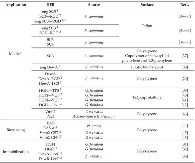
Platforms for protein immobilization were developed by using HGFI from Grifola frondosa native [31] and recombinant source [32]. On the other hand, the HFBs DewA and DewB from A. nidulans were fused to the laccase LccC from the same fungal source, developing an efficient system for laccase immobilization on polystyrene multiwell plates [33].
Table 1.Class I hydrophobins used in functionalization of plastic materials.
Application HFB Source Surface Refs.
Medical eng SC31 S. commune Teflon [34–36] SC3—RGD2 eng SC3—RGD1,2 eng SC31 S. commune [34–36] SC3—RGD2 SC3 S. commune [34–36] SC4 SC3 S. commune Polystyrene [37] Copolymer of benzoyl-1,4
phenylene and 1,3-phenylene
eng DewA1 A. nidulans Plastic biliary stent [38]
DewA A. nidulans Polystyrene [20] DewA–RGD2 DewA–LG32 HGFI—TPS2 G. frondosa Polycaprolattone [39] HGFI—VGF2 G. frondosa [40] HGFI—VGF2 G. frondosa [41] HGFI—PA12 G. frondosa [42] Vmh2 P. ostreatus Polystyrene [43] Pac3 Acremonium sclerotigenum
Biosensing EAS N. crassa Polystyrene [44] EAS-α2 Vmh2-GST2 P. ostreatus [45] Vmh2-GFP2 P. ostreatus [46] Immobilization HGFI G. frondosa Polystyrene [31] rHGFI3 G. frondosa [32] DewA–LccC2 A. nidulans [33] DewB–LccC2
1Recombinant engineered protein;2Recombinant fused protein;3Recombinant protein.
The exploitation of plastic surfaces coated with class I HFBs for biomedical devices was started by the pioneering work of Scholtmeijer et al. and Janssen and coworkers, who exploited the class I HFBs SC3 and SC4 (either native or recombinant engineered fused HFBs) in the functionalization of Teflon surfaces [34–36]. In that work, as well as in other examples, the main interest was in the development of biocompatible surfaces that allow both adhesion of human cells and tissue regeneration. Indeed, the HFBs were fused to different cell adhesion-mediating motifs and proved to be effective in the design of materials for regenerative medicine [20,39]. Moreover, Wang et al. and Zhao et al. [31,40] fused the HGFI to the vascular endothelial growth factor (VEGF), an effective molecule able to regulate the proliferation, migration, and survival pathways of endothelial cells [41].
Biomolecules 2017, 7, 45 5 of 11
Other examples of medical applications are the development of antibacterial devices [42] by the fusion of the bacteriocin pediocin PA-1 to HGFI. Furthermore, the layers formed by two fungal hydrophobins (Vmh2 and Pac3 from A. sclerotigenum [47]) reduce the biofilm formed by different strains of Staphylococcus epidermidis on polystyrene surfaces, without affecting the cell vitality [43]. Polymeric surfaces with enhanced lubricity and reduced surface friction were obtained using SC3 by Misra and coworkers [37]. Biliary plastic stents with delayed clogging process were developed thanks to the coating with the HFB, alone or in combination with heparin [38].
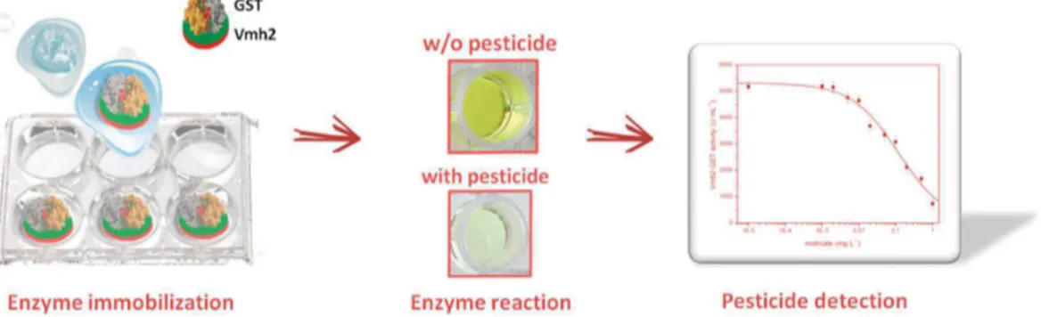
Considering biosensing applications, the EAS HFB from N. crassa fused to the yeast peptide pheromone α-factor was used in the detection and quantification of this pheromone [44]. Upon functionalization of polystyrene multiwell plate with a combination of HFBs either lacking or exposing the α-factor, an inverted enzyme-linked immunosorbent assay (ELISA) approach was developed yielding a novel kind of biosensor with the lowest limit of detection reported at the time of publication. Vmh2 from P. ostreatus fused to the enzyme glutathione-S-transferase (Vmh2-GST) was exploited for the quantification of the pesticides molinate and captan, acting as inhibitors of the enzymatic activity [45]. The fused protein efficiently functionalized the polystyrene multiwell plate for the development of high throughput analyses (Figure3).
ȱ ȱ ȱ ȱ ȱ ȱ ȱ ȱ ȱ ȱ ȱ ȱ ȱ ȱ ȱ ȱ ȱ ȱ ȱ ȱ ȱ ȱ ȱ ȱ ȱ ȱ ȱ ȱ ȱ ȱ ȱ ȱ ȱ ȱ ȱ ȱ ȱ ȱ ȱ ȱ ȱ ȱ ȱ ȱ ȱ ȱ ȱ ȱ ȱ ȱ ȱ ȱ ȱ ȱ ȱ ȱ ȱ ȱ ȱ ȱ ȱ ȱ ȱ ȱ ȱ ȱ ȱ ȱ ȱ ȱ ȱ ȱ ȱ ȱ ȱ ȱ ȱ ȱ ȱ ȱ ȱ ȱ ȱ ȱ ȱ ȱ ȱ ȱ ȱ ȱ ȱ ΅Ȭ ȱ ȱ ȱ ȱ ȱ ȱ ȱ ȱ ȱ ȱ ȱ ȱ ȱ ȱ ȱ ȱ ȱ ȱ ȱ ȱ ȱ ȱ ȱ ȱ ȱ ȱ ȱ ȱ ΅Ȭ ȱ ȱ ȱ Ȭ ȱ ȱ ȱ ȱ ȱ ȱ ȱ ȱ ȱ ȱ ȱ ȱ ȱ ȱ ȱ ȱ ȱ ȱ ȱ ȱ ȱ ȱ ȱ ȱ ȱ ȱ ȱ ȱ ȱ ȱ ȱ ȱ ȱ Ȭ Ȭ ȱ Ȭ ȱ ȱ ȱ ȱ ȱ ȱ ȱ ȱ ȱ ȱ ȱ ȱ ȱ ȱ ȱ ȱ ȱ ȱ ȱ ȱ ȱ ȱ ȱ ȱ ȱ ȱ ȱ ȱ ȱ ȱ ȱ ȱ ȱ ȱ ȱ ȱ ȱ ȱ ȱ ȱ ȱ ȱ ȱ ȱ ȱ ȱ ȱ ȱ ȱ ȱ ȱ Ȭ ȱ ȱ ȱȱ ȱ ȱ ȱ ȱ ȱ ȱ ȱ ȱ ȱ ȱ ȱ ȱ ȱ ȱ ȱ ȱ ȱ ȱ ȱ ȱ Ȭ ȱ ȱ ȱ ȱ ȱ ȱ ȱ ȱ ȱ ȱ ȱ ȱ ȱ ȱ ȱ Ȭ ȱ ȱ ȱ ȱ ȱ ȱ ȱ ȱ ȱ ȱ ȱ ȱ ȱ ȱ ȱ ȱ ȱ ȱ ȱ ȱ ȱ ȱ ȱ ȱ ȱ ȱ ȱ ȱ ȱ ȱ ȱ ȱ ȱ ȱ ȱ ȱ ȱ ȱ ȱ ȱ ȱ ȱ ȱ ȱ ȱ ȱ ȱ ȱ ȱ ȱ ȱ ȱ ȱ ȱ ȱ ȱ ȱ ȱ ȱ ȱ ȱ ȱ ȱ ȱ ȱ ȱ ȱ ȱ ȱ ȱ ȱ ȱ ȱ ȱ ȱ Ȭ ȱ ȱ ȱ
Figure 3. Pesticide biosensor developed on polystyrene multiwell plate coated with Vmh2-GST
fused proteins
Vmh2 adhesion ability was also combined with the fluorescence emission of the Green Fluorescent Protein (GFP) by genetic fusion [46]. Vmh2-GFP was proven to be a smart and effective tool for the study of Vmh2 self-assembling and was used as the active biological element in the realization of an ultrasensitive thrombin biosensor. Since the two proteins were linked by the specific cutting site of the thrombin, a decrease in the fluorescence intensity of the sample was observed due to the cleavage of the linker by thrombin and the subsequent desorption of the GFP from the surface (Figure4).
ȱ ȱ ȱ ȱ ȱ ȱ ȱ ȱ ȱ ȱ ȱ ȱ ȱ ȱ ȱ ȱ ȱ ȱ ȱ ȱ ȱ ȱ ȱ ȱ ȱ ȱ ȱ ȱ ȱ ȱ ȱ ȱ ȱ ȱ ȱ ȱ ȱ ȱ ȱ ȱ ȱ ȱ ȱ ȱ ȱ ȱ ȱ ȱ ȱ ȱ ȱ ȱ ȱ ȱ ȱ ȱ ȱ ȱ ȱ ȱ ȱ ȱ ȱ ȱ ȱ ȱ ȱ ȱ ȱ ȱ ȱ ȱ ȱ ȱ ȱ ȱ ȱ ȱ ȱ ȱ ȱ ȱ ȱ ȱ ȱ ȱ ȱ ȱ ȱ ȱ ȱ ΅Ȭ ȱ ȱ ȱ ȱ ȱ ȱ ȱ ȱ ȱ ȱ ȱ ȱ ȱ ȱ ȱ ȱ ȱ ȱ ȱ ȱ ȱ ȱ ȱ ȱ ȱ ȱ ȱ ȱ ΅Ȭ ȱ ȱ ȱ Ȭ ȱ ȱ ȱ ȱ ȱ ȱ ȱ ȱ ȱ ȱ ȱ ȱ ȱ ȱ ȱ ȱ ȱ ȱ ȱ ȱ ȱ ȱ ȱ ȱ ȱ ȱ ȱ ȱ ȱ ȱ ȱ ȱ ȱ Ȭ Ȭ ȱ Ȭ ȱ ȱ ȱ ȱ ȱ ȱ ȱ ȱ ȱ ȱ ȱ ȱ ȱ ȱ ȱ ȱ ȱ ȱ ȱ ȱ ȱ ȱ ȱ ȱ ȱ ȱ ȱ ȱ ȱ ȱ ȱ ȱ ȱ ȱ ȱ ȱ ȱ ȱ ȱ ȱ ȱ ȱ ȱ ȱ ȱ ȱ ȱ ȱ ȱ ȱ ȱ Ȭ ȱ ȱ ȱȱ ȱ ȱ ȱ ȱ ȱ ȱ ȱ ȱ ȱ ȱ ȱ ȱ ȱ ȱ ȱ ȱ ȱ ȱ ȱ ȱ Ȭ ȱ ȱ ȱ ȱ ȱ ȱ ȱ ȱ ȱ ȱ ȱ ȱ ȱ ȱ ȱ Ȭ ȱ ȱ ȱ ȱ ȱ ȱ ȱ ȱ ȱ ȱ ȱ ȱ ȱ ȱ ȱ ȱ ȱ ȱ ȱ ȱ ȱ ȱ ȱ ȱ ȱ ȱ ȱ ȱ ȱ ȱ ȱ ȱ ȱ ȱ ȱ ȱ ȱ ȱ ȱ ȱ ȱ ȱ ȱ ȱ ȱ ȱ ȱ ȱ ȱ ȱ ȱ ȱ ȱ ȱ ȱ ȱ ȱ ȱ ȱ ȱ ȱ ȱ ȱ ȱ ȱ ȱ ȱ ȱ ȱ ȱ ȱ ȱ ȱ ȱ ȱ Ȭ ȱ ȱ ȱ
Figure 4. Thrombin biosensor developed on polystyrene multiwell plate coated with Vmh2-GFP