A study of the properties, reactivity and anticancer activity of novel Nmethylated-thiazolyl or 3-1
thienyl carbazoles and their Pd(II) and Pt(II) complexes 2
3 4 5 6
Marta Reiga, Ramón Bosqueb, Mercè Font-Bardíac, Carme Calvisd, Ramon Messeguerd, 7
Laura Baldomàe,f, Josefa Badíae,f, Dolores Velascoa, Concepción Lópezb,⁎ 8 9 10 11 12 13 14 15 16 17
a Grup de Materials Orgànics, Institut de Nanociència i Nanotecnologia (IN2UB), Secció de Química 18
Orgànica, Departament de Química Inorgànica i Orgànica, Facultat de Química, Martí i Franquès 1-11, 19
E-08028 Barcelona, Spain 20
b Secció de Química Inorgànica, Departament de Química Inorgànica i Orgànica, Facultat de Química, 21
Martí i Franquès 1-11, E-08028 Barcelona, Spain 22
c Unitat de Difracció de Raigs-X, Centres Científics i Tecnològics (CCiT), Universitat de Barcelona, 23
Solé i Sabaris 1–3, E-08028 Barcelona, Spain 24
d Biomed Division, Health & Biomedicine Unit, LEITAT Technological Center, Parc Científic de 25
Barcelona, Edifici Hèlix, Baldiri i Reixach 13-21, E-08028 Barcelona, Spain 26
e Secció de Bioquímica i Biología Molecular, Departament de Bioquímica i Fisiologia, Facultat de 27
Farmàcia i Ciències de l'Alimentació, Universitat de Barcelona, Av. Joan XXIII 27-31, E-08028 28
Barcelona, Spain 29
f Institut de Biomedicina de la Universitat de Barcelona - Institut Recerca Sant Joan de Deu, Barcelona, 30 Spain 31 32 33 34 35 36 37 38 39 40 41 42 43 44 45 46 [email protected] (C. López). 47 48 49 50 51
ABSTRACT: 52
53
The synthesis and characterization of two hybrid N-methylated carbazole derivatives containing a 54
thiazolyl or a thienyl ring is reported. The thiazolyl derivative has been also characterised by X-ray 55
diffraction analysis. The study of its reactivity in front of [MCl2(dmso)2] (M=Pd or Pt) or Na2[PdCl4] 56
in methanol has allowed us to isolate and characterize its complexes. However, for the thienyl analogue, 57
the formation of any Pd(II) or Pt(II) complex was not detected, indicating that it is less prone to bind to 58
the M(II) ions than its thiazolyl analogue. Density Functional Theory (DFT) and Time-Dependent 59
Density Functional Theory (TD-DFT) calculations have also been carried out in order to rationalize the 60
influence of the nature of the thiazolyl or thienyl group on the electronic delocalization. Molecular 61
mechanics calculations show that the free rotation of the thiazolyl in relation to the carbazole requires a 62
greater energy income than for its thienyl analogue. Studies of the cytotoxic activity of the new 63
compounds on colon (HCT116) and breast (MDA-MB231 and MCF7) cancer cell lines show that the 64
thiazolyl carbazole ligand and its Pt(II) complex are the most active agents of the series and in the 65
MCF7 line their potency is higher than that of cisplatin. In the non-tumoral human skin fibroblast BJ 66
cell line, all the compounds were less toxic than cisplatin. Their potential ability to modify the 67
electrophoretic mobility of pBluescript SK+ plasmid DNA and to act as inhibitors of Topoisomerases I 68
and IIα or cathepsin B has also been investigated. 69
1. INTRODUCTION 71
72
Cancer is among the leading causes of morbidity and death that unfortunately affects millions of persons 73
worldwide (i.e. more than one million each year in USA [1]). The American Cancer Society estimates 74
an incidence of ca. 1.7 million new cases for 2017 and>0.6 million deaths [2] mainly produced by 75
colorectal, breast, lung and ovarian cancers [2–4]). Every cancer type needs a specific treatment protocol 76
that usually involves chemotherapy (CT) [5], radiotherapy and/or surgery. The development of new 77
antitumor drugs with improved activities and lower side effects than those used nowadays in CT is still 78
one of the main challenges of current research in medicinal chemistry. Among the variety of strategies 79
used nowadays in drug discovery [6–11], those with greater expectations are based on: a) natural 80
products and/or b) new synthetic products with several bioactive arrays (i.e. by incorporation of an 81
additional bioactive unit in the backbones of commercially available pharmaceuticals or known drugs 82
and commonly known as “molecular hybridization approach”) [6–11]. 83
On the other hand, it is well-known that heterocycles and their derivatives are one of the most important 84
types of organic compounds due to their outstanding physical and photo-optical properties, their rich 85
reactivity, their utility as ligands in Coordination and Organometallic Chemistry and their multiple 86
applications in a variety of fields, including medicinal chemistry [12–21]. Heterocyclic cores are present 87
in huge range of natural and marketed antimicrobial, anti-inflammatory, antiviral, anticancer, 88
antihypertensive, antimalarial, anti-HIV, antidepressant, antihelmintic drugs, among others. Their 89
relevance in new drugs design and development is undeniable [12–14]. For instance, among all the 90
anticancer drugs approved by FDA during the late five years ca. 75% have heterocyclic arrays with N 91
and/or S atoms or polycyclic aromatic compounds with heterocyclic fragments [15–22]. 92
Carbazole (Fig. 1), thiazole and thiophene are probably three of the most important scaffolds in drug 93
design and discovery [23–33]. These units are present in diverse bioactive synthetic -and even in 94
naturally occurring products. For instance, Ellipticine and Glybomine-C (Fig. 1) isolated from plants, 95
are potent cytotoxic agents in several cancer cell lines [34,35] and the discovery that N-alkylation of 96
Ellipticine enhanced inhibition growth activity has stimulated the interest on N-substituted carbazoles 97
[23–26,34,35]. The number of potent cytotoxic (substituted and/or anellated) carbazoles reported in the 98
last 2 years has grown exponentially and according to a recent review published by Caruso et al. [26]: 99
“Carbazoles are promising scenarios for breast cancer treatments”. 100
In addition to carbazoles, and in a lesser extent, thiazole and thiophene derivatives are gaining 101
increasing interest as “central cores” or as “pendant” groups in drug engineering and specially as 102
promising platforms or “templates” to build up new and more efficient antitumor agents that could 103
overcome or at least reduce the main problems (i.e. drug resistance, toxicity, or other undesirable side 104
effects) associated to drugs used currently. cis-[PtCl2(NH3)2] (cisplatin) and doxorubicin [4,36–40] are 105
two CT agents used in cancer treatments that may generate severe-side effects (i.e. nephrotoxicity, 106
neurotoxicity, the increase of blood pressure, severe nausea, vomiting, or diarrhoea produced by 107
cisplatin or severe heart problems (cardiomyopathy) associated to doxorubicin [36–40]). Since the 108
discovery of cisplatin, the development of metal coordination complexes as anticancer agents has 109
attracted a great deal of interest to obtain more effective and less toxic drugs [41]. For instance, trans-110
platinum(II) based complexes or those based on less toxic metals (ruthenium, gold or copper) are shown 111
to be promising candidates in safer cancer therapy. 112
Despite of the interest in electronics and materials science arisen by hybrid carbazole/thienyl derivatives 113
[42–52] and the potential synergic effect of the presence of two bioactive arrays [i.e. the N-substituted 114
carbazole and a thiazole (or a thienyl) unit] in the same molecule that could be relevant in drug design 115
[53,54] and in photodynamic therapy (PTD) [55], studies on their biological activities are scarce. 116
Moreover, it is well-known that heterocycles are valuable ligands in Coordination and Organometallic 117
Chemistry [15–17] and their binding to a transition metal ion (Mm+) commonly affects their properties 118
and their biological and/or catalytic activities. For instance, Pd(II) and Pt(II) complexes with 119
heterocyclic ligands (i.e. pyrazoles, indoles) showing greater cytotoxic activity than the free ligands 120
have been reported [56–60]. However, parallel studies on hybrid carbazole/thiazole or thiophene 121
derivatives have not been investigated yet. Therefore, there is a lack of information on their coordination 122
ability and the effect produced by the binding of these ions on their properties and especially on their 123
cytotoxic activity. 124
In this paper, we present two new carbazole derivatives: 9-methyl-3- (2-thiazolyl)-9H-carbazole (1a) 125
and 9-methyl-3-(2-thienyl)-9H-carbazole (1b) shown in Scheme 1, a study of their reactivity in front of 126
Pd (II) and Pt(II), their spectroscopic properties and the anticancer activity of the free ligands and the 127
new Pd(II) and Pt(II) complexes, trans- [PdCl2(1a)2] (2a) and trans-[PtCl2(1a)dmso] (3a). 128
2. EXPERIMENTAL 130 131 2.1. Chemistry 132 133
2.1.1. Materials and methods 134
[MCl2(dmso)2] {trans- for M=Pd or cis- for M=Pt} and 3-iodo-9H-carbazole were prepared as 135
described previously [61–63], and the remaining reagents were obtained from commercial sources and 136
used as received. The success of the synthesis of the Pt(II) compound 3a is strongly dependent on the 137
quality of the methanol; the presence of water produces the formation of metallic platinum, other 138
undesirable minor by-products and a significant decrease in the yield. Thus, the use of high quality 139
MeOH (HPLC grade) is required. The remaining solvents used were dried and distilled before use [64]. 140
During the preparation of the complexes (2a and 3a), the reaction flask was protected from the light with 141
aluminium foil. Elemental analysis were carried out at the Centres Científics i Tecnològics (CCiT, Univ. 142
Barcelona) with an Eager 1108 microanalyzer. Mass spectra (ESI+) were performed at the Servei 143
d'Espectrometria de Masses (Univ. de Barcelona) using a LC/MSD-TOF Agilent Technologies 144
instrument. UV–vis. spectra of CH2Cl2 solutions of the free ligands (1a and 1b) and complexes 2a and 145
3a were recorded at 298 K with a Varian Cary UV–Vis-NIR 500E spectrometer and their emission 146
spectra were obtained on a PTI fluorimeter equipped with a 220B lamp power supply, a 815 147
photomultiplier detection system and Felix 32 software at 298 K in CH2Cl2 solutions. 1,4-Bis(5-phenyl-148
2-oxazolyl) benzene (POPOP) dissolved in cyclohexane was used as a standard for the fluorescence 149
quantum yield determination (λexc=300 nm, ФPOPOP=0.93). 1H and 13C{1H}-NMR spectra were 150
recorded at 298 K in acetone-d6 for the precursor (3-iodo-9-methyl-9H-carbazole) or in CDCl3 (in the 151
remaining cases) with a Varian Mercury 400 MHz or a Bruker 400 MHz Avance III [for 1H and 152
13C{1H}] and a Bruker 250 MHz and a Bruker 400 Avance III HD (for 195Pt{1H}) spectrometers. 153
Chemical shifts are given in δ values (ppm) using the solvent peaks as internal references (1H and 13C) 154
and H2PtCl6 in D2O (195Pt{1H}) and coupling constants (J) are given in Hz. Abbreviations used for 155
the multiplicities are as follows: s (singlet), d (doublet), dd (doublet of doublets), t (triplet), q 156
(quadruplet) and m (multiplet). The atom numbering system used in the assignment of 1H and 157
13C{1H}-NMR data is shown in Fig. 2. 158
159 160
2.1.2. Synthesis of ligands 1a and 1b 161
2.1.2.1. Synthesis of the precursor 3-iodo-9-methyl-9H-carbazole. NaH (240 mg, 6.00 mmol, 60% 162
dispersion in mineral oil) was added to a solution of 3-iodo-9H-carbazole (1.60 g, 5.46 mmol) in 163
anhydrous DMF (10 mL) under nitrogen atmosphere. The solution was stirred at room temperature for 164
30 min. Then, iodomethane (374 μL, 6.00 mmol) was added and the mixture was stirred at room 165
temperature for 30 min and then treated with water. The aqueous layer was extracted with CH2Cl2 and 166
the organic layer was dried over Na2SO4, filtered off and the solvent was distilled off under reduced 167
pressure. The crude was purified by flash column chromatography using a mixture of hexane and ethyl 168
acetate (20:1 v/v) as the eluent to give 3-iodo-9-methyl-9H-carbazole (1.45 g, 86%). 1H NMR (400 169
MHz, acetone-d6) δ (ppm): 8.49 (d, 4JHH= 1.7, 1H, H4), 8.17 (d, H=7.6, 1H, H5), 7.74 (dd, 3JH-170
H=8.6, 4JH-H=1.7, 1H, H2), 7.55 (d, 3JH-H=8.2, 1H, H8), 7.53–7.48 (m, 1H, H7), 7.42 (d, 3JH-H=8.6, 171
1H, H1), 7.26–7.22 (m, 1H, H6), 3.91 (s, NMe, 3H). CI-MS (m/z): calc. For C13H11IN (M+H)+ 308.0, 172
found: 308.0. 173
174
2.1.2.2. Synthesis of 9-methyl-3-(2-thiazolyl)-9H-carbazole (1a). A mixture of 3-iodo-9-methyl-9H-175
carbazole (1.24 g, 4.04 mmol), 2- (tributylstannyl)thiazole (1.81 g, 4.84 mmol) and Pd(PPh3)4 (231 mg, 176
0.20 mmol) in anhydrous DMF (10 mL) was heated to 100 °C under a nitrogen atmosphere for 20 h. 177
Then, the reaction mixture was cooled down to room temperature, treated with water and the product 178
was extracted with dichloromethane. The organic layer was dried over Na2SO4, filtered off and the 179
solvent was distilled off under reduced pressure. The crude was purified by flash column 180
chromatography using a mixture of hexane and CH2Cl2 (5:1 v/v) as the eluent to give compound 1a 181 (420 mg, 39%). 1H NMR (400 MHz, CDCl3) δ (ppm): 8.73 (d, 4JH-H=1.7, 1H, H4), 8.16 (d, 3JH-182 H=8.0, 1H, H5), 8.10 (dd, 3JHH= 8.6, 4JH-H=1.7, 1H, H2), 7.87 (d, 3JH-H=3.3, 1H, H4’), 7.54–7.50 183 (m, 1H, H7), 7.44 (d, 3JH-H=8.6, 1H, H1), 7.43 (d, 3JH-H=8.2, 1H, H8), 7.31–7.27 (m, 2H, H6 and 184 H5’), 3.89 (s, 3H, NMe). 13C NMR (100 MHz, CDCl3) δ (ppm): 170.0 (C2’), 143.4 (C4’), 142.2 (C9a), 185
141.7 (C8a), 126.5 (C7), 125.1 (C3), 124.9 (C2), 123.3 (C4a), 123.0 (C5a), 120.8 (C5), 119.7 (C6), 186
119.0 (C4), 117.8 (C5’), 108.9 (2C, C1 and C8), 29.4 (NCH3). HRMS (ESI-MS) (m/z): calc. for 187
C16H13N2S (M+H)+: 265.0794, found: 265.0796. Elemental Anal. (%). Calc. for C16H12N2S 188
(MW=264.34). C, 72.70; H, 4.58; N, 10.60 and S, 12.13; found: C, 72.65; H, 4.65; N, 10.53; and S, 189
11.86. 190
191
2.1.2.3. Synthesis of 9-methyl-3-(2-thienyl)-9H-carbazole (1b). A mixture of 3-iodo-9-methyl-9H-192
carbazole (771 mg, 2.51 mmol), 2-(tributylstannyl)thiophene (1.12 g, 3.00 mmol) and Pd(PPh3)4 (139 193
mg, 0.12 mmol) in anhydrous DMF (10 mL) was heated to 100 °C under a nitrogen atmosphere for 24 h. 194
Then, the reaction mixture was cooled down to room temperature, treated with water and the product 195
was extracted with dichloromethane. The organic layer was dried over Na2SO4, filtered off and the 196
solvent was distilled off under reduced pressure. The crude was purified by flash column 197
chromatography using a mixture of hexane and dichloromethane (9:1 v/v) as the eluent to give 198 compound 1b (343 mg, 52%). 1H NMR (400 MHz, CDCl3) δ (ppm): 8.32 (d, 4JH-H=1.8, 1H, H4), 8.14 199 (d, 3JH-H=7.7, 1H, H5), 7.75 (dd, 3JH-H=8.5, 4JH-H=1.8, 1H, H2), 7.52–7.48 (m, 1H, H7), 7.41 (d, 200 3JH-H=8.1, 1H, H8), 7.40 (d, 3JH-H=8.5 Hz, 1H, H1), 7.35 (dd, 3JHH= 3.6, 4JH-H=1.0, 1H, H3’), 201 7.28–7.24 (m, 2H, H5’ and H6), 7.11 (dd, 3JH-H=5.1, 3JH-H=3.6, 1H, H4’), 3.87 (s, 3H, NMe). 13C 202 NMR (100 MHz, CDCl3) δ (ppm): 146.0 (C2’), 141.6 (C8a), 140.7 (C9a), 128.1 (C4’), 126.2 (C7), 203
125.9 (C3), 124.5 (C2), 123.8 (C5’), 123.3 (C4a), 122.9 (C5a), 122.2 (C3’), 120.6 (C5), 119.3 (C6), 204
118.0 (C4), 108.9 (C1 or C8), 108.8 (C1 or C8), 29.4 (NMe). HRMS (ESI-MS) (m/z): calc. for 205
C17H14NS (M+H)+ 264.0841, found: 264.0843; elemental Anal (%). Calc. for C17H13NS 206
(MW=263.34). C, 77.53; H, 4.98; N, 5.32 and S, 12.70; found: C, 77.45; H, 5.04; N, 5.25; and S, 12.61. 207
208
2.1.3. Preparation of the complexes 2a and 3a 209
2.1.3.1. Synthesis of compound 2a. This compound was obtained using two alternative procedures that 210
differ in the nature of the starting Pd(II) complex used as reagent: trans-[PdCl2(dmso)2] or Na2[PdCl4] 211
{methods a) and b), respectively}. Method b) allows the isolation of compound 2a with a higher yield 212
(and at lower temperatures) than using method a). Method a) trans-[PdCl2(dmso)2] (63 mg, 0.19 mmol) 213
was treated with 30 mL of methanol, refluxed until complete dissolution and filtered out. Then, 50 mg 214
(0.19 mmol) of carbazole 1a, were added to the hot filtrate and the mixture was refluxed for 1 h. After 215
this period, the resulting solution was allowed to cool down to room temperature and the solid formed 216
was collected by filtration, air-dried and later on dried in vacuum for 2 days. (Yield: 38 mg, 28%). 217
Method b) A solution containing Na2[PdCl4] (28 mg, 0.095 mmol) and 20 mL of methanol was added 218
to another one formed by ligand 1a (50 mg, 0.19 mmol) and 5 mL of methanol. The resulting mixture 219
was stirred for 24 h at 298 K. After this period, the solid formed was collected by filtration and dried as 220 in Method a) (Yield: 55 mg, 82%). 1H NMR (400 MHz, CDCl3) δ (ppm): 9.22 (d, 4JH-H=1.8, 2H, 221 2H4), 8.47 (dd, 3JH-H=8.5, 4JH-H=1.8, 2H, 2H2), 8.19 (d, 3JH-H=7.7, 2H, 2H5), 8.08 (d, 3JH-H=3.7, 222 2H, 2H4’), 7.58–7.44 (m, 8H, 2H1, 2H7, 2H8 and 2H5’), 7.32 (t, 3JH-H=7.7, 2H, 2H6), 3.93 (s, 6H, 223
2NMe). Elemental Anal. (%). Calc. for C32H24Cl2N4PdS2 (MW=706.01). C, 54.44; H, 3.43; N, 7.94 224
and S, 9.08; found: C, 54.50; H, 3.50; N, 8.03 and S, 8.85. 225
226
2.1.3.2. Synthesis of compound 3a. cis-[PtCl2(dmso)2] (80 mg, 0.19 mmol) was suspended in 30 mL of 227
methanol, until complete dissolution. Then, the hot solution was filtered out and the filtrate was poured 228
into a methanol solution (5 mL) of ligand 1a (50 mg, 0.19 mmol). The reaction flask was protected from 229
light with aluminium foil and the mixture was refluxed for 1 h and filtered. Then, the filtrate was 230
afterwards concentrated to dryness on a rotary evaporator and the residue was dried in vacuum for 24 h. 231
After this period, the solid was dissolved in the minimum amount of CH2Cl2 (ca. 15 mL) and passed 232
through a short SiO2 column (5.0 cm×1.5 cm). Elution with CH2Cl2 released a pale yellowish band that 233
was collected and concentrated to dryness on a rotary evaporator giving 3a (yield: 69 mg, 60%). 234 195Pt{1H}-NMR data (54 MHz, CDCl3) δ (ppm): −2983 (s). 1H NMR-data (400 MHz, CDCl3) δ 235 (ppm): 9.22 (d, 4JH-H=1.8, 1H, H4), 8.47 (dd, 3JH-H=8.5, 4JH-H=1.8, 1H, H2), 8.20 (d, 3JH-H=7.7, 236 1H, H5), 8.08 (d, 3JH-H=3.7, 1H, H4’), 7.58–7.54 (m, 2H, H1 and H7), 7.47 (d, 3JH-H=8.2, 1H, H8), 237
7.46 (d, 3JH-H=3.7, 1H, H5’), 7.34–7.30 (m, 1H, H6), 3.94 (s, 3H, NMe), 3.37 (s, 6H, dmso). ESI-MS 238
(m/z): calc. for C18H19Cl2N2OPtS2 (M+H)+ 608.0, found: 608.0. Elemental Anal. (%). Calc. for: 239
C18H18Cl2N2OPtS2 (MW=608.46): C, 35.53; H, 2.98; N, 4.60 and S, 10.54; found C, 35.59; H, 3.05; 240
N, 4.63 and S, 10.60. 241
242
2.1.3.3. Synthesis of the two isomers of [PtCl2(1a)(dmso)] {trans-(3a) and cis-(4a)}. NaAcO (16 mg, 243
0.19 mmol) was dissolved in 5 mL of methanol at 298 K and then added dropwise to a mixture formed 244
by carbazole 1a (50 mg, 0.19 mmol), cis-[PtCl2(dmso)2] (80 mg, 0.19 mmol) and 25 mL of toluene. The 245
flask was protected from the light with aluminium foil and refluxed for 3 days. After this period the deep 246
brown solution was filtered through a Celite pad, and the filtrate was concentrated on a rotary 247
evaporator. The dark residue was dried in vacuum for 24 h, dissolved in CH2Cl2 (ca. 30 mL) and finally 248
passed through a short (5.0 cm×1.5 cm) SiO2 column. Elution with CH2Cl2 released a wide pale yellow 249
band that was collected in portions (ca. 25 mL/each). The first collected fractions ca. 120 mL gave after 250
concentration 11 mg of 3a; while the remaining subsequent fractions eluted (ca. 200 mL) gave, after 251
concentration a solid (26 mg) containing isomers 3a and 4a (in a ca. equimolar ratio. %). 195Pt{1H}-252
NMR data (54 MHz, CDCl3, see also Fig. S1, A) δ (ppm): −2980 (s) (trans-isomer, 3a) and−2932 (s) 253
(cis-isomer, 4a); 1H NMR data (400 MHz, CDCl3) δ (ppm): (see also Fig. S1, B): 9.22 (d, 4JH-H=1.8, 254
1H, H4 of 3a), 8.47 (dd, 3JH-H=8.5, 4JH-H=1.8, 1H, H2 of 3a), 8.20 (d, 3JH-H=7.7, 1H, H5 of 3a), 255
8.08 (d, 3JH-H=3.7, 1H, H4’ of 3a), 7.58–7.54 (m, 2H, H1 and H7 of 3a), 7.47 (d, 3JH-H=8.2, 1H, H8 256
of 3a), 7.46 (d, 3JH-H=3.7, 1H, H5’), 7.34–7.30 (m, 1H, H6 of 3a), 3.98 (s, 3H, NMe of 4a); 3.94 (s, 257
3H, NMe of 3a), 3.37 (s, 6H, Me(dmso) of 3a); 3.25 [s, 3H, Me(dmso) of 4a]; and 2.29 [s, 3H, 258
Me(dmso) of 4a]. ESI-MS (m/z): calc. for C18H19Cl2N2OPtS2 (M+H)+=608.0; found: 608.0. 259
Elemental Anal. (%). Calc. for: C18H18Cl2N2OPtS2 (MW=608.46): C, 35.53; H, 2.98; N, 4.60 and S, 260 10.54; found C, 35.59; H, 3.15; N, 4.54 and S, 10.37. 261 262 2.2. Crystallography 263
A colourless prism-like specimen of C16H12N2S (1a) (sizes in Table 1) was used for the X-ray 264
crystallographic analysis. The X-ray intensity data were measured on a D8 Venture system equipped 265
with a multilayer monochromator and a Mo microfocus (λ=0.71073 Å). The frames were integrated with 266
the Bruker SAINT software package using a narrow-frame algorithm. The integration of the data using 267
an orthorhombic unit cell yielded a total of 7259 reflections to a maximum θ angle of 27.50° (0.77 Å 268
resolution), of which 2855 were independent (average redundancy 2.543, completeness=99.9%, 269
Rint=3.42%, Rsig=4.25%) and 2482 (86.94%) were greater than 2σ(F2). The final cell constants given 270
in Table 1 are based upon the refinement of the XYZ-centroids of reflections above 20 σ(I). The 271
calculated minimum and maximum transmission coefficients (based on crystal size) are 0.6711 and 272
0.7456. The structure was solved using the Bruker SHELXTL Software Package, and refined using 273
SHELXL [65], using the space group P212121, with Z=4 for the formula unit, C16H12N2S. The final 274
anisotropic full-matrix least-squares refinement on F2 with 173 variables converged at R1=3.58%, for 275
the observed data and wR2=8.01% for all data. The goodness-of-fit was 1.074. The largest peak in the 276
final difference electron density synthesis was 0.241 e-/Å3 and the largest hole was −0.234 e-/Å3 with 277
an RMS deviation of 0.053 e-/Å3. Further details concerning the resolution and refinement of the crystal 278
structure are presented in Table 1. CCDC-1560144 contains the crystallographic data of this paper. 279
These data can be obtained from the Cambridge Crystallographic Data Centre via: 280 www.ccdc.cam.ac.uk/data. request.cif. 281 282 2.3. Computational details 283
The conformational map has been searched at the molecular mechanics level using the augmented 284
MMFF94 method [66] as implemented in Spartan [67]. The dihedral angle S1-C2-C3-C4 (φ) has been 285
sampled every 5° and the remaining geometric parameters have been fully optimized. DFT [68] 286
calculations have been performed using the B3LYP functional [69,70] implemented in the Gaussian 03 287
software [71] and the 6-31G* basis set [72,73], including polarization functions for the non-hydrogen 288 atoms. 289 290 2.4. Biological studies 291 2.4.1. Cell culture 292
Colon adenocarcinoma (HCT116) cells (from the American Type Culture Collection) and breast cancer 293
(MDA-MB231 and MCF7) cells (from European Collection of Cell Cultures, ECACC) were used for all 294
the experiments. Cells were grown as a monolayer culture in DMEM-high glucose (Sigma, D5796) in 295
the presence of 10% heat-inactivated fetal calf serum and 0.1% streptomycin/penicillin in standard 296
culture conditions. 297
The human skin fibroblast cell line BJ was cultured in MEM (Sigma, M2279) in the presence of 10% 298
FBS, 4mM glutamine, and 0.5% streptomycin/penicillin. All the cells were incubated under standard 299
conditions (humidified air with 5% CO2 at 37 °C). The cells were passaged at 90% confluence by 300
washing once with cation-free HBSS followed by a 3 min incubation with trypsin ([0.5 μg/mL]/EDTA 301
[0.2 μg/ mL]) (Gibco-BRL, 15400054) solution in HBSS at 37 °C, and transferred to its medium. Prior 302
to seeding at a defined cell concentration, the cells were recovered from the medium by centrifugation 303
and counted. 304
305
2.4.2. Cell viability assays 306
For these studies, compounds were dissolved in 100% DMSO at 50mM as stock solution; then, 307
consecutive dilutions have been done in DMSO (1:1) (in this way DMSO concentration in cell media 308
was always the same); followed by 1:500 dilutions of the solutions of compounds on cell media. The 309
assay was carried out as described by Givens et al. [74]. In brief, MDA-MB231 and MCF7 cells were 310
plated at 5000 cells/well or 10,000 cells/well respectively, in 100 μL media in tissue culture 96 well 311
plates (Cultek). BJ cells were plated at 2500 cells per well. After 24 h, medium was replaced by 100 312
μL/well of serial dilution of drugs. Each point concentration was run in triplicate. Reagent blanks, 313
containing media plus colorimetric reagent without cells were run on each plate. Blank values were 314
subtracted from test values and were routinely 5–10% of uninhibited control values. Plates were 315
incubated for 72 h. Hexosamidase activity was measured according to the following protocol: the media 316
containing the cells was removed and cells were washed once with phosphate buffer saline (PBS) 60μL 317
of substrate solution (p-nitrophenol-N-acetyl-β-D-glucosamide 7.5mM [Sigma N-9376], sodium citrate 318
0.1 M, pH=5.0, 0.25% Triton X-100) was added to each well and incubated at 37 °C for 1–2 h; after this 319
incubation time, a bright yellow colour appeared; then, plates could be developed by adding 90 μL of 320
developer solution (Glycine 50 mM, pH=10.4; EDTA 5 mM), and absorbance was recorded at 410 nm. 321
322
2.4.3. DNA migration studies 323
A stock solution (10 mM) of each compound was prepared in high purity DMSO. Then, serial dilutions 324
were made in MilliQ water (1:1). Plasmid pBluescript SK+ (Stratagene) was obtained using a QIAGEN 325
plasmid midi kit as described by the manufacturer. Interaction of drugs with pBluescript SK+ plasmid 326
DNA was analysed by agarose gel electrophoresis following a modification of the method described by 327
Abdullah et al. [75]. Plasmid DNA aliquots (40 μg/mL) were incubated in TE buffer (10mM Tris-HCl, 328
1mM EDTA, pH 7.5) with different concentrations of compounds 1a, 1b, 2a and 3a ranging from 0 to 329
200 μM at 37 °C for 24 h. Final DMSO concentration in the reactions was always lower than 1%. For 330
comparison, cisplatin (1–10 μM) and ethidium bromide (EB, 10μM) were used as reference controls. 331
Aliquots of 20 μL of the incubated solutions of compounds containing 0.8 μg of DNA were subjected to 332
1% agarose gel electrophoresis in TAE buffer (40mM Trisacetate, 2mM EDTA, pH 8.0). The gel was 333
stained in TAE buffer containing ethidium bromide (ET, 0.5 mg/mL) and visualized and photographed 334
under UV light. 335
336
2.4.4. DNA topoisomerase I and topoisomerase IIα inhibition assays Topoisomerase I-based 337
experiments were performed as described previously [76]. Supercoiled pBluescript DNA, obtained as 338
described above, was treated with Topoisomerase I in the absence or presence of increasing 339
concentrations of compounds 1a, 1b, 2a and 3a. Assay mixtures contained supercoiled pBluescript DNA 340
(0.8 μg), calf thymus Topoisomerase I (3 units) and compounds 1a, 1b, 2a or 3a (0–200 μM) in 20 μL of 341
relaxation buffer Tris-HCl buffer (pH 7.5) containing 175mM KCl, 5mM MgCl2 and 0.1mM EDTA. 342
Ethidium bromide (EB, 10 μM) was used as a control of intercalating agents and etoposide (E, 100 μM) 343
as a control of the non-intercalating agent. Reactions were incubated for 30 min at 37 °C and stopped by 344
the addition of 2 μL of agarose gel loading buffer. Samples were then subjected to electrophoresis and 345
DNA bands stained with ethidium bromide as described above. 346
To distinguish whether compounds act as Topoisomerase inhibitors or DNA intercalators the conversion 347
of relaxed DNA to a supercoiled state caused by the compounds was analysed in the presence of 348
Topoisomerase I. Relaxed DNA was obtained by incubation of supercoiled DNA with Topoisomerase I 349
as described above. Assay mixtures (20 μL) contained: relaxed DNA, Topoisomerase I (3 units) and 350
compound (50 μM or 100 μM). Reactions were incubated 20 min at 37 °C and stopped as described 351
above. Ethidium bromide (10 μM) was used as a control of intercalative drug. 352
The DNA Topoisomerase IIα inhibitory activity of the compounds tested in this study was measured as 353
follows. Supercoiled pBluescript DNA was incubated with Topoisomerase IIα (Affymetrix) in the 354
absence or presence of increasing concentrations of compounds under analysis. Assay mixtures 355
contained supercoiled pBluescript DNA (0.3 μg), Topoisomerase IIα (4 units) and the tested compounds 356
(0–200 μM) in 20 μL of 1× Topo II reaction buffer (PN73592). Etoposide was used as a control of Topo 357
IIα inhibitor. Reactions were incubated for 45 min at 37 °C and stopped by the addition of 2 μL of 358
agarose gel loading buffer. Samples were then subjected to electrophoresis and DNA bands stained with 359
ethidium bromide as described before. 360
361
2.4.5. Cathepsin B inhibition assay 362
The colorimetric cathepsin B assay was performed as described by Casini et al. [77] with few 363
modifications. Briefly, the reaction mixture contained 100mM sodium phosphate (pH 6.0), 1mM EDTA 364
and 200 μM sodium N-carbobenzoxy-L-lysine p-nitrophenyl ester as the substrate. 365
To have the enzyme catalytically active before each experiment the cysteine in the active site was 366
reduced by treatment with dithiothreitol (DTT). For this purpose, 5mM DTT was added to the cathepsin 367
B sample, before dilution, and incubated 1 h at 30 °C. To test the inhibitory effect of the compounds on 368
cathepsin B, activity measurements were performed in triplicate using fixed concentrations of enzyme 369
(500 nM) and substrate (200 μM). The compounds were used at concentrations ranging from 5 to 100 370
μM. Previous to the addition of substrate, cathepsin B was incubated with the different compounds at 25 371
°C for 2 h. The cysteine proteinase inhibitor E-64 was used as a positive control of cathepsin B 372
inhibition. Complete inhibition was achieved at 10 μM concentration of E-64. Activity was measured 373 over 90 s at 326 nm on a UV-spectrophotometer. 374 375 376 377 378 379 380 381 382
3. RESULTS AND DISCUSSION 383
384
3.1. Synthesis and characterization 385
3.1.1. Synthesis of the ligands 386
The new carbazole derivatives: 9-methyl-3-(2-thiazolyl)-9H-carbazole (1a) and 9-methyl-3-(2-thienyl)-387
9H-carbazole (1b) were prepared from commercially available carbazole in a three-step-sequence of 388
reactions (Scheme 1), that involved the iodination of the 9H-carbazole [63] followed by the alkylation to 389
produce the 3-iodo-9-methyl-9Hcarbazole [78], that later on reacted with either 2-(tributylstannyl) 390
thiazole (for 1a) [79,80] or 2-(tributylstannyl)thiophene (for 1b) via Stille coupling reaction [81] to 391
produce the final products. All compounds were entirely characterized by 1H NMR and 13C{1H} NMR 392
spectroscopies, mass spectrometry and elemental analyses. 393
The crystal structure of compound 1a (Fig. 3) confirmed the presence of the thiazolyl group on position 394
3. In compound 1a, the nitrogen atom of the thiazolyl unit (N1) is on the same side as the Me group. As 395
a consequence of this arrangement of groups, the N1 atom is proximal to the hydrogen atom H12 of the 396
carbazole unit while the S1 atom is relatively close to the H4 atom. The distances N1⋯H12 (2.580 Å) 397
and S1⋯H4 (2.751 Å) are smaller than the sum of the van der Waals radii of the atoms involved (N, 398
1.55 Å; H, 0.95 Å and S, 1.85 Å) [82–87]. Thus suggesting the existence of non-conventional CeH⋯N 399
and CeH⋯S intramolecular hydrogen bonds [88], similar to those found in most 2-phenylthiazole 400
derivatives [82–86,89–92]. 401
The thiazolyl group is planar and slightly twisted (ca. 13.9°) in relation to the main plane of the 402
carbazole array. In the crystal, the relative orientation of the molecules (Fig. 4, A) allows π⋯π 403
interactions between the heterocyclic array of a unit at (x, y, z) and the substituted phenyl ring of another 404
one at (1+x, y, z) (the distance between the centroids of these rings is 3.90 Å). In addition, one of the 405
hydrogen atoms of the methyl group is at only 2.77 Å from the centroid of the phenyl ring of a parallel 406
unit, indicating the existence of intermolecular CeH⋯π contacts. As a consequence of this, the assembly 407
of the molecules results in pillars (Fig. 4, A). These structural units are connected by additional CeH⋯π 408
short contacts (Fig. 4, B) involving the H3 atom of the heterocyclic units in one of the pillars and the 409
centroids of the thiazolyl groups of another one. 410
411
3.1.2. Coordination capability of the new hybrid carbazoles 1a and 1b In view of their potential 412
biological activities (i.e. anticancer, antibacterial), we decided to evaluate the coordination abilities of 413
the new carbazoles in front of the Pd(II) and Pt(II) ions. In a first stage, we selected ligand 1a and 414
studied its reactivity with [MCl2(dmso)2] {transfor M=Pd or cis- for M=Pt} or Na2[PdCl4] under 415
different experimental conditions [Table 2 (entries I-VI) and Scheme 2]. When trans-[PdCl2(dmso)2] 416
was treated with the equimolecular amount of ligand 1a or a two-fold excess in refluxing methanol for 1 417
h, a pale yellowish precipitate (hereinafter referred to as 2a) was formed with a yield of 28% in the case 418
of using a molar ratio of 1:1 (Table 2, entry I). Elemental analyses of 2a and NMR characterization 419
agreed with those expected for trans-[PdCl2(1a)2]. Molecular models suggest that a cisdisposition of the 420
ligands will introduce strong steric hindrance between the two close carbazole ligands 1a and on this 421
basis, we assume that the isolated solid is the trans- isomer. Compounds [PdX2(L)2] with bulky 422
monodentate N-donor ligands, including heterocycles such as benzothiazolyl derivatives, tend to adopt 423
this configuration in solution and in the solid state [93–95]. Compound 2a is a stable solid at room 424
temperature and exhibits low solubility in CHCl3 or CH2Cl2. Compound 2a can be obtained with a 425
higher yield of 82% and at room temperature using Na2[PdCl4], instead of the trans-[PdCl2(dmso)2], a 426
two-fold excess of carbazole 1a and methanol as solvent (Table 2, entry II). 427
In order to compare the effect of the binding of the M(II) ion to the carbazole 1a on the anticancer 428
activity, we also studied the reactivity of 1a in front of Pt(II). Treatment of equimolar amounts of 1a and 429
cis- [PtCl2(dmso)2] in methanol (HPLC grade) under reflux for 1 h, followed by the work-up of a SiO2 430
column chromatography gave a yellowish solid (3a) (Table 2, entry III and Scheme 2). Its elemental 431
analyses were consistent with those expected for [PtCl2(1a)(dmso)]. Moreover, the position of the 432
singlet observed in the 195Pt{1H}-NMR spectrum of 3a (δ=−2983 ppm) agrees with those of related 433
Pt(II) complexes with a “PtCl2(Nheterocycle)(Sdmso)” core [57–59,96–98]. Its 1H-NMR spectrum 434
(Fig. S2) showed two singlets of relative intensities 1:2 in the high field region. The less intense one is 435
assigned to the methylic protons of the ligand at δ=4.0 ppm; while the other corresponds to the six 436
protons of the dmso ligand. This finding is characteristic of trans- isomers of [PtCl2(N-donor 437
ligand)(dmso)] [57–59,96–98], thus indicating that compound 3a is trans-[PtCl2(1a)(dmso)]. It should 438
be noted that when the reaction was performed using longer reaction times no evidences of the 439
formation of any other Pt(II) compound were detected by 1H-NMR. 440
Since it is well-known that the presence of a base such as NaOAc and mixtures of toluene / methanol 441
(5:1) as solvent may induce the formation of the cis- isomers of compounds [PtCl2(N-donor 442
ligand)(dmso)] or even cycloplatinated complexes [57–60,96–101], we also investigated whether for 443
ligand 1a the addition of NaOAc could affect the nature of the final Pt(II) product. When equimolar 444
amounts of 1a, cis-[PtCl2(dmso)2] and NaOAc were refluxed in a mixture of toluene: methanol (5:1) for 445
72 h (Table 2, entry VI and Scheme 2), the 1H-NMR spectrum of the raw material in CDCl3 at 298 K 446
(Fig. S4) revealed the coexistence of 3a and a minor product (hereinafter referred to as 4a). The work-up 447
of a column chromatography allowed us to isolate complex 3a and a solid containing a mixture of 3a 448
and 4a. The 195Pt{1H} NMR spectrum of the solid dissolved in CDCl3 at 298 K (Fig. S1, A) showed 449
two singlets (one at δ=−2980 ppm (due to 3a) and the other at δ = −2932 ppm assigned to compound 450
4a). Their chemical shifts suggest that the environment of the Pt(II) atoms in 3a and 4a should be very 451
similar. Moreover, the separation between the two singlets (ca. 41 ppm), falls in the typical range 452
reported for trans- and cis- isomers of [Pt(N-donor)Cl2(dmso)] compounds. Besides that, its 1H-NMR 453
spectrum (Fig. S1, B) revealed that for 4a the resonances due to the protons of the dmso ligand appeared 454
as two singlets, in good agreement with a cisdisposition of the Cl− ligands. On these basis we assumed 455
that 4a is the cis- isomer of [PtCl2(1a)(dmso)]. Unfortunately, attempts to isolate 4a in its pure form, by 456
fractional crystallization or subsequent column chromatography failed. 457
Comparison of 1H-NMR spectra of the new complexes (2a, 3a and 4a) with that of the parent ligand 1a 458
reveals that the resonances due to the H2 and H4 protons of the carbazole array were highly affected by 459
the binding of the nitrogen to the Pt(II) ion. It should be noted that: a) the formation of the Pt-460
N(thiazole) bond requires the cleavage of the intramolecular C11eH12⋯N hydrogen bond, b) in cis- and 461
trans- isomers of [PtCl2(N-heterocycle)(dmso)] complexes, the heterocycle is orthogonal to the main 462
coordination plane of the ligand [57–60,93,102,103], and c) frequently ancillary ligands (Cl− or dmso) 463
are involved in additional CeH ⋯X [X=Cl or O(dmso)] contacts with the neutral N-donor ligand 464
[93,102,103]. All these findings could explain the variations observed in the chemical shifts of the 465
protons adjacent to position 3 in complexes 3a and 4a. 466
In order to compare the potential coordination ability of the two new carbazoles, the reactivity of the 467
thienyl derivative 1b with [MCl2(dmso)2] and Na2[PdCl4] was studied under identical conditions as for 468
1a (described above and shown in Scheme 2) and using identical conditions as those shown in Table 2 469
(entries I - IV). However, none of these studies allowed us neither the isolation or even the detection by 470
1H-NMR of any Pd(II) or Pt(II) complex, thus suggesting that thiazolesubstituted carbazole 1a has a 471
greater coordination ability than the thienyl analogue 1b. 472
473
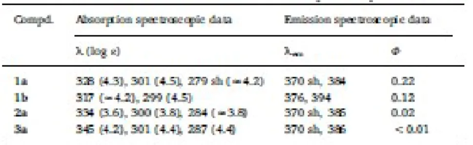
3.2. Electronic spectra and optical properties 474
Absorption spectra of CH2Cl2 solutions of 1a and 1b at 298 K (Table 3 and Fig. S5, A) showed two 475
intense bands in the range 250–450 nm that are characteristic of carbazoles. The corresponding UV–vis 476
spectra of the complexes 2a and 3a (Fig. S5, B and Table 3) exhibited two intense absorption bands in 477
the range 300≤λ < 350 nm. One of them shifted to lower energies in relation to the free ligand being for 478
the Pt(II) complex (3a) the magnitude of the shift bigger than for the Pd(II) complex 2a (Table 3). These 479
findings suggest that this absorption band is due to a metal perturbed intraligand electronic transition 480
(MPILET). The second absorption band, at higher energies, is practically coincident with that of the free 481
ligand. The spectra of compounds 2a and 3a (Fig. S5, B) also exhibited an additional and poorly 482
resolved absorption band at lower wavelengths [280≤λ < 290 nm]. 483
The emission spectra of 1a, 1b, 2a and 3a were recorded in CH2Cl2 solution at 298 K. Upon excitation 484
at λexc=300 nm, the free ligands 1a and 1b exhibited emission bands in the range 370–395 nm (Fig. S6 485
and Table 3). The thienyl-based derivative 1b showed a bathochromic shift of the wavelength of 486
maximum emission of 10 nm in comparison to the thiazolyl-based ligand 1a, according to the strongest 487
electron-donating character of the thienyl unit. It should be noted that the quantum yield of 1a (Table 3) 488
is significantly higher than that of 1b. Complexes 2a and 3a exhibited also emission bands consistent 489
with that of ligand 1a, but their fluorescence quantum yields [104] decreased considerably in relation to 490
the free ligand (1a). 491
3.3. Computational studies 493
In order to elucidate the effect produced by the thiazolyl or thienyl groups of compounds 1a and 1b on 494
the electronic delocalization, computational calculations based on the density functional theory (DFT) 495
methodology were undertaken [68]. Calculations were carried out using the B3LYP hybrid functional 496
[69,70] and the 6-31G* basis set [72,73] implemented in the Gaussian03 program [71]. In a first stage, 497
geometries of compounds 1a and 1b were optimized without imposing any restriction. Final atomic 498
coordinates for the optimized geometries are included as supplementary information (Tables S1–S2). 499
Bond lengths and angles of the optimised geometry of 1a were consistent with those obtained from the 500
X-ray studies (the differences did not clearly exceed 3σ) and those of 1b are in the range reported for 501
related carbazoles with mono or polythienyl units on position 3 [50–52,93,103]. 502
Molecular orbital (MO) calculations of the optimized geometries revealed that highest occupied 503
molecular orbital (HOMO) (Fig. 5) of 1a and 1b are very similar except for a tiny difference in the 504
contribution of the atomic orbitals of the sulphur atom. The LUMO (Fig. 5) of 1a is mainly centred on 505
the thiazolyl unit and two of the rings of the carbazole; while in 1b, the contribution of the thienyl 506
decreases in relation to that of the thiazole in 1a. Moreover, the HOMO-LUMO gap (ΔE) of 1b (4.32 507
eV) is higher than for 1a (4.14 eV). These findings suggest that the replacement of the thiazolyl ring of 508
1a by the thienyl in 1b reduces the electronic delocalization mentioned above. 509
In the optimized geometry of 1b the intramolecular separation between the S1 atom and the hydrogen 510
atom of phenyl ring (2.848 Å) is larger than in 1a [S1⋯H: 2.748 Å (optimized geometry) or 2.751 Å 511
(from the crystal structure)] and the angle formed by the heterocycle and the carbazole is 30.3° bigger 512
than in 1a. Since it is well-known that deviations from planarity affects the electronic delocalization, the 513
properties of the compounds and their potential utility, we also calculated the energy of the molecules 514
for different orientations of the attached heterocycle versus the carbazole unit using molecular 515
mechanics. These arrangements were generated by modifying the torsion angle defined by the set of 516
atoms S1-C2’-C3-C4 (hereinafter referred to as φ) from 0° to 360°. The results shown in Fig. 6, reveal 517
that for 1a the minimum energy corresponds to φ values in the ranges (0°≤φ≤16° and 344°≤φ≤360°), 518
that is to say close to co-planarity, similar to that found in the crystal structure φ=13.5° and with the S1 519
atom and NMe group located in opposite sides. 520
The energy barrier to achieve an orthogonal arrangement of the thiazole (φ=90° or 270°) is rather high 521
(9.1 kJ/mol). The conformer with the N1 and N2 atoms on opposite sides, [φ values between 164° and 522
196°] is slightly less stable than for φ=0 ± 16° (Fig. 6). The differences between the energies of both 523
conformers determined from molecular mechanics calculations and DFT are 0.4 and 0.5 Kcal/mol, 524
respectively. 525
In contrast with the results obtained for 1a, in 1b the most favourable orientation of the thienyl unit is far 526
away from co-planarity and corresponds to φ values in the ranges 128°–133° and 232°–237°. The 527
energy barriers to achieve coplanar arrangements (Fig. 6) are smaller than that obtained for 1a (9.1 528
kJ/mol); consequently, from an energetic point of view, the free rotation of the thienyl unit, that requires 529
a smaller energy income, is more likely to occur than that of the thiazolyl ring of 1a. In addition, time 530
dependent DFT (TD-DFT) calculations were performed to achieve the assignment of the bands observed 531
in the UV–vis spectra (Table S4 and Fig. S7). 532
Besides that and in order to compare the stability of the two isomers of the platinum(II) complexes (3a 533
and 4a), we optimized their geometries (Tables S5 and S6) and afterwards we calculated their relative 534
energies. The results revealed (Table S7) that in vacuum the transisomer (3a) is ca. 4.3 kcal/mol more 535
stable than 4a (cis- isomer), but in methanol (MeOH) the difference between their calculated free 536
energies decreased to −0.30 kcal/mol. This may explain the formation of both isomers in a similar molar 537 ratio. 538 539 3.4. Biological studies 540 3.4.1. Antiproliferative assay 541
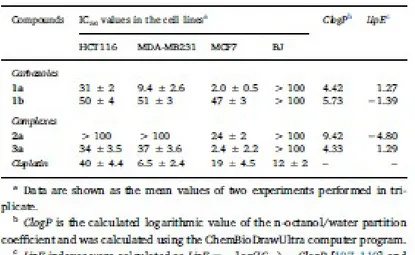
We have evaluated the cytotoxic activity of ligands 1a and 1b and the new complexes 2a and 3a in front 542
of the colon cell line HCT116 and two breast cancer cell lines [the triple negative (ER, PR and no HER2 543
over expression) MDA-MB231 and the MCF7]. The effects of the new products on the growth of the 544
three cell lines and that of cisplatin, used as positive control, were assessed after 72 h and the results are 545
presented in Table 4 and Fig. 7. 546
The comparison of the in vitro cytotoxic activities of the free carbazoles 1a and 1b in the HCT116 cell 547
line revealed that the replacement of the thienyl ring (in 1b) by the thiazolyl unit of 1a produced a 548
significant enhancement of the cytotoxic potency. This trend is practically identical to those observed in 549
the two breast (MDA-MB231 and the MCF7) cancer cell lines and could be attributed to several factors. 550
One of these could be the lipophilicity that, as shown in Table 4, is expected to be slightly different for 551
the two systems. It should be noted that in the MCF7 cell line ligand 1a is (ca. 9.5 times) more potent 552
than cisplatin, the non-alkylated 9H-carbazole (IC50 > 40μM) and similar to doxorubicin (IC50=2.3 μM 553
[105] or 2.43 ± 0.24 μM [106]). 554
In order to compare the effect produced by the binding of the Pd(II) or Pt(II) we also examined the 555
effect produced by the complexes 2a and 3a on identical cell lines. As shown in Table 4 and Fig. 7, the 556
Pd(II) complex 2a did not show any relevant antiproliferative activity (IC50 > 100 μM) in the HCT116 557
and MDA-MB231 cell lines. In the MCF7 it was more active, but its potency was close (IC50=24 ± 2 558
μM) to that of cisplatin (IC50=19 ± 4.5 μM). In contrast, the Pt(II) complex (3a) exhibited a higher 559
inhibitory growth effect, being 9 times more potent than the reference drug in the MCF7 breast cancer 560
cell line. 561
It is well-known that the preparation of new products with improved cytotoxic potency is not the unique 562
requirement in medicinal chemistry and drug design, other factors such as the lipophilicity that 563
contributes to the ADMET (absorption, distribution, metabolism excretion and toxicity) properties of 564
drugs also plays a crucial role. Nowadays, the lipophilic efficiency (LipE) index [107–110] that includes 565
lipophilicity and potency is becoming more and more popular in drug design and optimization, because 566
it allows to normalize the observed potency with changes in the lipophilicity. 567
In view of this, we calculated the Clog P values for the new compounds and their LipE index in the 568
MCF7 cell line. The results (Table 4) reveal that for the compounds characterized in this work the LipE 569
index increases as follow 2a≪1b < 1a≤3a. The Pd(II) complex 2a that shows low solubility it is 570
simultaneously the most lipophilic and the less potent compound of the series. In contrast with these 571
results, for the carbazole-thiazole ligand 1a and its trans-[PtCl2(1a)(dmso)] complex 3a there is an 572
effective combination of their cytotoxic potency in MCF7 and lipophilicity, and on these basis they are 573
promising scaffolds in the search of optimized drugs. Chemical modifications of the core of ligand 1a, 574
its binding to the Pt(II) atom or even changes on the ancillary ligands bound to it in 3a may allow to tune 575
the lipophilicity and to improve the lipophilic efficiency. 576
577 578
3.4.2. Additional studies to elucidate the mechanism of action 579
In the majority of the described cases of cytotoxic carbazoles, they act as DNA-intercalators or as 580
Topoisomerase I/II (or telomerases) inhibitors [24,111], although other mechanisms of action involving 581
different targets {i.e. estrogen receptors (ER) or cyclin dependent kinases (CDK), among others} have 582
also been postulated [22–26,112]. To examine whether the presence of the thiazole (in 1a) or the thienyl 583
unit (in 1b) in the free ligand and the binding of 1a to the Pd(II) or Pt(II) ions in complexes 2a and 3a, 584
could have an important role in the mechanism of action, additional experiments were performed. 585
To elucidate whether compounds 1a, 1b, 2a and 3a act as DNA intercalators or as Topoisomerase I or II 586
inhibitors, three different sets of experiments were undertaken. In a first stage it was examined if the 587
new compounds could induce changes in the electrophoretic mobility of the supercoiled closed form 588
(ccc) of pBluescript SK+ plasmid DNA. For DNA migration studies, the plasmid was incubated with 589
compounds 1a, 1b, 2a and 3a at increasing concentrations ranging from 0 to 200 μM. For comparison 590
purposes, incubation of DNA with cisplatin or ethidium bromide (EB) was also performed. As expected, 591
cisplatin greatly altered the electrophoretic mobility of pBluescript DNA at all concentrations tested. As 592
depicted in Fig. 8, the free ligands (1a and 1b) and the Pd(II) compound (2a) were not effective. Only 593
the Pt(II) complex (3a) produced a significant effect on the electrophoretic mobility of native 594
pBluescript DNA at concentrations>100 μM. Secondly, a Topoisomerase based gel assay was performed 595
to evaluate the ability of compounds 1a, 1b, 2a and 3a to intercalate into DNA or to act as DNA 596
Topoisomerase I inhibitors. For that, supercoiled pBluescript plasmid DNA was incubated with 597
Topoisomerase I in the presence of increasing concentrations of the compounds under study. The results 598
are presented in Fig. 9, where ethidium bromide (EB) was used as an intercalator control. The analysed 599
compounds did not prevent unwinding of DNA indicating that they are neither intercalators nor 600
Topoisomerase I inhibitors. 601
As mentioned above, another important target for anticancer agents is the Topoisomerase II, which is 602
associated with solving the topological constraints of DNA by transiently cleaving both strands of the 603
double helix [24,111]. In humans there are two Topoisomerase II isoenzymes, IIα and IIβ. Here we 604
study the capability of compounds 1a, 1b, 2a and 3a as catalytic inhibitors of Topoisomerase IIα. The 605
inhibitory activity was evaluated by measuring the extent of enzyme mediated relaxed DNA after 606
treatment with 100 μM of 1a, 1b, 2a and 3a compounds. Only the Pd(II) complex 2a showed at this 607
concentration inhibitory activity (Fig. 10A). The inhibitory effect of 2a was further examined at different 608
concentrations, from 50 to 200 μM. As it is shown in Fig. 10B, compound 2a showed inhibition at 100 609
μM but not at 50 μM. 610
Other mechanism of action implies Cathepsin B, which is a cysteine metaloprotease that could be 611
involved in metastasis, angiogenesis and tumour progression. Examples of Pd(II) and Pt(II) complexes 612
as inhibitors of Cathepsin B have been reported [113]. However, none of the new compounds presented 613
in this work (1a, 1b, 2a and 3a) inhibited the enzyme activity at 100 μM concentration. 614
Overall the biological studies undertaken with the new compounds 1a, 1b, 2a and 3a provide conclusive 615
evidences. The DNA migration studies revealed that only Pt(II) complex (3a) modifies the 616
electrophoretic mobility of the plasmid in a similar way as cisplatin but at higher concentrations. 617
Experimental results also revealed that neither compounds 1a, with higher cytotoxic activity than 618
cisplatin in the tested cancer lines HCT116 and MCF7, nor compounds 1b or 2a operate as intercalators 619
and none of them are inhibitors of Topoisomerase I or cathepsin B. However, the Pd(II) complex (2a) 620
inhibits the activity of Topoisomerase IIα (at 200 μM concentration). 621
All the new compounds show lower toxicity on the normal and nontumoral human skin fibroblast BJ 622
cell line than cisplatin (Table 4). Among the new compounds, 1a and its Pt(II) complex 3a are the most 623
active in the assayed HCT116, MDA-MB231 and MCF7 cancer cell lines. Moreover, compound 1a, 624
clearly more potent than 3a, has additional interest because it does not contain Pt(II) and consequently 625
might not produce the typical and undesirable side effects of conventional Pt(II)-based drugs. In 626
addition, compound 1a shows a remarkable high stability in the solid state and also in dmso or in 627
mixtures dmso: D2O (Figs. S9–S15) at 298 K. These findings enhance the interest and relevance of 628
carbazole 1a for further and additional biological studies. 629
630 631
4. CONCLUSIONS 632
633
Here we have presented two new N-methylated and 3-substituted carbazoles with a thiazolyl (1a) or a 634
thienyl (1b) unit and comparative studies of their properties and reactivity in front of Na2[PdCl4] or 635
[MCl2(dmso)2] {trans- for M=Pd or cis- for M=Pt} and biological activities. The obtained results 636
proved that compound 1a is clearly more reactive than 1b and has a greater coordination capability 637
towards the Pd(II) and Pt(II) ions, leading to trans-[PdCl2(1a)2] (2a) and the geometrical isomers {trans-638
(3a) or cis-(4a)} of [PtCl2(1a)(dmso)]. DFT studies confirmed the different reactivity of 1a and 1b and 639
the formation of the isomers 3a and 4a. 640
In vitro studies on the cytotoxic activity of compounds 1a, 1b, 2a and 3a in the cancer cell lines 641
(HCT116, MDA-MB231 and MCF7) and in the normal and non-tumoral human skin fibroblasts BJ cell 642
line show that: a) the replacement of the thiazole (of 1a) by the thienyl (to give 1b) reduces the 643
inhibitory growth effect, b) the binding of 1a to the Pt (II) atom (3a) reduces its cytotoxic activity, c) 644
compound 2a is less active than the Pt(II) complex 3a and d) all compounds are less toxic than cisplatin 645
in the BJ cell line. Additional biological studies revealed that: a) only the Pt(II) complex (3a) induced 646
significant changes on the electrophoretic mobility of the pBluescript DNA, but at higher concentrations 647
than the cisplatin and b) neither the ligands (1a and 1b) nor the Pd(II) (2a) or Pt(II) complex (3a) acted 648
as intercalators or inhibitors of Topoisomerase I or Cathepsin B. However, the Pd(II) complex (2a) with 649
an inhibitory growth activity in the MCF7 cell line similar to that of cisplatin inhibited the 650
Topoisomerase IIα activity. These findings suggest that the binding of the Pd(II) or the Pt(II) to the 651
carbazole 1a not only produces significant changes in their cytotoxic activity but also on their 652
mechanism of action. 653
To sum up, among the new compounds, 1a with high stability, low toxicity, potent cytotoxic activity and 654
photophysical properties is an excellent candidate for further studies on: a) their effect on a wider panel 655
of cancer cell lines, b) its mechanism of action, c) its potential use in combined therapies even in 656
photodynamic therapy, and d) other biological activities (i.e. antibacterial, antifungal, etc.) maybe 657
relevant in new drug design and development. 658
ACKNOWLEDGEMENTS 660
661
This work was supported by the Ministerio de Ciencia e Innovación of Spain (MICINN) (Grant numbers 662
CTQ2015-65040-P and CTQ2015- 65770-P MINECO/FEDER). 663
APPENDIX A. SUPPLEMENTARY DATA 665
Tables containing: final atomic coordinates of the optimised geometries of carbazoles 1a and 1b (Tables 666
S1 and S2, respectively), calculated energies of the HOMO and LUMO orbitals, energy gaps for the new 667
carbazoles and calculated values of the Mulliken charges of selected atoms (Table S3), summary of the 668
results obtained from the computational studies showing electronic transitions with greater contributions 669
in the absorption bands for 1a and 1b (Table S4), calculated final atomic coordinates for isomers 3a and 670
4a (Tables S5–S6) and calculated energies for 3a and 4a (Table S7) and additional Figures (Figs. S1– 671
S12) showing: the 1H-NMR spectrum of compound 3a (Fig. S2); an expansion of the 1H-NMR 672
spectrum of the raw material obtained after 24 h under reflux, showing the presence of complex 3a and 673
another minor product (Fig. S3); the 1H-NMR spectrum of the crude material obtained after 72 h that 674
shows the coexistence of 3a and an additional product 4a (Fig. S4); The 195Pt{1H} and 1H-NMR 675
spectra of the mixture of the two isomers of [PtCl2(1a)(dmso)] {trans- (3a) and cis-(4a)}, isolated from 676
the column (Fig. S1); UV–Vis spectra of compounds 1a, 1b, 2a and 3a (Fig. S5), emission spectra of 677
compounds 1a, 1b, 2a and 3a (Fig. S6); calculated absorption spectra of compounds 1a and 1b (Fig. S7); 678
the HOMO−1 and LUMO+1 orbitals for carbazoles 1a and 1b (Fig. S8); Figures (Figs. S9–S12); 679
showing the 1H-NMR spectra of a freshly prepared solution of compound 1a in dmso-d6 (Fig. S9) or in 680
mixtures dmso-d6: D2O (from 4:1 to 1:1) (Figs. S10–S12) after several periods of storage at 298 K; the 681
1H-NMR spectra of a freshly prepared solution of compounds 1b, 2a and 3a in dmso-d6 after several 682
periods of storage at 298 K (Figs. S13–S15); and ESI-MS spectra of compounds 1a, 1b and 3a (Figs. 683
S16–S18). Supplementary data to this article can be found online at 684
https://doi.org/10.1016/j.jinorgbio.2018.03.008. 685
REFERENCES 687
[1] Cancer Facts & Figures 2017, American Cancer Society, 2017, http://www.cancer. 688
org/research/cancerfactsstatistics/cancerfactsfigures2017/index , Accessed date: May 2017. 689
[2] Cancer statistics center, American Cancer Society, https://cancerstatisticscenter. 690
cancer.org/?_ga=1.48498790.1307978637.1460725255#/, (2017) , Accessed date: May 2017. 691
[3] A.B. Ryerson, C.R. Eheman, S.F. Altekruse, J.W. Ward, A. Jemal, R.L. Sherman, S.J. Henley, 692
D. Holtzman, A. Lake, A.-M. Noone, R.N. Anderson, J. Ma, K.N. Ly, K.A. Cronin, L. 693
Penberthy, B.A. Kohler, Cancer 122 (2016) 1312–1337. 694
[4] Updated Information on Colorectal and Breast Cancer can be Obtained from the American 695
Cancer Society, Atlanta, Georgia, USA (http://www.cancer.org/acs) Through (a) 696
www.cancer.org/acs/groups/content/documents/document/acspc- 042280.pdf and (b) 697
www.cancer.org/acs/groups/content/documents/document/ acspc-04638.pdf (for Colorectal 698
Cancer and Breast Cancer, Respectively) (2017) (accessed May 2017). 699
[5] G.F. Weber, Molecular Therapies of Cancer, Springer, Germany, 2015. 700
[6] A.C. Flick, H.X. Ding, C.A. Leverett, R.E. Kyne Jr., K.K.-C. Liu, S.J. Fink, C.J. O'Donnell, 701
Bioorg. Med. Chem. 24 (2016) 1937–1980. 702
[7] N.A. Meanwell, Chem. Res. Toxicol. 29 (2016) 564–616. 703
[8] A.L. Harvey, R.L. Clark, S.P. Mackay, B.F. Johnston, Expert Opin. Drug Discovery 5 (2010) 704
559–568. 705
[9] E.A.G. Blomme, Y. Will, Chem. Res. Toxicol. 29 (2016) 473–504. 706
[10] S.A. McKie, Future Med. Chem. 8 (2016) 579–602. 707
[11] I. Ali, M.N. Lone, Z.A. Al-Othman, A. Al-Warthan, M.M. Sanagi, Curr. Drug Targets 16 (2015) 708
711–734. 709
[12] A.R. Katritzky, C.A. Ramsden, E.F.V. Scriven, R.J.K. Taylor (Eds.), Comprehensive 710
Heterocyclic Chemistry III, Elsevier, Oxford (UK), 2008. 711
[13] R.K. Parashar, Chemistry of Heterocyclic Compounds, CRC Press, 2014. 712
[14] A.F. Pozharskii, A.T. Soldatenkov, A.R. Katritzky, Heterocycles in Life and Society: An 713
Introduction to Heterocyclic Chemistry, Biochemistry and Applications, second ed., Wiley-714
VCH, Weinheim (Germany), 2011. 715
[15] J.A. McCleverty, T.J. Meyer (Eds.), Comprehensive Coordination Chemistry II: From Biology 716
to Nanotechnology, Elsevier, Amsterdam, 2003. 717
[16] R.H. Crabtree, D.M.P. Mingos (Eds.), Comprehensive Organometallic Chemistry III, second 718
ed., Elsevier, Oxford, UK, 2007. 719
[17] G. Wilkinson, R.D. Gillard, J.A. McCleverty (Eds.), Comprehensive Coordination Chemistry: 720
The Synthesis, Reactions, Properties and Applications of Coordination Compounds, Pergamon 721
Press, Oxford, UK, 1987. 722
[18] J.J. Li, Heterocyclic Chemistry in Drug Discovery, John Wiley & Sons, Hoboken, USA, 2013. 723
[19] T.Y. Zhang, Chapter one - the evolving landscape of heterocycles in drugs and drug candidates, 724
in: E.F.V. Scriven, C.A. Ramsden (Eds.), Advances in Heterocyclic Chemistry, 121 Academic 725
Press, 2017, pp. 1–12. 726
[20] A. Gomtsyan, Chem. Heterocycl. Compd. 48 (2012) 7–10. 727
[21] P. Martins, J. Jesús, S. Santos, L.R. Raposo, C. Roma-Rodrigues, P.V. Baptista, A.R. Fernandes, 728
Molecules 20 (2015) 16852–16891. 729
[22] L.S. Tsutsumi, D. Gündisch, D. Sun, Carbazole scaffold in medicinal chemistry and natural 730
products: a review from 2010–2015, Curr. Top. Med. Chem. 16 (2016) 1290–1313. 731
[23] K.N. Mounika, A.N. Jyothy, G.N. Raju, R.R. Nadendla, World J. Pharm. Pharm. Sci. 4 (2015) 732
420–428. 733
[24] M. Bashir, A. Bano, A.S. Ijaz, B.A. Chaudhary, Molecules 20 (2015) 13496–13517. 734
[25] C. Asche, M. Demeunynck, Anti Cancer Agents Med. Chem. 7 (2007) 247–267. 735
[26] A. Caruso, D. Iacopetta, F. Puoci, A.R. Cappello, C. Saturnino, M.S. Sinicropi, Mini-Rev. Med. 736
Chem. 16 (2016) 630–643. 737
[27] T. Paneer, G. Saravanan, M. Palanivelu, Anticancer Evaluation of Thiazole Based Heterocycles 738
- A Review, Lambert Academic Publishing, Saarbrücken, Germany, 2014. 739
[28] A. Ayati, S. Emami, A. Asadipour, A. Shafiee, A. Foroumadi, Eur. J. Med. Chem. 97 (2015) 740
699–718. 741
[29] A. Chawla, H. Kaur, P. Chawla, U.S. Baghel, J. Glob. Trends Pharm. Sci. 5 (2014) 1641–1648. 742
[30] A. Leoni, A. Locatelli, R. Morigi, M. Rambaldi, Expert Opin. Ther. Pat. 24 (2014) 201–216. 743
[31] K.K. Jha, S. Kumar, I. Tomer, R. Mishra, J. Pharm. Res. 5 (2012) 560–566. 744
[32] M.M. Ghorab, M.S. Bashandy, M.S. Alsaid, Acta Pharma. 64 (2014) 419–431. 745
[33] D. Gramec, L.P. Mašič, M.S. Dolenc, Chem. Res. Toxicol. 27 (2014) 1344–1358. 746
[34] N.C. Garbett, D.E. Graves, Curr. Med. Chem. Anticancer Agents 4 (2004) 149–172. 747
[35] C.M. Miller, F.O. McCarthy, RSC Adv. 2 (2012) 8883–8918. 748
[36] R.K. Mehmood, Oncol. Rev. 8 (2014) 256. 749
[37] F.M. Muggia, A. Bonetti, J.D. Hoeschele, M. Rozencweig, S.B. Howell, J. Clin. Oncol. 33 750
(2015) 4219–4226. 751
[38] S. Amptoulach, N. Tsavaris, Chemother. Res. Pract. (2011) 843019, , http://dx. 752
doi.org/10.1155/2011/843019. 753
[39] M.A. Jordan, Curr. Med. Chem. Anticancer Agents 2 (2002) 1–17. 754
[40] K. Chatterjee, J. Zhang, N. Honbo, J.S. Karliner, Cardiology 115 (2010) 155–162. 755
[41] F. Trudu, F. Amato, P. Vanhara, T. Pivetta, E.M. Peña-Méndez, J. Havel, J. Appl. Biomed. 13 756
(2015) 79–103. 757
[42] H. Jiang, J. Sun, J. Zhang, Curr. Org. Chem. 16 (2012) 2014–2025. 758
[43] M. Reig, G. Bagdziunas, D. Volyniuk, J.V. Grazulevicius, D. Velasco, Phys. Chem. Chem. 759
Phys. 19 (2017) 6721–6730. 760
[44] M. Reig, C. Gozálvez, R. Bujaldón, G. Bagdziunas, K. Ivaniuk, N. Kostiv, D. Volyniuk, J.V. 761
Grazulevicius, D. Velasco, Dyes Pigments 137 (2017) 24–35. 762
[45] M. Reig, G. Bubniene, W. Cambarau, V. Jankauskas, V. Getautis, E. Palomares, E. Martínez-763
Ferrero, D. Velasco, RSC Adv. 6 (2016) 9247–9253. 764
[46] M. Reig, J. Puigdollers, D. Velasco, J. Mater. Chem. C 3 (2015) 506–513. 765
[47] J.L. Díaz, A. Dobarro, B. Villacampa, D. Velasco, Chem. Mater. 13 (2001) 2528–2536. 766
[48] B.-B. Ma, Y.-X. Peng, T. Tao, W. Huang, Dalton Trans. 43 (2014) 16601–16604. 767
[49] Y.-X. Li, X.-T. Tao, F.-J. Wang, T. He, M.-H. Jiang, Org. Electron. 10 (2009) 910–917. 768
[50] S.-I. Kato, S. Shimizu, A. Kobayashi, T. Yoshihara, S. Tobita, Y. Nakamura, J. Org. Chem. 79 769
(2014) 618–629. 770
[51] S.-I. Kato, S. Shimizu, H. Taguchi, A. Kobayashi, S. Tobita, Y. Nakamura, J. Org. Chem. 77 771
(2012) 3222–3232. 772
[52] P. Wang, Y. Ju, S.Y. Tang, J.Y. Wu, H.P. Zhou, Acta Cryst., Sect. E 63 (2007) o3671. 773
[53] M.A.T. Nguyen, A.K. Mungara, J.-A. Kim, K.D. Lee, S. Park, Phosphorus Sulfur Silicon Relat. 774
Elem. 190 (2015) 191–199. 775
[54] M.S. Shaikh, M.B. Palkar, H.M. Patel, R.A. Rane, W.S. Alwan, M.M. Shaikh, I.M. Shaikh, 776
G.A. Hampannavar, R. Karpoormath, RSC Adv. 4 (2014) 62308–62320. 777
[55] M.H. Adbel-Kader (Ed.), Photodynamic Therapy from Theory to Applications, Springer, 778
Heildelberg, Germany, 2014. 779
[56] J.U. Chukwu, C. López, A. González, M. Font-Bardía, M.T. Calvet, R. Messeguer, C. Calvis, J. 780
Organomet. Chem. 766 (2014) 13–21. 781
[57] E. Guillén, A. González, C. López, P.K. Basu, A. Ghosh, M. Font-Bardía, C. Calvis, R. 782
Messeguer, Eur. J. Inorg. Chem. (2015) 3781–3790. 783
[58] A. González, J. Granell, C. López, R. Bosque, L. Rodríguez, M. Font-Bardía, T. Calvet, X. 784
Solans, J. Organomet. Chem. 726 (2013) 21–31. 785
[59] M. Tomé, C. López, A. Gonzalez, B. Ozay, J. Quirante, M. Font-Bardía, T. Calvet, C. Calvis, R. 786
Messeguer, L. Baldomà, J. Badia, J. Mol. Struct. 1048 (2013) 88–97. 787
[60] C. López, A. González, R. Bosque, P.K. Basu, M. Font-Bardía, T. Calvet, RSC Adv. 2 (2012) 788
1986–2002. 789
[61] Z. Szafran, R.M. Pike, M.M. Singh, Microscale Inorganic Chemistry, A Comprehensive 790
Laboratory Experience, John Wiley & Sons, New York, USA, 1991, p. 218. 791
[62] J.H. Price, A.N. Williamson, R.F. Schramm, B.B. Wayland, Inorg. Chem. 11 (1972) 1280– 792
1284. 793
[63] S.H. Tucker, J. Chem. Soc. 129 (1926) 546–553. 794
[64] D.D. Perrin, W.L.F. Armarego, Purification of Laboratory Chemicals, fourth ed., Butterworth– 795
Heinemann, Oxford, UK, 1996. 796
[65] G.M. Sheldrick, Acta Cryst A64 (2008) 112–122. 797
[66] T.A. Halgren, J. Comput. Chem. 17 (1996) 490–519. 798
[67] Spartan '14 v. 1.1.0, Wavefunction, Inc, Irvine, CA, USA, 2013. 799
[68] P. Hohenberg, W. Kohn, Phys. Rev. 136 (1964) B864–B871. 800
[69] A.D. Becke, J. Chem. Phys. 98 (1993) 5648–5652. 801
[70] C. Lee, W. Yang, R.G. Parr, Phys. Rev. B 37 (1988) 785–789. 802
[71] M.J. Frisch, G.W. Trucks, H.B. Schlegel, G.E. Scuseria, M.A. Robb, J.R. Cheeseman, J.A. 803
Montgomery, T. Vreven, K.N. Kudin, J.C. Burant, J.M. Millam, S.S. Iyengar, J. Tomasi, V. 804
Barone, B. Mennucci, M. Cossi, G. Scalmani, N. Rega, G.A. Petersson, H. Nakatsuji, M. Hada, 805
M. Ehara, K. Toyota, R. Fukuda, J. Hasegawa, M. Ishida, T. Nakajima, Y. Honda, O. Kitao, H. 806
Nakai, M. Klene, X. Li, J.E. Knox, H.P. Hratchian, J.B. Cross, V. Bakken, C. Adamo, J. 807
Jaramillo, R. Gomperts, R.E. Stratmann, O. Yazyev, A.J. Austin, R. Cammi, C. Pomelli, J.W. 808
Ochterski, P.Y. Ayala, K. Morokuma, G.A. Voth, P. Salvador, J.J. Dannenberg, V.G. 809
Zakrzewski, S. Dapprich, A.D. Daniels, M.C. Strain, O. Farkas, D.K. Malick, A.D. Rabuck, K. 810
Raghavachari, J.B. Foresman, J.V. Ortiz, Q. Cui, A.G. Baboul, S. Clifford, J. Cioslowski, B.B. 811
Stefanov, G. Liu, A. Liashenko, P. Piskorz,I. Komaromi, R.L. Martin, D.J. Fox, T. Keith, M.A. 812
Al-Laham, C.Y. Peng, A. Nanayakkara, M. Challacombe, P.M.W. Gill, B. Johnson, W. Chen, 813
M.W. Wong, C. Gonzalez, J.A. Pople, Gaussian 03 (Revision C.02), Gaussian, Inc, Wallingford, 814
CT, 2004. 815
[72] P.C. Hariharan, J.A. Pople, Theor. Chim. Acta 28 (1973) 213–222. 816
[73] M.M. Francl, W.J. Pietro, W.J. Hehre, J.S. Binkley, M.S. Gordon, D.J. DeFrees, J.A. Pople, J. 817
Chem. Phys. 77 (1982) 3654–3665. 818
[74] K.T. Givens, S. Kitada, A.K. Chen, J. Rothschiller, D.A. Lee, Invest. Ophthalmol. Vis. Sci. 31 819
(1990) 1856–1862. 820