Behavioral/Systems/Cognitive
Action Dominates Valence in Anticipatory Representations
in the Human Striatum and Dopaminergic Midbrain
Marc Guitart-Masip,
1,2Lluis Fuentemilla,
1Dominik R. Bach,
2Quentin J. M. Huys,
2,3Peter Dayan,
3Raymond J. Dolan,
2and Emrah Duzel
1,41Institute of Cognitive Neuroscience, University College London, London, W1CN 4AR, United Kingdom,2Wellcome Trust Centre for Neuroimaging,
Institute of Neurology, University College London, London WC1N 3BG, United Kingdom,3Gatsby Computational Neuroscience Unit, University College
London, London, W1CN 4AR, United Kingdom, and4Institute of Cognitive Neurology and Dementia Research, Otto von Guericke University, 39120
Magdeburg, Germany
The acquisition of reward and the avoidance of punishment could logically be contingent on either emitting or withholding particular
actions. However, the separate pathways in the striatum for go and no-go appear to violate this independence, instead coupling affect and
effect. Respect for this interdependence has biased many studies of reward and punishment, so potential action– outcome valence
interactions during anticipatory phases remain unexplored. In a functional magnetic resonance imaging study with healthy human
volunteers, we manipulated subjects’ requirement to emit or withhold an action independent from subsequent receipt of reward or
avoidance of punishment. During anticipation, in the striatum and a lateral region within the substantia nigra/ventral tegmental area
(SN/VTA), action representations dominated over valence representations. Moreover, we did not observe any representation associated
with different state values through accumulation of outcomes, challenging a conventional and dominant association between these areas
and state value representations. In contrast, a more medial sector of the SN/VTA responded preferentially to valence, with opposite signs
depending on whether action was anticipated to be emitted or withheld. This dominant influence of action requires an enriched notion of
opponency between reward and punishment.
Introduction
In instrumental conditioning, particular outcomes are realized,
or obviated, through discrete action choices, controlled by
out-come valence. Rewarded (appetitive) action choices are repeated
and punished (aversive) action choices are deprecated, although
the nature of the opponency between appetitive and aversive
sys-tems remains the subject of debate (Gray and McNaughton,
2000). Aside from valence or affect opponency between reward
and punishment, a key role in instrumental conditioning is also
played by a logically orthogonal spectrum of effect, spanning
invigoration to inhibition of action (Gray and McNaughton,
2000; Niv et al., 2007; Boureau and Dayan, 2011; Cools et al.,
2011). This effect spectrum is enshrined in the structure of parts
of the striatum that are involved in instrumental control, in
which partially segregated direct and indirect pathways are
de-scribed for go (invigoration) and no-go (inhibition), respectively
(Gerfen, 1992; Frank et al., 2004).
Although instrumental behavior thus seems to arise through
an interaction of valence and action spectra, our understanding
of their association remains partial. There is evidence for a close
coupling of reward and go and some evidence for a coupling
between punishment and no-go (Gray and McNaughton, 2000).
In contrast, there is intense theoretical debate concerning how
instrumental behavior is generated for the opposite
associa-tions, namely reward–no-go and punishment– go (Gray and
McNaughton, 2000).
A conventional coupling between reward and go responses in
human functional neuroimaging studies on instrumental
condi-tioning has led to important findings, such as an encoding of
various forms of temporal difference prediction errors for future
reinforcement in the ventral and dorsal striatum (O’Doherty et
al., 2004) and the identification of brain regions engaged in
an-ticipation of wins and losses (Delgado et al., 2000; Knutson et al.,
2001; Guitart-Masip et al., 2010). Overall, these studies have
con-tributed to a view that the striatum, especially its ventral
subdi-vision, and the midbrain regions harboring dopamine neurons
are associated with the representation of rewards, prediction
er-rors for rewards, and reward-associated stimuli (Haber and
Knutson, 2010). However, in these experiments, the requirement
to act (i.e., to go) is typically constant, and so a possible
organi-zational principle of the striatum along an action spectrum has
not been fully explored. Thus, in this study, we examined valence
together with anticipation of a requirement to either act or
in-Received Dec. 7, 2010; revised April 4, 2011; accepted April 5, 2011.
Author contributions: M.G.-M., L.F., D.R.B., Q.J.M.H., P.D., R.J.D., and E.D. designed research; M.G.-M. and L.F. performed research; M.G.-M. analyzed data; M.G.-M., P.D., R.J.D., and E.D. wrote the paper.
The authors declare no competing financial interests.
This work was supported by Wellcome Trust Programme Grant 078865/Z/05/Z (R.J.D.), the Gatsby Charitable Foundation (P.D.), Marie Curie Fellowship PIEF-GA-2008-220139 (M.G.-M.), and Deutsche Forschungsgemeinschaft SFB 779 and TP A7. We thank Dr. Chris Lambert, Dr. Jo¨rn Diedrichsen, and Dr. John Ashburner for discussion about midbrain normalization, Dr. Chloe Hutton for help with physiological noise correction, and Dr. Tali Sharot, Dr. Estela Camera, Dr. Molly Crocket, and Dr. Regina Lopez for comments on previous versions of this manuscript.
Correspondence should be addressed to Marc Guitart-Masip, Institute of Cognitive Neuroscience, University College London, 17 Queen Square, London, WC1N 3AR, UK. E-mail: [email protected].
DOI:10.1523/JNEUROSCI.6376-10.2011
hibit action, thereby disentangling these
from an associated appetitive or aversive
outcome delivery.
We orthogonalized action and valence in
a balanced 2 (reward/punishment)
⫻ 2 (go/
no-go) design. A key difference between our
protocol and those of previous studies
ad-dressing the relationship between action
and valence (Elliott et al., 2004; Tricomi et
al., 2004) is that it allowed us to separate
activity elicited by anticipation, action
per-formance, and obtaining an outcome. Thus,
unlike previous experiments, we could
ana-lyze outcome valence and action effects
dur-ing anticipation as separate factors. We
focused our analysis on the striatum and the
putatively dopaminergic midbrain because
of the close association between this
neuro-modulator, reward, go, and indeed vigor
(Schultz et al., 1997; Berridge and Robinson,
1998; Salamone et al., 2005; Niv et al., 2007).
Materials and Methods
Subjects. Eighteen adults participated in the ex-periment (nine female and nine male; age
range, 21–27 years; mean⫾ SD, 23 ⫾ 1.72
years). All participants were healthy,
right-handed, and had normal or corrected-to-normal visual acuity. None of the participants reported a history of neurological, psychiatric, or any other current medical problems. All experiments were run with each subject’s written informed consent and according to the local ethics clearance (University College London, London, UK).
Experimental design and task. The goal of our experimental design was to disentangle neural activity related to the anticipation of action and valence. To investigate the relationship between the two predominant spectra in ventral and dorsal striatum, we had to include both punish-ment (losses) and reward (gains), along with go and no-go. With one notable exception (Crockett et al., 2009), the bulk of the human literature into instrumental conditioning has focused on rewards that are only available given an overt response (O’Doherty, 2004; Daw et al., 2006). These are well aligned with a tight coupling between reward and invigo-ration and thus do not address our critical questions. Alternatively, stud-ies including punishment have systematically included a motor response as a means of its avoidance (Delgado et al., 2000; Knutson et al., 2001) but did not include no-go conditions.
Similarly, in other studies that address the role of action or salience on reward processing, subjects had to perform a motor response as part of the task (Zink et al., 2003, 2004; Elliott et al., 2004; Tricomi et al., 2004). Although explicit foil actions were used to control for the overall require-ment to act, they did not study the case of controlled no-go, for which a lack of action itself constitutes the instrumental requirement.
Finally, it is important to note that it is not possible merely to use a comparison between classical and instrumental conditioning. Al-though in classical conditioning experiments, rewards or punish-ments are obtained without regard to a motor response, this form of conditioning is associated with the generation of conditioned antici-patory responses such as licking, approach, salivation, etc. These an-ticipatory responses, which generally result in increased biological efficiency in the interaction between an organism and unconditioned stimuli (Domjan, 2005), can in principle confound any attempt to isolate pure anticipation of valence.
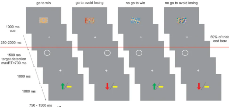
Our trials consisted of three events: a fractal cue, a target detection task, and an outcome. The trial timeline is displayed in Figure 1. In each trial, subjects saw one of four abstract fractal cues for 1000 ms. The fractal cues indicated, first, whether the participant would sub-sequently be required to emit a button press (go) or omit a button press (no-go), in the target detection task. The cues also indicated the
potential valence of the outcome related to performance in the target detection task (reward/no reward or punishment/no punishment). After a variable interval (250 –2000 ms) after offset of the fractal image, the target detection task started. The target was a circle dis-played on one side of the screen for 1500 ms. At this point, partici-pants had the opportunity to press a button within a time limit of 700 ms to indicate the target side for go trials or not to press for no-go trials. The requirement to make a go or a no-go response was dependent on the preceding fractal cue. At 1000 ms after the offset of the circle, subjects were presented with the outcome implied by their response. The out-come was presented for 1000 ms: a green arrow pointing upward meant they had won £1, a red arrow pointing downwards meant that they had lost £1, and a yellow horizontal bar indicated they did not win or lose any money. The outcome was probabilistic so that 70% of correct responses were rewarded in win trials, and 70% of correct responses were not punished in lose trials.
Thus, there were four trial types depending on the nature of the fractal cue presented at the beginning of the trial: (1) press the correct button in the target detection task to gain a reward (“go to win”); (2) press the correct button in the target detection task to avoid punish-ment (“go to avoid losing”); (3) do not press a button in the target detection task to gain a reward (“no-go to win”); and (4) do not press a button in the target detection task to avoid punishment (“no-go to avoid losing”).
Critically, on half the trials, target detection and outcome were omit-ted (Fig. 1). Therefore, at the beginning of the trial, fractal images spec-ified action requirements (go vs no-go) and outcome valence (reward vs punishment), but the actual target detection and potential delivery of an outcome only happened in half the trials. We implemented this manip-ulation because it allowed us to decorrelate activity related to an antici-pation phase cued by the fractal stimuli from activity related to actual motor performance in the target detection task and obtaining an out-come. One additional benefit of this design is that we could avoid the suboptimality of having to introduce long jitters between distinct task components. If every trial had been followed by the target detection task, anticipation of action would have been followed by action execution and anticipation of inaction by action inhibition in all correct trials. This would have resulted in highly correlated regressors for the anticipation and execution or withholding of a motor response, making it impossible
Figure 1. Experimental design. On each trial, one of four possible fractal images indicated the combination between action (making a button press in go trials or withholding a button press in no-go trials) and valence at outcome (win or lose). Actions were required in response to a circle that followed the fractal image after a variable delay. In go trials, subjects indicated via a button press on which side of the screen the circle appeared. In no-go trials, they withheld a response. After a brief delay, outcome was signaled in which a green upward arrow indicated a win of £1, a downward red arrow indicated a loss of £1, and a horizontal bar indicated the absence of a win or a loss. In go to win trials, a correct button press was rewarded. In go to avoid losing trials, a correct button press avoided punishment. In no-go to win trials, withholding a button press led to reward. In no-go to avoid losing trials, withholding a button press avoided punishment. The outcome was probabilistic so that 70% of correct responses were rewarded in win trials, and 70% of correct responses were not punished in lose trials. The red line indicates that half of the trials did not include the target detection task and the outcome. Subjects were trained in the task and fully learned the contingencies between the different fractal images and task requirements before scanning.
to separate activity elicited by anticipation, action performance, and the delivery of an outcome.
Scanning was divided into four 8 min sessions comprising 20 trials per condition, 10 trials in which the target detection task and the outcome was displayed and 10 trials in which only the fractal image was displayed. Subjects were told that they would be paid their earnings from the task up to a maximum of £35. To ensure that subjects learned the meaning of the fractal images and performed the task correctly during the scanning, we instructed them as to the meaning of each fractal image before the actual scanning began. Moreover, subjects performed one block of the task with 10 trials per condition in which the outcome of each trial also included text providing feedback whether the executed response was correct or not and whether the response was on time. Finally, after this initial training session and before actual scanning, subjects performed another run of the task that was identical to the task performed during the scanning. This en-sured that subjects experienced the possibility of the absence of the target detection task. Therefore, the presence of trials without target detection and outcome was not surprising during the crucial acquisition of functional magnetic resonance imaging (fMRI) data. Both training sessions were per-formed inside the scanner while the structural scans were acquired.
Behavioral data analysis. The behavioral data were analyzed using the statistics software SPSS, version 16.0. The number of correct on time button press responses per condition was analyzed with a two-way repeated-measures ANOVA with action (go/no-go) and valence (win/ lose) as factors. Response speed in go trials was analyzed by considering the button press reaction times (RTs) to targets and the proportion of trials in which button press RTs exceeded the response deadline. To further analyze these effects, we performed post hoc t tests.
fMRI data acquisition. fMRI was performed on a 3 tesla Siemens Al-legra magnetic resonance scanner with echo planar imaging (EPI). Func-tional data were acquired in four scanning sessions containing 117 volumes with 40 slices, covering a partial volume that included the
stria-tum and the midbrain (matrix, 128⫻ 128; 40 oblique axial slices per
volume angled at⫺30° in the anteroposterior axis; spatial resolution,
1.5⫻ 1.5 ⫻ 1.5 mm; TR, 4000 ms; TE, 30 ms). The fMRI acquisition
protocol was optimized to reduce susceptibility-induced blood oxygen level-dependent (BOLD) response sensitivity losses in inferior frontal and temporal lobe regions (Weiskopf et al., 2006). Six additional volumes at the beginning of each series were acquired to allow for steady-state magnetization and were subsequently discarded. Anatomical images of each subject’s brain were collected using multi-echo 3D fast, low-angle shot sequence (FLASH) for mapping proton density, T1 and
magnetiza-tion transfer (MT) at 1 mm3resolution, and by T1-weighted inversion
recovery prepared EPI sequences (spatial resolution, 1⫻ 1 ⫻ 1 mm).
Additionally, individual field maps were recorded using a double-echo
FLASH sequence (matrix size, 64⫻ 64; 64 slices; spatial resolution, 3 ⫻
3⫻ 3 mm; gap, 1 mm; short TE, 10 ms; long TE, 12.46 ms; TR, 1020 ms)
for distortion correction of the acquired EPI images. Using the FieldMap toolbox, field maps were estimated from the phase difference between the images acquired at the short and long TE.
fMRI data analysis. Data were analyzed using SPM8 (Wellcome Trust Centre for Neuroimaging, University College London). Preprocessing included realignment, unwrapping using individual field maps, and spa-tial normalization to the Montreal Neurological Institute (MNI) space
with spatial resolution after normalization of 1⫻ 1 ⫻ 1 mm. We used the
unified segmentation algorithm available in SPM to perform normaliza-tion. This has been shown to achieve good intersubject coregistration for brain areas such as caudate, putamen, and brainstem (Klein et al., 2009). Moreover, successful coregistration of the substantia nigra/ventral teg-mental area (SN/VTA) was also checked by manually drawing a region of interest (ROI) for each subject, in native space, and inspecting the over-lap of ROIs after applying the same normalization algorithm (data not shown). Finally, data were smoothed with a 6 mm FWHM Gaussian kernel. The fMRI time series data were high-pass filtered (cutoff, 128 s) and whitened using an AR(1)-model. For each subject, a statistical model was computed by applying a canonical hemodynamic response function combined with time and dispersion derivatives.
Our 2⫻ 2 factorial design included four conditions of interest that
were modeled as separate regressors in a general lineal model (GLM): go
to win trials, go to avoid losing trials, no-go to win trials, and no-go to avoid losing trials. We also modeled the onset of the target detection task separately for trials in which subjects performed a button press and for trials in which subjects did not perform a button press, respectively; the onset of the outcome, which could be win £1, lose £1, or no monetary consequences. Finally, we modeled separately the onsets of fractal images that were followed by incorrect performance. Note that the model used to analyze the data pooled together neutral outcomes from win trials (go to win and no-go to win conditions) together with neutral outcomes from lose trials (go to avoid losing and no-go to avoid losing conditions). Because the values of outcomes are assessed relative to expectations and the neutral outcomes have different effects if the alternative outcome is a win or a loss, the resulting analysis cannot be optimal for charac-terizing brain responses to the outcomes. This is because the goal of the present work was to study brain responses during the anticipatory phase and the experimental design, together with the GLM, optimized the detection of brain responses to the fractal images. To capture residual movement-related artifacts, six covariates were included (the three rigid-body translation and three rotations resulting from re-alignment) as regressors of no interest. Regionally specific condition effects were tested by using linear contrasts for each subject and each condition (first-level analysis). The resulting contrast images were entered into a second-level random-effects analysis. For the anticipa-tory phase, the hemodynamic effects of each condition were assessed
using a 2⫻ 2 ANOVA with the factors “action” (go/no-go) and
valence (win/lose). For the outcome onset, we assessed the hemody-namic effect of each condition using a one-way ANOVA with valence as a factor (win, lose, or neutral).
Results are reported familywise error (FWE) corrected for small
vol-ume in areas of interest at p⬍ 0.05. The predicted activations in the
midbrain and the striatum were tested using small volume correction (SVC) using anatomically defined regions of interest: the striatum as whole, the ventral striatum, and the SN/VTA of the midbrain (main origin of dopaminergic projections). The striatum as a whole ROI was defined using Marsbar (Brett et al., 2002) and included the caudate and the putamen. The ventral striatum ROI was drawn with Marsbar as two spheres of 8 mm around the coordinates referred to as right [MNI space
coordinates (shown as x,y,z throughout), 11.11, 11.43,⫺1.72] and left
(MNI space coordinates,⫺11.11, 11.43, ⫺1.72) nucleus accumbens in
previous publication (Knutson et al., 2005). This resulted in an ROI that incorporated the nucleus accumbens and ventral striatum as described in a recent review (Haber and Knutson, 2010). The SN/VTA ROI was man-ually defined, using the software MRIcro and the mean MT image for the group. On MT images, the SN/VTA can be distinguished from surround-ing structures as a bright stripe (Bunzeck and Du¨zel, 2006). It should be noted that, in primates, reward-responsive dopaminergic neurons are distributed across the SN/VTA complex, and it is therefore appropriate to consider the activation of the entire SN/VTA complex rather than, a priori, focusing on its subcompartments such as the VTA (Du¨zel et al.,
2009). For this purpose, a resolution of 1.5 mm3, as used in the present
experiment, allows sampling over 200 voxels of the SN/VTA complex,
which has a volume of 350 – 400 mm3. This does not imply that the whole
complex responds as a unit, and we have previously highlighted (Du¨zel et al., 2009) the possible existence of gradients in the functional anatomy of the SN/VTA in nonhuman primates (Haber et al., 2000) and the useful-ness of high-resolution imaging of the entire SN/VTA to detect these functional gradients (Du¨zel et al., 2009).
Results
Anticipation of losses impairs task performance when action
is required
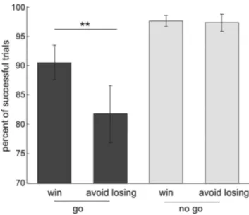
A two-way repeated-measures ANOVA on the percentage of
suc-cessful target response trials, with action (go/no-go) and valence
(win/lose) as factors, revealed a main effect of action (F
(1,17)⫽ 22.88,
p
⬍0.001),amaineffectofvalence(F
(1,17)⫽13.2,p⫽0.002),andan
action
⫻ valence interaction (F
(1,17)⫽ 12.28, p ⫽ 0.003). As
illus-trated in Figure 2 A, anticipation of punishment decreased the
percentage of successful (correct on time response to targets)
trials in the go conditions (repeated-measures Student’s t test,
t
(17)⫽ 3.79, p ⫽ 0.001) but did not affect task performance in
no-go conditions (t
(17)⫽ 0.33, NS). Note that errors in the go
trials included incorrect no-go responses and RTs that
ex-ceeded the requisite response window (700 ms). The
percent-age of incorrect (no-go) responses in go trials was higher for
the lose condition (mean
⫾ SEM percentage of incorrect
no-go responses for the win condition, 1.11
⫾ 0.36; for the
lose condition, 4.17
⫾ 1.43; t
(17)⫽ 2.5, p ⫽ 0.023). The
per-centage of trials in which RTs exceeded the response deadline
in go trials was also higher for the lose condition (mean
⫾
SEM percentage of trials with too slow responses for the go to
win trials, 8.06
⫾ 1.75; for the go to avoid losing trials, 13.89 ⫾
2.69; t
(17)⫽ 2.61, p ⫽ 0.018). Furthermore, mean RTs were
slower for correct go responses in the lose condition (mean
⫾
SEM RT for go to win trials, 529.24
⫾ 13.5; mean ⫾ SEM RT
for go to avoid losing trials 557.81
⫾ 18.1; t
(17)⫽ 3, p ⫽ 0.008).
Thus, despite high levels of response accuracy throughout the
scanning session (correct responses
⬎95% for all conditions),
anticipation of loss had a negative impact on task performance
whenever a go response was required. There was no evidence
for a similar effect of valence in the no-go condition, whereas
anticipation of gains exerted no deleterious effect on an ability
to withhold responses in no-go trials.
These data are strongly indicative of a
behavioral asymmetry between actions
for gains and losses.
Anticipatory brain responses for action
and valence
We focused our fMRI analysis on
re-sponses evoked by the onset of fractal
im-ages because these cues predicted both
valence (win/lose) and response
require-ment (go/no-go) in each trial. To examine
whether the striatum responded to action
anticipation, valence, or both, we
con-ducted an ROI analysis on this region
us-ing a second-level two-way ANOVA with
action (go/no-go) and valence (win/lose)
as factors within anatomically defined
ROIs in the striatum. All six ROIs within
the striatum (for details, see Fig. 3, Table
1) showed a main effect of action but no
effect of valence. Only in the right
puta-men did we find an action
⫻ valence
in-teraction, an effect driven by action effects
(a difference between go and no-go) in the
lose conditions but none in the win
con-ditions. To increase the power of our
anal-ysis, we pooled the data from all striatal
ROIs and performed a three-way ANOVA with ROI (six different
subdivisions), action (go/no-go), and valence (win/lose). This
re-vealed a main effect of action alone (F
(1,17)⫽ 11.87, p ⫽ 0.001)
without any main effect of valence (F
(1,17)⫽ 2.21, p ⫽ 0.155) or any
action
⫻ valence interaction (F
(1,17)⫽ 1.18, p ⫽ 0.292). These
re-sults demonstrate in an unbiased manner that, in our
para-digm, action anticipation was widely represented within the
striatum. This contrasted with the absence of significant
va-lence anticipation effects. Although the second part of this
general conclusion is based on a failure to reject the null
hy-pothesis, it is nevertheless important to highlight the contrast
with the consistent difference between the go to win and the
no-go to win conditions. These two conditions had the same
value expectation, but post hoc pairwise t tests showed that
they elicited markedly different BOLD responses in left
puta-men (t
(17)⫽ 2.22, p ⫽ 0.04) and left ventral striatum (t
(17)⫽
2.69, p
⫽ 0.016). In the left caudate and right ventral striatum,
this difference between the go to win and the no-go to win
conditions approached significance (t
(17)⫽ 1.9, p ⫽ 0.075 and
t
(17)⫽ 1.8, p ⫽ 0.089, respectively). Conversely, we emphasize
that none of the pairwise comparisons between the go to win
and the go to avoid losing conditions was significant.
We next conducted a whole-brain, voxel-based analysis that
revealed a simple main effect of action (go
⬎ no-go) in three local
Figure 2. Behavioral results. Mean percentage of trials in which subjects did a correct re-sponse within the rere-sponse deadline for the go trials (blue) and did not emit any rere-sponse on the no-go trials (red). Post hoc comparisons were implemented by means of repeated-measures t test: **p⬍ 0.005.
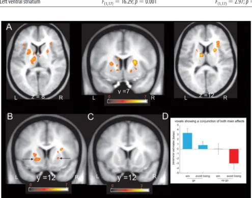
Figure 3. Response to anticipation of action and valence within anatomically defined ROIs in the striatum. Fractal images indicating go trials elicited higher activity than fractal images indicating no-go trials in all three bilateral ROIs: caudate, putamen, and ventral striatum (main effect of action, p⬍ 0.05) (for details, see Table 1). In the right putamen, fractal images indicating go trials elicited higher activity than fractal images indicating no-go trials only in the lose conditions (action⫻ valence interaction, p⬍ 0.05) (for details, see Table 1).
maxima within dorsal striatum that survived SVC within the
an-atomical whole-striatum ROI (Fig. 4 A). These foci were located
in the right putamen (MNI space coordinates, 23, 7, 12; peak Z
score, 4.92; p
⫽ 0.001 FWE), right caudate (MNI space
coordi-nates, 21, 7, 13; peak Z score, 4.75; p
⫽ 0.003 FWE), and left
putamen (MNI space coordinates,
⫺23, 11, 13; peak Z score,
4.07; p
⫽ 0.04 FWE). The first two belonged to a single cluster that
extended between the right caudate and putamen but was segregated
into a caudate and putamen portion in the ROI analysis because the
dividing internal capsule white matter tract, which separates these
structures, was not part of the ROI. When we constrained our
anal-ysis to an ROI restricted to the ventral striatum (Fig. 4B), we found
significant action anticipation-related activation in the left (MNI
space coordinates,
⫺17, 12, ⫺2; peak Z score, 3.99; p ⫽ 0.007 FWE)
and right (MNI space coordinates, 16, 7,
⫺5; peak Z score, 3.71; p ⫽
0.018 FWE) ventral putamen.
In keeping with previous studies of reward (Delgado et al.,
2000; Knutson et al., 2001; O’Doherty et al., 2002), the only
stri-atal region showing a main effect of valence (win
⬎ lose) was
located in the left ventral putamen (MNI space coordinates,
⫺17,
12,
⫺39) (Fig. 4C). However, this main
effect only approached significance when
the search volume was restricted to the
ventral striatum (peak Z score, 3.23; p
⫽
0.076 FWE). Because this cluster
over-lapped with the cluster showing a main
effect of action, we extracted betas for the
conjunction cluster (Fig. 4 D). Even in this
ventral striatal cluster, the dominant
ac-tivity pattern was an effect of action (go
⬎
no-go), with greater activity in go to win
compared with the no-go to win
condi-tion. The difference between the go to win
and go to avoid losing on one hand, and
the no-go to win and the no-go to avoid
losing on the other hand, is reminiscent of
the previously reported valence effects in
the fMRI literature in which action
re-quirements were not manipulated
(Del-gado et al., 2000; Knutson et al., 2001). A
weak effect of valence, however, is
com-patible with recent evidence that ventral
striatal activation to wins and losses is less
differentiable than individual valence
re-sponses compared with neutral trials
(Wrase et al., 2007; Cooper and Knutson,
2008). Note that all our experimental
con-ditions were highly salient by virtue of
their affective significance, and, on this
basis, we do not consider the signals we
find are likely to reflect mere salience
(Redgrave et al., 1999). An intriguing
pos-sibility is that increased ventral striatum
activity in the go to win relative to go to
avoid losing condition might be related to our behavioral finding
of better performance in the go to win compared with the go to
avoid losing condition.
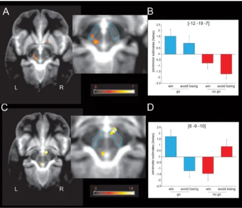
Midbrain activity (Fig. 5 A, B) showed a simple main effect of
action (go
⬎ no-go) within a left lateral region of SN/VTA that
survived SVC within our a priori ROI (MNI space coordinates,
⫺12, ⫺19, ⫺7; peak Z score, 3.33; p ⫽ 0.039 FWE). This
con-trasted with the response profile within a right medial SN/VTA
region (Fig. 5C,D), which showed a significant interaction of
ac-tion and valence that survived SVC within our a priori ROI (MNI
space coordinates, 8,
⫺9, ⫺10; peak Z score, 3.85; p ⫽ 0.008
FWE), with anticipation of action inducing activation in win
trials but deactivation in lose trials. These findings also survived
physiological noise correction for cardiac and respiratory phases
(data not shown). This dissociable pattern is strikingly similar to
findings from a recent electrophysiological study in monkeys
(Ma-tsumoto and Hikosaka, 2009), which distinguished between the
re-sponse profiles of two distinct groups of dopaminergic neurons. One
group, located in dorsolateral substantia nigra/VTA complex,
re-Figure 4. Voxel-based results within the striatum in response to anticipation of action and valence. A, Fractal images indicating go trials elicited higher dorsal striatal activity than fractal images indicating no-go trials ( p⬍ 0.001 uncorrected; p ⬍ 0.05 SVC within the whole-striatum ROI). The color scale indicates t values. B, Fractal images indicating go trials elicited higher ventral putamen activity than fractal images indicating no-go trials ( p⬍0.001uncorrected;p⬍0.05SVCwithintheROIrestrictedtothe ventral striatum). The color scale indicates t values. C, Fractal images indicating that win trials elicited higher ventral putamen activity than fractal images indicating lose trials ( p⬍ 0.001 uncorrected; did not survive SVC within the ROI restricted to the ventral striatum). The color scale indicates t values. D, Parameter estimates at the peak coordinates confirm that activation of the ventral putamen signals the anticipation of action. Although the anticipation of valence also seems to have an effect in the left ventral putamen, the effect did not survive SVC. Coordinates are given in MNI space. Error bars indicate SEM (note that these parameter estimates were not used for statistical inference). L, Left; R, right.
Table 1. Summary results within anatomically defined ROIs in the striatum
Main effect of action Main effect of valence Action⫻ valence interaction
Right caudate F(1,17)⫽ 7.06; p ⫽ 0.017 F(1,17)⫽ 2.02; p ⫽ 0.17 F(1,17)⫽ 1.09; p ⫽ 0.31
Left caudate F(1,17)⫽ 11.74; p ⫽ 0.003 F(1,17)⫽ 1.35; p ⫽ 0.26 F(1,17)⫽ 0.3; p ⫽ 0.66
Right putamen F(1,17)⫽ 5.78; p ⫽ 0.03 F(1,17)⫽ 0.64; p ⫽ 0.44 F(1,17)⫽ 5.9; p ⫽ 0.027
Left putamen F(1,17)⫽ 16.4; p ⫽ 0.001 F(1,17)⫽ 1.25; p ⫽ 0.28 F(1,17)⫽ 2.41; p ⫽ 0.14
Right ventral striatum F(1,17)⫽ 9.37; p ⫽ 0.007 F(1,17)⫽ 2.83; p ⫽ 0.11 F(1,17)⫽ 2.16; p ⫽ 0.65
sponded to both reward and punishment
predictive cues, whereas the other, located
more ventromedially, responded
preferen-tially to reward-predictive stimuli. We note
that heterogeneity within dopaminergic
midbrain is also described at a cellular level
(Lammel et al., 2008) and in rat
electrophys-iological recordings (Brischoux et al., 2009),
although the anatomical location of
dopa-mine neurons responsive to punishment
within the SN/VTA complex might differ
between rats and monkeys.
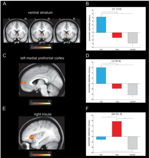
Brain responses at outcome
Although our statistical model was
subop-timal for studying brain responses at the
time of the outcome, we performed a
one-way ANOVA with valence as a factor (win,
lose, or neutral) to confirm whether we
could detect stronger BOLD responses in
the ventral striatum for win than loss
out-comes, an effect that has been described in
many studies (for a recent review, see
Haber and Knutson, 2010). As shown in
Figure 6, our analysis revealed a simple
main effect of valence in the right insula
(whole-brain FWE, p
⬍ 0.05), the left
me-dial prefrontal cortex (whole-brain FWE,
p
⬍ 0.05), and ventral striatum (SVC, p ⬍
0.05). We did not find any activated voxels
in SN/VTA. A post hoc t test analysis on
peak voxels showed that the insula
re-sponded more to loss whereas ventral
striatum responded more to wins, results
broadly consistent with the existing
liter-ature (Haber and Knutson, 2010),
find-ings that show that our imaging protocol
was indeed sensitive to BOLD responses
in the ventral striatum. Although our
de-sign was not optimal for studying
out-come responses, this result demonstrates that the striatum
responded to winning outcomes when consequences of an action
were evaluated. This is in sharp contrast to the activation pattern
seen during an anticipation period, which captured the influence
of action requirements rather than valence. This pattern also fits
well with the known role of striatum and dopaminergic system in
reward-guided action learning (Robbins and Everitt, 2002; Frank
et al., 2004).
Discussion
Participants were faster and more successful in the go to win than
the go to avoid losing condition. This suggests an asymmetric link
between opponent response tendencies (go and no-go) and
out-come valence (win and lose), consistent with a mandatory
cou-pling between valence and action. Our parallel fMRI data showed
that activation in striatum and lateral SN/VTA elicited by
antic-ipatory cues predominantly represented a requirement for a go
versus no-go response rather than the valence of the predicted
outcome (Figs. 3, 4). Finally, activity in the medial SN/VTA
mir-rored the asymmetric link between action and valence (Fig. 5).
An essential backdrop to our results, and indeed the rationale
for our experimental design, is the contrast between a seemingly
ineluctable tie between valence and action spectra and their
log-ical independence. In particular, it is widely reported that
dopa-mine neurons report a prediction error for reward (Montague et
al., 1996; Schultz et al., 1997; Bayer and Glimcher, 2005) in the
striatum (McClure et al., 2003; O’Doherty et al., 2003; O’Doherty
et al., 2004). However, dopamine also invigorates action
(Salam-one et al., 2005), regardless of its instrumental appropriateness,
with dopamine depletion being linked to decreased motor
activ-ity (Ungerstedt, 1971) and decreased vigor or motivation to work
for rewards in demanding reinforcement schedules (Salamone and
Correa, 2002; Niv et al., 2007). This coupling between action and
reward in the dopaminergic system is exactly why a signal associated
with go versus no-go might be confused with a signal associated with
reward versus punishment.
The role of action in a modified theory of opponency:
striatum and lateral SN/VTA
Many previous fMRI experiments involving pavlovian and
in-strumental conditioning have reported BOLD signals in both
striatum (McClure et al., 2003; O’Doherty et al., 2003, 2004) and
SN/VTA (D’Ardenne et al., 2008), correlating with putative
pre-diction error signals. In most studies [although not all, including
those involving the anticipation of pain (Seymour et al., 2004,
2005) and monetary loss (Delgado et al., 2008)], these signals are
Figure 5. Midbrain response to anticipation of action and reward. A, Fractal images indicating that go trials elicited higher left lateral midbrain (or substantia nigra compacta) activity than fractal images indicating no-go trials ( p⬍ 0.001 uncorrected; p ⬍ 0.05 SVC). The color scale indicates t values. B, Parameter estimates at the peak coordinates in the left lateral midbrain confirm that activation at this location signals the anticipation of action regardless of the valence of the outcome of the action (reward or punishment avoidance). Coordinates are given in MNI space. Error bars indicate SEM (note that these parameter estimates were not used for statistical inference). C, An action⫻ valence interaction was observed in the right medial midbrain or ventral tegmental area ( p⬍ 0.001 uncorrected; p ⬍ 0.05 SVC). The color scale indicates F values. D, Parameter estimates at the peak coordinates in the right medial midbrain confirm that activation at this location signals the anticipation of action if the outcome of the action is rewarding. The anticipation of actions that avoid punishment, conversely, is associated with a relative deactivation of this region. The inverse pattern of activation is observed for the no-go trials: the anticipation of a passive response that wins a reward is associated with relative deactivation, whereas the anticipation of a passive response that avoids punishment is associ-ated with activation. Coordinates are given in MNI space. Error bars indicate SEM (note that these parameter estimates were not used for statistical inference). L, Left; R, right.
positive when the prediction of future gains is greater than
ex-pected and negative when the prediction of future losses is greater
than expected. Our task was not designed to test the existence of
such prediction errors (McClure et al., 2003; O’Doherty et al.,
2003, 2004). Nevertheless, according to temporal difference
learning, the prediction errors associated with the appearance of
cues are the same as the predictions itself, and, given that
perfor-mance of the task was indeed very stable throughout the period of
fMRI data acquisition, we can reasonably assume that any brain
region encoding a reward prediction error to presentation of cues
might be expected to express a main effect of valence (go to win
⫹
no-go to win
⬎ go to avoid losing ⫹ no-go to avoid losing).
Despite the clear effect of valence on behavioral performance, we
did not find a main effect of valence in the fMRI data during
anticipation, apart from a small cluster within left ventral
puta-men. Even there, cue-evoked activity for the go to win and no-go
to win conditions differed significantly, despite both having the
same expected value. We interpret these results as valence losing
out to invigoration and thus as motivating the direct
incorpora-tion of acincorpora-tion into theories of opponency.
In the one area of the ventral striatum
in which we observed a main effect of
va-lence, the BOLD response took a form
that was more akin to the value (called a Q
value) associated with the go action as
op-posed to one associated with a reward
pre-diction or prepre-diction error. That is, there
was a single available action in our
exper-iment, namely to generate a go response.
The Q value of this response was high
when an action was rewarded (go to win),
zero when go responses led to avoidance
of punishment (go to avoid losing), or
omis-sion of reward (no-go to win) and negative
when actions were punished (no-go to
avoid losing). The observation that the
ven-tral striatum showed an action-dependent
prediction was unexpected, given its
associ-ation with the affective critic, which is
gov-erned by valence rather than some form of
actor (O’Doherty et al., 2004). Although
vi-sual inspection of Figure 3 seems to suggest
that this kind of signal is widely represented
in most of our anatomical ROIs, especially
the ventral subdivision of the striatum,
sta-tistical analyses do not support the presence
of a systematic difference between go to win
and go to avoid losing. However, we cannot
entirely rule out the presence of such a
signal.
The main effect in the striatum and
lat-eral SN/VTA related most strongly to
ac-tion (go to win
⫹ go to avoid losing ⬎
no-go to win
⫹ no-go to avoid losing).
There are at least three possible
interpre-tations for this dominance. First, it could
be argued that the no-go condition
quires inhibition of a prepotent motor
re-sponse, and a relative deactivation in the
striatum might reflect action suppression.
However, there are good empirical grounds
to believe this is not the case, including
evidence from previous fMRI studies that
action suppression activates inferior frontal gyrus (Rubia et al.,
2003; Aron and Poldrack, 2006) and subthalamic nucleus (Aron
and Poldrack, 2006). To our knowledge, suppression of neuronal
responses in the striatum has not been systematically reported,
although we note some evidence suggesting that striatal activity is
enhanced by a need for action suppression (Aron et al., 2003;
Aron and Poldrack, 2006). A second possibility arises from an
alternative computational implementation for action choice in
reinforcement learning. In the purest form of actor, the
propen-sities to perform a given action are detached from the values of
the states in which they are taken (Sutton and Barto, 1998). Thus,
invigorating (or inhibiting) an action requires a positive (or
neg-ative) propensity for go, with the scale of the propensities being
detached from any consideration of state values. Finally, a third
possibility is that the striatum represents the advantage of making
a go action as in advantage reinforcement learning (Dayan,
2002). In this model, action selection results from comparing the
advantage of different options in which the advantage is the
dif-ference between the action value and the state value. Whereas state
values would be positive in the win conditions and negative in the
Figure 6. Brain responses to the outcome. A, Activation in the ventral striatum revealed by a one-way ANOVA with valence as factor ( p⬍ 0.001 uncorrected; p ⬍ 0.05 SVC). The color scale indicates F values. B, Post hoc t test on the peak voxel in ventral striatum revealed that the main effect was driven by higher activation for the win trials when compared with the loss and neutral trials (*p⬍0.005;**p⬍0.001).C,Activationinleftmedialprefrontalcortexrevealedbyaone-wayANOVAwithvalenceasfactor ( p⬍ 0.001 uncorrected; p ⬍ 0.05 whole-brain FWE). The color scale indicates F values. D, Post hoc t test on the peak voxel in left medial prefrontal cortex revealed a main effect driven by greater activation for win compared with loss and neutral trials, respec-tively (**p⬍ 0.001). E, Activation in right insula revealed by a one-way ANOVA with valence as factor ( p ⬍ 0.001 uncorrected; p⬍ 0.05 whole-brain FWE). The color scale indicates F values. F, Post hoc t test on the peak voxel in the right insula revealed that the main effect was driven by higher activation for loss trials compared with win and neutral trials (*p⬍ 0.005; **p ⬍ 0.001). L, Left; R, right.
lose conditions, the advantage of performing a go action would be
positive but small in the go conditions (because action values are
positive or neutral) and negative in the no-go condition (because
action value is neutral or negative). However, if the observed
re-sponses in the striatum and SN/VTA represented advantages, these
would be the advantage of the go action even when participants
successfully choose a no-go response.
Our results show that, during the anticipatory phase, striatal
representations are dominated by actions rather than state values
independent from action. These results are not incompatible
with previous studies reporting reward prediction errors for state
values under experimental conditions controlling action
require-ments indirectly through the use of explicit foil actions (Delgado
et al., 2000, 2003, 2004; O’Doherty et al., 2003; Seymour et al.,
2004; Tricomi et al., 2004). This is because, in those studies,
re-ward prediction errors were isolated by comparing actions
lead-ing to rewards with foil actions that did not result in reward.
Hence, those studies were suitable to isolate reward components
that could be observed in addition to action representations in
the striatum and SN/VTA. However, they were less suitable to
highlight the predominant role of action representations in these
regions. In fact, our results show that, when the action axis is
explicitly incorporated within the experimental design, a refined
picture of striatal and SN/VTA representations emerges. Our
de-sign allowed us to show that the predominant coding reflects
anticipation of action. Reward prediction errors for state values
may be superimposed either when an instrumental action is
re-quired to gain a reward or when an action tendency is
automat-ically generated in response to a reward-predicting cue as in
classical (pavlovian) conditioning.
In light of these results and within the limitation of fMRI
studies of the SN/VTA (Du¨zel et al., 2009), theories implicating
dopaminergic system in valence opponency (Daw et al., 2002)
may need to be modified (Boureau and Dayan, 2011). The
dopa-minergic system would have to play a critical role in punishment
as well as reward processing, whenever an action is required. That
is, the semantics of the dopamine signal should be changed to
reflect loss avoidance by action (Dayan and Huys, 2009) as well as
the attainment of reward through action, indeed as in classical
two-factor theories (Mowrer, 1947). In fact, some reinforcement
learning models of active avoidance code the removal of the
pos-sibility of punishment (i.e., the achievement of safety) as akin
(dopaminergically coded) to a reward (Grossberg, 1972;
Schma-juk and Zanutto, 1997; Johnson et al., 2002; Moutoussis et al.,
2008; Maia, 2010). Compatible with this two-factor view is the
ob-servation that dopamine depletion impairs the acquisition of active
avoidance behavior (McCullough et al., 1993; Darvas et al., 2011).
Paralleling the case for dopamine, this modification from valence
opponency toward action opponency motivates a search for an
iden-tifiable neurotransmitter system that promotes the other end of the
action spectrum, namely inhibition. Serotonin has been thought to
serve as such a neurotransmitter (Deakin and Graeff, 1991; Gray and
McNaughton, 2000). Interestingly, one study that inspired ours
(Crockett et al., 2009) showed that tryptophan depletion abolished
punishment-induced inhibition, which is similar to the
disadvan-tage that we observed in the go to avoid losing condition.
Medial SN/VTA
Unlike the case for the lateral SN/VTA, valence had opposite
effects for go and no-go in the medial SN/VTA: for go, neural
activity was higher for the win condition, whereas for no-go,
activity was higher for the avoid-losing condition. One way to
interpret this pattern is in terms of prediction errors relative to
the mandatory couplings between action and reward and
be-tween inhibition and punishment. That is, go is mandatorily
as-sociated with reward, and so the relevant prediction error, which
could stamp in appropriate actions, favors reward over
punish-ment. Conversely, no-go is associated with punishment, and so
the relevant prediction error favors punishment over reward.
In-deed, punishment prediction errors have been reported
previ-ously (Seymour et al., 2004; Delgado et al., 2008) in pavlovian
conditions in which actions are irrelevant. Future studies could
usefully target the functional interactions between action and
valence in the medial SN/VTA, taking account also of anatomical
and physiological findings regarding the involvement of
dopa-mine in processing punishment. Unexpected punishment leads
to supra-baseline dopamine activity in some microdialysis
exper-iments in rats (Pezze et al., 2001; Young, 2004). Furthermore,
unconditioned avoidance responses can only be elicited from
topographically appropriate regions of the shell region of the
nucleus accumbens given appropriately high levels of dopamine
(Faure et al., 2008). Thus, one possibility is that the signal we
observed in medial SN/VTA was more akin to one that organizes
unconditioned responses in a valence-dependent manner.
Conclusions
Our study expands on conventional views regarding the nature of
signals reported from both STN/VTA and striatum. Although the
striatum responded to wins more than losses at outcome, a
pri-mary form of coding in both the striatum and lateral SN/VTA
complex during anticipation reflected action requirement rather
than state values. These results indicate that the status of an action
in relation to approach or withdrawal may be best captured in a
modified opponent theory of dopamine function.
References
Aron AR, Poldrack RA (2006) Cortical and subcortical contributions to Stop signal response inhibition: role of the subthalamic nucleus. J Neu-rosci 26:2424 –2433.
Aron AR, Schlaghecken F, Fletcher PC, Bullmore ET, Eimer M, Barker R, Sahakian BJ, Robbins TW (2003) Inhibition of subliminally primed re-sponses is mediated by the caudate and thalamus: evidence from func-tional MRI and Huntington’s disease. Brain 126:713–723.
Bayer HM, Glimcher PW (2005) Midbrain dopamine neurons encode a quantitative reward prediction error signal. Neuron 47:129 –141. Berridge KC, Robinson TE (1998) What is the role of dopamine in reward:
hedonic impact, reward learning, or incentive salience? Brain Res Brain Res Rev 28:309 –369.
Boureau YL, Dayan P (2011) Opponency revisited: competition and coop-eration between dopamine and serotonin. Neuropsychopharmacology 36:74 –97.
Brett M, Anton J-L, Valabregue R, Poline J-B (2002) Region of interest anal-ysis using an SPM toolbox. Presented at the Eighth International Confer-ence on Functional Mapping of the Human Brain, Sendai, Japan, June. Brischoux F, Chakraborty S, Brierley DI, Ungless MA (2009) Phasic
excita-tion of dopamine neurons in ventral VTA by noxious stimuli. Proc Natl Acad Sci U S A 106:4894 – 4899.
Bunzeck N, Du¨zel E (2006) Absolute coding of stimulus novelty in the hu-man substantia nigra/VTA. Neuron 51:369 –379.
Cools R, Nakamura K, Daw ND (2011) Serotonin and dopamine: unifying affective, activational, and decision functions. Neuropsychopharmacol-ogy 36:98 –113.
Cooper JC, Knutson B (2008) Valence and salience contribute to nucleus accumbens activation. Neuroimage 39:538 –547.
Crockett MJ, Clark L, Robbins TW (2009) Reconciling the role of sero-tonin in behavioral inhibition and aversion: acute tryptophan deple-tion abolishes punishment-induced inhibideple-tion in humans. J Neurosci 29:11993–11999.
D’Ardenne K, McClure SM, Nystrom LE, Cohen JD (2008) BOLD re-sponses reflecting dopaminergic signals in the human ventral tegmental area. Science 319:1264 –1267.
Darvas M, Fadok JP, Palmiter RD (2011) Requirement of dopamine signal-ing in the amygdala and striatum for learnsignal-ing and maintenance of a conditioned avoidance response. Learn Mem 18:136 –143.
Daw ND, Kakade S, Dayan P (2002) Opponent interactions between sero-tonin and dopamine. Neural Netw 15:603– 616.
Daw ND, O’Doherty JP, Dayan P, Seymour B, Dolan RJ (2006) Cortical substrates for exploratory decisions in humans. Nature 441:876 – 879. Dayan P (2002) Motivated reinforcement learning. In: Advances in neural
in-formation processing systems 14 (Dietterich TG, Becker S, Ghahramani Z, eds). Cambridge, MA: MIT.
Dayan P, Huys QJ (2009) Serotonin in affective control. Annu Rev Neurosci 32:95–126.
Deakin JFW, Graeff FG (1991) 5-HT and mechanisms of defence. J Psycho-pharmacol 5:305–315.
Delgado MR, Nystrom LE, Fissell C, Noll DC, Fiez JA (2000) Tracking the hemodynamic responses to reward and punishment in the striatum. J Neurophysiol 84:3072–3077.
Delgado MR, Locke HM, Stenger VA, Fiez JA (2003) Dorsal striatum re-sponses to reward and punishment: effects of valence and magnitude manipulations. Cogn Affect Behav Neurosci 3:27–38.
Delgado MR, Stenger VA, Fiez JA (2004) Motivation-dependent responses in the human caudate nucleus. Cereb Cortex 14:1022–1030.
Delgado MR, Li J, Schiller D, Phelps EA (2008) The role of the striatum in aversive learning and aversive prediction errors. Philos Trans R Soc Lond B Biol Sci 363:3787–3800.
Domjan M (2005) Pavlovian conditioning: a functional perspective. Annu Rev Psychol 56:179 –206.
Du¨zel E, Bunzeck N, Guitart-Masip M, Wittmann B, Schott BH, Tobler PN (2009) Functional imaging of the human dopaminergic midbrain. Trends Neurosci 32:321–328.
Elliott R, Newman JL, Longe OA, William Deakin JF (2004) Instrumental responding for rewards is associated with enhanced neuronal response in subcortical reward systems. Neuroimage 21:984 –990.
Faure A, Reynolds SM, Richard JM, Berridge KC (2008) Mesolimbic dopamine in desire and dread: enabling motivation to be generated by localized gluta-mate disruptions in nucleus accumbens. J Neurosci 28:7184 –7192. Frank MJ, Seeberger LC, O’Reilly RC (2004) By carrot or by stick: cognitive
reinforcement learning in parkinsonism. Science 306:1940 –1943. Gerfen CR (1992) The neostriatal mosaic: multiple levels of
comportamen-tal organization. Trends Neurosci 15:133–139.
Gray JA, McNaughton M (2000) The neuropsychology of anxiety: an inquiry into the function of the septohippocampal system, Ed 2. Oxford: Oxford UP. Grossberg S (1972) A neural theory of punishment and avoidance. Math
Biosci 15:39 – 67.
Guitart-Masip M, Bunzeck N, Stephan KE, Dolan RJ, Du¨zel E (2010) Con-textual novelty changes reward representations in the striatum. J Neurosci 30:1721–1726.
Haber SN, Knutson B (2010) The reward circuit: linking primate anatomy and human imaging. Neuropsychopharmacology 35:4 –26.
Haber SN, Fudge JL, McFarland NR (2000) Striatonigrostriatal pathways in primates form an ascending spiral from the shell to the dorsolateral stria-tum. J Neurosci 20:2369 –2382.
Johnson J, Li W, Li J, Klopf A (2002) A computational model of learned avoid-ance behavior in a one-way avoidavoid-ance experiment. Adapt Behav 9:91–104. Klein A, Andersson J, Ardekani BA, Ashburner J, Avants B, Chiang MC,
Christensen GE, Collins DL, Gee J, Hellier P, Song JH, Jenkinson M, Lepage C, Rueckert D, Thompson P, Vercauteren T, Woods RP, Mann JJ, Parsey RV (2009) Evaluation of 14 nonlinear deformation algorithms applied to human brain MRI registration. Neuroimage 46:786 – 802. Knutson B, Adams CM, Fong GW, Hommer D (2001) Anticipation of
in-creasing monetary reward selectively recruits nucleus accumbens. J Neu-rosci 21:RC159(1–5).
Knutson B, Taylor J, Kaufman M, Peterson R, Glover G (2005) Distributed neural representation of expected value. J Neurosci 25:4806 – 4812. Lammel S, Hetzel A, Ha¨ckel O, Jones I, Liss B, Roeper J (2008) Unique
properties of mesoprefrontal neurons within a dual mesocorticolimbic dopamine system. Neuron 57:760 –773.
Maia TV (2010) Two-factor theory, the actor-critic model, and conditioned avoidance. Learn Behav 38:50 – 67.
Matsumoto M, Hikosaka O (2009) Two types of dopamine neuron dis-tinctly convey positive and negative motivational signals. Nature 459: 837– 841.
McClure SM, Berns GS, Montague PR (2003) Temporal prediction errors in a passive learning task activate human striatum. Neuron 38:339 –346. McCullough LD, Sokolowski JD, Salamone JD (1993) A neurochemical and
behavioral investigation of the involvement of nucleus accumbens dopa-mine in instrumental avoidance. Neuroscience 52:919 –925.
Montague PR, Dayan P, Sejnowski TJ (1996) A framework for mesen-cephalic dopamine systems based on predictive Hebbian learning. J Neu-rosci 16:1936 –1947.
Moutoussis M, Bentall RP, Williams J, Dayan P (2008) A temporal differ-ence account of avoidance learning. Network 19:137–160.
Mowrer OH (1947) On the dual nature of learning: a reinterpretation of conditioning and problem solving. Harv Educ Rev 17:102–148. Niv Y, Daw ND, Joel D, Dayan P (2007) Tonic dopamine: opportunity costs
and the control of response vigor. Psychopharmacology (Berl) 191:507–520. O’Doherty JP (2004) Reward representations and reward-related learning in the human brain: insights from neuroimaging. Curr Opin Neurobiol 14:769 –776.
O’Doherty JP, Deichmann R, Critchley HD, Dolan RJ (2002) Neural responses during anticipation of a primary taste reward. Neuron 33:815– 826. O’Doherty JP, Dayan P, Friston K, Critchley H, Dolan RJ (2003) Temporal
difference models and reward-related learning in the human brain. Neu-ron 38:329 –337.
O’Doherty J, Dayan P, Schultz J, Deichmann R, Friston K, Dolan RJ (2004) Dissociable roles of ventral and dorsal striatum in instrumental condi-tioning. Science 304:452– 454.
Pezze MA, Heidbreder CA, Feldon J, Murphy CA (2001) Selective respond-ing of nucleus accumbens core and shell dopamine to aversively condi-tioned contextual and discrete stimuli. Neuroscience 108:91–102. Redgrave P, Prescott TJ, Gurney K (1999) Is the short-latency dopamine
response too short to signal reward error? Trends Neurosci 22:146 –151. Robbins TW, Everitt BJ (2002) Limbic-striatal memory systems and drug
addiction. Neurobiol Learn Mem 78:625– 636.
Rubia K, Smith AB, Brammer MJ, Taylor E (2003) Right inferior prefrontal cortex mediates response inhibition while mesial prefrontal cortex is re-sponsible for error detection. Neuroimage 20:351–358.
Salamone JD, Correa M (2002) Motivational views of reinforcement: impli-cations for understanding the behavioral functions of nucleus accumbens dopamine. Behav Brain Res 137:3–25.
Salamone JD, Correa M, Mingote SM, Weber SM (2005) Beyond the reward hypothesis: alternative functions of nucleus accumbens dopamine. Curr Opin Pharmacol 5:34 – 41.
Schmajuk N, Zanutto B (1997) Escape avoidance and imitation: A neural network approach. Adapt Behav 6:63–129.
Schultz W, Dayan P, Montague PR (1997) A neural substrate of prediction and reward. Science 275:1593–1599.
Seymour B, O’Doherty JP, Dayan P, Koltzenburg M, Jones AK, Dolan RJ, Friston KJ, Frackowiak RS (2004) Temporal difference models describe higher-order learning in humans. Nature 429:664 – 667.
Seymour B, O’Doherty JP, Koltzenburg M, Wiech K, Frackowiak R, Friston K, Dolan R (2005) Opponent appetitive-aversive neural processes underlie predictive learning of pain relief. Nat Neurosci 8:1234 –1240.
Sutton RS, Barto AG (1998) Reinforcement learning: an introduction. Cambridge, MA: Massachusetts Institute of Technology.
Tricomi EM, Delgado MR, Fiez JA (2004) Modulation of caudate activity by action contingency. Neuron 41:281–292.
Ungerstedt U (1971) Adipsia and aphagia after 6-hydroxydopamine in-duced degeneration of the nigro-striatal dopamine system. Acta Physiol Scand Suppl 367:95–122.
Weiskopf N, Hutton C, Josephs O, Deichmann R (2006) Optimal EPI pa-rameters for reduction of susceptibility-induced BOLD sensitivity losses: a whole-brain analysis at 3 T and 1.5 T. Neuroimage 33:493–504. Wrase J, Kahnt T, Schlagenhauf F, Beck A, Cohen MX, Knutson B, Heinz A
(2007) Different neural systems adjust motor behavior in response to reward and punishment. Neuroimage 36:1253–1262.
Young AM (2004) Increased extracellular dopamine in nucleus accumbens in response to unconditioned and conditioned aversive stimuli: studies using 1 min microdialysis in rats. J Neurosci Methods 138:57– 63. Zink CF, Pagnoni G, Martin ME, Dhamala M, Berns GS (2003) Human striatal
response to salient nonrewarding stimuli. J Neurosci 23:8092– 8097. Zink CF, Pagnoni G, Martin-Skurski ME, Chappelow JC, Berns GS (2004)
Human striatal responses to monetary reward depend on saliency. Neu-ron 42:509 –517.