http://ats.ctsnetjournals.org/cgi/content/full/83/6/1978
located on the World Wide Web at:
The online version of this article, along with updated information and services, is
Print ISSN: 0003-4975; eISSN: 1552-6259.
Southern Thoracic Surgical Association. Copyright © 2007 by The Society of Thoracic Surgeons.
is the official journal of The Society of Thoracic Surgeons and the
Transxiphoid Hand-Assisted Videothoracoscopic
Surgery
Tommaso Claudio Mineo,
MD,
Vincenzo Ambrogi,
MD,
Davide Mineo,
MD,
and Eugenio Pompeo,
MD
Thoracic Surgery Division, Policlinico Tor Vergata University, Rome, Italy
Background. We have performed transxiphoid
hand-assisted videothoracoscopy since 1995 to allow manual palpation in bilateral lung metastasectomy. This ap-proach was extended to other thoracoscopic procedures requiring a handport. No extensive report about early and late results has yet been published.
Methods. We retrospectively reviewed the first 100
consecutive patients undergoing transxiphoid hand-as-sisted videothoracoscopy. Acute and chronic postopera-tive pain, respiratory function, patient’s satisfaction score (1 to 5), quality of life (Short Form-36), and survival rate were evaluated.
Results. Seventy-four patients had lung metastases, 5
had primary lung cancers, 16 had benign nodules, and 5 had Morgani’s hernia. Five patients needed conversion to thoracotomy, whereas 7 successfully underwent a second transxiphoid operation. Sixty-five metastatic patients were bilaterally explored, 44 were without radiologic evidence of controlateral lesions, discovering 23 occult metastases and 10 patients with occult controlateral dis-ease. A total of 207 minimal resections and 11
lobecto-mies were performed. Mean operative time was 103ⴞ 35 minutes. We had no intraoperative mortality or major complications. Thirty-day postoperative morbidity docu-mented arrhythmia (nⴝ 4) and acute pneumonia (n ⴝ 4). Visual Analogue Scale pain, C-reactive protein, fibrino-gen, and serum interleukin-6, -8, and -10 normalized within 72 hours. Respiratory function and most of the Short Form-36 domains recovered within 3 months. Six-month mean patient satisfaction score was 4.0 ⴞ 0.8. Three- and 5-year survival rates for metastatic patients were 52% and 43%, respectively. Mean disease-free inter-val was 12ⴞ 5.8 months.
Conclusions. Transxiphoid hand-assisted
videothora-coscopy proved a good alternative to conventional ap-proaches, and provided rapid recovery without affecting the survival rate in those patients with metastatic lesions. We recommend it whenever a handport during video-assisted procedure is required.
(Ann Thorac Surg 2007;83:1978 – 85) © 2007 by The Society of Thoracic Surgeons
V
ideo-assisted thoracic surgery (VATS) was perceived to be less invasive than the conventional open thoracotomy with respect to less pain, better preservation of pulmonary function, and less impairment of the shoul-der girdle [1–3]. Minimal resection by the VATS ap-proach is a widespread and accepted therapeutic method for treating small peripheral lung lesions [4, 5]. Never-theless, having one hand inside the chest may simplify the identification of subcentimetric nodules detected by spiral computed tomography (CT) scan, facilitate the resection for nodules sited in posterior segments of the lung, and permit the exploration of the controlateral hemithorax[6].Transxiphoid hand-assisted videothoracoscopy was developed to face these obstacles without increasing operative morbidity [7]. This incision was useful for bilateral approaches during the same operation, required by the metastatic nature of the disease. We also found the access useful to facilitate video-assisted procedures
such as resection of deeply located nodules, lower lobectomies, and Morgagni’s hernia repair. Our expe-rience has now reached 100 patients and a significant follow-up is available. The purpose of this study was to analyze the short- and long-term results of using this approach.
Patients and Methods
We retrospectively reviewed our experience with the first 100 consecutive patients who underwent transxiphoid hand-assisted videothoracoscopy in our division. Early and long-term postoperative results were collected from clinical reports or during outpatient clinics. The study was approved by the Ethics Committee of our University. Patient’s permission for use of personal data was asked and obtained.
Indication for the Procedure
Since 1995, when we first introduced this approach for pulmonary metastases, we limited its use to some se-lected conditions. Complete control of the primary tumor and absent or resectable extrapulmonary metastases were considered the two mandatory prerequisites.
Fur-Accepted for publication Feb 7, 2007.
Address correspondence to Dr Mineo, Cattedra di Chirurgia Toracica, Policlinico Tor Vergata, Viale Oxford 81, Rome, 00133, Italy; e-mail: [email protected].
thermore, we excluded patients with metastases at more than 2 cm from the visceral pleura at the CT, or greater than 3 cm or anyway requiring lobectomy, or with a history of pleuritis or pleurodesis, or in the presence of cardiomegaly or arrhythmia. Nevertheless, through the transxiphoid approach, we performed controlateral man-ual palpation even in the case of negative imaging. Neither previous thoracic nor abdominal surgery repre-sented absolute contraindication to this operation in itself. Four patients had a previous VATS for metastasec-tomy; another 4 had an abdominal operation through a subcostal access, and transxiphoid incision was made along the middle trait of this scar. Finally, in 7 patients, we performed a successful redo transxiphoid operation. Following the same technique, we developed a new approach for resection of pulmonary and hepatic me-tastasis, combining the subcostal transverse laparot-omy with the transxiphoid hand-assisted approach. Thereafter, we extended this approach to facilitate video-assisted procedures such as lobectomy or resec-tion of nodules difficult to localize by instrumental palpation. In addition, we found it very useful in surgery of the anterior leaflet of the diaphragm such as Morgagni’s hernia repair.
Preoperative Studies
After the discovery of lung metastases, all patients un-derwent further examination (total body CT scan, ultra-sonography, bone scan) to exclude primary tumor recur-rence or extrapulmonary relapse. The lung lesions were also restaged immediately before planned surgery by means of a helical chest CT scan (Tomoscan SR 7000; Philips Medical Systems, Eindhoven, Netherlands, and more recently, LightSpeed16
scanner; General Electric Medical Systems, Milwaukee, Wisconsin) without intra-venous contrast medium. This also allowed determina-tion of the stability of pulmonary lesions. The mean interval between helical CT scan and surgical exploration was 8 days (range, 4 to 15).
Operative Details
Transxiphoid hand-assisted videothoracoscopy tech-nique has been described elsewhere [7], and the main
steps of the procedure are reported inTable 1and and shown in Figure 1. General anesthesia was always in-duced with a double-lumen endotracheal tube. Explora-tion started from the side deemed more technically demanding, either because of the greater number of lesions or a more difficult resection. The entire lung, both partially inflated or totally deflated, was carefully pal-pated between thumb and forefinger according to a precise sequence regardless of radiologic findings and thoracoscopic appearance. Thereafter, all areas predicted by helical CT to be site of metastases were specifically explored until the predicted lesions could be identified and evaluated for resection. All palpated nodules were excised with minimal resection by an endoscopic sta-pler or laser beam, taking care to spare the surround-ing healthy parenchyma. After the resection, the lung was manually reinflated until visible collapse had disappeared.
The same surgical maneuvers were usually repeated on the opposite hemithorax. Bilateral exploration and manual palpation of both hemithoraces were performed on a routine basis. The only reason to refrain from bilateral exploration was the presence of pleural adhe-sions in the side radiologically determined as free of metastases. At the end of the procedure, one or two chest tubes were inserted. Size and location of the resected nodules were immediately recorded. All resected nod-ules were sent separately for histologic analysis.
Postoperative Treatment
All patients were extubated in the operatory room. Epi-dural anesthesia (fentanyl and bupivacaine) adminis-tered for the first 48 hours after surgery was used until 1999. Thereafter, postoperative pain was treated only by tramadole 300 mg and ketorolac 90 mg in continuous intravenous infusion until chest tube removal.
Acute Pain Evaluation
Evaluation of acute postoperative pain was assessed using the Visual Analogue Scale (VAS) that graded from 0 (no pain) to 10 (most severe imaginable pain) as an
Fig 1. Position of the patient and setup of surgical approach.
Table 1. Steps of Transxiphoid Hand-Assisted Thoracoscopy (HATS) Approach
● Patient side decubitus in a 60-degree off-center position ● Insertion of three thoracoscopic trocars
● Midtransverse arcuate skin incision and retraction of rectus abdominis muscles along the linea alba
● Resection of the xiphoid appendix
● Under thoracoscopic visualization, introduction of one hand below the sternum and blunt dissection of retrosternal areolar tissue
● Incision of mediastinal pleura, allowing the whole hand to enter in the cavity
● Bidigital palpation of the entire lung, either partially inflated or totally deflated
● Resection by stapler, guided by hand, of all palpated nodules
● Drainage positioning and closure of incisions
GENERAL
index of the patient’s perception of pain[8]. The VAS was explained to patients preoperatively and consisted of patients marking a grade of pain on a graded ruler. Pain was quantified both at basal level and when provoked by particular movements. Assessments were made twice daily for the first 2 days, then once daily until the discharge.
Acute Pain Stress Markers
It is known that elective thoracic surgery provokes an increase in acute pain stress serum markers such as C-reactive protein, fibrinogen, white blood cell count, and interleukin-6, -8, and -10 using comercially available assays. The magnitude of the elevation is related directly to the degree of tissue injury [9]. Blood samples were drawn preoperatively, at the end of the procedure, and every 12 hours postoperatively for the first 48 hours, centrifuged at 3,000g for 5 minutes, and then stored in deep refrigeration at⫺70°C until serum measurements were performed.
Patient Satisfaction
Patient satisfaction with the operation was assessed 6 months later by asking the patient to choose one of the five possible responses: poor, fair, good, very good, or excellent, assigning them a point score from 1 to 5.
Quality of Life Evaluation
We used the Medical Outcomes Study Short-Form 36-Item Questionnaire (SF-36) [10], which consists of 36 multiple-choice questions that cover eight health con-cepts: physical functioning, social functioning, physical role, emotional role, vitality, body pain, mental health, and general health perception (best score⫽ 100, worst ⫽ 0). To simplify the evaluation, the Physical and Mental Component Summary (Health Assessment Lab, New England Medical Center, Boston, Massachusetts,1994) was also used. We chose this questionnaire because of its relative simplicity, wide use in chronic pulmonary dis-eases, specific presence of body pain domain, and
avail-Table 2. Demographics, and Intraoperative and Postoperative Results Classified for Pathology
Variable Lung Metastasis
Primary Lung Cancers
Benign
Nodules Morgagni’s Hernia Total
Patients 74 5 16 5 100
Age, years (mean⫾ SD) 54.2⫾ 14.4 65.3⫾ 5.9 49.4⫾ 6.8 57.4⫾ 11.2 54.7⫾ 14.0
Sex (male:female) 40:34 4:1 8:8 3:2 55:45
Procedures 81a 5 16 5 107
Procedures converted to open 3 2 — — 5
Operative time (min) 102⫾ 34 145⫾ 44 70⫾ 12 85⫾ 8 103⫾ 35
Operative bleeding (mL) 160⫾ 83 230⫾ 75 90⫾ 36 76⫾ 56 161⫾ 81
Bilateral exploration (no. of patients) 65 — 2 — 67
Bilateral resection (no. of patients) 55 — 2 — 57
Occult controlateral disease 10 — — — 10
Minimal resections 189 — 18 — 207
Lobectomies 8 3 — — 11
Resected lesions 211 3 18 — 228
Number per patients (mean⫾ SD) 2.97⫾ 1.0 1 1.12⫾ 0.3 — 2.78⫾ 0.9
Size, cm (mean⫾ SD) 1.75⫾ 1.1 3.4⫾ 1.6 1.1⫾ 0.7 — 1.77⫾ 1.2
Side (right:left) 118:103 2:1 10:8 — 130:112
Malignant lesions 178 3 — — 181
Benign lesions 33 — 18 — 51
Occult metastases 23 — — — 23
Occult metastases (no. of patients) 18 — — — 18
Air leak time (days) 2.1⫾ 1.1 3.4⫾ 2.1 1.5⫾ 0.7 — 2.2⫾ 1.1
Hospital stay (days) 4.1⫾ 1.4 6.5⫾ 1.2 2.7⫾ 1.5 2.6⫾ 0.5 4.3⫾ 1.4
Morbidity rate (%) 5 1 2 — 8
1-week basal VAS (1–10) 1.0⫾ 0.8 2.3⫾ 1.3 0.9⫾ 0.5 1.0⫾ 0.9 1.0⫾ 1.3
1-week provoked VAS (1–10) 2.2⫾ 1.7 3.1⫾ 1.6 1.9⫾ 1.1 1.5⫾ 1.0 2.2⫾ 1.4
1-month⌬FEV1(%) ⫺5.1 ⫾ 4.7 ⫺9.2 ⫾ 5.7 ⫺4.3 ⫾ 3.2 ⫺1.2 ⫾ 0.3 ⫺5.0 ⫾ 4.1
1-month basal VAS (1–10) 0.3⫾ 0.2 1.0⫾ 0.6 0.3⫾ .01 0.2⫾ 0.1 0.3⫾ 0.2
1-month provoked VAS (1–10) 0.9⫾ 0.3 1.3⫾ 0.5 0.4⫾ 0.2 0.4⫾ 0.1 0.9⫾ 0.3
3-month⌬FEV1(%) ⫺4.1 ⫾ 3.4 ⫺9.2 ⫾ 5.7 ⫺3.1 ⫾ 1.2 ⫺1.2 ⫾ 0.3 ⫺4.2 ⫾ 3.2
6-month acceptance (1–5) 4.0⫾ 0.8 4.1⫾ 1.2 4.3⫾ 1.1 4.3⫾ 0.5 4.0⫾ 0.8
a74 first operations and 7 second operations.
⌬FEV1⫽ change forced expiratory volume in one second; VAS⫽ Visual Analog Scale.
ability of a validated version for the Italian population
[11]. The questionnaire was administrated preoperatively and at 1 month and 6 months after the operation. Further evaluations were not included in the present study, as the natural history of patients’ disease greatly affects their quality of life.
Follow-Up
Follow-up included clinical visit, timed spirometry, se-rum biochemical, and chest radiography to be performed every 6 months (every 3 months for the neoplastic pa-tients) for the first 2 years and thereafter once per year. Total body CT scan was done twice per year for the first 2 years and then yearly. For neoplastic patients, func-tional assessment (ie, electrocardiogram, clearance creat-inine, blood urea nitrogen, bilirubin, and liver enzymes) was repeated before each chemotherapic cycle.
Statistical Analysis
Data were expressed throughout as mean⫾ SD. Postop-erative changes were assessed by the Student t test. All tests were two-tailed with a significant level of 0.05. Overall survival and disease-free survival curves were estimated using the Kaplan-Meier method, and the sig-nificance test was based on the log-rank test as given by Mantel. The survival time was counted from the day of surgery. Recurrence was considered any new neoplastic lesion, second primaries included. All causes of death were treated as final events.
Results
Early Results
General details of the operated patients are summarized inTable 2. Seventy-four patients had lung metastases, 13 of whom had simultaneous liver metastasectomy through the same access. Four patients had already undergone a previous thoracic procedure in other insti-tutions. Bilateral transxiphoid exploration was performed in a total of 65 patients, 44 without CT evidence of controlateral lesions. In 6 patients with CT evidence of unilateral disease, contralateral palpation was impossible for the presence of tenacious adherences.
Furthermore, we used the same access to approach 5 primary lung cancers, 16 benign nodules deeply sited
into the parenchyma or difficult to localize without bi-digital palpation, and 5 Morgagni’s hernia.
Table 2 shows operative and postoperative results. Mean operative time was 103⫾ 35 minutes, decreasing from a mean time of 138 minutes at the beginning of our experience to 91 minutes in the last patients (p⬍ 0.01). A total of 207 minimal resections and 11 lobectomies were performed in 102 operations.
We converted the procedure to open lateral thoracot-omy in 5 instances: 4 on the left and 1 on the right. In 2 cases, there were two primary lung cancers requiring lobectomy in which we found the hilar dissection diffi-cult. In the other 3 cases, we performed a rib-spreading minithoracotomy of 6 to 8 cm through which it was possible to better circumscribe the lesion and separate it from adjacent major vessels; this was necessary owing to the presence of deeply located metastasis. This incision was usually accomplished by prolonging a trocar port.
In the metastatic patients subset, we performed a total of 189 minimal resections and 8 lobectomies, removing a total of 211 lesions: 87 were located in the upper lobes (45 right, 42 left), 97 were found in the lower lobes (46 right,
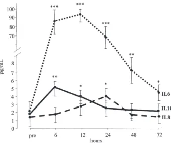
Fig 2. Interleukin (IL) changes in the first 72 hours after surgery. Intragroup significance: ***pⱕ 0.001; **p ⱕ 0.01; *p ⱕ 0.05. (Bars⫽ standard error of the mean; dotted line ⫽ IL-6; dashed line⫽ IL-8; solid line ⫽ IL-10.)
Table 3. Mean Values of the Main Acute Pain Stress Markers Assessed at Timed Intervals From the Operation
Preoperative End Procedure 12 Hours 24 Hours 48 Hours 96 Hours Basal VAS (1–10) 0.3⫾ 0.1 3.1⫾ 2.5a 3.3⫾ 1.2a 2.15⫾ 0.9b 1.71⫾ 1.2b 0.5⫾ 0.8 Provoked VAS (1–10) 0.8⫾ 0.2 4.5⫾ 3.2a 5.4⫾ 3.5a 4.1⫾ 2.4a 2.5⫾ 1.9b 1.5⫾ 1.4c C-reactive protein (mg/L) 1.8⫾ 1.5 10⫾ 3.2a 73⫾ 33a 105⫾ 34a 86⫾ 23a 7⫾ 4b Fibrinogen (mg/dL) 288⫾ 55 350⫾ 41c 420⫾ 57b 462⫾ 134a 347⫾ 103b 291⫾ 37 Glycemia (mg/dL) 82⫾ 31 102⫾ 34b 105⫾ 41b 109⫾ 34b 88⫾ 19 83⫾ 24 Leukocytes count (n/mm3) 5,320⫾ 1,340 6,700⫾ 590 7,920⫾ 1,440b 8,964⫾ 1,320b 6,340⫾ 1,050c 5,550⫾ 1,210 paO2(mm Hg) 82⫾ 9 80⫾ 17c 77⫾ 12b 75⫾ 12b 80⫾ 8c 83⫾ 4 Intragroup significance:apⱕ 0.001; bpⱕ 0.01; cpⱕ 0.05.
VAS⫽ Visual Analog Scale.
GENERAL
51 left), and 27 were detected in the middle lobe. The maximum number of resected nodules per patient was 10. One hundred and seventy-eight lesions were identi-fied as metastasis at histologic examination. Twenty-three CT-documented occult metastases were discovered in 18 patients, and controlateral occult disease in 10. The size of occult lesions ranged between 3 and 12 mm (mean, 6.7⫾ 2.8 mm). There were 33 benign nodules with a mean diameter of 7.5⫾ 4.4 mm, 25 of which had been judged metastasis by the imaging studies. Seven nodules diagnosed using the CT scan were not detected at palpa-tion, and they were interpreted as small blood vessels.
We did not report intraoperative mortality. Mild and transitory hypotension or arrhythmia from right ventricle compression could be avoided by raising the sternum. Neither substernal bleeding nor phrenic nerve palsy were recorded. Intraoperative cardiac arrhythmia was experienced in 15.4% of the right and 34.5% of the left approaches. We were never compelled to stop the oper-ation or defibrillate the patient. Eight patients presented with a significant morbidity: atrial fibrillation (n⫽ 4) and acute pneumonia (n⫽ 4).
After the expected increment, 24 hours postopera-tively, all acute pain variables presented a significant decrement at 48 hours and a return to basal value after 96 hours (Table 3) in almost all measures.Figure 2 docu-ments the timed variations of the three interleukins, showing a normalization after 72 hours.
Chest drainage was removed after a mean of 2.26⫾ 1.12 days (range, 2 to 7). Four patients presented a
prolonged air leakage requiring discharge with Heimlich valve. Mean hospital stay was 4.33⫾ 1.48 days (range, 2 to 10).
Despite parenchyma loss, changes of forced expiratory volume in one second at 3 months were unsignificant (⫺4.2% ⫾ 3.2%, p ⫽ 0.1).
All domains of the SF-36 questionnaire and both phys-ical and mental component summaries did not signifi-cantly decrease at 1 month postoperatively (Fig 3). All SF-36 domains, including also the physical component summary and mental component summary, approxi-mated the preoperative value within 3 months from the operation. Interestingly, vitality demonstrated a marginal significant improvement compared with the baseline value. Data about all SF-36 domains are reported in Figure 2.
Fig 3. Mean values of Short Form-36 (SF-36) domains at 1 and 6 months and relative polar graphic. (BP⫽ bodily pain; GH ⫽ gen-eral health; MCS⫽ mental component summary; MH ⫽ mental health; n. pts⫽ number of patients; NS ⫽ not significant; PCS ⫽ physical component summary; PF⫽ physical function; RE ⫽ role emotional; RP⫽ role physical; SF ⫽ social function; VT ⫽ vitality.)
Fig 4. Kaplan-Meier curve for patients with lung metastases.
Table 4. Site of First Recurrence According to the Approach for Metastatic Patients
Recurrence Pattern No. of Patients Second Disease-Free Interval (Months Mean⫾ SD) Patients alive 25 29⫾ 12
Patients dead of disease 43 11⫾ 4
Patients relapsed 59 12⫾ 5.8 Explored lung 22 21⫾ 12 Nonpalpated lung 4a 6⫾ 1.8 Brain 13 12⫾ 7 Liver 8 13⫾ 8 Disseminated 9 9⫾ 3 Primary site 3 14⫾ 9 Total 71 12⫾ 5.8 aOf 6 unexplored lungs. THORACIC
Long-Term Results
Median follow-up for transxiphoid hand-assisted video-thoracoscopy patients was 62⫾ 33.1 months (range, 0 to 118). During this period, we did not experience incisional hernias. At 6 months, patient satisfaction was 4.2⫾ 0.5.
Data divided for each kind of operation are summarized inTable 2.
Figure 4describes overall survival of patients with lung metastases. Three- and 5-year overall survival rates for metastatic disease were 52% and 43%, respectively. Among patients who relapsed, mean disease-free inter-val was 12⫾ 5.8 months. First site of relapse (Table 4) showed a significant shorter interval in nonpulmonary sites or in nonpalpated lungs. There were 4 patients with pulmonary recurrences of 6 lungs not palpated for tech-nical reasons, after a mean interval of 6 ⫾ 1.8 months. Conversely, only 22 of 65 bilaterally explored patients recurred after a mean disease-free interval of 21 ⫾ 12 months. Seven patients had a second transxiphoid ap-proach. In none of them did we find adhesion dense enough to impede the second procedure. Furthermore, no recurrence was found in the transxiphoid incision, along the thoracoscopic ports, or adjacent to previous resection margins. Although selection biases do not allow statistical comparison, overall recurrence rate in the lungs was lower than that of the patients operated on with a unilateral pure thoracoscopic approach durign the same period. That was likely attributable to later findings in the nonexplored lung.
Comment
Since the first presentation of our transxiphoid hand-assisted approach[7], we were quite confident that this route would be adopted worldwide. We believe there are many advantages in combining a mini-invasive approach with one hand inside the thorax for the surgery of nodules and metastases sited in the lung parenchyma. As a matter of fact, the need for palpating the lung during video-assisted procedures has been a constant and de-manding problem in thoracic surgery [12–17]. Our first description of the transxiphoid approach, dated 1999[7], delineated an extra–rib cage access to the thorax. We chose the term “transxiphoid,” already widely employed in cardiac surgery[18], because we found the removal of this appendix extremely advantageous to facilitate hand insertion into the thorax with a marginal discomfort for patient and surgeon.
In 2003, Wright and colleagues[19]reported a modified approach using a transdiaphragmatic route. This combi-nation of approaches was very appropriately defined by the same authors as “hand-assisted thoracoscopic sur-gery.” In comparison with the transxiphoid, the transdia-phragmatic route allows easier resection of lesions sited in the left lower lobe. However, it implies the dissection of the diphragmatic fibers with potential damages of phrenic branches and creation of a locus minoris resistentiae, predis-posing to diaphragmatic hernias.
In a recent article, Detterbeck and Egan [20] repro-posed the transxiphoid approach through which they performed 24 consecutive operations, including pulmo-nary metastasectomy, biopsy, and lung cancer resection. They demonstrated that this substernal handport al-lowed biopsy and resection of lesions otherwise not amenable to a minimally invasive approach. They also evaluated short- and long-term results with less postop-erative pain and morbidity and shorter hospital stay compared with the sternotomy, with equivalent relapse rate and long-term survival.
The advantages in using the transxiphoid hand-assisted videothoracoscopy approach can be summarized as follows. At the moment, the hand is the best device to detect small metastases or deeply located nodules in lung parenchyma. With the refinement of imaging devices, the concept of CT-documented occult metastases is going to become obsolete[21]. Indeed, actual CT allows the dis-covery of very small lesions. Nevertheless, we still con-sider palpation to be fundamental in finding such a small nodule within the parenchyma and to simplify resection procedure. Even the introduction of positron emission tomography[22]does not seem to change our policy. The scant predictive power for subcentimetric lesions of this technique still allows us to consider resection under palpation as the preferable option.
The confidence and reliability that we have acquired with transxiphoid hand-assisted videothoracoscopy for lung metastasectomy allows us to propose routine man-ual palpation of both lungs [23], making this technique really comparable with median sternotomy or bilateral thoracotomy. Comparing VAS values with those mea-sured after median sternotomy performed in the same period for thoracic pathologies, there were significant differences at day 1 (mean basal pain, 2.15 versus 2.7; p⬍ 0.05), 1 week (mean basal, 1.0 versus 1.8; p⫽ 0.001), and 1 month (mean provoked by movement, 0.9 versus 1.5;
p⫽ 0.01). Similarly, acute stress makers were lower and
recovered earlier than those recorded after sternotomy. Finally, serum interleukin-6, -8, and -10 were lower than those registered for median sternotomy in other studies [24]. Although cardiac arrhythmia is still one of the most important contraindications to this procedure, we found that in cardiac healthy patients, rhythm disorders in-duced by the arm insertion are mild and transitory. Our conversion rate was 5%: this is comparable with the conversion rate for any other VATS and may be justified by an accurate preoperative selection. In our experience, we had no right phrenic palsy as reported by other authors[19]. We would also like to stress the oncologic benefits of the procedure, which are usually the more criticized aspects of thoracoscopic approaches. We found a risk of recurrence comparable with that of the other conventional bilateral accesses, with an analogous over-all and disease-free survival.
We acknowledge that no statistical comparison with a control group was made in this study, but results are anyway remarkable and should be an inducement to authorize a multicentric study done on a prospective randomized basis.
GENERAL
We conclude that transxiphoid hand-assisted video-thoracoscopy proved less invasive than conventional bilateral approaches and improved long-term quality of life without affecting the survival rate. On these bases, transxiphoid hand-assisted videothoracoscopy deserves to be included in the “standard armamentarium” of approaches for thoracic surgery. Hence, we recommend it whenever manual palpation during video-assisted pro-cedure is required.
This study was supported by grants from the Ministry of Health and Regione Lazio 2002 “Profilo genetico associato al fenotipo metastastico ed alla prognosi nei tumori polmonari” and par-tially (60%) by the Ministry of Health.
References
1. Landreneau RJ, Hazelrigg SR, Mack MJ, et al. Postoperative pain-related morbidity: video-assisted thoracic surgery ver-sus thoracotomy. Ann Thorac Surg 1993;56:1285–9. 2. Tschernko EM, Hofer S, Bieglmayer C, Wisser W, Haider W.
Early postoperative stress. Video-assisted wedge resection/ lobectomy versus conventional axillary thoracotomy. Chest 1996;109:1636 – 42.
3. Demmy T, Curtis J. Minimally invasive lobectomy directed toward frail and high-risk patients: a case-control study. Ann Thorac Surg 1999;68:194 –200.
4. Hazelrigg SR, Magee MJ, Cetindag IB. Video-assisted tho-racic surgery for diagnosis of the solitary lung nodule. Chest Surg Clin North Am 1998;8:763–74.
5. Allen MS, Deschamps C, Lee RE, Trastek VF, Daly RC, Pairolero PC. Video-assisted thoracoscopic stapled wedge excision for indeterminate pulmonary nodules. J Thorac Cardiovasc Surg 1993;106:1048 –52.
6. Ambrogi V, Paci M, Pompeo E, Mineo TC. Transxiphoid video-assisted pulmonary metastasectomy: relevance of helical CT occult lesions. Ann Thorac Surg 2000;70:1847– 52.
7. Mineo TC, Pompeo E, Ambrogi V, Pistolese C. Video-assisted approach for tranxiphoid bilateral pulmonary me-tastasectomy. Ann Thorac Surg 1999;67:1808 –10.
8. Price DD, McGrath PA, Rafii A, Buckingham B. The valida-tion of visual analogue scales as ratio scale measures for chronic and experimental pain. Pain 1983;17:45–56. 9. Nagahiro I, Andou A, Aoe M, Sano Y, Date H, Shimizu N.
Pulmonary function, postoperative pain, and serum cytokine
level after lobectomy: a comparison of VATS and conventional procedure. Ann Thorac Surg 2001;72:362–5.
10. Ware JE, Snow KK, Kosinski M. SF-36® Health Survey.
Manual and interpretation guide. Lincoln, RI: Quality Metric Incorporated, 1993.
11. Apolone G, Mosconi P. The Italian SF-36 Health Survey: translation, validation and norming. J Clin Epidemiol 1998; 51:1025–36.
12. Daniel TM, Kern JA, Tribble CG, Kron IL, Spotnitz WB, Rodgers BM. Thoracoscopic surgery for diseases of the lung and pleura. Effectiveness, changing indications, and limita-tions. Ann Surg 1993;217:566 –74.
13. Kaiser LR. Video-assisted thoracic surgery. Current state of the art. Ann Surg 1994;220:720 –34.
14. McCormack PM, Ginsberg KB, Bains MS, et al. Accuracy of lung imaging in metastases with implications for the role of thoracoscopy. Ann Thorac Surg 1993;56:863– 6.
15. Habicht JM, Stulz P, Gradel E. Costotomy and “hand inside”: a useful adjunct to video-assisted thoracic surgery or just a silly idea? Thorac Cardiovasc Surg 1994;42:345–9.
16. Kido T, Hazama K, Inoue Y, Tanaka Y, Takao T. Resection of anterior mediastinal masses through an infrasternal ap-proach. Ann Thorac Surg 1999;67:263–5.
17. Kido T, Fukui S, Hamanaka Y, Utsumi T, Nakahara M. Video-assisted thoracic lobectomy by the substernal hand-assisted method for a metastatic lung tumor. Surg Endosc 2001;15:1228.
18. Barbero-Marcial M, Tanamati C, Jatene MB, Atik E, Jatene AD. Transxiphoid approach without median sternotomy for the repair of atrial septal defects. Ann Thorac Surg 1998;65: 771– 4.
19. Wright GM, Clarke CP, Paiva JM. Hand assisted thoracos-copy surgery. Ann Thorac Surg 2003;75:1665–7.
20. Detterbeck FC, Egan TM. Thoracoscopic using a sub-sternal handport for palpation. Ann Thorac Surg 2004;78: 1031– 6.
21. Parsons AM, Detterbeck FC, Parker LA. Accuracy of helical CT in the detection of pulmonary metastases: is intraopera-tive palpation still necessary? Ann Thorac Surg 2004;78: 1910 – 6.
22. Bryant AS, Cerfolio RJ. The maximum standardized uptake values on integrated FDG-PET/CT is useful in differentiating benign from malignant pulmonary nodules. Ann Thorac Surg 2006;82:1016 –20.
23. Mineo TC, Ambrogi V, Paci M, Iavicoli N, Pompeo E, Nofroni I. Transxiphoid bilateral palpation in video-assisted thoracoscopic lung metastasectomy. Arch Surg 2001;136: 783– 8.
24. Friscia ME, Zhu J, Kolff JW, et al. Cytokines response is lower after lung volume reduction through bilateral thoracoscopy versus sternotomy. Ann Thorac Surg 2007;83:252– 6.
INVITED COMMENTARY
Mineo and colleagues[1] deserve credit for introduc-ing and developintroduc-ing a hybrid approach that overcomes one of the primary limitations of video-assisted tho-racic surgery (VATS), namely the inability to directly palpate the lung. The current article nicely describes their now extensive experience over 11 years with the transxiphoid approach and provides objective data of their results [1]. As their experience shows, it is a well-tolerated approach that results in minimal mor-bidity and rapid recovery.
It is surprising this technique has not been more widely adopted. Perhaps it is because many thoracic surgeons have not ventured very far beyond the simple wedge resection or pleural procedure. Perhaps it is
because of the perception (generally well-deserved) that anything more than this requires a great deal of patience and is associated with a major learning curve. Perhaps it is because, in the case of metastasectomy, the temptation is too strong to only take out one or two lesions that one can see. However, multiple studies have shown that palpation still finds significantly more metastases than modern helical computed tomo-graphic scanners. Perhaps it is because we have simply accepted that VATS approaches preclude any real palpation.
Perhaps it is more simply because it is always easier to stick to your own routine than to adopt something new. However, after 11 years of experience, it is not