Antioxidants 2020, 9, 951; doi:10.3390/antiox9100951 www.mdpi.com/journal/antioxidants
Review
Nutrition and microRNAs: Novel Insights to
Fight Sarcopenia
Alessandra Barbiera
1, Laura Pelosi
2, Gigliola Sica
1and Bianca Maria Scicchitano
1,*
1 Sezione di Istologia ed Embriologia, Dipartimento di Scienze della Vita e Sanità Pubblica, Fondazione
Policlinico Universitario A. Gemelli IRCCS, 00168 Rome, Italy; [email protected] (A.B.); [email protected] (G.S.)
2 DAHFMO-Unità di Istologia ed Embriologia Medica, Sapienza Università di Roma, 00161 Rome, Italy;
* Correspondence: [email protected]
Received: 29 July 2020; Accepted: 29 September 2020; Published: 2 October 2020
Abstract: Sarcopenia is a progressive age-related loss of skeletal muscle mass and strength, which
may result in increased physical frailty and a higher risk of adverse events. Low-grade systemic
inflammation, loss of muscle protein homeostasis, mitochondrial dysfunction, and reduced number
and function of satellite cells seem to be the key points for the induction of muscle wasting,
contributing to the pathophysiological mechanisms of sarcopenia. While a range of genetic,
hormonal, and environmental factors has been reported to contribute to the onset of sarcopenia,
dietary interventions targeting protein or antioxidant intake may have a positive effect in increasing
muscle mass and strength, regulating protein homeostasis, oxidative reaction, and cell autophagy,
thus providing a cellular lifespan extension. MicroRNAs (miRNAs) are endogenous small
non-coding RNAs, which control gene expression in different tissues. In skeletal muscle, a range of
miRNAs, named myomiRNAs, are involved in many physiological processes, such as growth,
development, and maintenance of muscle mass and function. This review aims to present and to
discuss some of the most relevant molecular mechanisms related to the pathophysiological effect of
sarcopenia. Besides, we explored the role of nutrition as a possible way to counteract the loss of
muscle mass and function associated with ageing, with special attention paid to nutrient-dependent
miRNAs regulation. This review will provide important information to better understand
sarcopenia and, thus, to facilitate research and therapeutic strategies to counteract the
pathophysiological effect of ageing.
Keywords: ageing; autophagy; fructose; hormesis; inflammation; nutrition; oxidative stress; skeletal
muscle; TNF; uric acid; vitagene
1. Introduction
It is generally accepted that the progressive age-related reduction in skeletal muscle mass and
strength, a condition known as sarcopenia [1], is implicated in an increased incidence of falls,
disability, and loss of independence [2–4]. Moreover, decreased muscle strength is also highly
predictive of adverse outcomes and may cause mortality in older persons [5]. The mechanisms that
underlie sarcopenia are not yet completely elucidated, but it is likely that sarcopenia is the result of
multifactorial events, such as a reduction in number and activity of satellite cells [6], mitochondrial
dysfunction [7,8], elevated level of inflammation [9], increased ROS production [10] and imbalance
between protein synthesis and breakdown [11–14] (Figure 1). Indeed, in the elderly, the proteolytic
processes are not accompanied by an adequate protein synthesis within the physiological turnover,
and muscle cells lose progressively the sensitivity to anabolic stimuli, thus manifesting the so-called
“anabolic resistance” [15,16]. Protein balance is regulated by different factors, each susceptible to
alterations during ageing; among them are changes in hormone levels [17,18], a decreased physical
activity, and inadequate food intake [19,20]. Food intake falls by around 25% between 40 and 70 years
of age [21], and there is growing evidence that correlates poor nutrition and adverse effects on muscle
in the elderly, suggesting that the maintenance of adequate nutritional intake could be an effective
strategy for preventing or treating sarcopenia [22].
MicroRNAs (miRNAs) are endogenous small non-coding RNAs, containing approximately 22
nucleotides, which control gene expression by targeting mRNAs and triggering either the translation
repression or RNA degradation [23,24]. MiRNAs are required for many biological processes, such as
intercellular communication, differentiation, and proliferation [25,26]. In skeletal muscle, a range of
miRNAs, named myomiRNAs, has been identified, and includes 1, 133a,
miRNA-133b, miRNA-206, miRNA-208b, miRNA-486, miRNA-499 [27–29]. MyomiRNAs regulate multiple
aspects of skeletal muscle, since they are involved in many physiological processes, such as growth,
development, and maintenance of muscle mass and function [30–32]. Consequently, alterations of
miRNAs expression may occur during ageing, and can be associated with pathological conditions
[30,33–36].
This review aims to present and to discuss some of the most relevant molecular mechanisms
related to the pathophysiological effect of sarcopenia. Besides, we explored the role of nutrition as a
possible way to counteract the loss of muscle mass and function associated with ageing, lading a
special focus on nutrient-dependent miRNAs regulation, which represents an important component
to fight sarcopenia. This review will provide essential information in a general attempt to better
understand sarcopenia and, thus, facilitate research and therapeutic strategies in the future.
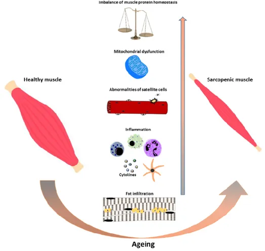
Figure 1. Schematic representation of cellular processes involved in the onset of sarcopenia during
ageing. During ageing multifactorial events such as protein synthesis/degradation imbalance, satellite cell number/activity impairment, chronic inflammation, mitochondrial dysfunction, and fat infiltration increase contributing to the onset of sarcopenia.
2. Nutrition-Dependent microRNA Regulation of Skeletal Muscle Regeneration
Although adult skeletal muscle is composed of fully differentiated fibers, it retains the capacity
to regenerate in response to injury. Muscle regeneration is a highly coordinated process that leads to
a morpho-functional recovery of injured tissue through the activation and differentiation of muscle
stem cells, maturation of newly formed muscle fibers, and remodeling of extracellular matrix [37,38].
The decrease of skeletal muscle regenerative capacity has been observed in both human and mice
sarcopenic muscle, and it seems to be the primary consequence of satellite cells ageing [39,40]. The
severe alteration in the functionality of satellite cells in senescent muscle can be caused by either
extrinsic factors or intrinsic events, including defects in self-renewing mechanisms, exhaustion by
forced differentiation, as well as apoptosis and alteration of muscle environment [41,42].
An elegant study by Conboy et al. demonstrated the rejuvenation of aged progenitor cells by
exposure to a young systemic environment, supporting the heterochronic transplantation
experiments, in which satellite cells, in aged mice that had been paired with young mice, showed
marked improvements in functionality [43]. Similarly, specific nutrients may also promote a more
rejuvenating systemic milieu enhancing satellite cell function and favoring healthy aging both in in
vivo and in vitro experimental models [44]. Likewise, satellite cells in young mice that had been
paired with old mice showed a decline in functionality [43,45,46]. These data suggest that there is a
strong contribution of the environment to the satellite cell ageing phenotype, including the
dysregulation of signals from either the myofibers or the circulatory system. Nevertheless, additional
experimental evidence revealed that ageing induces intrinsic alterations in muscle stem-cell
regenerative functions, which cannot be rejuvenated by a young host environment [39]. This is due
to the modulation of the transcriptional and epigenetic network that regulates distinct fates of
stem-cell progeny during ageing.
Among other factors, miRNAs play an important role in the modulation of stem cell function
and activity, muscle homeostasis, and have been involved in different neuromuscular diseases. In
particular, Chuang et al. elegantly demonstrated that the ablation of miRNAs in satellite cells leads
to a reduced number of these cells, mild atrophy with ageing, and an impaired regenerative capability
of muscle fibers upon injury [47]. Recently, several studies demonstrated that 1, 206,
miR-133, miR-188, and miR-27 are potential regulators of the muscle regeneration process. In particular,
miR-1, miR-133, and miR-206 are induced upon satellite cell commitment and differentiation, and
their increased expression promotes the differentiation of these cells [48,49]. Besides, the local
injection of a cocktail of miRNAs, including miR-1, miR-133, and miR-206, in rat skeletal muscle
injury model, enhanced regeneration, and prevented fibrosis [50]. Moreover, in regenerating muscle,
miR-27 plays a crucial role in downregulating Pax3 expression in order to stimulate myogenesis,
while its inhibition in injured muscle delays muscle regeneration [51].
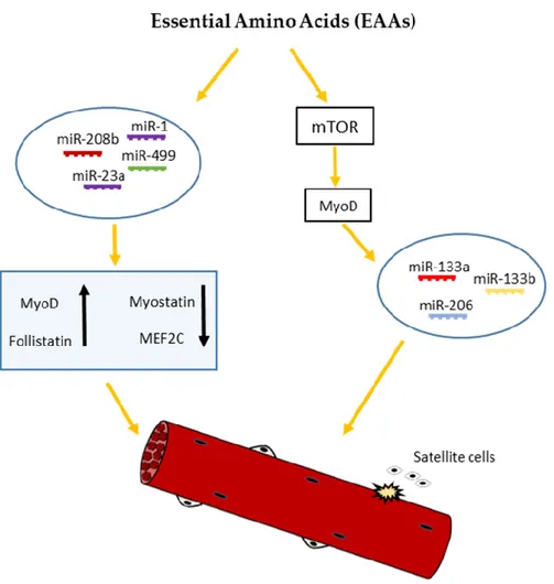
It has been well established that several nutrients such as amino acids and glucose may alter the
expression of miRNAs [12,52–55]. Leucine has been shown to induce the proliferation of satellite cells
and to increase the size and strength of regenerating fibers [56,57]. Moreover, Drummond et al. (2009)
showed that acute essential amino acids (EAAs) ingestion elicited robust increases in 1,
miR-23a, miR-208b, and miR-499 expression, with an accompanying increase in MyoD1 and Follistatin
Like 1 mRNA expression, and a decrease in myostatin and MEF2C mRNA expression in human
skeletal muscle [52]. It has been also reported by Iannone et al. [58] that miR-133a/b and miR-206
appear to be directly or indirectly regulated by the mammalian target of rapamycin (mTOR) [59], the
main mediator of cellular nutrient sensing, and a key regulator of skeletal muscle growth and
hypertrophy [60]. According to these studies, Zhang and al. [36] proposed a model for nutrient—
mTOR-myomiR signaling, where mTOR may affect the expression of miR-133a/b and miR-206,
through the regulation of MyoD transcription factor. In agreement with this model, under low
nutrient conditions such as amino acid and glucose starvation, mTOR is inactive and, consequently,
unable to induce MyoD resulting in the downregulation of miR-133a/b and miR206 (Figure 2).
Although further experiments are needed to elucidate the molecular mechanisms that regulate
the effect of specific nutritional compounds on miRNAs expression in skeletal muscle regeneration,
these findings demonstrate a significant miRNAs response to essential amino acid supplementation,
and suggest a key role for these molecules in regulating the homeostasis of muscle tissue.
Figure 2. Schematic representation of nutrient-dependent-miRNA signaling in skeletal myogenesis.
Nutrients such as essential amino acids (EAAs) may affect the expression of miR-133a/b and miR-206 through mTOR-dependent regulation of MyoD mRNA levels. On the other hand, EAAs may elicit robust increases in miR-1, miR-23a, miR-208b, and miR-499 expression, with an accompanying increase in MyoD and follistatin mRNA and decrease in myostatin and MEF2C mRNA expression, regulating skeletal muscle growth and differentiation.
3. Nutrition-Dependent microRNA Regulation of Inflammageing
Ageing is associated with a chronic low-grade inflammatory state known as “inflammageing”,
characterized by a 2- to 3-fold elevation in circulating inflammatory mediators [61]. Pro-inflammatory
cytokines, such as TNF, IL-6, and C-reactive protein (CRP), are key components in this chronic
inflammatory condition. Recently, experimental evidence demonstrates that the above mentioned
pro-inflammatory cytokines significantly increase ageing in skeletal muscle cells, and play a key role
in the complex network that connects inflammatory signals with ageing-related disability and
mortality [62–64].
In particular, elevated serum levels of IL-6 and TNF are markers of functional frailty and
predictors of poor prognosis in the elderly [65], and increased levels of cellular IL-6 production are a
significant predictor of sarcopenia [66]. Besides, elevated levels of CRP predict mortality and
functional decline in older subjects [67]. Notably, the chronic inflammatory ageing process depends
not only on the increased concentration of pro-inflammatory cytokines, but also on a reduction in the
levels of anti-inflammatory cytokines [68].
Although the molecular signaling involved in the interaction between inflammageing and
muscle loss is not yet completely understood, recent in vivo studies demonstrate that the increased
low-grade inflammation may result in the activation of catabolic pathways favoring protein
breakdown and inhibiting protein synthesis, ultimately leading to age-related muscle wasting [69–
72].
In muscle, pro-inflammatory cytokines such as TNF regulate sarcopenia through the activation
of the nuclear factor kappa B (NF-κB) transcription factor which, in turn, may activate the
ubiquitin-proteasome system [73]. NF-kB is maintained in the inactive state by the binding with a family of
inhibitory proteins called IκB. The increase in the TNF level induces activation of an IκB kinase (IKK)
complex that phosphorylates IκB, which, in turn, leads to its degradation mediated by the
proteasome system. This degradation of IκB allows for NF-κB to translocate to the nucleus and to
activate the transcription of several κB-dependent genes [74]. In particular, under conditions of
chronic inflammation, high levels of NF-κB expression activate the ubiquitin-proteasome system
which involves an enzymatic cascade that begins with the ubiquitination of protein substrates and
terminates with the hydrolysis of targeted protein to small peptides or amino acids, resulting in
protein degradation and muscle wasting [71,75].
As mentioned previously, specific miRNAs, named myomiRNAs, are known to be associated
with the skeletal muscle [27,29,76], where they play a crucial role, by targeting genes involved in
different processes such as development, differentiation, and regeneration [77]. Experimental
evidence demonstrated that the expression of these myomiRNAs can be dysregulated during ageing
and contribute to the resistance of older muscles to anabolic stimuli [78]. It has been reported that the
cytokine named TNF-weak-inducer of apoptosis can induce muscle wasting through the regulation
of miRNAs including miRNA-1, miRNA-133a, and miRNA133b, involved in the growth of mouse
skeletal muscle [79]. Besides, a down-regulation of miRNA-133b and miRNA-206 was observed by
Georgantas et al. in the muscle of patients with inflammatory myopathy [80], and the expression of
these miRNAs has been correlated by Iannone et al. with the nutritional status, revealing a mediating
effect of nutrition on the relationship between sarcopenia and myomiRNAs [58].
In addition to the myomiRNAs, other miRNAs are critical regulators for both pro-inflammatory
cytokines and skeletal muscle function [81,82]. For instance, miRNA-155 significantly increases upon
muscle injury and in mdx mice, the mouse model of Duchenne muscular dystrophy. By a genetic
approach, M. Nie et al. demonstrated that the ablation of miRNA-155 expression severely
compromised skeletal muscle regeneration, largely owing to aberrant macrophage activation and
disrupted balance between the expression of pro- and anti-inflammatory cytokines [83].
A recent RNA sequencing study performed by Mercken et al. (2013) revealed the differential
expression of miRNAs in the skeletal muscles of old and young rhesus monkeys [84]. Besides, Xie et
al. (2013) observed that the expression of miR-181a was downregulated in old muscle and its
reduction resulted in an increased expression of the cytokines TNF, IL-6, IL-1b in skeletal muscle
during the ageing process [85].
It has been well established that malnutrition and sarcopenia are closely correlated with
inflammation [9]. Nutrients such as glucose and amino acids can modulate the expression of miRNAs
[52–55], and caloric restriction can revert the level of miR-181a, suggesting a significant role of
nutrition in the modulation of the inflammatory pathway.
Importantly, significant positive or negative correlations were found between miR-133b and
miR-206 levels and albumin and ferritin, respectively [80], where decreased albumin and elevated
ferritin levels are characteristic features of inflammation, besides being markers of nutritional status
[86–88].
Although so far there is only a restricted number of studies regarding the molecular mechanisms
involved in the interconnection between miRNAs, nutrition, and sarcopenia, it is reasonable to
assume that dietary interventions may represent an efficient strategy to help prevent or counteract
the loss of muscle mass and functionality that occurs in ageing.
4. Nutrition-Dependent microRNA Regulation of Mitochondrial Dysfunction
4.1. Autophagy
Autophagy is a highly evolutionarily conserved catabolic process through which misfolded
proteins and dysfunctional organelles are degraded and recycled by autophagosomes that are then
delivered to the lysosomal machinery to prevent waste accumulation [89,90]. While the basal level of
autophagy is essential for the physiological turnover of old or damaged organelles, the dysregulation
of autophagy signaling may cause cellular stress and death as a result of cellular atrophy or,
alternatively, of apoptotic program induction [91].
Several findings indicate that autophagy becomes progressively dysfunctional during ageing,
and this effect seems to be related to the accumulation of damaged cellular components such as
defective mitochondria, which in turn may induce increased levels of reactive oxygen species (ROS)
and trigger apoptotic events [92,93]. In particular, it has been demonstrated that in aged muscles,
both excessive and defective autophagy may result in the onset of sarcopenia [94]. Indeed, the
deficiency of basal autophagy can result in the abnormal aggregation of misfolded proteins, while
excessive autophagy can also cause cellular stress and induce the loss of skeletal muscle mass due to
increased protein degradation [95].
One of the most important proteins involved in the regulation of skeletal muscle autophagy is
mTOR, a highly conserved serine/threonine kinase required for numerous aspects of cellular
homeostasis [96]. MTOR phosphorylates several transcription factors involved in the autophagy
process, thereby preventing their translocation to the nucleus [97]. An example is represented by the
helix-loop-helix transcription factor TFEB, a member of the MITF (microphthalmia-associated
transcription factor) family [98–103], that has been demonstrated to have a role in all the stages of
autophagy process, from lysosomal biogenesis to autophagosome formation [102]. Under
nutrition-rich conditions, mTOR phosphorylates TFEB that consequently is retained in the cytosol and is unable
to stimulate autophagy gene expression [98,100,101]. Conversely, in response to nutrient deprivation,
TEFB translocates to the nucleus to activate transcriptional targets leading to autophagy stimulation
[104]. As reported by Lapierre LR et al., there is a TFEB homolog in C. elegans, named HLH-30, that
plays a role similar to TFEB in the modulation of autophagy process [105]. HLH-30 translocates to
the nucleus as a response of mTOR inhibition or nutrient deprivation, and it can regulate several
genes involved in the autophagy process, supporting the concept that increased autophagic flux is
likely critical to ensure a long lifespan [105,106]. Since mTOR-dependent regulation of TFEB activity
is an evolutionarily conserved mechanism of the autophagic flux, there is an attempt to speculate that
this process could provide a vital source of metabolites during periods of nutrient deprivation.
Another family of transcriptional factors involved in the regulation of the autophagy process,
with a conserved role in ageing, is the Forkhead transcription factors (FoxO), which play a crucial
role in the activation of the ubiquitin-proteasome system, but they are also involved in the activation
of the autophagic/lysosomal pathway [107]. In particular, it has been demonstrated that several
nutrient-signaling pathways can modulate FoxO activity [108]. Indeed, reduced INS-IGF1 signaling
activates FoxO-dependent expression of genes involved in autophagy and proteostasis in several
species [109,110] and extends longevity [108]. In 2015, Brown et al. demonstrated that FoxO3 might
be post-transcriptionally regulated by miR-182, with a consequent modulation of genes involved in
the autophagy/lysosome system. Moreover, they showed a critical role for miR-182 in the control of
fuel usage and glucose homeostasis in skeletal muscle [111].
Among miRNAs involved in the regulation of the autophagy process in different species,
miR-34 is up-regulated during ageing and may contribute to ageing process, by directly modulating the
expression of autophagy-related proteins [112–114]. Recently, Yan Li et al. reported that miR-378
promotes autophagy through targeting PDK1, which is crucial in the activation of the PI3K/Akt
signaling, but it also inhibits mitochondria-mediated intrinsic apoptosis by targeting Caspase 9. Since
miR-378 is highly expressed in skeletal muscle, it is possible to speculate that failure to maintain the
high levels of miR-378 in skeletal muscle would lead to increased vulnerability to cell death observed
in muscle dystrophy or in sarcopenia. Notably, under metabolic stress conditions such as nutrient
deprivation, miR-378 dramatically increases, suggesting its significant role in the cellular adaptation
to dwindling nutrient resources [115].
4.2. ROS Imbalance
Mitochondria are important cellular organelles, with key regulatory functions in energy
production, reactive oxygen species (ROS) balance, and in the control of cell death [116,117].
Mitochondrial function may be affected by cumulative damage to mitochondrial DNA, which occurs
during ageing. The damaged mitochondrial DNA leads to an impairment of key electron transport
enzymes and subsequent ROS generation, thus causing a decrease in energy production [118].
Although adequate levels of ROS play an important role in the maintenance of tissue homeostasis
[119], age-related ROS overproduction has been proposed as one of the major contributors of the
skeletal muscle decline that occurs with ageing [120,121]. Indeed, it not only generates oxidative
damage of muscle, but it is also involved in regulating intracellular signal transduction pathways
that play, directly or indirectly, a role in the impairment of skeletal muscle strength and functionality
[62,72,122–124]. The opposite effects exerted by different concentrations of ROS can be justified
considering the concept of hormesis, which is a process characterized by a biphasic response to
environmental agent with a low-dose stimulation and high-dose inhibition [125]. Thus, skeletal
muscle benefits from low doses of free radicals, whereas excessive free radicals concentration can
impair its functions. Hence, efficient mechanisms of antioxidant defense have to be developed,
especially in those tissues like skeletal muscle highly exposed to the oxidation process.
Antioxidants are present in different forms; some of them include enzymes such as superoxide
dismutase (SOD), catalase (CAT), and glutathione peroxidase (GSH-Px), which converts free radicals
into nontoxic forms, and others, represented by vitamins, carotenoids, and polyphenols, are
introduced by the diet [10,126,127].
Vitamin C is a water-soluble antioxidant introduced in humans by dietary intake. Elevated levels
of vitamin C are associated with a lower risk of hypertension, heart disease, and stroke [128]. This
vitamin also promotes the regeneration of fat-soluble vitamin E in the cell membrane [129]. A
protective effect of vitamin C supplementation against exercise-induced muscle damage was
demonstrated by Jakeman and Maxwell. They also reported that vitamin E exerts antioxidant
properties by scavenging ROS and boosting cellular anti-oxidative capacity to reduce oxidative
damage [130]. Similarly, vitamin C and E supplementation has been shown to reduce muscle damage
by Shafat and colleagues [131]. In a mouse model of muscle atrophy, the MLC/SOD1 G93A mice,
characterized by progressive muscle atrophy associated with a significant reduction of muscle
strength, alteration in the contractile apparatus, and mitochondrial dysfunction, the treatment with
a derivate of vitamin E significantly reduced the toxic effect of ROS, partially rescuing muscle
phenotype and muscle performance [132]. Moreover, a mixture of antioxidants, including vitamin E,
vitamin A, zinc, and selenium has been shown to increase the anabolic response of all the muscles to
leucine and the leucine-induced inhibition of protein degradation in rats [133].
Recently, particular attention has been paid to the polyphenols. These molecules, which are
produced as secondary metabolites by the plants for protection against bacteria, fungi, and insects,
display remarkable antioxidant properties [134]. Experimental studies performed in animal models
showed that the dietary administration of polyphenols, such as resveratrol, in combination with
treadmill exercise, exert beneficial effects which improve mitochondrial function, and reduce
age-related decline in physical performance [135]. Similarly, the supplementation of another polyphenol
represented by curcumin ameliorates exercise performance in rats [136]. Moreover, the Geny group
demonstrated that intake of polyphenols starting at a young age restored muscle maximal
mitochondrial oxidative capacity, normalized production of ROS, and enhanced antioxidant defense,
therefore protecting aged muscle [137].
However, controversial data have been published regarding the relationship between
antioxidant supplementation and muscle performance. In fact, human trials did not confirm the
positive results obtained in animals. It has been shown that undesirable effects, such as the disruption
of the endogenous antioxidant levels, may result from prolonged antioxidant supplementation, thus
failing to counteract exercise-induced oxidative stress, and interfering with muscle adaptation to
exercise [138–140]. Moreover, the long-term administration of vitamin C has been observed to
prevent mitochondrial biogenesis, decreasing the expression of endogenous antioxidant enzymes
[141].
Several reasons can be responsible for these contradictory results. In particular, it has been
demonstrated that ROS are required for cellular adaptation to exercise and for the insulin-sensitizing
capabilities of physical exercise in healthy humans. Besides, the health-promoting effects of physical
exercise are abrogated by antioxidants such as vitamin C and E, and polyphenols. A potentially
health-promoting process may be derived from transiently increased levels of oxidative stress,
whereas an uncontrolled accumulation of oxidative stress may have pathological implications.
Recent studies revealed that the direct antioxidant properties of polyphenols are not the major
mechanism of their action [142,143]. In fact, there is a poor bioavailability and very low concentrations
of active polyphenols in target tissues. It seems likely that the antioxidant effects of polyphenols are
mediated via the activation of various transcription factors, signaling pathways, and vitagenes.
Vitagenes encode components of the heat shock protein (HSP), thioredoxin, and sirtuin protein
systems, that show antioxidant and antiapoptotic activities [144–148]. In particular, the effects of
polyphenols in the vitagene network can be demonstrated using silymarin (SM), a plant extract
containing polyphenols. In fact, as reported by Surai et al., SM was shown to improve antioxidant
defenses by upregulating heme oxygenase-1 (HO-1). In addition, SM consumption has been shown
to be associated with decreased HSP70 expression in stressed cells, which indicates an improvement
in anti-oxidant defenses. Finally, SM-related activation or the prevention of inhibition of sirtuins in
stress conditions might be an essential adaptive mechanism responsible for maintaining the
redox-regulated homeostasis in the cell and the whole body [149]. SIRT1 has been identified as a link
between caloric restriction and longevity, and its overexpression is linked to increased lifespans for
several organism models [150]. SIRT1 activation inhibits NF-κB signaling and increases oxidative
metabolism, favoring the resolution of inflammation. SIRT1 exerts this effect directly by deacetylating
the p65 subunit of NF-κB complex. SIRT1 activates AMPK, PPARα, and PGC-1α stimulating
oxidative energy production; these factors inhibit NF-κB signaling and suppress inflammation. On
the other hand, the expression of miR-34a, IFNγ, and ROS, induced by NF-κB signaling,
down-regulates SIRT1 activity. The inhibition of SIRT1 disrupts oxidative energy metabolism and
stimulates the NF-κB-induced inflammatory responses present in many chronic metabolic and
age-related diseases [151].
Several miRNAs play a crucial role in the regulation of mitochondrial gene expression. For
example, miR-1, a microRNA specifically induced during myogenesis, efficiently enters the
mitochondria, where it stimulates the translation of specific mitochondrial genome-encoded
transcripts. Moreover, miR-696 negatively affects fatty acid oxidation and mitochondrial function by
targeting the transcription factor peroxisome proliferator-activated receptor γ coactivator 1α
(PGC-1α), a master regulator of mitochondrial biogenesis and ROS removal [152].
In a recent study of the Nie group, it has been demonstrated that the deficiency of miR-133a in
mice leads to low levels of PGC-1α and nuclear respiratory factor-1(Nrf1), and lower mitochondrial
mass and exercise tolerance [83]. Since this phenotype is similar to the sarcopenia phenotype, the
authors speculate that miR-133a might have a significant role in maintaining skeletal muscle
mitochondrial functionality. Other miRNAs, such as miR-340-5p and miR-206, have also been shown
to regulate ROS generation in skeletal muscle via Nrf2, which is a key factor in regulating redox
homeostasis, although the molecular mechanisms involved in its effect in the onset of sarcopenia are
still unknown [153,154].
Since it has been widely demonstrated that nutrients may influence the expression of
endogenous miRNAs involved in different cellular processes, the manipulation of miRNAs profiles
through dietary modifications and supplements can be proposed as a potential future therapeutic
intervention or prevention strategy against sarcopenia.
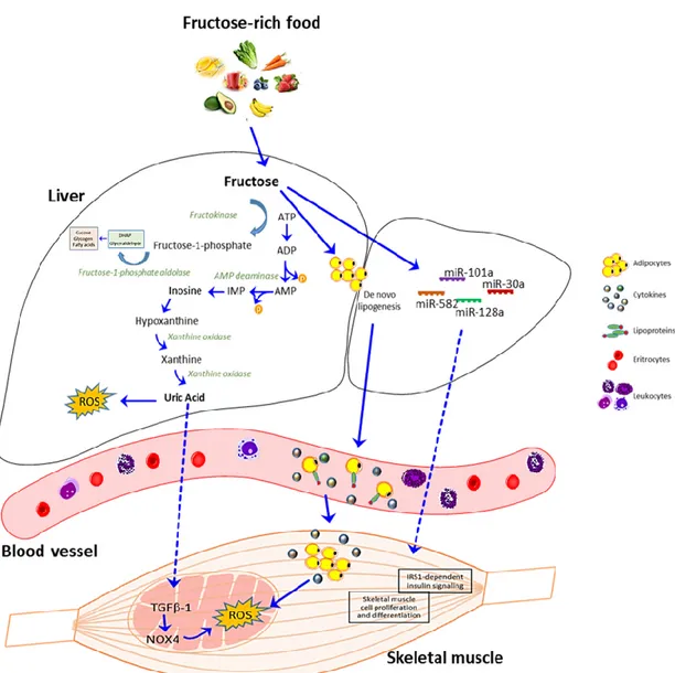
5. High Fructose Diet Modulation of miRNAs Expression in Sarcopenia
Among the nutritional factors that have been reported to play a crucial role to increase
inflammation [155], mitochondrial dysfunction and ROS production in skeletal muscle is fructose
[156–159]. Fructose is one of the major constituents of the modern diet, since it is highly expressed in
fruits and vegetables [160] and it is also used as a sweetener for food and drinks, and as an excipient
in pharmaceutical preparations, syrups, and solutions [161].
Although low doses of fructose have beneficial effects on glycemic control without increasing
cardiometabolic risk [162] and blood pressure [163], several studies demonstrated that a high level of
fructose can stimulate ROS production in the mitochondria in a variety of tissues including kidney,
liver, small intestine and skeletal muscle [164–169]. Fructose can exert these effects in different ways,
including increased blood uric acid (UA) concentration [170,171], with a consequent upregulation of
TGF-β1 expression and NOX4 activation [172] and through the induction of de novo lipogenesis [173–
176].
In recent years, a gradual increment in blood UA concentration has been demonstrated,
especially in people of Western countries, where the increased consumption of fructose has been
revealed [177,178]. Excessive fructose consumption has also been associated with hepatic steatosis,
cellular stress, and inflammation [179]. This is responsible for the release by the liver of lipids,
methyglyoxal, UA, and hepatokines leading to alterations in the communication between the liver
and the gut, muscles, and adipose tissue. Fructose and muscle/liver axis has been reported in several
studies that showed how a high-fructose diet is associated with modifications in muscle function
[180] in humans [181] and rodents [182]. In particular, mechanisms involved in diet-induced
sarcopenia may be (i) a decrease in the mechanistic target of rapamycine complex (mTORC) 1 activity
and thereafter in protein synthesis; and (ii) inflammation. Moreover, recent studies in fructose-fed
rats have shown an association between nonalcoholic-fatty liver disease and sarcopenia [183]. This is
a key factor involved in disease progression to NASH (nonalcoholic steatohepatitis), as the muscle
heavily contributes to energy homeostasis [184].
In the inter-organs crosstalk caused by excessive fructose intake, it is absorbed primarily in the
gut, and then metabolized in the liver, where it stimulates UA production [185,186]. The increased
levels of intracellular UA are followed by an acute rise in circulating levels of UA, which is likely due
to its release from the liver [170,171]. Besides, fructose may stimulate UA synthesis from amino acid
precursors such as glycine [187], and it has been reported that long-term fructose administration
suppresses the renal excretion of UA, resulting in elevated serum UA levels [188]. Interestingly,
Kaneko and colleagues found that a single administration of fructose affects the excretion of UA to
the intestinal lumen, inducing the reactive oxygen species (ROS)-derived production of dinucleotide
phosphate (NADPH) oxidase activation [189].
Besides, experimental evidence shows that fructose-dependent UA production stimulates the
upregulation of TGFβ-1, leading to NOX4 activation and ROS generation in mitochondria in skeletal
muscle [172,190] (Figure 3).
As mentioned above, fructose consumption increases de novo lipogenesis in the liver, that is
accompanied by an increased release of lipids in the bloodstream, which are then uptaken by
different tissues, such as skeletal muscle [173–176]. In skeletal muscle, intracellular lipids
accumulation increases the production of ROS and reactive nitrogen species [191–193]. Furthermore,
the excessive production of lipoproteins induces an inflammatory response and, consequently, an
elevation in circulating fatty acids and inflammatory cytokines, that may cause insulin resistance in
peripheral tissues, leading to whole-body insulin resistance [194–196] (Figure 3). In a recent paper,
Tamrakar group reported that, in myogenic cells, the fructose-dependent ROS production results in
the activation of the stress/inflammation markers c-Jun N terminal kinase (JNK) and extracellular
signal-regulated kinase 1/2 (ERK1/2), and the degradation of inhibitor of NFκB (IκBα), leading to
impaired insulin signaling and attenuated glucose utilization in skeletal muscle cells [156].
Although the role of fructose in causing energy alterations and metabolic disorders has been
well documented, the molecular mechanisms that regulate these effects have not yet been elucidated.
In recent years, a growing interest has been directed to the role of miRNAs, since they are known to
be dysregulated in several metabolic disorders and sarcopenia, and can be controlled by dietary
factors [197,198]. A study of Su group demonstrated that a set of miRNAs are altered by high fructose
diet; among them, miRNA-101a, miRNA-30a, and miRNA-582 have been reported to be involved in
other cellular processes than energy metabolic signaling [199]. For example, fructose induces the
expression of miRNA-101 involved in skeletal muscle cell proliferation and differentiation [200], and
of miR-30a, which belongs to a miRNAs family, that promotes skeletal muscle differentiation. Both
miRNAs are down-regulated in in vivo models of muscle injury and muscle disuse atrophy [201].
Besides, a high fructose diet may regulate a set of miRNAs involved in the hepatic insulin
signaling. Among them, miR-128a can regulate insulin receptor substrate 1 (IRS1), ultimately
affecting glucose and lipid metabolism [202]. Interestingly, the modulation of IRS1 by miR-128a has
been reported in skeletal muscle, where it regulates myoblast proliferation and myotube hypertrophy
and provides a novel mechanism, through which IRS1-dependent insulin signaling is regulated in
skeletal muscle [199].
In summary, these data demonstrate that a high fructose diet can induce metabolic dysfunctions
and modulate several processes, including oxidative stress and inflammation, that are also
characteristic of sarcopenic muscles. In this contest, a crucial role is played by miRNAs, that can be
altered by a high fructose diet, providing novel insights to counteract the physio-pathological effect
of aging in different tissues (Figure 3).
Figure 3. Schematic representation of fructose metabolism and uric acid effect on skeletal muscle. In
the liver, fructose is phosphorylated into fructose 1-phosphate by fructokinase in a reaction that decreases the levels of intracellular phosphate and ATP. Subsequently, the enzyme
fructose-1-phosphate aldolase gives rise to dihydroxyacetone fructose-1-phosphate (DHAP) and glyceraldehyde. When fructose 1-phosphate accumulates, intracellular phosphate decreases, stimulating AMP deaminase, which catalyzes the degradation of AMP to inosine monophosphate (IMP). IMP is metabolized to inosine, which is further degraded to xanthine and hypoxanthine by xanthine oxidase, ultimately generating uric acid (UA). UA can induce ROS production in the liver and other tissues, such as skeletal muscle via TGFβ-1-NOX4 signaling. Alternatively, fructose can stimulate de novo lipogenesis in the liver with increased release of lipids and lipoproteins in the bloodstream that are then uptaken by different tissues including skeletal muscle, with consequent cytokines and ROS production. Besides, high levels of fructose can modulate the expression of miRNAs that may affect skeletal muscle cell proliferation, differentiation, and insulin signaling.
6. Circulating miRNAs
Circulating miRNAs (c-miRNAs) represent a category of non-coding RNAs detectable in
different bio-fluids, such as saliva, breast milk, urine, plasma, and serum [203,204]. Several
mechanisms have been demonstrated for c-miRNAs packing and secretion, avoiding their
degradation by serum ribonuclease. These include exosomes [205,206], high-density lipoprotein
[207], RNA-binding proteins [208,209], and apoptotic bodies [210]. C-miRNAs can actively participate
in cell-cell communication in different organs and tissues and, since it has been reported that their
expression can be altered in pathological conditions and ageing, they have been suggested as
potential biomarkers for the diagnosis and treatment of several diseases [211–216].
In particular, several papers in the last years have demonstrated a differential expression of
c-miRNAs in sarcopenic compared to non-sarcopenic patients [58,217]. Besides, a correlation between
nutrition, c-miRNAs, and sarcopenia has been recently revealed [58,218], highlighting a new role for
the c-miRNAs as potential noninvasive biomarkers for the diagnosis of sarcopenia and the
involvement of nutrition in this contest.
A number of studies have shown that miRNAs can be derived not exclusively from endogenous
synthesis, but might also be obtained from dietary sources such as plants and animal origin food.
These miRNAs are known as xeno-miRNAs [219–222]. Recently, it has been revealed by Zhang et al.
that an exogenous plant-derived miRNA, miR168a, is one of the most highly enriched exogenous
plant miRNA found in the serum of Chinese subjects. By using both in vivo and in vitro experimental
models, they demonstrated that miR168a, packaged into microvesicles (MVs), can pass through the
mouse gastrointestinal tract and might be released in the circulatory system, decreasing the plasma
level of low-density lipoprotein [219]. In the same years, further research has shown that about 100
miRNAs are present in bovine milk that are resistant and stable to both industrial procedures and
harsh conditions (low pH and RNase treatment). These miRNAs, encapsulated into MVs, can be
diffused among animal species by dietary means, and are able to regulate a variety of metabolic
pathways in humans and rats [223,224].
These data suggest that xeno-miRNAs may contribute to the circulating miRNAs population
and, thanks to their effect in the modulation of target gene expression and the maintenance of tissue
homeostasis, can represent novel biomarkers of age-related muscle mass and functionality.
7. Conclusions
Since sarcopenia dramatically affects the quality of life of older adults, therapeutic strategies are
needed to prevent and/or counteract the progressive age-related reduction in skeletal muscle mass
and functionality. Accumulating evidence suggests that nutrients such as amino acids, vitamins, and
antioxidants represent key tools to elicit anabolic signaling and protein turnover, favoring the
maintenance of muscle function. Dietary compounds have been shown to influence miRNAs levels
in skeletal muscle and, given the importance of miRNAs as crucial regulators of skeletal muscle mass,
composition and function, they may represent diagnostic or prognostic biomarkers of age-related
muscle dysfunctions (Table 1).
Table 1. Dietary compounds that have been shown to influence miRNAs levels in skeletal muscle. Nutraceuticals Positive MyomiRs modulation Negative MyomiRs modulation
Final effect References
EAAs
miR-1, miR-23a, miR-208b, miR-499,
miR-27a
Skeletal muscle regeneration,
proliferation and differentiation [52,55]
Serum iron miR-133b Skeletal muscle differentiation
[58]
Albumin miR-133b, miR-206 Skeletal muscle regeneration and
differentiation [58]
Ferritin 133b,
miR-206
Downregulation of skeletal muscle regeneration and
differentiation [58] Insulin 1, miR-133a, miR-206, 29a, miR-29c Downregulation of skeletal muscle regeneration, differentiation and insulin
resistance
[225]
Resveratrol miR-21, miR-27b 133b, miR-20b, miR-149
Modulation of skeletal muscle
differentiation [198,226,227]
Palmitic Acid miR-29a Insulin resistance and diabetes
[228]
Vitamin D miR-26a Skeletal muscle regeneration and
differentiation [229]
Fructose miR-101, miR-30a miR-128a
Skeletal muscle proliferation and differentiation, insulin signaling,
insulin resistance [199]
Author Contributions: B.M.S.: literature research and interpretation, conceptualization, writing original draft,
and editing. A.B.: writing, literature research, G.S.: validation and critical review. L.P.: critical review and editing. All authors have read and agreed to the published version of the manuscript.
Funding: This work was supported by: Progetto di ricerca di interesse di Ateneo—Linea D.3.2—Anno 2015,
Università Cattolica del Sacro Cuore to BMS.
Conflicts of Interest: The authors declare no conflict of interest.
References
1. Cruz-Jentoft, A.J.; Bahat, G.; Bauer, J.; Boirie, Y.; Bruyère, O.; Cederholm, T.; Cooper, C.; Landi, F.; Rolland, Y.; Sayer, A.A.; et al. Sarcopenia: Revised European consensus on definition and diagnosis. Age Ageing
2019, 48, 16–31.
2. Carmeli, E.; Reznick, A.Z.; Coleman, R.; Carmeli, V. Muscle strength and mass of lower extremities in relation to functional abilities in elderly adults. Gerontology 2000, 46, 249–257.
3. Rolland, Y.; Czerwinski, S.; Van Kan, G.A.; Morley, J.E.; Cesari, M.; Onder, G.; Woo, J.; Baumgartner, R.; Pillard, F.; Boirie, Y.; et al. Sarcopenia: Its assessment, etiology, pathogenesis, consequences and future perspectives. J. Nutr. Heal. Aging 2008, 12, 433–450.
4. Ryall, J.G.; Schertzer, J.D.; Lynch, G.S. Cellular and molecular mechanisms underlying age-related skeletal muscle wasting and weakness. Biogerontology 2008, 9, 213–228.
5. Metter, E.J.; Talbot, L.A.; Schrager, M.; Conwit, R. Skeletal muscle strength as a predictor of all-cause mortality in healthy men. Journals Gerontol. - Ser. A Biol. Sci. Med. Sci. 2002, 57, B359–B365.
6. Marzetti, E.; Calvani, R.; Cesari, M.; Buford, T.W.; Lorenzi, M.; Behnke, B.J.; Leeuwenburgh, C. Mitochondrial dysfunction and sarcopenia of aging: From signaling pathways to clinical trials. Int. J.
Biochem. Cell Biol. 2013, 45, 2288–2301.
7. Marzetti, E.; Hwang, J.C.Y.; Lees, H.A.; Wohlgemuth, S.E.; Dupont-Versteegden, E.E.; Carter, C.S.; Bernabei, R.; Leeuwenburgh, C. Mitochondrial death effectors: Relevance to sarcopenia and disuse muscle atrophy. Biochim. Biophys. Acta - Gen. Subj. 2010, 1800, 235–244.
8. Alway, S.E.; Mohamed, J.S.; Myers, M.J. Mitochondria Initiate and Regulate Sarcopenia. Exerc. Sport Sci.
Rev. 2017, 45, 58–69.
9. Dalle, S.; Rossmeislova, L.; Koppo, K. The role of inflammation in age-related sarcopenia. Front. Physiol.
2017, 8.
10. Meng, S.J.; Yu, L.J. Oxidative stress, molecular inflammation and sarcopenia. Int. J. Mol. Sci. 2010, 11, 1509– 1526.
11. Churchward-Venne, T.A.; Breen, L.; Phillips, S.M. Alterations in human muscle protein metabolism with aging: Protein and exercise as countermeasures to offset sarcopenia. BioFactors 2014, 40, 199–205.
12. Scicchitano, B.M.; Sica, G. The Beneficial Effects of Taurine to Counteract Sarcopenia. Curr. Protein Pept. Sci.
2018, 19, 673–680.
13. Short, K.R.; Nair, K.S. Muscle protein metabolism and the sarcopenia of aging. I Int J Sport Nutr Exerc Metab.
2001 Dec;11 Suppl:S119-27.
14. Welle, S.; Thornton, C.; Jozefowicz, R.; Statt, M. Myofibrillar protein synthesis in young and old men. Am.
J. Physiol. 1993, 264, E693-8.
15. Morton, R.W.; Traylor, D.A.; Weijs, P.J.M.; Phillips, S.M. Defining anabolic resistance: Implications for delivery of clinical care nutrition. Curr. Opin. Crit. Care 2018, 24, 124–130.
16. Haran, P.H.; Rivas, D.A.; Fielding, R.A. Role and potential mechanisms of anabolic resistance in sarcopenia.
J. Cachexia. Sarcopenia Muscle 2012, 3, 157–162.
17. Dillon, E.L.; Durham, W.J.; Urban, R.J.; Sheffield-Moore, M. Hormone treatment and muscle anabolism during aging: Androgens. Clin. Nutr. 2010, 29, 697–700.
18. Basualto-Alarcón, C.; Varela, D.; Duran, J.; Maass, R.; Estrada, M. Sarcopenia and androgens: A link between pathology and treatment. Front. Endocrinol. (Lausanne). 2014, 5.
19. Moore, D.R. Keeping Older Muscle “Young” through Dietary Protein and Physical Activity. Adv. Nutr.
2014, 5, 599S–607S.
20. Walker, D.K.; Dickinson, J.M.; Timmerman, K.L.; Drummond, M.J.; Reidy, P.T.; Fry, C.S.; Gundermann, D.M.; Rasmussen, B.B. Exercise, amino acids, and aging in the control of human muscle protein synthesis.
Med Sci Sports Exerc. 2011 Dec;43(12):2249-58.
21. Nieuwenhuizen, W.F.; Weenen, H.; Rigby, P.; Hetherington, M.M. Older adults and patients in need of nutritional support: Review of current treatment options and factors influencing nutritional intake. Clin.
Nutr. 2010, 29, 160–169.
22. Robinson, S.; Granic, A.; Sayer, A.A. Nutrition and muscle strength, as the key component of sarcopenia: An overview of current evidence. Nutrients 2019, 11.
23. Bernstein, E.; Kim, S.Y.; Carmell, M.A.; Murchison, E.P.; Alcorn, H.; Li, M.Z.; Mills, A.A.; Elledge, S.J.; Anderson, K. V; Hannon, G.J. Dicer is essential for mouse development. Nat. Genet. 2003, 35, 215–217. 24. Wienholds, E.; Koudijs, M.J.; van Eeden, F.J.M.; Cuppen, E.; Plasterk, R.H.A. The microRNA-producing
enzyme Dicer1 is essential for zebrafish development. Nat. Genet. 2003, 35, 217–218.
25. Winter, J.; Jung, S.; Keller, S.; Gregory, R.I.; Diederichs, S. Many roads to maturity: microRNA biogenesis pathways and their regulation. Nat. Cell Biol. 2009, 11, 228–234.
26. Ambros, V.; Lee, R.C. Identification of microRNAs and Other Tiny Noncoding RNAs by cDNA Cloning. In RNA Interference, Editing, and Modification; Humana Press: Totowa, NJ, 2004; Vol. 265, pp. 131–158. 27. Callis, T.E.; Deng, Z.; Chen, J.-F.; Wang, D.-Z. Muscling Through the microRNA World. Exp. Biol. Med.
2008, 233, 131–138.
28. McCarthy, J.J.; Esser, K.A. MicroRNA-1 and microRNA-133a expression are decreased during skeletal muscle hypertrophy. J. Appl. Physiol. 2007, 102, 306–313.
29. van Rooij, E.; Quiat, D.; Johnson, B.A.; Sutherland, L.B.; Qi, X.; Richardson, J.A.; Kelm, R.J.; Olson, E.N. A Family of microRNAs Encoded by Myosin Genes Governs Myosin Expression and Muscle Performance.
Dev. Cell 2009, 17, 662–673.
30. Zacharewicz, E.; Lamon, S.; Russell, A.P. MicroRNAs in skeletal muscle and their regulation with exercise, ageing, and disease. Front. Physiol. 2013, 4, 266.
31. Luo, W.; Nie, Q.; Zhang, X. MicroRNAs Involved in Skeletal Muscle Differentiation. J. Genet. Genomics 2013,
40, 107–116.
32. Williams, A.H.; Liu, N.; van Rooij, E.; Olson, E.N. MicroRNA control of muscle development and disease.
Curr. Opin. Cell Biol. 2009, 21, 461–469.
33. McGregor, R.A.; Poppitt, S.D.; Cameron-Smith, D. Role of microRNAs in the age-related changes in skeletal muscle and diet or exercise interventions to promote healthy aging in humans. Ageing Res. Rev. 2014, 17, 25–33.
34. Sharma, M.; Juvvuna, P.K.; Kukreti, H.; McFarlane, C. Mega roles of microRNAs in regulation of skeletal muscle health and disease. Front. Physiol. 2014, 5 JUN.
35. Yin, J.; Qian, Z.; Chen, Y.; Li, Y.; Zhou, X. MicroRNA regulatory networks in the pathogenesis of sarcopenia.
J. Cell. Mol. Med. 2020, 24, 4900–4912.
36. Zhang, Y.; Yu, B.; He, J.; Chen, D. From nutrient to MicroRNA: A novel insight into cell signaling involved in skeletal muscle development and disease. Int. J. Biol. Sci. 2016, 12, 1247–1261.
37. Barberi, L.; Scicchitano, B.M.; De Rossi, M.; Bigot, A.; Duguez, S.; Wielgosik, A.; Stewart, C.; McPhee, J.; Conte, M.; Narici, M.; et al. Age-dependent alteration in muscle regeneration: The critical role of tissue niche. Biogerontology 2013, 14.
38. Forcina, L.; Miano, C.; Pelosi, L.; Musarò, A. An Overview About the Biology of Skeletal Muscle Satellite Cells. Curr. Genomics 2019, 20, 24–37.
39. Sousa-Victor, P.; Gutarra, S.; García-Prat, L.; Rodriguez-Ubreva, J.; Ortet, L.; Ruiz-Bonilla, V.; Jardí, M.; Ballestar, E.; González, S.; Serrano, A.L.; et al. Geriatric muscle stem cells switch reversible quiescence into senescence. Nature 2014, 506, 316–321.
40. Zwetsloot, K.A.; Childs, T.E.; Gilpin, L.T.; Booth, F.W. Non-passaged muscle precursor cells from 32-month old rat skeletal muscle have delayed proliferation and differentiation. Cell Prolif. 2013, 46, 45–57.
41. Sousa-Victor, P.; García-Prat, L.; Serrano, A.L.; Perdiguero, E.; Muñoz-Cánoves, P. Muscle stem cell aging: Regulation and rejuvenation. Trends Endocrinol. Metab. 2015, 26, 287–296.
42. Scicchitano, B.M.; Sica, G.; Musarò, A. Stem Cells and Tissue Niche: Two Faces of the Same Coin of Muscle Regeneration. Eur. J. Transl. Myol. 2016, 26, 6125.
43. Conboy, I.M.; Conboy, M.J.; Wagers, A.J.; Girma, E.R.; Weismann, I.L.; Rando, T.A. Rejuvenation of aged progenitor cells by exposure to a young systemic environment. Nature 2005, 433, 760–764.
44. Murphy, T.; Thuret, S. The systemic milieu as a mediator of dietary influence on stem cell function during ageing. Ageing Res. Rev. 2015, 19, 53–64.
45. Brack, A.S.; Rando, T.A. Intrinsic changes and extrinsic influences of myogenic stem cell function during aging. Stem Cell Rev. 2007, 3, 226–237.
46. Villeda, S.A.; Luo, J.; Mosher, K.I.; Zou, B.; Britschgi, M.; Bieri, G.; Stan, T.M.; Fainberg, N.; Ding, Z.; Eggel, A.; et al. The ageing systemic milieu negatively regulates neurogenesis and cognitive function. Nature 2011,
477, 90–96.
47. Cheung, T.H.; Quach, N.L.; Charville, G.W.; Liu, L.; Park, L.; Edalati, A.; Yoo, B.; Hoang, P.; Rando, T.A. Maintenance of muscle stem-cell quiescence by microRNA-489. Nature 2012, 482, 524–528.
48. Chen, J.F.; Mandel, E.M.; Thomson, J.M.; Wu, Q.; Callis, T.E.; Hammond, S.M.; Conlon, F.L.; Wang, D.Z. The role of microRNA-1 and microRNA-133 in skeletal muscle proliferation and differentiation. Nat. Genet.
2006, 38, 228–233.
49. Hak, K.K.; Yong, S.L.; Sivaprasad, U.; Malhotra, A.; Dutta, A. Muscle-specific microRNA miR-206 promotes muscle differentiation. J. Cell Biol. 2006, 174, 677–687.
50. Nakasa, T.; Ishikawa, M.; Shi, M.; Shibuya, H.; Adachi, N.; Ochi, M. Acceleration of muscle regeneration by local injection of muscle-specific microRNAs in rat skeletal muscle injury model. J. Cell. Mol. Med. 2010,
14, 2495–2505.
51. Ling, Y.H.; Sui, M.H.; Zheng, Q.; Wang, K.Y.; Wu, H.; Li, W.Y.; Liu, Y.; Chu, M.X.; Fang, F.G.; Xu, L.N. MiR-27b regulates myogenic proliferation and differentiation by targeting Pax3 in goat. Sci. Rep. 2018, 8, 1–12.
52. Drummond, M.J.; Glynn, E.L.; Fry, C.S.; Dhanani, S.; Volpi, E.; Rasmussen, B.B. Essential Amino Acids Increase MicroRNA-499, -208b, and -23a and Downregulate Myostatin and Myocyte Enhancer Factor 2C mRNA Expression in Human Skeletal Muscle. J. Nutr. 2009, 139, 2279–2284.
53. Druz Aliaksandr, Betenbaugh Michael, S.J. Glucose Depletion Activates mmu-miR-466h-5p Expression Through Oxidative Stress and Inhibition of Histone Deacetylation. Nucleic Acids Res 2012.
54. Dey, N.; Das, F.; Mariappan, M.M.; Mandal, C.C.; Ghosh-Choudhury, N.; Kasinath, B.S.; Choudhury, G.G. MicroRNA-21 orchestrates high glucose-induced signals to TOR complex 1, resulting in renal cell pathology in diabetes. J. Biol. Chem. 2011, 286, 25586–25603.
55. Chen, X.; Huang, Z.; Chen, D.; Yang, T.; Liu, G. MicroRNA-27a is induced by leucine and contributes to leucine-induced proliferation promotion in C2C12 cells. Int. J. Mol. Sci. 2013, 14, 14076–14084.
56. Pereira, M.G.; Silva, M.T.; da Cunha, F.M.; Moriscot, A.S.; Aoki, M.S.; Miyabara, E.H. Leucine supplementation improves regeneration of skeletal muscles from old rats. Exp. Gerontol. 2015, 72, 269–277. 57. Rogulska, A.; Kurasz, S. Regeneration of crushed skeletal muscles in experimental animals and the effect
of leucine on the course of this process in white rat. Pol Med Sci Hist Bull. 1975 Mar-Apr;15(2):245-8. 58. Iannone, F.; Montesanto, A.; Cione, E.; Crocco, P.; Caroleo, M.C.; Dato, S.; Rose, G.; Passarino, G. Expression
patterns of muscle-specific miR-133b and miR-206 correlate with nutritional status and sarcopenia.
Nutrients 2020, 12.
59. Horak, M.; Novak, J.; Bienertova-Vasku, J. Muscle-specific microRNAs in skeletal muscle development.
Dev. Biol. 2016, 410, 1–13.
60. Ge, Y.; Chen, J. Mammalian target of rapamycin (mTOR) signaling network in skeletal myogenesis. J. Biol.
Chem. 2012, 287, 43928–43935.
61. Calçada, D.; Vianello, D.; Giampieri, E.; Sala, C.; Castellani, G.; de Graaf, A.; Kremer, B.; van Ommen, B.; Feskens, E.; Santoro, A.; et al. The role of low-grade inflammation and metabolic flexibility in aging and nutritional modulation thereof: A systems biology approach. Mech. Ageing Dev. 2014, 136–137, 138–147. 62. Forcina, L.; Miano, C.; Scicchitano, B.M.; Rizzuto, E.; Berardinelli, M.G.; De Benedetti, F.; Pelosi, L.; Musarò,
A. Increased Circulating Levels of Interleukin-6 Affect the Redox Balance in Skeletal Muscle. Oxid. Med.
Cell. Longev. 2019, 2019.
63. Visser, M.; Pahor, M.; Taaffe, D.R.; Goodpaster, B.H.; Simonsick, E.M.; Newman, A.B.; Nevitt, M.; Harris, T.B. Relationship of interleukin-6 and tumor necrosis factor-α with muscle mass and muscle strength in elderly men and women: The health ABC study. Journals Gerontol. - Ser. A Biol. Sci. Med. Sci. 2002, 57. 64. Molanouri Shamsi, M.; Hassan, Z.H.; Gharakhanlou, R.; Quinn, L.S.; Azadmanesh, K.; Baghersad, L.;
Isanejad, A.; Mahdavi, M. Expression of interleukin-15 and inflammatory cytokines in skeletal muscles of STZ-induced diabetic rats: Effect of resistance exercise training. Endocrine 2014, 46, 60–69.
65. Bruunsgaard, H.; Ladelund, S.; Pedersen, A.N.; Schroll, M.; Jørgensen, T.; Pedersen, B.K. Predicting death from tumour necrosis factor-alpha and interleukin-6 in 80-year-old people. Clin. Exp. Immunol. 2003, 132, 24–31.
66. Payette, H.; Roubenoff, R.; Jacques, P.F.; Dinarello, C.A.; Wilson, P.W.F.; Abad, L.W.; Harris, T. Insulin-like growth factor-1 and interleukin 6 predict sarcopenia in very old community-living men and women: The Framingham heart study. J. Am. Geriatr. Soc. 2003, 51, 1237–1243.
67. Reuben, D.B.; Cheh, A.I.; Harris, T.B.; Ferrucci, L.; Rowe, J.W.; Tracy, R.P.; Seeman, T.E. Peripheral blood markers of inflammation predict mortality and functional decline in high-functioning community-dwelling older persons. J. Am. Geriatr. Soc. 2002, 50, 638–644.
68. Wang, B.; Yang, G.; Liang, X.; Zhu, M.; Du, M. Grape seed extract prevents skeletal muscle wasting in interleukin 10 knockout mice. BMC Complement. Altern. Med. 2014, 14.
69. Argilés, J.M.; Busquets, S.; Stemmler, B.; López-Soriano, F.J. Cachexia and sarcopenia: Mechanisms and potential targets for intervention. Curr. Opin. Pharmacol. 2015, 22, 100–106.
70. Beyer, I.; Mets, T.; Bautmans, I. Chronic low-grade inflammation and age-related sarcopenia. Curr. Opin.
Clin. Nutr. Metab. Care 2012, 15, 12–22.
71. Scicchitano, B.M.; Faraldi, M.; Musarò, A. The Proteolytic Systems of Muscle Wasting. Recent Adv. DNA
gene Seq. 2015, 9, 26–35.
72. Scicchitano, B.M.; Dobrowolny, G.; Sica, G.; Musarò, A. Molecular Insights into Muscle Homeostasis, Atrophy and Wasting. Curr. Genomics 2018, 19, 356–369.
73. Thoma, A.; Lightfoot, A.P. Nf-kb and inflammatory cytokine signalling: Role in skeletal muscle atrophy. In Advances in Experimental Medicine and Biology; Springer New York LLC, 2018; Vol. 1088, pp. 267–279.
74. Li, H.; Malhotra, S.; Kumar, A. Nuclear factor-kappa B signaling in skeletal muscle atrophy. J. Mol. Med.
2008, 86, 1113–1126.
75. Sandri, M.; Barberi, L.; Bijlsma, A.Y.; Blaauw, B.; Dyar, K.A.; Milan, G.; Mammucari, C.; Meskers, C.G.M.; Pallafacchina, G.; Paoli, A.; et al. Signalling pathways regulating muscle mass in ageing skeletal muscle. the role of the IGF1-Akt-mTOR-FoxO pathway. Biogerontology 2013, 14, 303–323.
76. Small, E.M.; O’Rourke, J.R.; Moresi, V.; Sutherland, L.B.; McAnally, J.; Gerard, R.D.; Richardson, J.A.; Olson, E.N. Regulation of PI3-kinase/Akt signaling by muscle-enriched microRNA-486. Proc. Natl. Acad. Sci. U. S.
A. 2010, 107, 4218–4223.
77. Chen, J.F.; Callis, T.E.; Wang, D.Z. microRNAs and muscle disorders. J. Cell Sci. 2009, 122, 13–20.
78. Drummond, M.J.; McCarthy, J.J.; Fry, C.S.; Esser, K.A.; Rasmussen, B.B. Aging differentially affects human skeletal muscle microRNA expression at rest and after an anabolic stimulus of resistance exercise and essential amino acids. Am. J. Physiol. - Endocrinol. Metab. 2008, 295.
79. Panguluri, S.K.; Bhatnagar, S.; Kumar, A.; McCarthy, J.J.; Srivastava, A.K.; Cooper, N.G.; Lundy, R.F.; Kumar, A. Genomic profiling of messenger RNAs and microRNAs reveals potential mechanisms of TWEAK-induced skeletal muscle wasting in mice. PLoS One 2010, 5.
80. Georgantas, R.W.; Streicher, K.; Greenberg, S.A.; Greenlees, L.M.; Zhu, W.; Brohawn, P.Z.; Higgs, B.W.; Czapiga, M.; Morehouse, C.A.; Amato, A.; et al. Inhibition of myogenic microRNAs 1, 133, and 206 by inflammatory cytokines links inflammation and muscle degeneration in adult inflammatory myopathies.
Arthritis Rheumatol. 2014, 66, 1022–1033.
81. van Rooij, E.; Liu, N.; Olson, E.N. MicroRNAs flex their muscles. Trends Genet. 2008, 24, 159–166.
82. Baltimore, D.; Boldin, M.P.; O’Connell, R.M.; Rao, D.S.; Taganov, K.D. MicroRNAs: New regulators of immune cell development and function. Nat. Immunol. 2008, 9, 839–845.
83. Nie, Y.; Sato, Y.; Wang, C.; Yue, F.; Kuang, S.; Gavin, T.P. Impaired exercise tolerance, mitochondrial biogenesis, and muscle fiber maintenance in miR-133a-deficient mice. FASEB J. 2016, 30, 3745–3758. 84. Mercken, E.M.; Majounie, E.; Ding, J.; Guo, R.; Kim, J.; Bernier, M.; Mattison, J.; Cookson, M.R.; Gorospe,
M.; de Cabo, R.; et al. Age-associated miRNA alterations in skeletal muscle from rhesus monkeys reversed by caloric restriction. Aging (Albany. NY). 2013, 5, 692–703.
85. Xie, W.; Li, Z.; Li, M.; Xu, N.; Zhang, Y. MiR-181a and inflammation: MiRNA homeostasis response to inflammatory stimuli in vivo. Biochem. Biophys. Res. Commun. 2013, 430, 647–652.
86. Keller, U. Nutritional Laboratory Markers in Malnutrition. J. Clin. Med. 2019, 8, 775.
87. Moen, I.W.; Bergholdt, H.K.M.; Mandrup-Poulsen, T.; Nordestgaard, B.G.; Ellervik, C. Increased plasma ferritin concentration and low-grade inflammation—a mendelian randomization study. Clin. Chem. 2018,
64, 374–385.
88. Soeters, P.B.; Wolfe, R.R.; Shenkin, A. Hypoalbuminemia: Pathogenesis and Clinical Significance. J.
Parenter. Enter. Nutr. 2019, 43, 181–193.
89. He, C.; Klionsky, D.J. Regulation Mechanisms and Signaling Pathways of Autophagy. Annu. Rev. Genet.
2009, 43, 67–93.
90. Cuervo, A.M.; Bergamini, E.; Brunk, U.T.; Dröge, W.; Ffrench, M.; Terman, A. Autophagy and aging: the importance of maintaining “clean” cells. Autophagy 2005, 1, 131–140.
91. Maiuri, M.; Galluzzi, L.; Vicencio, J.; Kepp, O.; Tasdemir, E.; Kroemer, G. To Die or Not to Die: That is the Autophagic Question. Curr. Mol. Med. 2008, 8, 78–91.
92. Marzetti, E.; Wohlgemuth, S.E.; Lees, H.A.; Chung, H.Y.; Giovannini, S.; Leeuwenburgh, C. Age-related activation of mitochondrial caspase-independent apoptotic signaling in rat gastrocnemius muscle. Mech.
Ageing Dev. 2008, 129, 542–549.
93. Terman, A.; Brunk, U.T. Oxidative stress, accumulation of biological “garbage”, and aging. Antioxidants
Redox Signal. 2006, 8, 197–204.
94. Petrovski, G.; Das, D.K. Does autophagy take a front seat in lifespan extension? J. Cell. Mol. Med. 2010, 14, 2543–2551.
95. Fan, J.; Kou, X.; Jia, S.; Yang, X.; Yang, Y.; Chen, N. Autophagy as a Potential Target for Sarcopenia. J. Cell.
Physiol. 2016, 231, 1450–1459.
96. Jung, C.H.; Ro, S.H.; Cao, J.; Otto, N.M.; Kim, D.H. MTOR regulation of autophagy. FEBS Lett. 2010, 584, 1287–1295.
97. Beck, T.; Hall, M.N. The TOR signalling pathway controls nuclear localization of nutrient- regulated transcription factors. Nature 1999, 402, 689–692.