TABLE OF CONTENTS
INTRODUCTION
3Rotavirus
3Historical background 3 Classification 5 Genome structure 6 Rotavirus proteins 7 Capsid architecture 9
VP7 layer and VP4 spikes 9
Aqueous channels 10 VP6 layer 10 VP2 layer and transcription enzyme complex 11 Cell entry 13 Endogenous transcription 13 Genome replication and packaging 14 Maturation and release 16 Transmission and epidemiology 17 Pathogenesis 19
Diagnosis 22 Immunity 22 Vaccine development 24
Rotavirus-like particles 27 Update on recommendations for the use of Rotavirus vaccine (FDA) 28 Communication of AIFA (Agenzia Italiana del Farmaco) 29
Herpesvirus-1
30Virion structure 30 HSV-1 lytic and latent cycles 31
HSV-1 and its derived vectors 33 Defective recombinant vectors 35
HSV-1-based vectors for vaccination 37
PURPOSE
39MATERIALS AND METHODS
41Plasmids 41
Plasmids constructions 41
Bacterial transformation and plasmid purification 43
Cell lines and culture conditions 43
Construction of recombinant HSV-1 vectors by homologous recombination in
eukaryotic cells 44
Viral DNA extraction and Southern blot analysis 46
Extraction and purification of recombinant viruses 46
Western blotting 47
Immunofluorescence 47
RESULTS
49HSV-1 based vectors used in this project 49
Plasmid construction 50
Construction of replication-defective HSV-1 vectors 53
Analysis of Rotavirus protein expression 57
VP6 Rotavirus protein expression 57
Evaluation of T0VP6RRV ability to express the VP6 product in non-permissive cells 60
VP2 Rotavirus protein expression 60
VP7 Rotavirus protein expression 61
Immunofluorescence 62
Evaluation of immune responses against HSV-based Rotavirus vaccines 64 Testing of immunogenicity and protection induced by HSV-1 based vectors expressing
Rotavirus proteins 65
DISCUSSION
69INTRODUCTION
ROTAVIRUS
Historical Background
Rotaviruses are members of the Rotavirus genus of the Reoviridae family. Rotaviruses are recognized as the most important cause of severe viral gastroenteritis in humans and animals. Rotaviruses cause diarrheal disease primarily in the young people, but infection and disease in older children and adults can occur.
Before 1972 no virus had been implicated as an important etiologic agent of gastroenteritis; techniques to identify nonbacterial agents (including viruses) were still rudimentary.
In 1965, a retrospective study of hospital and laboratory records of patients was published (Ferris, 1965) that demonstrated that most gastroenteritis in summer months could be attributed to infection with Salmonella sp. or Shigella sp., but there was clear evidence of winter epidemic peaks in children under 5 years of age, from whom no enteric pathogens (bacterial or viral) could be identified. The inability to identify a pathogen was considered to indicate a different (unknown) etiologic agent in winter months.
In 1972, it was reported visualization of a small (27 nm) particle in faecal extracts from adult volunteers who had ingested faecal filtrates from adults with acute nonbacterial gastroenteritis. This virus was subsequently identified as a calicivirus, the Norwalk virus (Kapikian et al., 1972).
In 1973, ultrathin sections of duodenal mucosa from children with acute gastroenteritis were examined, using electron microscopy. Abundant viral particles were identified in the epithelial cells lining the upper villous surface (Bishop et al., 1973). The virus was identified as being reovirus-like/orbivirus-like, with a close resemblance to viruses already implicated in causation of diarrhea in neonatal mice (Adams et al., 1963) and in calves (Mebus et al., 1969). The virus could readily be identified (by electronmicroscopy of negatively stained faecal extracts) as 70-nm particles (Bishop et al., 1974). This virus was clearly different from the virus identified from adults.
The 70-nm virus from children was initially referred to by several names, including reovirus-like, orbivirus-like, duovirus (because of its double layered structure), infantile gastroenteritis virus, or a
“new” virus. The wheel-like structure seen by electronmicroscopy led to agreement to accept the name Rotavirus (rota = Latin for wheel). Human Rotaviruses were quickly linked to previous descriptions in the literature of similar viruses causing severe diarrhea in newborn mice and calves, and to a virus identified from a rectal swab of a healthy monkey, SA11 (Malherbe et al., 1967). Rotaviruses have now been shown to be a cause of diarrhea in the young of many mammalian and avian species (Kapikian et al., 2001).
One of the important consequences of this study was the successful initiative that removed antibiotic treatment from the Pharmaceutical Benefit List for pediatric gastroenteritis.
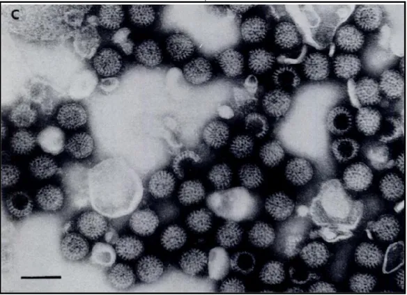
Figure 1. Particles observed by electronmicroscopy in filtrates made from a stool of a child with
Classification
Human Rotaviruses display diverse and complex serotypic specificities. In Rotavirus particles three concentric protein layers surround the double-stranded RNA viral genome and the viral capsid proteins are the majors determinants of antigenic properties of these viruses –group, subgroup, and serotypes.
Viral protein (VP) 2 represents the core or inner capsid, whereas VP6 constitutes the middle capsid and is an important antigenic determinant that gives serogroup antigen specificity.
Figure 2. Structure of Rotavirus. A cut-away of the viral structure is shown, with the proteins designated that
make up each concentric protein layer [adapted from Estes, 2001].
The outer capsid is composed by VP7 (the glycoprotein or G-protein) and VP4 (the protease-sensitive protein or P-antigen). These two proteins determine the serotype specificity and are the basis of the binary classification (G and P type) of Rotaviruses. Various combinations of VP4 and VP7 types have been observed in natural Rotavirus isolates. Virtually all Rotavirus genes are involved in reassortment events (Maunula et al., 2002). The extensive genomic and antigenic diversity of Rotaviruses of both human and animal origins has led to the proposal to classify Rotaviruses according to the composition of the whole genome (Matthijnssens et al., 2008; Maes et al., 2009).
Currently Rotaviruses are classified into seven major groups (A to G). Group A, B and C Rotaviruses have been found in both human and animals; group D, E, F and G Rotaviruses have been found only in animals (Kapikian et al., 2001). Group A Rotaviruses have clearly been
established as causing severe diarrheal disease in the young (Hrdy, 1987); group B Rotaviruses include viruses that have been associated with annual epidemics of severe diarrhea primarily in adults (Yamamoto et al., 2010); group C viruses have been found in sporadic cases and outbreaks of diarrhea in piglets and children (Moon et al., 2010). VP6, which is a major determinant of group reactivity, is the target of common diagnostic assays and is used to further classify Rotavirus into subgroups I and II. VP7 (G) and VP4 (P) as previous reported are essential for serotype classification. 10 of 14 G serotypes and 8 P serotypes of Rotavirus occur in humans.
Genome Structure
Rotaviruses are members of the Rotavirus genus of the Reoviridae family. Member of this family are nonenveloped, with complex capsid containing several concentric protein layers displaying icosahedral symmetry.
Rotavirus genome is composed of 11 segments of double-stranded RNA. The segments range in size from 667 (segment 11) to 3302 base pairs (segment 1), with the total genome containing approximately 18522 base pairs. Each segment is a gene that code for one protein, except gene 11 which had two open reading frames and codified for NSP5 and NSP6 proteins.
The nucleotide sequences of the 11 RNA segments from at least 53 different virus strains are known. There are general features about the structure of each of the 11 genome segments and sequences common to all RNA segments. Each RNA segment starts with a 5’ guanidine followed by a set of conserved sequences that are part of the 5’ noncoding sequences and another set of noncoding sequences, which contains a different subset of conserved 3’-terminal sequences and ends with a 3’-terminal cytidine, is found after the stop codon. The length of the 5’ and 3’ noncoding sequences vary for different genes; however, these lengths are conserved among strains for a given gene. Because of the conserved sequences of this 5’ and 3’ part of the genes, these terminal sequences are thought to contain signals important for genome transcription, replication, and possibly assembly of the viral genome segments. A polyadenylation signal is not been found at the 3’ end of the genes (Estes et al., 1989).
Figure 3. General scheme of Rotavirus genes structure [adapted from Estes, 2001].
Deproteinized, purified Rotavirus dsRNAs are not infectious, reflecting the fact that virus particles contain their own RNA-dependent RNA polymerase required to transcribe the individual RNA segments into active messenger RNAs (Cohen, 1977).
Rotavirus Proteins
The 11 dsRNA segments of the Rotavirus genome code for six structural (VP1-VP4, VP6 and VP7) and six non-structural proteins (NSP1-NSP6).
The naming of the structural proteins (viral proteins, VP) is based on their molecular weights, with VP1, the largest at 125 kDa, and VP8, one of the two proteolytic fragments of VP4, the smallest at 28 kDa. The six structural proteins form the multy-layered capsid of the mature Rotavirus particle. The nonstuctural proteins (NSPs) are synthesized in infected cells and function in some aspect of the viral replication cycle or interact with host proteins to influence pathogenesis or the immune response to infection.
Figure 4. Genome segments, proteins and structure of Rotavirus.
The RNA segments are numbered in order of gel migration, and they are correlated with their encoded protein products by arrows. Gene segments 7, 8, 9 are very close in length and tend to migrate nearly on top of one another. Gene 11 is alternatively processed to produce NSP5 and NSP6. On the right, locations of the viral proteins in a Rotavirus particle [adapted from Prasad et al., 1996].
The development and application of RNA interference techniques offers an important tool to study the functional roles of rotaviral proteins during the process of infection (Arias et al., 2004; Campagna et al., 2005; Lopez et al., 2005). Furthermore, X-ray crystallography has been applied to determine the atomic structures of several structural and non-structural proteins of Rotavirus (Groft et al., 2002; Dormitzer et al., 2004).
Capsid Architecture
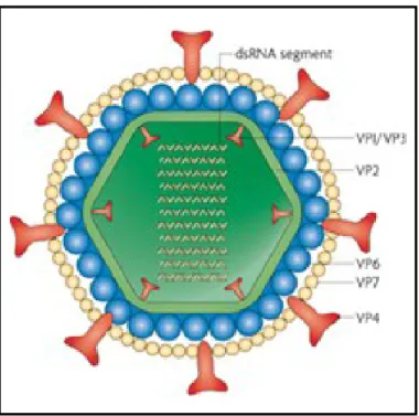
The mature infectious Rotavirus particle (1000 Å in diameter, including the spikes) is made of three concentric icosahedral protein layers that encapsidate the genome. The complete virion is called a triple-layered particle (TLP).
Figure 5. Rotavirus virion. The internal layer, or core, surrounds the viral genome, and contains the
scaffolding protein VP2, the RNA-dependent RNA polymerase VP1, and the guanylyltransferase and methylase VP3. The intermediate layer is made of VP6, the major structural protein. The external layer is made up of VP7 and is decorated with spikes of VP4 [adapted from Angel et al., 2007].
VP7 Layer and VP4 Spikes
The outer layer of the TLP is composed of two structural proteins: VP7 and VP4. VP7 (34 kDa), the major constituent of the outer layer, is a glycoprotein, although glycosylation is not required for capsid assembly (Estes, 2001). Seven hundred eighty copies of VP7 are grouped as 260 trimers. The outer layer is decorated by 60 spikes, each of which is formed by a dimmer of VP4 (88 kDa); thus each Rotavirus particle has 120 copies of VP4.
From their locations in the structure of Rotavirus, VP7 and VP4 are obvious candidates to be implicated in the cell entry processes. Although early studies have implicated VP7 in the cell entry process (Fukuhara et al., 1988), subsequent studies have increasingly indicated the involvement of
VP4 not only in cell attachment and cell penetration, but also in hemagglutination, neutralization, virulence, and host range (Burns et al., 1988; Fiore et al., 1991; Ludert et al., 1998). Prior to its interaction with host cell, VP4 is proteolytically cleaved for efficient internalization of Rotavirus into cells. This is particularly relevant considering that Rotavirus replication takes place in enterocytes in the small intestine, an environment rich in proteases. Proteolytic cleavage of VP4 enhances viral infectivity by several folds (Arias et al., 1996) and facilitates virus entry into cells (Kaljot et al., 1988). Proteolysis of VP4 generates two fragments, VP8 (aa 1-247, 28 kDa) and VP5 (aa 248-776, 60 kDa), and these fragments remain associated with the virion (Fiore et al., 1991).
Aqueous Channels
One of the distinctive features of the Rotavirus architecture is the presence of large channels that penetrate through the VP7 outer layer and VP6 intermediate layer. These 132 channels allow for the passage of aqueous materials and biochemical substrates into and out of the capsid (Pesavento et al., 2003).
VP6 Layer
The intermediate layer is formed by the VP6 protein (41 kDa). Particles carrying VP6 on the outside are called double-layered particles (DLPs). The VP6 layer maintains the same icosahedral symmetry as the VP7 layer with 780 copies of VP6 arranged as 260 trimers. These trimers are located right below the VP7 trimers such that the channels in the VP7 and VP6 layers are in register.
The DLP is the transcriptionally competent form of the virus during the replication cycle. VP6 is the major protein of the Rotavirus particle by weight. It plays a key role in the overall organization of the Rotavirus architecture by interacting with the outer layer proteins, VP7 and VP4, and the inner most layer protein VP2. Thus it may integrate two principal functions of the virus: cell entry (outer layer) and endogenous transcription (inner layer). The X-ray structure of VP6 has been determined (Mathieu et al., 2001) and it shows that VP6 has two domains: the distal domain with an eight-stranded antiparallel β-sandwich fold makes contact with VP7 layer, and the lower domain, consisting of a cluster of α-helices, makes contact with the inner VP2 layer.
VP2 Layer and Transcription Enzyme Complex
Underneath the VP6 layer is the innermost protein layer of Rotavirus structure. The particle structure at this level is referred as single-layer particle (SLP). The SLP houses the dsRNA genome in a protein layer composed of 120 copies of VP2 protein. VP2 is the only structural protein shown to possess nucleic acid-binding activity (Boyle et al., 1986). This internal layer, or core, contains also VP1 protein (an RNA-dependent RNA polymerase) and VP3 protein (a guanylyltransferase and methylase); VP1 and VP3 together form transcription enzyme complex. The question of how the dsRNA segments are arranged inside the capsid is particularly interesting considering that they are transcribed simultaneously and repeatedly in the confines of the capsid. A plausible model that emerges from the available biochemical and structure data for Rotaviruses and other dsRNA viruses, is that each genome segment is spooled around a transcription complex (consisting of VP1 and VP3) that is anchored to the inner surface of the VP2 layer. Such a model allows for up to 12 independent transcription complexes, each associated with an individual dsRNA segment for concurrent transcription (Pesavento et al., 2003).
Cell Entry
Viral replication occurs in the mature epithelial cells of the small intestine, where Rotavirus infects nondividing differentiated enterocytes near the tips of the villus.
Triple-layered particles (TLP) attach to host cell and enter by receptor-mediated endocytosis or direct penetration. The cellular receptors of RVs have not been fully characterized, but the consensus opinion that emerged from recent studies is that Rotavirus cell entry is a coordinated multistep process involving sequential interactions with sialic acid (SA)-containing receptors in the initial cell attachment step. Next, interactions occur with hsp70, and integrins during the subsequent post-attachment steps (Lopez et al., 2004). In the entry process, the VP8 domain is involved in the interactions with SA, whereas VP5 is implicated in the interactions with integrins. Involvement of SA during Rotavirus infections is not an essential step in all Rotavirus strains. For many of the Rotavirus strain, including human Rotaviruses, cell entry is SA-independent (Ciarlet et al., 2001), suggesting that cell entry is mediated mainly by the VP5.
Endogenous Transcription
During the process of cell entry, the outer layer is removed from TLPs by cellular enzymes and a low intracellular Ca++ level; in this way double-layered particles (DLPs) emerge. DLPs in the
cytoplasm become transcriptionally active, and large number of positive-stranded RNA molecules (capped but not polyadenylated) is transcribed from all 11 RNA segments within the structural confines of the DLP (Estes et al., 2001). The nascent transcripts exit through channels that penetrate the inner VP2 and outer VP6 capsid layers of the DLP (Lawton et al., 1997).
The DLP possesses the complete enzymatic activities needed to synthesize not only mRNA transcripts but also to properly guanylate and methylate the cap structure at the 5’ end of each mRNA to facilitate translation by the cellular translation machinery. These enzymatic functions are carried out by VP1, the RNA-dependent-RNA polymerase (Valenzuela et al., 1991), and VP3, a guanylyltransferase and methyltransferase (Chen et al., 1999).
Figure 6. A DLP with mRNA transcripts exiting out by the proposed pathway through the capsid channels.
The transcripts are represented as gray strands. On the right, a close-up view of a transcribing DLP [adapted from Lawton et al., 1997].
Genome Replication and Packaging
The Rotavirus replication cycle consists of three subsequent major stages: 1) translation and synthesis of the viral proteins; 2) replication, genome packaging and DLP assembly; 3) budding of the newly formed DLPs into endoplasmatic reticulum (ER) and assembly of outer layer to form mature TLPs.
The non-structural protein NSP3 is implicated in the specific recognition of rotaviral mRNAs and in facilitating their translation using the cellular machinery (Vende et al., 2000); while the N-terminal domain of NSP3 interacts with the 3’-consensus sequence of the rotaviral mRNAs, the C-terminal domain enables their delivery to the ribosomes for viral protein synthesis.
Replication, genome packaging and assembly of the DLP occur in perinuclear inclusions called viroplasms, which appear 2-3 hours after infection. In the viroplasms, viral messenger RNAs are replicated to produce new genomic RNA. The proteins in the core of the incoming particles possess all the enzymatic activities required to produce the viral transcripts from the viral genome double-stranded RNA because eukaryotic cells do not express RNA polymerases that transcribe mRNA from dsRNA templates (Greenberg et al., 2009). In particular, VP1 protein is an RNA-dependent RNA polymerase (Valenzuela et al., 1991).
NSP2 and NSP5 proteins are the major components of viroplasms, and they are strongly implicated not only in the formation of the viroplasm, but also in genome replication and packaging. NSP5 is a
NTPase (nucleotide triphosphatase), ssRNA binding, and helix destabilizing activities (Taraporewala et al., 2001); based on these properties, NSP2 may function as a molecular motor using the energy derived from NTP hydrolysis to facilitate genome packaging. The replication complex may be organized around the NSP2 octamer providing a platform or a scaffold (Jayaram et al., 2004). Co-expression of NSP2 and NSP5 in uninfected cells forms viroplasm-like structures and experiments of co-transfection with NSP5 and NSP2 have shown that NSP2 upregulates phosphorylation of NSP5 (Afrikanova et al., 1998).
The exact order of events during early morphogenesis and the molecular interactions and control mechanisms by which genome replication, packaging and reassortment of RNA segments into cores occur are at present unknown (Desselberger et al., 2009).
Also NSP6 is active in this process, but its role is still unclear; NSP6 interacts with NSP5 and it might have a regulatory role in the self-association of NSP5 (Torres-Vega et al., 2000).
Maturation and Release
Currently is still not entirely clear how the set of 11 segments of dsRNA get encapsidates into each virion. The encapsidation could be concurrent with the capsid assembly (Pesavento et al., 2003). Thus, the capsid assembly begins with the association of 12 units, each unit consisting of pentamers of VP2 dimers in complex with a transcription enzyme complex (VP1/VP3) and a genome segment to form the SLP and provide a scaffold for the subsequent assembly of the VP6 trimers, leading to the assembly of a DLP.
Figure 8. A working model for genome encapsidation in Rotavirus [adapted from Pesavento et al., 2003].
Once formed, DLPs bud from the viroplasms into the proximally located endoplasmatic reticulum (Estes, 2001) and, by a mechanism that is not clear, DLPs acquire the outer layer consisting of VP7 and VP4. The non-structural protein NSP4, which has a binding site for VP6 in its C-terminal sequence, facilitates the budding process (O’Brien et al., 2000). Both NSP4 and VP7 are synthesized on the ER-associated ribosomes.
VP7 does not affect the assembly of DLPs but leads to the accumulation of enveloped DLPs in the ER, suggesting that VP7 is required for removal of the lipid envelope (Lopez et al., 2005). Where and how the spike protein VP4, which is synthesized on cytosolic ribosomes, is assembled onto the particles is unclear.
Triple-layered infectious virions are released by lysis of non-polarized cells or by exit from polarized cells, before a cytopathic effect becomes obvious (Estes, 2001).
Transmission and Epidemiology
Rotaviruses spread mainly via the faeco-oral route; there is also evidence to suggest that they can be transmitted in respiratory droplets (Parashar et al., 1998). Water, fomites and occasionally food may act as vehicles. Rotavirus particles are very resistant to environmental conditions. Primary infection is occurred before 5 years of age with Rotavirus peaks between 9 and 23 months of age with or without evidence of symptoms, (Parashar et al., 2006). Adults exposed to infected children have a high risk for contracting Rotavirus; although infection in adults is more frequently asymptomatic, Rotavirus is still shed in their stools and can therefore be transmitted to other people (Anderson et al., 2004). The high particle number in the faeces of children with acute Rotavirus disease and the very small 50% diarrhoea-causing dose [1 DD50 = 10 plaque forming units (pfu)] lead to wide
spread to any susceptible host. While there are marked seasonal peaks (winter/spring) in countries with temperate climates, in tropical regions Rotavirus infections and disease occur throughout the year.
According to the estimates based on studies carried out worldwide during 1986-2000, Rotaviruses cause 100 million episodes of acute gastroenteritis per annum. While the incidence of Rotavirus disease is similar for developed and developing countries, death from Rotavirus disease is most frequent in developing countries of sub-Saharan Africa and Asia (Parashar et al., 2003). In developing countries, Rotavirus gastroenteritis is responsible for more than 500.000 deaths each year, likely related to limited access to medical provisions (Parashar et al., 2006). Studies from Europe found that 50% of cases of gastroenteritis in children younger than 5 years of age were caused by Rotavirus and that the infection resulted in 230 deaths per year (Van Damme et al., 2007).
Figure 9. Deaths due to diarrhoea per 100000 children younger than 5 years [adapted from Black et al.,
2008; WHO datas].
The epidemiology of Group A of Rotavirus infections is complex, since Rotaviruses of different G and P types co-circulate in a geographical region at any one time. The relative incidence of different G types also changes over time in the same location. Approximately 95% of co-circulating strains are G1-G4 in most regions of temperate climate, but other G types may be represented at high frequencies, particularly in tropical areas (Desselberger et al., 2001). Recently, G9 Rotaviruses have been isolated as the predominant outbreak strains in several locations in the USA and in Europe.
Besides being acquired in the community, Rotavirus infections are increasingly recognized as the cause of a significant proportion of nosocomial infections and diarrheal disease (Gleizes et al., 2006). Group B Rotavirus causes severe diarrhea primarily in adults; it has been detected recently in India, Bangladesh and Myanmar (Yamamoto et al., 2010). Group C Rotaviruses are associated with small outbreaks in humans worldwide (Moon et al., 2010).
Apart from the accumulation of point mutations (genomic drift), gene reassortment (genomic shift) plays a major role in generating the high diversity of Rotaviruses (Iturriza-Gomara et al., 2001; Maunula et al., 2002). Animals of different mammalian species are increasingly recognized as significant reservoirs for human Rotavirus infections as animal Rotaviruses have been found to infect humans directly and to form reassortants with human Rotaviruses (Matthijnssens et al., 2008; Steyer et al., 2008).
Pathogenesis
Rotavirus infection can result in asymptomatic or symptomatic infection.
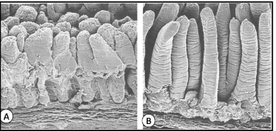
In case of symptomatic infection, extensive cellular necrosis of epithelium of the small intestine develops, leading to villous atrophy, loss of digestive enzymes, reduction in absorption and increased osmotic pressure in the gut lumen and the onset of diarrhea. This is followed by a reactive crypt cell hyperplasia accompanied by increased fluid secretion, which also contributes to the severity of diarrhea. Rotavirus infection can lead to death.
Figure 11. (A) Electron microscopy image of calf small intestine infected by Rotavirus. (B) Calf small
Both host and viral factors affect the outcome of infection. The most prominent host factor that affects the clinical outcome of infection is age. Neonates infected with Rotavirus rarely have symptomatic disease; this protection is thought to be mediated by transplacental transfer of maternal antibodies (Ray et al., 2007). Reductions in these antibodies coincide with the age of maximum susceptibility of infants to severe Rotavirus-induced disease (range, three months to two years). Rotavirus can infect adults, but severe symptomatic disease is relatively uncommon and can result from infections with an unusual virus strain or extremely high doses of virus.
Viral factors determining the pathogenicity of RVs have been investigated in several animal models, such as piglets, mice, and rabbits (Burke et al., 1996). The protein product of RNA segment 4, VP4, is the major determinant of pathogenicity in several systems, but products of other structural (VP3, VP7) and non-structural genes (NSP1, NSP2, NSP4) are also implicated. NSP4 is the first described virus-encoded enterotoxin. It produces an increase in intracellular Ca++ concentration (Berkova et al., 2003) and perturbs cellular electrolyte homeostasis. A peptide of NSP4, an active enterotoxin, is secreted from infected cells; the secreted protein binds cellular receptors and initiates signalling cascades in uninfected cells (Seo et al., 2008).
Rotavirus can also stimulate the enteric nervous system (ENS), inducing secretory diarrhea and increasing intestinal motility (Lundgren et al., 2000). Drugs that inhibit the ENS are useful in treating Rotavirus diarrhea in children.
Moreover Rotavirus infection is not limited to the intestine, but all infected individuals and animals undergo at least a short period of viremia and virus can be detected in the several other tissues of immunocompetent hosts; while viremia in Rotavirus infection appears to be frequent, systemic disease is rare (Ramig, 2007).
Figure 12. Mechanisms by which Rotaviruses cause diarrhea. (A) Events that occur after Rotavirus infection
of enterocytes are shown in order from left to right. Not all events are shown in each cell. (1) Infection of the initial cell by luminal virus leads to virus entry, uncoating, transcription, translation of viral proteins, formation of viroplasms (Vi), and apical release of virus and viral protein. NSP4 (red triangle) and virus particles are released by a nonclassic secretory pathway. Intracellular NSP4 also induces the release of
Ca2++, from internal stores, primarily the ER, leading to increasing [Ca++]i. (2) Another outcome can result
from a cell being infected with virus. NSP4 produced by the infection disrupts tight junctions, allowing paracellular flow of water and electrolytes (blue arrow). (3) NSP4 released from previously infected cells binds to a specific receptor and triggers a signaling cascade through phospholipase C (PLC) and inositol
phosphatase (IP)3 that results in release of Ca2++ and an increase in [Ca2++]i. Intracellular expression of
NSP4 increases [Ca2++]i through a PLC-independent mechanism. The increase in [Ca2++]i also disrupts the
microvillar cytoskeleton. (4) A crypt cell (brown) can be acted on directly by NSP4, or NSP4 can stimulate
the enteric nervous system (ENS), which in turn signals an increase in [Ca2++]i that induces Cl- secretion. (B)
Normal architecture of the small intestine, without the circulatory system shown. The ENS and its ganglia in the different submucosal levels are shown. (C) Reflex arc in the ENS that can receive signals from the villus epithelium and activate the crypt epithelium. Inset 1 shows a whole-mount of an adult mouse small intestinal villus stained with antibody to the gene product 9.5 neuroendocrine marker to reveal the rich innervation (yellow). Inset 2 shows that infected villus enterocytes can stimulate the ENS by the basolateral release of NSP4 or other effector molecules. The integrin α2β1 can bind NSP4 and elicit diarrhoea in neonatal mice [adapted from Ramig, 2004].
Diagnosis
Diagnosis of a Rotavirus infection is relatively easy as large numbers of virus particles (up to 1011 particles/ml of faeces) are shed at acute stage of the disease. The main techniques are enzyme-linked immunoassays (ELISAs) and, when searching comprehensively for diarrhoeagenic viruses, electronic microscopy (EM). G and P types of Rotavirus isolates can be determined serologically, but molecular techniques are increasingly being applied for both detection and genotyping. Rotavirus-specific oligonucleotide primers complementary to VP6, VP7 and VP4 sequences allow sensitive detection, subgroup determination and typing for both G and P types by RT-PCR (Maunula et al., 2002; Simpson et al., 2003; Matthijnssens et al., 2008).
Immunity
Rotavirus-induced immune responses, especially the T and B cell responses, have been extensively characterized; however, little is known about innate immune mechanism involved in the control of Rotavirus infection.
Protective Rotavirus immunity is multifactorial, achieved through the combined action of secretory antibodies, humoral and cell-mediated immunity. In adult mice have been shown that, after infection with a homologous murine Rotavirus, CD8+ T cells have a role in the timely resolution of
primary infection and B cells are necessary for long-term robust protection against Rotavirus (Franco et al., 2001). CD4+ T cells have an important, but non essential, role in supplying help to CD8+ T cells and B cells; in particular, CD4+ T cells contribute to generate Rotavirus-specific intestinal immunoglobulin A (IgA), which is the principal effector of long-term protection against Rotavirus infection (Franco et al., 1999). As would be expected by the fact that Rotavirus infection includes a viraemic phase (Fenaux et al., 2006; Blutt et al., 2007), both intestinal and systemic Rotavirus-specific B cell responses are observed in mice. However, only Rotavirus-specific plasma cells that reside in the intestine seem to have an antiviral effect, suggesting that mucosal, but not systemic, antibodies provide protection in this model (Franco et al., 2006). Humoral antibodies, directed towards VP2 and particularly VP6 proteins, are produced to high titers after Rotavirus infection (Burns et al., 1996). The induction of neutralizing antibodies in neonatal mice (Smiley et al., 2007) and pigs (Yuan et al., 2000) after infection with homologous Rotavirus is relatively weak; it could, in part, be due to the immaturity of their immune system.
In agreement with the animal studies, the levels of Rotavirus-specific serum IgA, measured shortly after natural infection in children, generally correlate with intestinal IgA levels and in many, but not all studies, the serum IgA level provides a good correlate of protection (Franco et al., 2006). Furthermore, T-cell responses to Rotavirus are related to the development of protective antibodies (Offit et al., 1993), even if the T-cell response to Rotavirus in humans, as in animal models, seems to be transient and of low intensity, especially in Rotavirus-infected children, in fact, the T-cell response appears to be more robust in adults (Kaufhold et al., 2005).
The innate immune response is also very important. Rotavirus infection results in the secretion of type I interferon (IFNα and IFN β), presumably from a plasmacytoid subset of dendritic cells, pDCs (Mesa et al., 2007). While increased levels of IFNα have also been correlated with a positive clinical outcome in infected children (Mangiarotti et al., 1999), several Rotaviruses have recently been demonstrated to antagonize the production of type I IFN through the degradation of interferon regulatory factors (IRF) 3, IRF5, and IRF7 (Barro et al., 2007). NSP1 recognizes a common element of IRF proteins, thereby allowing NSP1 to act as a broad-spectrum antagonist of IRF function. IRF5 also has roles in stimulating the expression of cytokines and chemokines that cause the recruitment of T lymphocytes (Yanai et al., 2007); interaction between NSP1 and IRF5 can downregulate the activation of genes producing proinflammatory cytokines that initiate events leading to apoptosis, so that the Rotavirus can exist longer in infected cells.
This response is dependent on the presence of the viral dsRNA genome. As IFNα production by pDCs is classically trigged in response to ssRNA or DNA, the induction of this response by a dsRNA virus indicates a potentially novel mechanism of viral sensing by pDCs (Deal et al., 2010). Rotavirus NSP1 also inhibits nuclear factor κB (NFκB) activation (Graff et al., 2009). NFκB induces the production of IFNβ and chemokines, both of which have antiviral effects; thus Rotavirus bloks NFκB activity to delay the innate immune response.
Figure 13. Potential mechanisms of Rotavirus pathogenesis and immunity. The mechanisms of Rotavirus
pathogenesis and immunity are not completely understood and vary depending on the animal species studied. In step 1, neutralizing antibodies directed against VP4 and/or VP7 can prevent viral binding and penetration, inducing viral exclusion. If this mechanism fails, as shown in step 2, Rotavirus replication inside enterocytes causes altered metabolism of enterocyte membrane proteins inducing malabsorptive or osmotic diarrhoea. Rotavirus also increases the concentration of intracellular calcium, which disrupts the cytoskeleton and the tight junctions, raising the paracellular permeability. During step 3, intracellular viral replication can be inhibited by secretory anti-VP6 immunoglobulin A (IgA) during transcytosis across enterocytes. In step 4, cytokine-secreting Rotavirus specific T cells can also inhibit viral replication. If viral replication is not stopped, as shown in step 5, replicating Rotavirus produces non-structural protein 4 (NSP4), a toxin which induces a secretory non-cystic fibrosis transmembrane conductance regulator (CFTR)-mediated diarrhoea. By an unknown mechanism Rotavirus can also stimulate the enteric nervous system (ENS) (as shown in step 6), inducing secretory diarrhoea and increasing intestinal motility. Drugs that inhibit the ENS are useful in treating Rotavirus diarrhoea in children. Antibodies against NSP4 could potentially have an effect against the last two mechanisms. Late in the infectious process, Rotavirus kills the host cell (as shown in step 7), further contributing to malabsorptive or osmotic diarrhoea [adapted from Angel et al., 2007].
Natural infection or appropriate vaccination seems to protect from severe disease in subsequent infections (Velazquez, 2009), even if the serotype of the challenging virus differs from that of previous infections or those in a prior vaccine.
Vaccine Development
The worldwide burden of disease because of Rotavirus quickly led to initiatives supported by the World Health Organization (WHO) to develop vaccines to prevent this disease. The vaccine initiative was developed in parallel with initiatives aimed at widespread extension of oral rehydration therapy in developing countries.
The development of vaccines against Rotavirus started in the early 1980s. Animal Rotaviruses (of simian or bovine origin) were used as live attenuated vaccines for humans.
A tetravalent vaccine contained a rhesus Rotavirus (RRV) of G3 type and three mono-reassortants, which individually carry the VP7 gene of human serotypes G1, G2 and G4 in the RRV genetic background (Rotashield®). This vaccine was highly effective (80-100%) in preventing severe diarrheal disease, even if the immunologic basis for this efficacy was unclear. It received Food and Drug Administration approval as a universal vaccine in the USA in 1998. More then 1.5 million doses were administrated in the followed 10 months. During that time, several cases of intussusception were observed in vaccinees, particularly within 3-7 days after the first dose (Murphy et al., 2001); these observations led to withdraw the recommendation of Rotashield for use in infants, and vaccine production ceased. A recent re-analysis of the data indicated that the age of vaccinees is a critical factor for intussusception, as a disproportionately high number (>80%) of children who developed intussusception after the first dose of Rotavirus vaccine were older than 90 days leading to the consideration those vaccinations in children older than 3 months contraindicated.
It took another 7 years before new Rotavirus vaccine candidates were available.
In 2006 two new live-attenuated Rotavirus vaccines were licensed in the United States, the European Union, as well as many countries in Central and South America.
In one of these new vaccines, a bovine Rotavirus strain (WC3), isolated in the United States, was used as a backbone to create a pentavalent vaccine that contained 5 separate viruses that expressed either human G1, G2, G3, or G4 VP7s, and human P(8) VP4 on the bovine WC3 backbone. This vaccine is manufactured by Merck and it is called RotaTeq®. It was found to be highly efficacious in preventing severe Rotavirus gastroenteritis (98%) caused by G1, G2, G3, G4 and G9 strains (Vesikari et al., 2006).
RotaTeq® is given in a three-dose schedule and preliminary data indicate that at least two doses are required to generate significant levels of protection (Vesikari, 2008). The vaccine is given by mouth; the first dose is given from 6 to 12 weeks of age, the second dose is given 4 to 10 weeks later. The last (third) dose should be given by 32 weeks of age. No link between RotaTeq® and intussusception was found (Vesikari et al., 2006).
Another vaccine was licensed in 2006. GlaxoSmithKline manufactures this vaccine under the trade name Rotarix®. It’s a monovalent vaccine derived from an attenuated human Rotavirus isolate of the G1P1A[8] type. The rationale underlying the development of Rotarix® was that a single natural
Rotavirus infection provides protective immunity against subsequent severe disease, irrespective of serotype (Velazquez et al., 1996). Therefore, it seemed logical to predict that an attenuated human Rotavirus strain might do the same. The virulent G1 human Rotavirus strain was passaged for multiple rounds in monkey kidney cell cultures to achieve attenuation. The initial passaged material possessed virulence, but after subsequent additional passages and plaque purification, a highly attenuated product was attained. As with RotaTeq®, the molecular basis for the attenuation of the Rotarix® vaccine is unknown. Rotarix® provided good heterologous protection against G2, G3, G4 and G9 type, and obviously against G1 type.
Rotarix® requires only two doses, probably because it is better adapted to replication in the human gastrointestinal tract than the bovine-based vaccine, and it can be administered at a dose approximately 100-fold lower than that of RotaTeq®. The vaccination series consists of two 1-ml
doses administered by mouth. The first dose can be given beginning at six weeks of age and the second at least four weeks after the first dose, but before the child reaches 24 weeks of age.
Rotarix® is 85% effective against preventing severe diarrhea and no association between this vaccine and intussusception is known (Ruiz-Palacios et al., 2006).
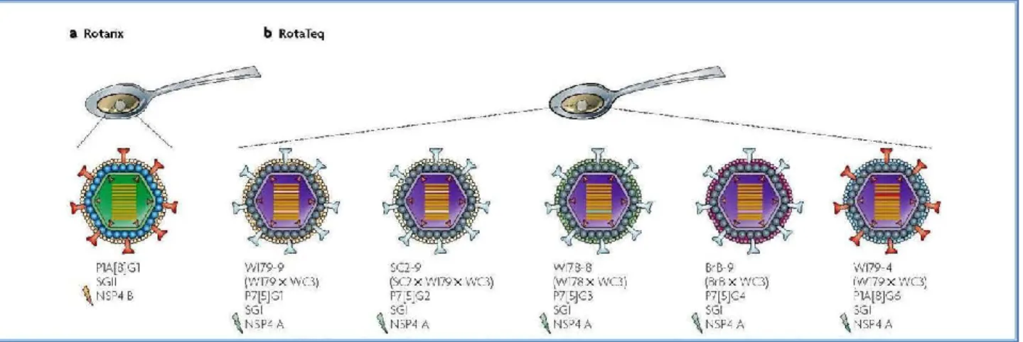
Figure 14. The Rotarix® and Rotateq® vaccines. a. Rotarix® is an attenuated human Rotavirus vaccine made
of a tissue culture-adapted human P1A[8]G1, VP6 and NSP4 geno-group B strain. b. RotaTeq® is a bovine
(WC3)–human reassortant vaccine composed of the five strains shown, each containing a human Rotavirus gene encoding the VP7 neutralizing protein from different serotypes. Notably, in the WI79-9 and SC2-9 viruses (the last was used to create the first), genes 3 (VP3) and 9 (VP7) are of human origin. Although VP6 and NSP4 can potentially be the targets of protective antibodies, their role in immunity against disease in humans is unknown [adapted from Angel et al., 2007].
Nowadays the risk of desirable effects of live attenuated vaccines (clinical complications, reversion to virulence, genetic interaction and reassortment with co-circulating wildtype Rotavirus strains, etc.) is considered to be low, but not zero. Thus several third-generation Rotavirus vaccines are in development because of possible safety issues associated with the use of RotaTeq® and Rotarix®, and several groups are pursuing recombinant virus-like particles approaches.
Rotavirus-Like Particles (VLPs)
Virus-like particles (VLPs) are a highly effective type of subunit vaccines that mimic the overall structure of virus particles without any requirement that they contain infectious genetic material. Indeed, many VLPs lack the DNA or RNA genome of the virus altogether, but have the authentic conformation of viral capsid proteins, without any of the risks associated with virus replication or inactivation (Roy et al., 2008).
The fact that VLPs mimic the structure of the virus particles usually means that VLPs elicit strong humoral response. In addition to their ability to stimulate B cell mediated immune responses, VLPs have also been demonstrated to be highly effective at stimulating CD4 proliferative and cytotoxic T lymphocyte (CTL) responses (Schirmbeck et al., 1996).
To date, VLPs have been produced for many different viruses that infect humans and other animals. VLPs have also been produced for Rotavirus. VLPs formed from the two inner structural proteins of the Rotavirus capsid (VP2 and VP6) have been shown to be effective immunogens in animal models (Parez et al., 2006). The expression of VP2 alone results in the production of pseudo-core particles (Zeng et al., 1994). VP6 alone can form spherical or tubular aggregates (Lepault et al., 2001); VP6 self-assembles into different types of particles depending on conditions such as pH, ionic strength and divalent cation concentration.
Moreover virus-like particles provide the spatial structure for the repetitive, high density display of conformational epitopes and can be exploited as platforms for the presentation of foreign epitopes or targeting molecules on chimeric VLPs (Peralta et al., 2009). It has been reported that heterologous proteins as green fluorescent protein (GFP) could genetically fused to VP2 and incorporated into VLP (Charpilienne et al., 2001); to assure the correct VLP assembly, it is considered that molecules smaller than 250 amino acids could easily be integrated into this delivery system by fusion to VP2 protein. A foreign epitope could also fused to VP6, because the ability of
VP6 to form multimeric structures and the strong immune responses that VP6 can elicit in different species (Peralta et al., 2009).
Rotavirus exhibits a marked tropism for intestinal epithelium. As Rotavirus-like particles display properties very similar to those of Rotavirus, their natural tropism and nonreplicative properties make these VLPs a viable alternative to the live virus vaccine for Rotavirus and a promising safe candidate for drug delivery to intestine in pathologies such as inflammatory bowel diseases (IBD) (Cortes-Perez et al., 2010).
Update on Recommendations for the Use of Rotavirus Vaccines (by Food and
Drug Administration, FDA)
On March 2010, FDA provided an early communication regarding Rotarix® (GlaxoSmithKline) while the agency and manufacturer investigated the finding of DNA from porcine circovirus type 1 (PCV1) in the vaccine. Since that time, both FDA and GSK have confirmed the presence of PCV1 in the vaccine, FDA recommended that clinicians and public health professionals in the United States temporarily suspend the use of Rotarix®.
On May 2010, FDA provided information about RotaTeq® (Merck). FDA indicated that preliminary studies conducted by Merck identified fragments of DNA from PCV1 and from a related porcine circovirus type 2 (PCV2) in RotaTeq®.
PCV1 and PCV2 are both small viruses composed of a single strand of circular DNA. Both viruses are common in pigs. Neither PCV1 nor PCV2 are known to infect or cause illness in humans, however PCV2 may cause illness in pigs.
At the end of May 2010, based on careful evaluation of a variety of scientific informations, FDA has determined it is appropriate for clinicians and health care professionals to resume the use of Rotarix® and to continue the use of RotaTeq® (because there is no evidence that these findings pose a safety risk in humans).
On October 2010, Merck and FDA’s laboratories determined that there is no evidence of the presence of PCV2 in RotaTeq®.
Communication of AIFA (Agenzia Italiana del Farmaco)
On July 2010, AIFA (Agenzia Italiana del Farmaco) overturns the prohibition to use Rotarix® and RotaTeq® (decided on April 2010 and July 2010 respectively) because the ratio benefit/risk of these vaccines continues to be positive. However the presence of exogenous DNA in these vaccines must be eliminated.
HERPESVIRUS-1
Virion Stucture
The Herpes simplex virus type 1 (HSV-1) is an enveloped, double-stranded (ds) DNA virus. The DNA virus genome is enclosed into an icosahedric capsid, which is surrounded by the tegument, a rather unstructured layer containing some twenty virus-encoded proteins with structural and regulatory roles. The tegument is delimited by the envelope, which is a lipid membrane of cellular origin, containing a dozen virus-encoded glycoproteins.
HSV-1 enters epithelial cells and neurons by fusion of the virus envelope with the plasma or endosomal membranes, and the virus capsid are transported to the nuclear pores through association with microtubules (Marozin et al., 2004), from where the viral DNA is then released into the nucleus. The HSV genome can be viewed as consisting of two covalently linked components, designated as L (long) and S (short); each component consists of unique sequences bracketed by inverted repeats. During lytic infection, the virus 153-kilobase pairs (kbp) double-stranded DNA genome expresses at least 84 genes that are temporarily regulated in a cascade fashion, giving rise to three phases of gene expression. The expression cascade, which is regulated mainly at the transcriptional level, begins with the expression of the immediate-early (IE) genes. Five viral IE genes are expressed first, and four of these encode regulatory proteins (ICP0, ICP4, ICP22 and ICP27) that are responsible for controlling viral gene expression during subsequent, early (E) and late (L) phases of the replication cycle and for inducing shutoff of cellular protein synthesis.
stimulated by a virion protein known as VP16, which is a powerful transcription factor that, in conjunction with cellular proteins, acts on DNA motifs present only in the IE regulatory regions to up-regulate expression. The early (E) gene products that are synthesized next include enzymes that, like thymidine kinase and ribonucleotide reductase, act to increase the pool of deoxynucleotides of the infected cells, and several replication proteins that are directly involved in viral DNA synthesis. The last functions to be expressed are the late (L) genes, which encode proteins involved in the packaging of virus DNA, as well as the structural proteins involved in the assembly of the virion particle, including the capsid, the tegument, and the envelope. Some of these structural proteins, like the tegumentary VP16, play major regulatory roles in the next infectious cycle. Capsids are assembled in the nucleus but the tegument and the envelope are acquired in the cytoplasm, most probably by budding into endosomes, and are released by exocytosis at cell membranes (Skeeper et al., 2001).
HSV-1 Lytic and Latent Cycles
After initial infection and lytic multiplication at the body periphery, generally at oral or genital epithelial cells, HSV enters the sensory neurons that innervate the infected epithelia and, following retrograde transport of the capsid to the cell bodies, establishes a lifelong latent infection in sensory ganglia. Periodic reactivation from latency usually leads to the return of the virus to epithelial cells, where it produces secondary lytic infections resulting in mild illness symptoms, such as cold sores.
Figure 16. (A) Genome structure of HSV-1. TR: Terminal Repeats; IR: Internal Repeats. (B) Schematic
representation of cascade mechanism of genic expression in HSV-1. IE: Immediate Early genes; E: Early genes; L: Late genes.
In rare cases, HSV-1 can spread centripetally into the central nervous system, to cause devastating encephalitis (Roizman et al., 2001).
Lytic viral replication results in the impairment of host macromolecular synthesis, the release of newly assembled viral progeny particles and the ultimate death of the host cell. A striking property of the virus life strategy is the high number of functions devoted to avoid or inhibit inimical cellular responses that could eventually block or diminish virus expression and multiplication. These functions, which actually represent more than 10% of the virus genome, include many proteins that act to counteract the innate immune responses, to inhibit the proapoptotic response of the cells to virus infection, and to facilitate escaping from the adaptive immune system (Mossman et al., 2005).
Figure 17. Schematic representation of HSV-1 replication cycle.
The attachment of the virion to the surface of the cell is followed by viral entry into cell. Then the virion is transported to the cell nucleus, where the cascade mechanism of gene expression takes place. In the nucleus capsid assembly and the cleavage of HSV-1 progeny DNA concatamers into unit-length monomers occur. After encapsidation of full-length DNA molecules, the nucleocapsid is capable of budding through the inner nuclear membrane, and then the virus particle is released through secretory vesicles.
During latency in sensory neurons, the viral genome remains as a circular chromatinized episome (Deshmane et al., 1989) within the cell nuclei, and undergoes dramatic changes resulting in an almost complete silencing of transcription. Only one region of the viral genome, known as the LAT locus, is actively transcribed during latency, due to the presence of a latency associated promoter (LAP) that remains active during this phase of the infection, resulting in the synthesis of non-messenger RNA molecules of unknown function (the latency associated transcripts, or LATs), which accumulates in the nucleus of the latently infected neurons (Farrell et al., 1991). Very recently, the LAT locus has been shown to express one class of miRNA molecules that can down-regulate expression of cellular proteins like TGF-1 and SMAD 3, and which seems to play an anti-apoptotic role during latency or reactivation (Gupta et al., 2006). The latent virus genome can reactivate in responses to a wide variety of stimuli that allow it to enter the lytic phase of the HSV-1 life cycle (Preston, 2000).
HSV-1 and its Derived Vectors
The uniqueness of HSV-1-based vectors stems from outstanding properties of HSV-1, not shared with any other viral system. HSV displays a broad host cell range, the very large capacity of the virus particle, which allows packaging and efficiently delivery of up to 150 kbp of DNA to the
nuclear environment of mammalian cells. The complexity of the virus genome, which contains some 40 genes that are not essential for virus replication, allows deletion of these genes without disturbing virus production in culture conditions, yet they are required for expression of a fully virulent phenotype in vivo. Other interesting property is that the viral DNA will not integrate into host chromosomes, thus reducing the risk of insertional mutagenesis, and the genes carried by these molecules will be expressed from the episomic genome.
Three different types of vectors can be derived from HSV-1, which attempt to exploit one or more of the previously described properties. Recombinant HSV-1 vectors are created by replacing one or several virus genes with transgene sequences. Depending on the virus genes that are replaced, recombinant HSV-1 vectors can be replication-competent, -conditional, or –defective. The choice of the replicative state of a vector depends on the purpose of gene delivery and the target tissue. Attenuated recombinant vectors are replication-competent HSV-1, generally carrying attenuating mutations that restrict spread and lytic viral replication to a limited number of tissues, without causing major toxicity to the inoculated organism. Defective recombinant vectors are disabled, replication incompetent and non-pathogenic HSV-1 mutants lacking one or more essential genes. These vectors retain many advantageous features of wild-type HSV-1, particularly the ability to express transgenes after having established latent infections in central and peripheral neurons, but cannot replicate in, and therefore cannot disseminate out of, the infected cells. Lastly, amplicon vectors are defective, helper-dependent vectors that take advantage essentially of the large transgenic capacity of the virus particle. Actually, there is no other mammalian vector available that could equal the ability of herpesvirus-based amplicons to deliver 150 kbp of foreign DNA with no simultaneous delivering of viral genes.
In all three cases, the vector particles are basically identical to the wild type HSV-1, which are complex particles made up of some 40 different virus encoded structural proteins. These proteins will be delivered into the cell during infection and can trigger cell signalling and cellular responses and, consequently, may have a transient impact on the cell homeostasis and gene expression. However, at least when using defective vectors not expressing virus genes, such as amplicons, the imported structural proteins will soon disappear and the cells will resume their normal functions, including the ability to divide and to respond to physiological stimuli. Although the HSV-1-based vectors have been used mainly for gene transfer to neurons, they can efficiently deliver genes t o other cell types, including epithelial cells, myoblasts, myotubes, embryonic and adult
cardiomyocytes, hepatocytes, and cell lines derived from gliomas, hepatocellular carcinomas, osteosarcomas, epidermoid carcinomas and many other human and murine malignancies.
Defective Recombinant Vectors
These vectors are disabled, replication-incompetent and non-pathogenic mutant viruses that lack one or more genes encoding essential proteins, but retain many advantageous feature of wild-type virus, particularly the ability to express transgenes. Various aspects of these vectors are attractive when considering the design of gene therapy vectors. These include a broad host cell range, the ability to efficiently transduce dividing and non-dividing cells, the large capacity of their genomes, allowing introduction of more than 30 kbp of foreign DNA, and the latent behaviour of the virus, which may be exploited for the stable long-term expression of therapeutic transgenes in neurons. Non-replicative HSV-1 recombinants are potential vectors for several applications in human health. These include delivery and expression of human genes to central nervous system cells, selective destruction of cancer cells, prophylaxis and immunotherapy against tumors and prophylaxis against HSV-1 and other infectious disease. Each application requires a different kind of vector and, in principle, different kinds of genetic engineering. Some gene therapy applications will require short-term transgene expression, while others will demand a long-short-term or regulated expression of the exogenous gene. Although delivery of a single gene should be adequate in many cases, other applications will likely require the delivery of multiple transgenes. One of the major advantages of non-replicative vectors is the possibility, due to their capacity to accommodate multiple transgenes in one vector, to achieve a synergistic therapeutic effect in gene therapy applications of multifactorial diseases.
In HSV-1 virus immediate-early genes (IE), which encode ICP0, ICP4, ICP22, ICP27 and ICP47, are expressed shortly after viral entry into host cell and are required for initiation of the cascade of early (E) and late (L) viral gene transcription. ICP4 and ICP27 are essential for replication and the deletion of one or both of them requires adequate complementing cell lines that provide in trans the missing function (Marconi et al., 1996; Wu et al., 1996). The “first generation” of replication-defective HSV-1 based vectors consisted of mutants deleted in the single essential IE encoding ICP4, namely dl20 and S4TK vectors (Krisky et al., 1997). Although these vectors show reduced pathogenicity, they possess some residual cytotoxicity that probably results from the expression of the other four IE genes. ICP4, ICP27, ICP0, and ICP22 have all been shown to be toxic in stable
transfection assays (Wu et al., 1996), so deletion of these genes in combination is required to eliminate toxicity (Krisky et al., 1998; Moriuchi et al., 2000). Cell lines that complement ICP4 and ICP27 have permitted the construction of a “second generation” of highly defective mutants (Marconi et al., 1999). To date, several replication-defective vectors have been constructed in which ICP0, ICP4, ICP27, ICP22 and ICP47 genes have been deleted in various combinations. The second generation of replication-defective vectors is characterized by absence of early and late viral gene expression and provide enough space to introduce distinct and independently regulated expression cassettes for different transgenes (Krisky et al., 1998). Deletion of all five IE genes prevents virus cytotoxicity at high multiplicity of infection, allowing the vector genome to persist in cells for long periods of time. However, these vectors grow poorly in culture and express transgenes at very low levels in the absence of ICP0 transactivator (Samaniego et al., 1997).
Figure 19. Defective recombinant vectors lack one or more genes encoding essential proteins and they grow
HSV-1-Based Vectors for Vaccination
Replication defective HSV-1 virus makes attractive vaccine vectors in that they are highly infectious for an extremely broad host cell range, including dendritic cells. They express transgenes within cells, so the antigens can be presented efficiently by both MHC class I and class II pathways, and can activate the innate immune system through Toll-like receptors (Kurt-Jones et al., 2004). Various non-replicative HSV-1 viruses, deleted in one or more of the immediate-early genes, have been successfully used as vaccine vectors in murine and simian models.
Due to the high prevalence of HSV-1 infection within the human population, a possible drawback to the use of HSV-1-based vectors is the presence of pre-existing anti-HSV-1 immunity and its potential ability to reduce vector efficacy. The effect of pre-existing immunity on HSV-1 vectors remains controversial, with some studies showing strong immune response in the face of anti-HSV-1 immunity (Brockmann et al., 2002), while another study showed a reduction in the immune response to a transgene, with the intensity of the reduction depending on the route of inoculation (Lauterbach et al., 2005). .
PURPOSE
Rotaviruses are the most common cause of severe gastroenteritis among young children worldwide. These viruses are responsible for more than 600 000 deaths each year (Parashar et al., 2003). While the incidence of Rotavirus disease is similar for developed and developing countries, death from Rotavirus disease is most frequent in developing countries of sub-Saharan Africa and Asia, where patients may not always receive adequate medical attention quickly enough. The burden of Rotavirus infections highlights the urgent need for the development of an effective prophylactic anti-Rotavirus vaccine that would have universal application as part of childhood immunization programs.
In 2006 two Rotavirus vaccines was licensed: Rotarix® and Rotateq®. Rotarix® is manufactured by GlaxoSmithKline and it is based on a live-attenuated G1P[8] human Rotavirus; Rotateq® is produced by Merck and it is a mixture of five human-bovine reassortant Rotaviruses that expressed either human G1, G2, G3 or G4 VP7s, and a human P[8] VP4 on the bovine WC3 backbone (Vesikari, 2008).
These vaccines have performed better in developed than in non-developed countries, probably due to inhibition by higher natural exposition before vaccination, or atypical serotypes prevalence in less developed countries (Bresee et al., 1999). In addition, adverse events correlated with the use of human vaccines based on infectious animal Rotaviruses have already been described; in fact, a previous human-simian reassortant Rotavirus vaccine, Rotashield®, was withdrawn from the market because of an association with intestinal intussusception (Murphy et al., 2001). Finally, Rotaviruses are constantly and rapidly evolving, due both to their segmented genomes and to the fact that genes can reassort between strains coinfecting a same host (Maunula et al., 2002; Steyer et al., 2008). This fact is an important threat to the rational of using live attenuated Rotavirus strains as vaccines, both because these strains can revert to more virulent phenotypes and because these vaccine strains are being released to the nature in the faeces of the inoculated person, raising a considerable ecological concern.
It is therefore critical to develop alternatives to this classical approach, both to generate a deeper understanding and to explore the potential of novel vaccination strategies.
This experimental work is part of the HEVAR project. HEVAR (Herpesvirus-based vaccines against Rotavirus infections) is a collaborative project involving four academic laboratories from
four European countries (France, Switzerland, Germany, Italy) and four academic laboratories belonging to three South American countries (Argentine, Brazil, Uruguay).
The goal of HEVAR project was the development of innovative genetic vaccines to fight Rotavirus infection, based on the use of replication-defective HSV-1 vectors. A lot of advantages are related with the use of HSV-1 defective recombinant vectors; these include a broad host cell range, the ability to efficiently infect a lot of mammalian species, including humans, mice, rabbits and pigs, and the very large transgene capacity of these vectors, which allow the simultaneous delivery of multiple transgenes expression (as required for virus-like particles construction).
The aim of this work was to produce HSV-1 based vectors expressing Rotavirus antigens.
As first step Rotavirus genes encoding for structural proteins VP2, VP4, VP6 and VP7 were cloned from mouse Rotavirus strain (EC), human strains (Wa and Ds), and from simian strain (RRV) in basic plasmids such as pcDNA Hygro 3.1(+) or 3.1(-) or in pBSSK. In a second step, they were cloned in plasmids that have HSV-1 sequences (pB41, pB5 or pgJHE plasmids), where the Rotavirus cassettes were inserted between these Herpes sequences in order to recombine them into the viral genomes. Homologous recombinations were carried out using replication-incompetent HSV-1 viruses that lack one or more genes encoding essential proteins: they are two triple mutants viral DNAs (T0ZGFP and THZ, deleted on three immediate early genes, that encoding for ICP4, ICP27, and ICP22 proteins) and the single mutant S0ZgJGFP viral DNA (deleted on the gene which encodes for ICP4 protein). ICP4 and ICP27 proteins are essential for replication of the virus.
Finally the expression of the Rotavirus transgenes was evaluated by immunofluorescence and western blot techniques with specific antibodies.
Future studies will cross the recombinant HSV-1 vectors carrying a single Rotavirus transgene in order to generate empty Rotavirus-like particles (VLPs). VLPs mimic the overall structure of virus particles without any requirement that they contain infectious genetic material (Roy et al., 2008), making them a promising safe alternative to the live virus vaccines for Rotavirus.

![Table 1. Properties of Rotavirus structural and non-structural proteins [adapted from Pesavento et al., 2006]](https://thumb-eu.123doks.com/thumbv2/123dokorg/4708693.45181/14.893.130.629.121.1027/table-properties-rotavirus-structural-structural-proteins-adapted-pesavento.webp)
![Figure 8. A working model for genome encapsidation in Rotavirus [adapted from Pesavento et al., 2003]](https://thumb-eu.123doks.com/thumbv2/123dokorg/4708693.45181/18.893.121.778.355.745/figure-working-model-genome-encapsidation-rotavirus-adapted-pesavento.webp)