Anti-DFS70 antibodies detected by
specific methods in patients with
thrombosis or recurrent pregnancy
loss: no evidence of an association
Nicola Bizzaro
1,24✉, Giampaola pesce
2,3,24, Maria Teresa trevisan
4, Manuela Marchiano
2,
Luigi cinquanta
5, Maria infantino
6, Giusy paura
7, Marilina tampoia
8, Maria Grazia Alessio
9,
Giulia previtali
9, Magda Marchese
10, Clelia Zullo
11, Danilo Villalta
12, Ignazio Brusca
13,
Mario Laneve
14, Caterina castiglione
15, Teresa carbone
16, Carmela curcio
17, Laura invernizzi
18,
Fabrizio Montecucco
19,20, Daniele Saverino
2,21, Fabio ferretti
22& Brunetta porcelli
23A dense fine speckled pattern (DFS) caused by antibodies to the DFS70 kDa nuclear protein is a relatively common finding while testing for anti-nuclear antibodies (ANA) by indirect
immunofluorescence (IIF) on HEp-2 cells. However, despite many efforts and numerous studies, the clinical significance of anti-DFS70 antibodies is still unknown as they can be found in patients with various disorders and even in healthy subjects. In this study we aimed at verifying whether these antibodies are associated with thrombotic events or with unexplained recurrent pregnancy loss (RPL). We studied 443 patients with venous or arterial thrombosis or RPL and 244 controls by IIF on HEp-2 cells and by a DFS70-specific chemiluminescent immunoassay (CIA). The DFS pattern was observed in IIF in 31/443 (7.0%) patients and in 6/244 (2.5%) controls (p = 0.01) while anti-DFS70 specific antibodies were detected by CIA in 11 (2.5%) patients and in one (0.4%) control (p = 0.06). Positive samples, either by IIF or by CIA, were then assayed by a second DFS70-specific line-immunoassay (LIA) method: 83.3% of the CIA positive samples were confirmed DFS70 positive versus only 29.7% of the IIF positive samples. These findings show that IIF overestimates anti-DFS70 antibody frequency and that results obtained by specific CIA and LIA assays do not indicate that venous or arterial thrombosis or RPL are linked to a higher prevalence of anti-DFS70 antibodies.
1Laboratorio di Patologia Clinica, Ospedale San Antonio, Azienda Sanitaria Universitaria Integrata di Udine,
Tolmezzo, Italy. 2Laboratorio Diagnostico di Autoimmunologia, IRCCS Ospedale Policlinico San Martino, Genova,
Italy. 3Dipartimento di Medicina Interna e Specialità Mediche (DIMI), Università degli Studi di Genova, Genova,
Italy. 4Unità Operativa di Laboratorio, Ospedale G. Fracastoro, Verona, Italy. 5Laboratorio centralizzato SDN Spa,
Gruppo SYNLAB, Salerno, Italy. 6Laboratorio Immunologia Allergologia, Dipartimento di Medicina di Laboratorio,
Ospedale San Giovanni di Dio, Azienda Usl Toscana Centro, Firenze, Italy. 7Patologia Clinica, AOU San Giovanni di Dio
e Ruggi d’Aragona, Salerno, Italy. 8Laboratorio di Autoimmunologia UOC di Patologia Clinica Universitaria, Azienda
Ospedaliero-Universitaria, Policlinico di Bari, Italy. 9Laboratorio Analisi Chimico Cliniche, ASST Papa Giovanni XXIII,
Bergamo, Italy. 10Servizio di Patologia Clinica, Ospedale Santa Maria Delle Grazie, Asl Napoli 2 Nord, Pozzuoli-Napoli,
Italy. 11UOC di Procreazione Medicalmente Assistita, Ospedale S.M. delle Grazie, Pozzuoli, Italy. 12Immunologia e
Allergologia, Presidio Ospedaliero S. Maria degli Angeli, Pordenone, Italy. 13Patologia Clinica, Ospedale Buccheri La
Ferla FBF, Palermo, Italy. 14Patologia Clinica, Presidio Ospedaliero Annunziata, Taranto, Italy. 15Laboratorio Patologia
Clinica P.O. Santo Spirito, Pescara, Italy. 16IReL – Istituto Reumatologico Lucano – Ospedale San Carlo, Potenza,
Italy. 17Ostetricia e Ginecologia, Ospedale San Carlo, Potenza, Italy. 18Chirurgia Vascolare, Ospedale G. Fracastoro,
Verona, Italy. 19Clinica di Medicina Interna 1, Dipartimento di Medicina Interna e Centro di Eccellenza per le Ricerche
Biomediche (CEBR), Università degli Studi di Genova, Genoa, Italy. 20IRCCS Ospedale Policlinico San Martino, Genoa,
Italy. 21Dipartimento di Medicina Sperimentale DiMES, Università degli Studi di Genova, Genoa, Italy. 22Dipartimento
di Medicina, Chirurgia e Neuroscienze, Università degli Studi di Siena, Siena, Italy. 23Dipartimento Biotecnologie
Mediche, Università degli Studi di Siena, UOC Laboratorio Patologia Clinica, Policlinico S. Maria alle Scotte, AOU Senese, Siena, Italy. 24These authors contributed equally: Nicola Bizzaro and Giampaola Pesce. ✉e-mail: nic.
Testing for antinuclear antibodies (ANA) by indirect immunofluorescence (IIF) on HEp-2 cells is a useful tool for the serological diagnosis of systemic autoimmune rheumatic diseases. Among the numerous ANA-IIF patterns, a distinctive one is the dense fine speckled (AC-2 of the ICAP standardized nomenclature) which is character-ized by a nuclear speckled fluorescence of interphase cells and a positive chromatin staining in metaphase cells. The autoantibodies recognize the 70 kDa dense fine speckled protein (DFS70) (also known as the lens epithe-lium derived growth factor - LEDGFp75), a survival protein implicated in cellular protection against oxidative DNA damage and resistance to stress-induced cell death1–3. The protein may be over-expressed or altered during
inflammation, thus stimulating autoantibody responses4,5. Anti-DFS70 antibodies have aroused in recent years
a growing interest due to their frequency, which is around 2–4% of all ANA tests performed in the routine work up6,7, and especially because their clinical and biological significance remains undefined.
Initially described in patients with ocular, cutaneous or allergic diseases, soon it became evident that anti-DFS70 antibodies could be found in patients with many other disorders of autoimmune or non-autoimmune origin6–13, and even at a higher frequency in healthy subjects10,14,15. Thus, despite many efforts and numerous
studies, the search for their association with a specific disease has been frustrating so far.
To further complicate matters in the quest to establish their possible clinical association, it has been seen that their prevalence reported in different cohorts of diseased subjects as well as in healthy individuals is depend-ent on the detection method employed. For instance, their recognition by IIF is not standardized, being highly related to the characteristics (brand) of the HEp-2 substrates used and to the experience of the readers16,17. This is
confirmed by an international internet-based interpretative survey conducted by Bentow et al., who have shown that in samples with isolated anti-DFS70 positivity, the DFS70 pattern was correctly identified by only 50% of the participants18. No help comes from automated computer-aided digital systems for reading and interpreting ANA
on HEp-2 cells. In one study it was found that these systems are able to recognize 85% of the homogeneous pat-terns and 78% of the speckled patpat-terns, but none of the samples with the DFS70 pattern that are classified either as homogeneous or speckled19.
For these reasons, there is now a widespread consensus that for their proper identification a more specific method such as chemiluminescence20,21, line-immunoassay22, immunoabsorption21,23, or DFS70 knocked-out
HEp-2 cells24,25 is needed for confirmation of a DFS70-like pattern on the ANA-IIF test.
Interestingly, very recently it has been suggested that anti-DFS70 antibodies may be associated with throm-botic events and that their presence might be indicative of a thrombophilic status. Marlet and coworkers26 studied
two groups of patients: the first one consisted of 421 consecutive patients presenting a DFS70-like pattern at the routine ANA IIF screening test, referred by internists to diagnose connective tissue diseases or by hematologists to investigate a history of thrombosis. Unexpectedly, they found that 13.1% of their patients had had a throm-botic event or obstetric complications. This finding prompted the authors to study a second cohort of 63 patients with a history of confirmed idiopathic arterial thrombosis (myocardial infarction or ischemic stroke) or venous thromboembolism (deep vein thrombosis or pulmonary embolism), and patients with obstetric complications (≥3 miscarriages before the 10th week of gestation, or fetal death after the 10th week of gestation, or premature
birth with eclampsia before the 34th week of gestation); 11.1% of patients in this group displayed a DFS70-like
pattern. However, though the study was well designed and conducted by expert researchers, results were ques-tioned27 because identification of anti-DFS70 antibodies was based only on the IIF pattern and the specificity of
the immunofluorescence finding was not confirmed with a specific method.
Therefore, this study was undertaken to verify on a larger series of patients whether an association exists between thrombotic events (arterial or venous thrombosis) or recurrent pregnancy loss (RPL) and anti-DFS70 antibodies and whether the high prevalence of anti-DFS70 antibodies in these disease groups described by Marlet using the IIF method could be confirmed by DFS70 specific methods. In addition, since heparin plays a role in the trafficking and function of DFS70/LEDGFp7528 and no previous studies have investigated the possible
relation-ship between anti-DFS70 antibody expression and heparin, we decided to include in the study a cohort of patients and controls treated with heparin.
Materials and methods
Patients.
In this multicenter collaborative study of the Study Group on Autoimmune Diseases of the Italian Society of Clinical Pathology and Laboratory Medicine we collected sera from 443 subjects who had suffered a thrombotic event, including 116 with primary anti-phospholipid syndrome (APS) classified according to the international criteria of Sydney, 130 with venous thromboembolism (VTE) (116 with deep vein thrombosis and 14 with pulmonary embolism; 79 on oral anticoagulation treatment - OAT), 69 with arterial thrombosis (7 on OAT), 27 with arterial or venous thrombosis on heparin therapy. Patients were randomly selected among those attending the department of Internal Medicine or Thrombosis Centers. Selection criteria were based on the clinical diagnosis, in the absence of a known cause of thrombosis. Therefore, patients recognizing a specific post-traumatic or post-surgical thrombotic event, patients with cancer and cases in which thrombosis was the result of prolonged immobilization, were excluded to avoid possible confounding factors. We also included a cohort of 101 women with unexplained intrauterine RPL defined according to the European Society of Human Reproduction and Embryology (ESHRE) guidelines29 (>2 spontaneous pregnancy losses before the 24th weekof gestation excluding ectopic and molar pregnancies). Patients were recruited in the hospital departments of Obstetrics and Gynecology or in Centers for Medically Assisted Procreation. Besides the group with APS, the other patients had no history of autoimmune diseases.
The control group comprised 99 serum samples from patients with atrial fibrillation on OAT; 45 subjects without thrombosis who were undergoing prophylactic heparin treatment before orthopedic surgery; and 100 healthy blood donors without a history of thrombosis or of RPL. Inclusion of a group of patients and controls on heparin treatment was motivated by the overexpression of the DFS70/LEDGFp75 protein induced by heparin.
DFS70/LEDGFp75 has a high affinity for heparin and protects it from proteolytic degradation and from heat and acid inactivation28.
The median age of the 443 patients was 53 years (range, 21–92); that of the controls was 68 years (range, 21–92). Among patients, 314 (70.8%) were women, with a mean age of 51.2 years (median 49); in healthy con-trols, women were 71 (71%) with a mean age of 51.6 years and a median age of 51 (Table 1).
Data for anticardiolipin antibodies (aCL), anti-β2 glycoprotein I (β2GPI) antibodies and lupus anticoagulant (LAC) were obtained by the referring clinicians or patients records: data on tests for aCL were available in 294 patients (116 with APS, 101 with RPL, 22 with arterial thrombosis, 13 with thrombosis on heparin and 45 with VTE), in 239 patients for anti-β2GPI, and in 173 patients for LAC. The presence of genetic abnormalities or defects predisposing to thrombosis (Factor V Leiden; prothrombin G20210A mutation; MTHFR mutation with hyperhomocysteinemia; antithrombin III, protein S and / or protein C deficiency) was retrieved from clinical records and was available in 167 patients with thrombosis or recurrent pregnancy loss.
Ethical approval.
All procedures performed in studies involving human participants were in accord-ance with the ethical standards of the institutional and/or national research committee (Ethic Committee of the Hospital of Taranto/Brindisi (Authorization no. 42369) and by the Ethic Committee of Bergamo Hospital (Authorization no. 45104/III-1) for the healthy donors) and with the 1964 Helsinki declaration and its later amendments or comparable ethical standards.Informed consent.
Informed consent was obtained from all individual participants included in the study. Records and information were anonymized and de-identified prior to analysis, according to the Declaration of Helsinki and to the Italian legislation (Authorization of the Privacy Guarantor No. 9, December 12th, 2013).Research involving human participants and/or animals.
This article does not contain any studies with animals performed by any of the authors.Methods
All samples were tested both by IIF on HEp-2 cells (Euroimmun, Luebeck, Germany) and by a DFS70-specific chemiluminescence assay (CIA). The IIF test was performed in the Laboratory of the University of Genoa on HEp-2 cells at the screening dilution of 1:80 and the interpretation of the ANA-IIF test was done by two observers with more than 20-year experience in pattern reading. Specific research for anti-DFS70 antibodies was performed in the Laboratory of San Bonifacio (Verona) using the QUANTA Flash DFS70 CIA (Inova Diagnostics, San Diego, CA) on the Bioflash instrument (Biokit, Barcelona, Spain). Based on a previous study21 the cutoff determined by
receiver operating characteristic (ROC) curves was set at 20 chemiluminescent units (CU) which is identical to the cutoff recommended by the manufacturer. Serological analyses were performed blinded to clinical data.
Statistical analysis.
Descriptive statistics were used to analyse the variables’ main characteristics. Once the assumption of normal distribution for the quantitative measures was rejected by the Kolmogorov-Smirnov test, non-parametric tests were chosen to verify the research hypothesis. The one-sided Fisher’s exact test was used to assess the association between the categories of two dichotomous variables and the Mann-Whitney U test wasused to compare the median ages for the two groups of subjects (cases vs. controls). Statistical analyses were performed with SPSS-IBM v23 and the level of significance was set at p < 0.05.
Results
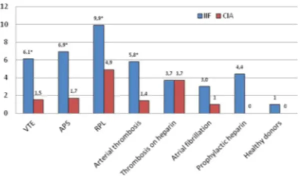
The dense fine speckled pattern was observed in IIF in 31/443 (7.0%) patients (27 women and 4 males) at a titer ranging from 1:80 to 1:2560: eight had APS, eight had VTE, four had arterial thrombosis, ten had RPL, one had thrombosis in treatment with heparin, and in 6/244 (2.5%; p = 0.01) controls (three with atrial fibrillation, two on prophylactic heparin and in one healthy donor). Conversely, anti-DFS70 antibodies were detected by CIA in 11/443 (2.5%) patients (9 women and 2 males; mean value, 118 CU; range, 23.9-450): two with APS, two with VTE, one with arterial thrombosis, five with RPL, one with thrombosis on treatment with heparin, and in 1/244 (0.4%) control with atrial fibrillation (43.8 CU) (p = 0.06) (Fig. 1).
Twenty two patients and five controls were positive only by IIF; two patients were positive only by CIA (at 24 and 33 CU); and nine patients and one control were positive by both methods. Thus, the prevalence rate of posi-tive results by IIF was nearly 3-fold higher than the prevalence detected by CIA. Distribution of results per clinical diagnosis and method are summarized in Table 2. To verify these findings, all positive samples (either by IIF or by CIA) were then tested by a DFS70-specific line-immunoassay (LIA) (ANA Profile 3 plus DFS70 - Euroimmun).
Patients Controls P
No. 443 244 —
Median age, years (range) 53 (21–92) 68 (21–92) <0.0001
Gender F/M 314/129 142/102 —
% females (patients vs. healthy) 70.8 71.0 0.44 Median age females (patients vs. healthy) 49 51 0.39 Table 1. Demographic characteristics of patients and controls.
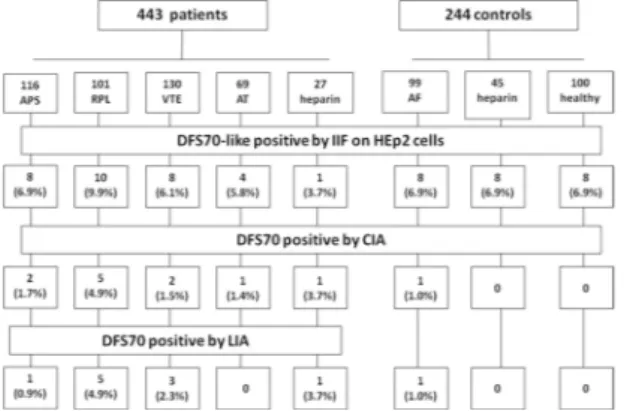
All DFS70 IIF + CIA + samples (9 patients and one control) were confirmed positive for DFS70 anti-bodies by LIA). Instead, of the 27 samples (22 patients and five controls) that were IIF + and CIA−, 25 (92.6%) were negative by LIA as were the two CIA + IIF- samples (Table 3 and Fig. 2).
Women were significantly more prevalent in anti-DFS70 antibody positive patients than in anti-DFS70 antibody-negative patients according to IIF (p = 0.001), but not to CIA (p = 0.17). Anti-DFS70 positive women were younger than anti-DFS70 negative ones both for IIF (p = 0.04) and for CIA (p = 0.001).
aCL (IgG and/or IgM; cutoff 40 GPL-MPL/ml) were detected in 157 of the 294 (53.4%) patients with throm-botic events or RPL that were tested for these antibodies; anti-β2GPI (IgG and/or IgM; cutoff 10 units/ml) in 53/196 (27%), and LAC in 45/173 (26.1%). aCL, anti-β2GPI antibody and LAC frequency was not different between anti-DFS70 antibody positive and negative patients (p = 0.54; 0.72; and 0.44, respectively, for IIF and
p = 0.60; 0.64; and 0.61, respectively, for CIA) (Table 4).
Among patients treated with heparin, 1/27 (3.7%) was DFS70 positive (both in IIF and in CIA, confirmed by LIA) and 2/45 (4.4%) were positive among controls (both positive in IIF and negative in CIA and LIA) (p = 0.68; ns). In the group of patients that were tested for the presence of a genetic abnormality predisposing to throm-bosis, 28/167 (16.8%) had a genetic mutation: nine were heterozygous for factor V Leiden, 10 had prothrombin G20210A mutation, three had homozygous MTHFR mutation with hyperhomocysteinemia, two had factor V Leiden heterozygosity and hyperhomocysteinemia, three had a deficit of protein S and one had prothrombin G20210A mutation and protein C deficiency. Two of these 28 patients (one with RPL and one with VTE) had anti-DFS70 antibodies (one detected only by IIF and one by both IIF and CIA, confirmed by LIA) without signif-icant difference (p = 0.55 for IIF and p = 0.33 for CIA).
Discussion
Anti-DFS70 has been defined as the enigmatic antibody30. Indeed, although anti-DFS70 antibodies have been
thoroughly studied, their clinical association and meaning remain elusive, as their presence does not appear to be linked to any autoimmune disease, but rather to still-undefined events or causes. The recent report by Marlet26,
which suggested the possibility that these antibodies could be associated with thrombosis or RPL, presented an intriguing hypothesis worthy of being studied more in depth. Therefore, in this study, we investigated a broad cohort of patients with venous or arterial thrombosis or with recurrent pregnancy loss, to ascertain whether an association of anti-DFS70 antibodies with thrombotic events or RPL could be confirmed by a DFS70-specific CIA method. Our data show that, when relying on IIF findings, the overall prevalence of the DFS pattern was Figure 1. Prevalence (%) of anti-DFS70 antibodies detected by indirect immunofluorescence (IIF) and by a chemiluminescence method (CIA) in patients and controls (VTE, venous thromboembolism; APS, anti-phospholipid syndrome; RPL, recurrent pregnancy loss). Significant differences are marked by *.
no. IIF + CIA + P IIF + / CIA− IIF− / CIA + IIF + / CIA + Total + Thrombosis/RPL group VTE 130 8 (6.1%) 2 (1.5%) 0.001* 6 (4.6%) 0 2 (1.5%) 8 (6.1%) Arterial thrombosis 69 4 (5.8%) 1 (1.4%) 0.04* 4 (5.8%) 1 (1.4%) 0 5 (7.2%) APS 116 8 (6.9%) 2 (1.7%) 0.03* 7 (6.0%) 1 (0.9%) 1 (0.9%) 9 (7.7%) RPL 101 10 (9.9%) 5 (4.9%) 0.02* 5 (5.0%) 0 5 (5.0%) 10 (9.9%) Thrombosis on heparin 27 1 (3.7%) 1 (3.7%) 1.0 0 0 1 (3.7%) 1 (3.7%) Total 443 31 (7.0%) 11 (2.5%) 0.0001* 22 (4.9%) 2 (0.5%) 9 (2.0%) 33 (7.4%) Control group Atrial fibrillation 99 3 (3.0%) 1 (1.0%) 0.15 2 (2.0%) 0 1 (1.0%) 3 (3.0%) Prophylactic heparin 45 2 (4.4%) 0 0.15 2 (4.4%) 0 0 2 (4.4%) Healthy donors 100 1 (1.0%) 0 0.31 1 (1.0%) 0 0 1 (1.0%) Total 244 6 (2.5%) 1 (0.4%) 0.07 5 (2.0%) 0 1 (0.4%) 6 (3.2%)
Table 2. Distribution of anti-DFS70 antibodies by the detection method, in patients with thrombosis or recurrent pregnancy loss (RPL) and in controls.
7%, which is consistent with the frequency found in many other diseases and approximately half the frequency reported by Marlet (this could be explained by the different selection criteria; a part of Marlet’s patients in the group of patients with thrombosis came from the hematology department to investigate unexplained thrombo-sis). In our series, the highest frequency as detected by IIF was observed in patients with RPL (9.9%), followed by APS (6.9%), VTE (6.1%) and arterial thrombosis (5.8%). However, when the same samples were analyzed with a DFS70-specific CIA method, only five (5.0%) of RPL, one APS (0.9%), two (1.5%) VTE and one (1.4%) arterial thrombosis were confirmed DFS70 positive.
The use of a third independent DFS70-specific LIA method indicated that all CIA-positive samples but two (those that were IIF-negative), contained antibodies targeting the DFS70 antigen.
If the ANA-IIF test alone is considered, an association between thrombotic events and the presence of anti-DFS70 antibodies is confirmed (p = 0.01). However, if the more specific CIA assay is considered
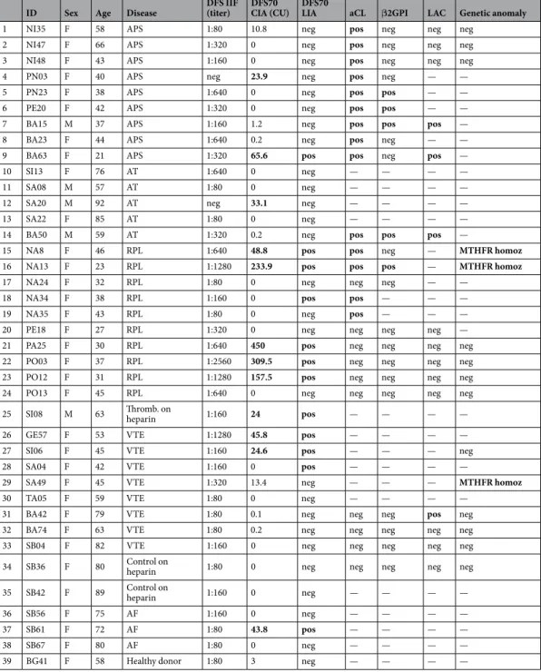
ID Sex Age Disease DFS IIF (titer) DFS70 CIA (CU) DFS70 LIA aCL β2GPI LAC Genetic anomaly
1 NI35 F 58 APS 1:80 10.8 neg pos neg neg neg
2 NI47 F 66 APS 1:320 0 neg pos neg neg neg
3 NI48 F 43 APS 1:160 0 neg pos neg neg neg
4 PN03 F 40 APS neg 23.9 neg pos neg — —
5 PN23 F 38 APS 1:640 0 neg pos pos — —
6 PE20 F 42 APS 1:320 0 neg pos pos — —
7 BA15 M 37 APS 1:160 1.2 neg pos pos pos —
8 BA23 F 44 APS 1:640 0.2 neg pos neg — —
9 BA63 F 21 APS 1:320 65.6 pos pos neg pos —
10 SI13 F 76 AT 1:640 0 neg — — — —
11 SA08 M 57 AT 1:80 0 neg — — — —
12 SA20 M 92 AT neg 33.1 neg — — — —
13 SA22 F 85 AT 1:80 0 neg — — — —
14 BA50 M 59 AT 1:320 0.2 neg pos pos pos —
15 NA8 F 46 RPL 1:640 48.8 pos pos neg — MTHFR homoz
16 NA13 F 23 RPL 1:1280 233.9 pos pos pos — MTHFR homoz
17 NA24 F 32 RPL 1:80 0 neg neg neg — —
18 NA34 F 38 RPL 1:160 0 pos pos — — —
19 NA35 F 43 RPL 1:80 0 neg pos — — —
20 PE18 F 27 RPL 1:320 0 neg neg neg neg —
21 PA25 F 30 RPL 1:640 450 pos neg neg neg neg
22 PO03 F 37 RPL 1:2560 309.5 pos neg neg neg neg
23 PO12 F 31 RPL 1:1280 157.5 pos neg neg neg neg
24 PO13 F 45 RPL 1:640 0 neg neg neg neg neg
25 SI08 M 63 Thromb. on heparin 1:160 24 pos — — — —
26 GE57 F 53 VTE 1:1280 45.8 pos — — — —
27 SI06 F 45 VTE 1:160 24.6 pos — — — neg
28 SA04 F 42 VTE 1:160 0 pos — — — —
29 SA49 F 45 VTE 1:320 13.4 neg — — — MTHFR homoz
30 TA05 F 59 VTE 1:80 0 neg — — — —
31 BA42 F 79 VTE 1:80 0.1 neg neg neg pos neg
32 BA74 F 63 VTE 1:80 0.2 neg neg neg neg neg
33 SB04 F 82 VTE 1:160 0 neg neg neg neg neg
34 SB36 F 80 Control on heparin 1:80 0 neg neg neg neg neg
35 SB42 F 89 Control on heparin 1:160 0 neg — — — —
36 SB56 F 75 AF 1:160 0 neg — — — —
37 SB61 F 72 AF 1:80 43.8 pos — — — —
38 SB67 F 80 AF 1:80 0 neg — — — —
39 BG41 F 58 Healthy donor 1:80 3 neg — — — —
Table 3. Clinical and immunoserological data of anti-DFS70 positive patients. (DFS, dense fine speckled; IIF, indirect immunofluorescence; CIA, chemoluminescence immunoassay; CU, chemiluminescence units; LIA, line-immunoassay; aCL, anticardiolipin antibodies; β2GPI, β2 glycoprotein I; LAC, lupus anticoagulant; APS, antiphospholipid syndrome; AT, arterial thrombosis; VTE, venous thromboembolism; RPL, recurrent pregnancy loss; AF, atrial fibrillation).
independently from IIF results, no significant correlation is observed (p = 0.06). Likewise, if we consider only samples that were confirmed anti-DFS70 positive by LIA (10/12 by CIA and 2/37 by IIF), no association between thrombotic events or RPL and anti-DFS70 antibodies was apparent (p = 0.08).
A possible explanation for the discrepancy between IIF and CIA/LIA results, showing a higher prevalence of DFS70-like pattern by IIF, may reside in the fact that not all antibodies displaying a DFS pattern on HEp-2 cells specifically target the DFS70/LEDGFp75 antigen. Indeed, it has been shown that the DFS70/LEDGF75 pro-tein has many cellular interacting partners which co-localize in the nucleus and that autoantibodies other than anti-DFS70 can produce a similar ANA-IIF pattern31,32. This pattern, recently defined as pseudo-DFS7033, may
explain the higher positive rate of supposed anti-DFS70 antibodies detected by IIF, both in Marlet’s study, as in ours, compared to the CIA and LIA methods. Our results underscore once again that the data on the prevalence of anti-DFS70 antibodies reported in the literature are greatly influenced by the analytical method used34. Hence, the
importance of using specific methods to avoid interpretative errors capable of misleading clinical interpretation. Other interesting data emerge from our study. Since it has been reported that anti-DFS70 are more prevalent in younger people34 and especially in women8,15 of younger age14,35, we have matched the patients with healthy
controls for sex and age: our findings confirm that anti-DFS70 antibodies detected by a specific method are more prevalent in younger women than in older women (p = 0.001) but not in females than males (p = 0.17).
Furthermore, Choi et al.36, in their study on an international inception cohort of 1137 lupus patients, found
that anti-β2GPI antibodies were more frequently associated with anti-DFS70 antibodies (odds ratio 2.17) than aCL and LAC. Though our patient cohorts differ from that of Choi, we cannot confirm an association between anti-β2GPI and anti-DFS70 antibodies as only 5/53 (9.4%) patients with anti-β2GPI were anti-DFS70 positive by IIF and 1/53 (1.9%) by CIA.
Finally, since heparin has been shown to be a stabilizing factor of DFS70/LEDGF, protecting it from prote-olytic degradation under various stress conditions29, to ascertain a possible role played by heparin treatment in
anti-DFS70 expression, we included in this series of patients 25 subjects who had a venous or arterial thrombosis currently under heparin therapy and 45 subjects without thrombosis on prophylactic treatment with heparin before orthopedic surgery. Results show that heparin has no effect on DFS70 antibody production, since only 1/72 subjects on heparin treatment was found to be anti-DFS70 antibody positive by CIA.
Limitations of this study may reside on the fact that data of APS-related antibodies as well as search for genetic anomalies were not available for all patients; also the number of patients treated with heparin was small. However, Figure 2. Flow diagram of study results showing number and percentage of samples that were found positive for anti-DFS70 antibodies by the methods used (IIF, indirect immunofluorescence on HEp2 cells; CIA, chemoluminescence immunoassay; LIA, line-immunoassay; APS, anti-phospholipid syndrome; RPL, recurrent pregnancy loss; VTE, venous thromboembolism; AT, arterial thrombosis; AT, atrial fibrillation).
No. DFS70 + (CIA) DFS70 - (CIA) P
No. 687 12 675 —
Median age (years) all cases 46.2 59.4 0.01
Gender F/M 456/231 10/2 446/229 0.17
Median age in F (years) 39 61 0.001
aCL positive (on 294 tested) 157 4/7 (57.1%) 153/287 (53.3%) 0.60 β2GPI positive (on 239 tested) 53 1/5 (20%) 52/234 (22.2%) 0.64 LAC positive (on 173 tested) 45 1/4 (25%) 44/169 (26.0%) 0.61
On heparin therapy 72 1/72 (1.4%) 71/72 (98.6%) 0.37
Genetic anomaly 32 2/32 (6.2%) 30/32 (93.8%) 0.33
Table 4. Characteristics of anti-DFS70 positive and negative patients by chemiluminescence immunoassay CIA.
to date this is the largest study on the prevalence of DFS70 antibodies in patients with APS and the first one to investigate the possible link between anti-DFS70 antibodies and heparin.
In conclusion, this study is the largest published to date on the prevalence of anti-DFS70 antibodies in patients with thrombotic events or recurrent pregnancy loss. Results obtained by specific CIA and LIA methods do not indicate that venous or arterial thrombosis or RPL are linked to a higher prevalence of anti-DFS70 antibodies. Further studies are needed to solve the DFS70 enigma.
Data availability
The datasets generated during and/or analysed during the current study are available from the corresponding author on reasonable request.
Received: 6 December 2019; Accepted: 17 April 2020; Published: xx xx xxxx
References
1. Singh, D. P., Ohguro, N., Chylack, L. T. Jr & Shinoara, T. Lens epithelium-derived growth factor: increased resistance to thermal and oxidative stresses. Invest. Ophtalmol. Visual Sci. 40, 1444–51 (1999).
2. Shinohara, T., Singh, D. P. & Fatma, N. LEDGF, a survival factor, activates stress-related genes. Prog. Retin. Eye Res. 21, 341–358 (2002).
3. Ganapathy, V., Daniels, T. & Casiano, C. A. LEDGF/p75: a novel nuclear autoantigen at the crossroads of cell survival and apoptosis.
Autoimmun. Rev. 2, 290–297 (2003).
4. Ganapathy, V. & Casiano, C. A. Autoimmunity to the nuclear autoantigen DFS70 (LEDGF): what exactly are the autoantibodies trying to tell us? Arthritis Rheum. 50, 684–688 (2004).
5. Ochs, R. L. et al. The significance of autoantibodies to DFS70/LEDGFp75 in health and disease: integrating basic science with clinical understanding. Clin. Exp. Med. 16, 273–293 (2016).
6. Conrad, K., Röber, N., Andrade, L. E. C. & Mahler, M. The clinical relevance of anti-DFS70 antibodies. Clin. Rev. Allerg. Immunol.
52, 202–216 (2017).
7. Carbone, T. et al. Prevalence and serological profile of anti-DFS70 positive subjects from a routine ANA cohort. Sci. Rep. 9, 2177 (2019).
8. Dellavance, A. et al. The clinical spectrum of antinuclear antibodies associated with the nuclear dense fine speckled immunofluorescence pattern. J. Rheumatol. 32, 2144–2149 (2005).
9. Bizzaro, N. et al. Antibodies to the lens and cornea in anti-DFS70-positive subjects. Ann. N.Y. Acad. Sci. 1107, 174–183 (2007). 10. Mahler, M. et al. Anti-DFS70/LEDGF antibodies are more prevalent in healthy individuals compared to patients with systemic
autoimmune rheumatic diseases. J. Rheumatol. 39, 2104–2110 (2012).
11. Fabris, M. et al. Anti-DFS70 antibodies: a useful biomarker in a pediatric case with suspected autoimmune disease. Pediatrics 134, e1706–1708 (2014).
12. Infantino, M. et al. The clinical impact of anti-DFS70 antibodies in undifferentiated connective tissue disease: case reports and a review of the literature. Immunol. Res. 65, 293–295 (2017).
13. Vazquéz-Del Mercado, M. V. et al. Detection of autoantibodies to DSF70/LEDGFp75 in Mexican Hispanics using multiple complementary assay platforms. Autoimmun. Highlights 8, 1, https://doi.org/10.1007/s13317-016-0089-7 (2017).
14. Watanabe, A. et al. Anti-DFS70 antibodies in 597 healthy hospital workers. Arthritis Rheum. 503, 892–900 (2004).
15. Mariz, H. A. et al. Pattern on the antinuclear HEp-2 test is a critical parameter for discriminating antinuclear antibody-positive healthy individuals and patients with autoimmune rheumatic diseases. Arthritis Rheum. 63, 191–200 (2011).
16. Muro, Y., Sugiura, K., Morita, Y. & Tomita, Y. High concomitance of disease marker autoantibodies in anti-DFS70/LEDGF autoantibody-positive patients with autoimmune rheumatic disease. Lupus 17, 171–176 (2008).
17. Bizzaro, N., Tonutti, E. & Villalta, D. Recognizing the dense fine speckled/lens epithelium-derived growth factor/p75 pattern on HEp-2 cells: Not an easy task! Arthritis Rheum. 63, 4036–4037 (2011).
18. Bentow, C., Fritzler, M. J., Mummert, E. & Mahler, M. Recognition of the dense fine speckled (DFS) pattern remains challenging: Results from an international internet based survey. Autoimmun. Highlights 7, 8, https://doi.org/10.1007/s13317-016-0081-2 (2016). 19. Bizzaro, N. et al. Automated antinuclear immunofluorescence antibody screening: a comparative study of six computer-aided
diagnostic systems. Autoimmun. Rev. 13, 292–298 (2013).
20. Mahler, M. & Fritzler, M. J. The clinical significance of the dense fine speckled immunofluorescence pattern on HEp-2 cells for the diagnosis of systemic autoimmune diseases. Clin. Dev. Immunol. 2012, 494356 (2012).
21. Bizzaro, N. et al. Specific chemoluminescence and immunoasdorption tests for anti-DFS70 antibodies avoid false positive results by indirect immunofluorescence. Clin. Chim. Acta 451, 271–277 (2015).
22. Bizzaro, N. et al. Anti-DFS70 antibodies detected by immunoblot methods: A reliable tool to confirm the dense fine speckles ANA pattern. J. Immunol. Methods 436, 50–53 (2016).
23. Bentow, C. et al. Development and multi-center evaluation of a novel immunoadsorption method for anti-DFS70 antibodies. Lupus
25, 897–904 (2016).
24. Malyavantham, K. & Suresh, L. Analysis of DFS70 pattern and impact on ANA screening using a novel HEp-2 ELITE/DFS70 knockout substrate. Autoimmun. Highlights 8, 3 (2017).
25. Bizzaro, N. & Fabris, M. New genetically engineered DFS70 knock-out HEp-2 cells enable rapid and specific recognition of anti-DFS70 antibodies. Autoimmunity 51, 152–156 (2018).
26. Marlet, J. et al. Thrombophilia associated with anti-DFS70 autoantibodies. PLoS ONE 10, e0138671 (2015).
27. Mahler, M. et al. Towards a better understanding of the clinical association of anti-DFS70 autoantibodies. Autoimmun. Rev. 15, 198–201 (2016).
28. Fatma, N., Singh, D. P., Shinoara, T. & Chylack, L. T. Jr Heparin’s role in stabilizing, potentiating, and transporting LEDGF into the nucleus. Invest. Ophtalmol. Vis. Sci. 41, 2648–2657 (2000).
29. ESHRE Guideline Group on RPL, et al. ESHRE Guideline: Recurrent Pregnancy Loss. Hum. Reprod. Open 2. https://doi. org/10.1093/hropen/hoy004 (2018).
30. Basu, A., Sanchez, T. W. & Casiano, C. A. DFS70/LEDGFp75: An enigmatic autoantigen at the interface between autoimmunity, AIDS, and cancer. Front. Immunol. 6, 116 (2015).
31. Basu, A. et al. Specificity of antinuclear autoantibodies recognizing the dense fine speckled nuclear pattern: Preferential targeting of DFS70/LEDGFp75 over its interacting partner MeCP2. Clin. Immunol. 161, 241–250 (2015).
32. Ortiz-Hernandez, G. L., Sanchez-Hernandez, E. S. & Casiano, C. A. Twenty years of research on the DFS70/LEDGF autoantibody-autoantigen system: many lessons learned but still many questions. Auto Immun. Highlights 11, 3 (2020).
33. Mahler, M., Andrade, L. E., Casiano, C. A., Malyavantham, K. & Fritzler, M. J. Anti-DFS70 antibodies: an update on our current understanding and their clinical usefulness. Expert Rev. Clin. Immunol. 15, 241–250 (2019).
34. Bonroy, C. et al. The importance of detecting anti-DFS70 in routine clinical practice: comparison of different care settings. Clin.
Chem. Lab. Med. 56, 1090–1099 (2018).
35. Albesa, R. et al. Increased prevalence of anti-DFS70 antibodies in young females: experience from large international multi-center study on blood donors. Clin. Chem. Lab. Med. 57, 999–1005 (2019).
36. Choi, M. Y. et al. The prevalence and determinants of anti-DFS70 autoantibodies in an international inception cohort of systemic lupus erythematosus patients. Lupus 26, 1051–1059 (2017).
Acknowledgements
We gratefully acknowledge Inova Diagnostics of the Werfen Group for providing reagents free of charge. Inova Diagnostics had no role in the collection, analysis, or interpretation of the data, the writing of the manuscript, or the decision to submit the manuscript for publication. They accept no responsibility for the contents.
Author contributions
N.B. and G.Pe. conceived and designed the study. G.Pe., M.T.T. and ManM performed the tests. F.F. B.P. N.B. G.Pe. analyzed the data. L.C., G.Pa., M.T., M.G.A., GPr., MagM, C.Z., D.V., I.B., M.L., C.C.a., T.C., C.C.u., L.I., F.M. and D.S. recruited the patients and retrieved clinical records. N.B., G.Pe., M.I. and M.T. wrote the paper. All authors approved the final version of the article.
Competing interests
N.B. has received consultation grants from the Werfen Group. The other authors declare that they have no conflict of interest.
Additional information
Correspondence and requests for materials should be addressed to N.B. Reprints and permissions information is available at www.nature.com/reprints.
Publisher’s note Springer Nature remains neutral with regard to jurisdictional claims in published maps and institutional affiliations.
Open Access This article is licensed under a Creative Commons Attribution 4.0 International License, which permits use, sharing, adaptation, distribution and reproduction in any medium or format, as long as you give appropriate credit to the original author(s) and the source, provide a link to the Cre-ative Commons license, and indicate if changes were made. The images or other third party material in this article are included in the article’s Creative Commons license, unless indicated otherwise in a credit line to the material. If material is not included in the article’s Creative Commons license and your intended use is not per-mitted by statutory regulation or exceeds the perper-mitted use, you will need to obtain permission directly from the copyright holder. To view a copy of this license, visit http://creativecommons.org/licenses/by/4.0/.


