D
D
D
II
I
P
P
P
A
A
A
R
R
R
T
T
T
II
I
M
M
M
E
E
E
N
N
N
T
T
T
O
O
O
D
D
D
II
I
SS
S
C
C
C
II
I
E
E
E
N
N
N
Z
Z
Z
E
E
E
C
C
C
H
H
H
II
I
M
M
M
II
I
C
C
C
H
H
H
E
E
E
,,
,
B
B
B
II
I
O
O
O
L
L
L
O
O
O
G
G
G
II
I
C
C
C
H
H
H
E
E
E
,,
,
F
F
F
A
A
A
R
R
R
M
M
M
A
A
A
C
C
C
E
E
E
U
U
U
T
T
T
II
I
C
C
C
H
H
H
E
E
E
E
E
E
D
D
D
A
A
A
M
M
M
B
B
B
II
I
E
E
E
N
N
N
T
T
T
A
A
A
L
L
L
II
I
C
C
C
O
O
O
R
R
R
S
S
S
O
O
O
D
D
D
II
I
D
D
D
O
O
O
T
T
T
T
T
T
O
O
O
R
R
R
A
A
A
T
T
T
O
O
O
D
D
D
II
I
R
R
R
II
I
C
C
C
E
E
E
R
R
R
C
C
C
A
A
A
II
I
N
N
N
B
B
B
II
I
O
O
O
L
L
L
O
O
O
G
G
G
II
I
A
A
A
A
A
A
P
P
P
P
P
P
L
L
L
II
I
C
C
C
A
A
A
T
T
T
A
A
A
E
E
E
M
M
M
E
E
E
D
D
D
II
I
C
C
C
II
I
N
N
N
A
A
A
S
S
S
P
P
P
E
E
E
R
R
R
II
I
M
M
M
E
E
E
N
N
N
T
T
T
A
A
A
L
L
L
E
E
E
X
X
X
X
X
X
II
I
X
X
X
C
C
C
II
I
C
C
C
L
L
L
O
O
O
Probing the molecular interactions between
human Carbonic Anhydrases (hCAs) and a
novel class of designed benzenesulfonamides
T
ESI DID
OTTORATO:
D
OTT.
SSAE
LVIRAB
RUNO
T
UTOR:
P
ROF.
SSAR
OSARIAG
ITTOC
OORDINATORE DELC
ORSO DID
OTTORATOC
HIAR.
MOP
ROF.
S
ALVATOREC
UZZOCREAT
Abbreviation
IV
Introduction
8
CHAPTER 1
10
In the search of new therapeutics acting as Carbonic Anhydrase
Inhibitors (CAIs) in human
10
1.1 Structure of Carbonic Anhydrase
12
1.2 Distribution of α-CA
16
1.3 Inhibition of the enzymatic activity
19
1.4 Characterization of the interactions between selected
known inhibitors and hCA isoforms
24
CHAPTER 2
34
Results of the present thesis and Discussion
34
2.1 “In vivo” and “in silico” profiling of isoquinolinesulfonamides 13-31
35
2.1.1 Synthesis of isoquinolinesulfonamide derivatives 13-31
36
2.1.2 Biochemical screening and anticonvulsant effects
39
2.1.3 Prediction of biopharmaceutical parameters for compounds 13-31
42
2.2 Development of new arylsulfonamides inhibitors
45
2.2.2 Carbonic anhydrase inhibition assays against hCA isoforms and
Structure-Activity Relationships (SARs) considerations
54
2.2.3 Enantiomeric separation and CA effects of each enantiomer
58
2.2.4 X-ray and docking studies
60
Conclusions
70
CHAPTER 3
72
Experimental section
72
3.1 Chemistry
72
3.2 CA inhibition assay
79
3.3 Anticonvulsant activity in DBA/2 mice
79
3.4 Enantiomeric resolution
80
3.5 X-ray studies
81
3.5.1 Co-crystal structures of inhibitors 70a, 70b, 70e, 71c and
71e in complex with hCAII
81
3.5.2. Co-crystal structures of inhibitors (R)-75d and
(S)-75d in complex with hCA II
82
3.6 Molecular docking
83
References
85
1.1 Recombinant protein
93
1.1.1
Procedure for protein expression in Escherichia coli - BL21(DE3) 95
1.1.2 Procedure to purify the protein
96
2.1 SDS-PAGE electrophoresis
97
3.1 Isothermal titration calorimetry (ITC)
99
References
100
ACC acetyl-coenzyme A carboxylase
Ac-CoA acetyl coenzyme A
ACE angiotensin converting enzyme
AZM acetazolamide
BBB brain blood barrier
BBr3 boron thribromide
CARPs carbonic anhydrase related proteins
CD circular dichroism
CNS central nervous system
CSPs chiral stationary phases
D&D Drug and Development
D2O deuterium oxide
DCM dichloromethane
DMF dimethylformamide
DMSO dimethyl sulfoxide
ED50 effective doses
Et2O diethyl ether
EtOAc ethyl acetate
EtOH ethanol
FC Flash Chromatography
GABA γ-aminobutyric acid
HBTU N,N,N′,N′-Tetramethyl-O-(1H-benzotriazol-1-yl)uronium
hexafluorophosphate
HPLC high performance liquid chromatography
IOP intraocular pressure
Ki inhibition constant
LE ligand efficiency
logBB brain-to-blood concentration ratio
MBG metal binding group
MMPs metalloproteinases
MW microwaves
nM nanomolar
NMR Nuclear Magnetic Resonance
PC pyruvate carboxylase enzyme
PDB Protein Data Bank
POCl3 phosphoryl chloride
PSA polar surface area
SARs structure-activity relationships
TEA Triethylamine
TFA trifluoroacetic acid
TLC Thin-layer chromatography
TPM topiramate
TY tyrosinase
ZBG Zinc-binding group
In the first chapter we have reviewed several previous works in the field of human Carbonic Anhydrase Inhibitors (hCAIs), which have been studied as innovative therapeutics for the treatment of several human diseases. The Chapter 2 describes the “results” of this research work concerning synthetic procedures, chemical characterization, structure-activity relationships (SARs), structural analysis of protein interaction of newer hCAIs. These compounds have been rationally designed on the basis of previous studies that have furnished suggestions about the key chemical features controlling inhibitory effects toward carbonic anhydrases. In this chapter we also discuss our main findings of our studies thus highlighting new suggestions for the development of hCAIs that could target specific and druggable CA isoforms.
In the Chapter 3 we report all experimental procedures for the synthesis and biochemical and pharmacological assays of the designed CA inhibitors. The “in vitro” biological assays have been performed at University of Florence in collaboration with Prof. Claudiu T. Supuran. The “in vivo “ properties of selected hCAIs have been tested in collaboration with Prof. Giovanbattista De Sarro from University of Catanzaro. The crystal structures of the synthesized compounds in complex with the hCA isoforms have been obtained in the laboratories of Prof. Pavlìna Řezáčová at Academy of Sciences of the Czech Republic, Prague and in collaboration with Dr. Giuseppina De Simone from Institute of Biostructures and Bioimaging-CNR, Naples. Finally, in collaboration with Dr. Roberto Cirilli from the Istituto Superiore di Sanità, Rome, HPLC studies have been carried out. In the last part of this dissertation, the Annex summarizes the main skills and competences that have been acquired during the six-months of external experience at University of Dundee, Division of Biological Chemistry & Drug Discovery, School of Life Sciences under supervision of Prof. Alessio Ciulli.
Key words: Carbonic Anhydrase, quinolines, isoquinolines, sulfonamides, HPLC, Docking studies,
X-ray.
Introduction
Carbonic Anhydrases (CAs, EC 4.2.1.1) are a family of monomeric Zinc metalloenzymes that
catalyze the reversible hydration of CO2. The relevant role of this class of enzymes is the regulation
of a broad range of physiological functions (gluconeogenesis, lipogenesis, and ureagenesis).[1]
The family of human carbonic anhydrases (hCAs) comprises 15 different α-carbonic anhydrase
isoforms: CA I-IV, CA VA and CA VB, CA VI-VII, CA IX, CA XII- XV and CARPs CA VIII, CA X, CA XI). In
the last years, the attention toward this class of metalloproteins is in continuous growth because it
was discovered their involvement in different diseases such as cancer,[2] glaucoma,[3] obesity,[4]
and epilepsy.[5] Among the hCAs, the hCA VII is one of the least investigated cytosolic CA isoforms;
it presents a limited distribution, being mainly expressed in the cortex, hippocampus, and thalamus regions within the mammalian brain where it is involved in generating neuronal excitation and seizures. Moreover hCA VII is expressed in other organs including the stomach,
duodenum, colon, liver and skeletal muscle of mice. [2, 5-7]
The hCA VII is currently considered to be involved in the mechanism of GABAergic generated
seizures. [5, 8] Recently, its involvement in neuropathic pain control has been proposed by a
mechanism which is not completely known. [6, 7, 9]Thereby, the investigation on hCA VII could also
represent an interesting tool for the design of new pain killers useful for therapeutic applications. Several CA inhibitors such as acetazolamide (AAZ) and topiramate (TPM) have a long history as anticonvulsants, but their molecular targets and mechanisms of action at the neuronal network level are still poorly understood.[10]
In the last years in the Department ChiBioFarAm – medicinal chemistry laboratories of the University of Messina, a collection of small molecules acting as carbonic anhydrase inhibitors had
been discovered. The most active inhibitors displayed ki values in the nanomolar range.
Unfortunately, some of them showed poor selectivity toward the more druggable isoforms.
These inhibitors possess the sulfonamide portion as key chemical portion that binds the Zinc2+ ion
that is located at the bottom of a deep cleft of catalytic site (Figure 1).[11]
Heterocycle SO2NH- Zn Glu Val Pro Leu Phe Gln Asn His His94 His96 His119
Figure 1. Representation of a hCA inhibitor with sulfonamide moiety.
The other moieties of these compounds control the major or minor interactions with the hydrophilic and/or hydrophobic regions of catalytic pocket. The degree of this pattern of contacts might be considered responsible of the inhibitory potency as well as selectivity toward specific CA isoforms.
During the three years doctoral course we chose to introduce new chemical moieties and modify these sulfonamides to obtain the new compounds. Specifically, we planned the synthesis of new compounds to improve inhibitory effects and isoforms selectivity.
Therefore, we planned the synthesis of a series of new sulfonamides bearing the quinoline and isoquinoline skeleton that has been variously decorated through the introduction of chemical fragment as hydrophilic/hydrophobic anchoring groups.
This series of novel hCAIs were in vitro tested against the hCA I, hCA II, hCA VII, hCA IX, hCA XII and hCA XIV. Structural and computational studies have been performed to explain the mechanism of inhibitory properties. Furthermore, in vivo studies were carried out to find new potential therapeutics for the treatment of brain pathologies.
CHAPTER 1
In the search of new therapeutics acting as Carbonic
Anhydrase Inhibitors (CAIs) in human
Several metalloenzymes are involved in a wide array of in vivo functions, regulating blood pH,
facilitating matrix degradation, modulating DNA transcription, and many others.[12]
Moreover, given the relevance of these functions, an imbalance of the levels of these metalloenzymes plays a significant role in several diseases. In detail, pathologies for which metalloenzymes are implicated include cancer, [13, 14] heart disease, [15] epilepsy[16] and obesity.[17]
For this reason the development of metalloenzymatic modulators as new drugs for the treatment of these diseases could result in a good and innovative strategy in the Drug and Development (D&D) process.
Typically, metalloenzyme inhibitors are drug-like small molecules that incorporate a metal binding group (MBG) able to coordinate the metal ion within active site. By inducing an impairment of catalytic function, the MBG is appended to the drug-like portion of the inhibitor via a linker that
can take on many forms, reflective of the diversity in enzyme active sites.[18]
To represent a diverse range of MBGs we choose to analyze a small array of metalloproteins.
Matrix metalloproteinases (MMPs) are a group of Zn2+-dependent endopeptidases known for their
potential to disrupt angiogenesis in malignant tumors and represent a prototypical metalloenzyme
target in medicinal chemistry. The MMP active site contains a catalytic Zn2+ ion coordinated by a
His3 motif and a water molecule. The discovery of several potent broad-spectrum MMP inhibitors, have not successfully completed clinical trials for some disease (e.g. cancer, arthritis, etc.), due to
problematic side effects, lack of efficacy in human trials, and other barriers.[18]
Angiotensin converting enzyme (ACE, EC 3.4.15.1) is a Zn2+ dipeptidyl carboxypeptidase in which
the catalytic Zn2+ ion is bound to the protein by a His2 Glu motif. ACE functions endogenously to
generate the biologically active polypeptide angiotensin II from angiotensin I. The cleaved product induces a vasoconstriction response that manifests as increased blood pressure. An important metalloenzyme inhibitor for treating hypertension, Captopril (Capoten, Squibb, patented in
1976),[19] incorporates a thiol group capable of coordinating the catalytic Zn2+ ion in the active site
of ACE. [18]
Mushroom tyrosinase (TY) is a dinuclear Cu2+ oxidase controlling the production of melanin. The
two active site Cu2+ ions are bound by six histidine residues (three to each metal ion). TY converts
tyrosine to L-DOPA and subsequently L-DOPA to dopaquinone. Then dopaquinone, a highly reactive compound determinant in the melanogenesis, rapidly and spontaneously evolves towards
the formation of various melanin pigments.[20] Consequently, TY inhibitors are of interest to the
cosmetics industry. [18]
Discovered in the 1930s, [21] Carbonic Anhydrases (CAs) represent the oldest known class of Zn
metalloenzyme. In many organisms these enzymes are involved in crucial physiological processes
connected with pH and CO2 homoeostasis/sensing; biosynthetic reactions, respiration and
transport of CO2/bicarbonate, electrolyte secretion in a variety of tissues/organs, bone resorption;
calcification, tumorigenicity and many other physiological or pathological processes (thoroughly
studied in vertebrates and some pathogens).[22] The attention toward this family of enzymes is
incremented due to the possibility to develop therapeutics in prevention and treatment of various diseases such as glaucoma, obesity, cancer, etc.
The family of carbonic anhydrases (CAs, EC 4.2.1.1) catalyse the interconversion between CO2 and
bicarbonate.[23-25]
The physio/pathological relevance of this reaction is that the exceedingly high amounts CO2 may
damage cellular components, while its conversion to water soluble ions (bicarbonate and protons),
may interfere with the pH balance of the cell through the generation of an acid (H+) and a
buffering base (HCO3−).[22]
To date the CA enzymatic families are: α-, β-, γ -, δ-, ζ -and η classes. The α-CAs are present in
vertebrates, protozoa, algae, cytoplasm of green plants and in many Gram negative bacteria;[26-28]
the β-CAs are found in both Gram negative and positive bacteria algae and chloroplasts of mono-
as well as dicotyledons, and also in many fungi and some Archaea. [23-25, 27] The γ -CAs were found
in Archaea, cyanobacteria and most types of bacteria,[26, 29] the δ- and ζ-CAs seem to be present
only in marine diatoms,[24, 30] whereas the η-CAs in protozoa.[23, 31]
CO2 + H2O HCO3- + H+
The class of α-CA is present in mammals; human carbonic anhydrase isoforms (hCAs) are subdivided in 15 different isoforms, that play an important physiological and patho-physiological functions “in vivo”.
1.1 Structure of Carbonic Anhydrase
The active site of the CA shows a conical shape; it displays a 15 Å diameter entrance that tapers into the center of the enzyme. In the depth of the catalytic site we can find the MBG in a tetrahedral geometry. There are three amino acid residues (His94, His96 and His119) generating coordination bonds. Additionally, there is a water molecule/hydroxide ion that is coordinated with the metal ion.
The presence of Zn2+ ion characterizes all six CA genetic families, but Cd2+ is interchangeable with
Zn2+ in the ζ-CAs, [25] Fe2+ seems to be present in γ-CAs, at least in anaerobic conditions, [32]
whereas Co2+ may substitute the Zinc ion in many α-CAs without a significant loss of the catalytic
activity. [23-25, 33-35]
The cavity is partitioned in to two very different environments. The area containing the zinc, in the depth of the active site, lies a cluster of hydrophobic amino acids (Val121, Phe131, Leu141, Val143, Leu198 and Val207), whereas on the other side of the zinc, leading out of the active site to the bulk solvent, the surface is lined with hydrophilic amino acids (Asn62, His64, Asn67, Gln92) (figure 2).[22, 36]
The bulky hydrophobic residue Phe131 divides the hydrophobic area in two sub-sites in which
various classes of inhibitors bind in a specific manner.[36-39] Indeed, the mechanism where the zinc
binds the CO2 is supposed that the hydrophobic part is used to entrap the CO2 molecule. [36]
The hydrophilic region facilitates the binding of the polar components generated from the CO2
hydration reaction (bicarbonate and protons) and their release from the cavity, towards the
environment.[22]
B -BH+ O N H H N HN His94 His96 His119 O H H H O H O O O H His64 Glu106 Thr199 Zn CO2 -CO2 O N H H N HN His94 His96 His119 O H H O H O O His64 Glu106 Thr199 Zn O C O O N H H N HN His94 His96 His119 O H H O H O O His64 Glu106 Thr199 Zn O C O -O N H H N HN His94 His96 His119 O H H O H O O His64 Glu106 Thr199 Zn H O H 2 H2O HCO3 -O N H H N HN His94 His96 His119 O H H O H O O His64 Glu106 Thr199 Zn H O H H H
Scheme 1. Catalytic mechanism for the α-CA catalyzed CO2 hydration. Hydrogen bonds are indicated by dashed lines
in red. The hydrophobic cleft is indicated by an line.[40]
The enzymatic mechanism of the CA is is shown in the scheme 1. Zinc is the key of the enzyme
reaction. The water bond to the Zn+2 ion is actually broken down to a proton and hydroxyl ion.
Since zinc is a positively charged ion it stabilizes the negatively charged hydroxyl ion.[40]
Phe131 Leu141 Val143 Leu198 Val207 a b c d
13
The active form of the enzyme is the basic one, with hydroxide bound to Zn2+ (a). This strong
nucleophile attacks the CO2 molecule that is bound in a hydrophobic pocket in its neighbourhood
(b), leading to the formation of bicarbonate coordinated to Zn2+ (c). The bicarbonate ion is then
displaced by a water molecule, leading to the acidic form of the enzyme, with water coordinated to Zn2+ (d), this form is catalytically inactive.[23]
Generally, in the catalytically very active isozymes such as CA II, CA IV, CA VII and CA IX, occurs the
regeneration of basic form (a), the His64 assists to the a proton transfer reaction from the active
site to the environment or as well as by a cluster of histidines, which protrudes from the rim of the active site to the surface of the enzyme, assuring thus a very efficient proton transfer process for
the most efficient CA isozyme, CA II.[41, 42] Another pathway can transform the Zn2+ from the
inactive form (d) to active form (a). Specifically, it can be by buffers present in the medium but this
is the rate-limiting step in catalysis.
Figure 2. Catalytic pocket with the hydrophobic side in red and the hydrophilic region in green. The zinc is shown in
grey with the three amino acid residues (His94, His96 and His119) and the Phe131 in blue. Picture modified from reference [36].
In the mechanism regenerating the zinc ion/hydroxyl in the catalytic process, there are several histidines called proton shuttling that are involved in this processes (such as the residue of His64).[32, 35, 36, 38, 39]
Indeed, His64 is one of the few amino acid residues that has a flexibility within the catalytic pocket. This phenomenon has been demonstrated through x-ray studies where the imidazole
moiety presents two conformations: an ‘in’ one, pointing towards the Zn2+ ion, and an ‘out’ one,
pointing towards the exit of the cavity.[43] The two conformations are part of the proton transfer
mechanism by which this residue shuttles protons between the active site and the reaction medium (see Figure 3).[38, 39, 43]
Figure 3. Catalytic pocket of hCA II, with zinc ion in grey, the molecules of water in red. His64 in both conformation,
“In” and “Out”. (PDB code 2CBA)[44]
As shown in the Figure 3, the catalytic Zn2+ ion is located at the bottom of this cavity, coordinated
by the three histidine residues and a water molecule/hydroxide ion. Moreover, the MBG is engaged in hydrogen bond interactions with another water molecule (called the deep water) and with the hydroxyl moiety of the threonine residue (Thr199), which in turn is bridged to the carboxylate moiety of a glutamic acid residue (Glu106) (figure 4).[23, 34, 38, 39, 43]
His64 “In” “Out” Zn His96 His94 His119
15
Figure 4.The ordered water network in the active site of hCA II. The zinc is represented by a grey sphere and the oxygen atoms of water molecules as smaller red spheres. Dotted lines are presumed hydrogen bonds with heavy atom
distances given. Stick figures are selected amino acids of the active.[45]
As displayed in the Figure 4, the presence of these two amino acids in the catalytic pocket results
in an enhancement of the nucleophilicity of the Zn2+ bound water molecule, and orients the CO
2
substrate in a location favorable for the nucleophilic attack.
The residues Thr199 and Glu106 are important elements in the catalytic side for all α-CAs, so they
are called gate-keeping residues.[22]
1.2 Distribution of α-CA
In mammals, the different CA isozymes and CA-related proteins (CARP) display different tissue distribution, expression levels, and subcellular locations. In more details, there are five cytosolic forms (CA I, CA II, CA III, CA VII and CA XIII), five membrane bound isozymes (CA IV, CA IX, CA XII,
CA XIV and CA XV), one mitochondrial forms (CA V), and a secreted CA isozyme (CA VI).[42]In
addition, there are the catalytically inactive isoforms of α-carbonic anhydrases known as carbonic
anhydrase related proteins (CARPs) and classified as CA VIII, CA X, and CA XI. The hCA I, II and IV are involved in respiration and regulation of the acid/base homeostasis. These processes involve both
the transport of CO2/bicarbonate between metabolizing tissues and excretion sites (lungs,
kidneys), they facilitate CO2 elimination in capillaries and pulmonary microvasculature, elimination
of H+ ions in the renal tubules and collecting ducts, as well as reabsorption of bicarbonate in the
brush border and thick ascending Henle loop in kidneys. [42]
Moreover, CAs are involved in vision and control the production of the bicarbonate-rich aqueous humor secretion (mediated by ciliary processes isozymes CA II, CA IV and CA XII) within the eye; so
their malfunctioning leads to high intraocular pressure, and glaucoma. [42, 46] Specifically, the hCA II
is also involved in the bone development and function, such as the differentiation of osteoclasts, or the provision of acid for bone resorption in osteoclasts.
CAs are involved in the secretion of electrolytes in many other tissues/organs, such as: saliva production in acinar and ductal cells; gastric acid production in the stomach parietal cells; bile
production, pancreatic juice production, intestinal ion transport. [42, 46]
CAs also play a role in gustation and olfaction, protection of gastro-intestinal tract from extreme pH conditions (too acidic or too basic), regulation of pH and bicarbonate concentration in the seminal fluid, muscle functions and adaptation to cellular stress. Some isozymes, such as CA V,
control molecular signaling processes, such as insulin secretion signaling in pancreas β cells. [42, 46]
Isozymes II and VA are involved in important metabolic processes, as they provide bicarbonate for gluconeogenesis, fatty acids de novo biosynthesis or pyrimidine base synthesis.
Finally, some isozymes (such as CA IX, CA XII, CARP VIII) are highly abundant in tumors, being involved in oncogenesis and tumor progression.
Currently, CA inhibitors are clinically used as antiglaucoma agents, diuretics, antiepileptics, in the management of mountain sickness, gastric and duodenal ulcers, neurological disorders, or
osteoporosis among others.[42, 47, 48]
The development of carbonic anhydrase inhibitors (CAIs) are of growing interest since various isoforms of the enzyme are identified as promising drug targets for treatment of diseases. The principal drawback of the clinically used CAIs is the lack of isoform selectivity, which may lead to observable side effects. [49]
Table 1. Classification of the hCA isoforms.
Isoform Sub-cellular localization Tissue/organ localization Disease Therapeutics
hCA I Cytosol Erythrocytes, GI tract cerebral edema retinal No drug
hCA II Cytosol
Erythrocytes, eye, GI tract, bone osteoclasts, kidney,
lung, testis, brain
glaucoma acetazolamide, methazolamide, ethoxzolamide, dichlorophenamide, dorzolamide edema epilepsy acetazolamide, topiramate, zonisamide altitude sickness No drug hCA III Cytosol Skeletal muscle, adipocytes oxidative stress No drug
hCA IV Membrane-bound Kidney, lung, pancreas, brain capillaries, colon, heart muscle glaucoma acetazolamide, methazolamide, ethoxzolamide, dichlorophenamide, dorzolamide Retinitis pigmentosa No drug
stroke No drug hCA VA/VB Mitochondria muscle, pancreas, kidney, Liver/Heart and skeletal
spinal cord, GI tract
obesity topiramate, zonisamide
hCA VI Secreted into saliva/milk Salivary and mammary glands cariogenesis No drug hCA VII Cytosol CNS epilepsy acetazolamide, topiramate,
zonisamide hCA VIII
(CARP VIII) Cytosol CNS
neurodegeneration No drug cancer Indisulam (E7070) hCA IX Transmembrane Tumours, GI mucosa cancer Indisulam (E7070)
hCA XII Transmembrane reproductive epithelia, eye, Renal, intestinal, tumours
cancer Indisulam (E7070)
glaucoma acetazolamide, methazolamide, ethoxzolamide, dichlorophenamide, dorzolamide hCA XIII Cytosol Kidney, brain, lung, gut, reproductive tract sterility No drug
hCA XIV Transmembrane Kidney, brain, liver
epilepsy acetazolamide, topiramate, zonisamide retinopathy No drug
1.3 Inhibition of the enzymatic activity
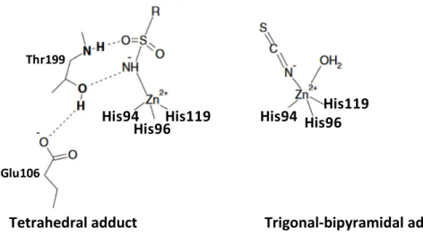
For a long period, the only mechanism of CA inhibition known was the interaction between the metal ion in tetrahedral geometry with the zinc-binding group (ZBG as MBG) or by adding to the metal coordination sphere and generating trigonal bipyramidal geometry, as reported in the Figure 5.[22]
Glu106 Thr199
His94
His96His119 His94 His96 His119
Figure 5. α-CA inhibition mechanism between the metal ion with the ZBG in two different geometries.
The anions may bind either in tetrahedral geometry of the metal ion or as trigonal–bipyramidal adducts, such as for instance the thiocyanate adduct shown in Figure 5.
Others examples of the inorganic anions are generally represented by halogens, such as chloride, iodide and bromide, or cyanate and the sulphur and anions characterized by less affinity for metal ions in solution (tetrafluoraborate, perchlorate, nitrate, fluoride and heavier halides, among
others).[42, 50] The application in therapy of anions is not possible due of the many side effects and
even their use is limited by the small size which makes it difficult the optimization of their structure.
The tetrahedral geometry is formed by the unsubstituted sulfonamides that bind the Zn2+ ion
replacing the non-protein ligand. Sulfonamides and their isosteres
Tetrahedral adduct Trigonal-bipyramidal adduct
(sulfamates, sulfamides),[23, 29, 39] are the most important CAIs binding in a tetrahedral
geometry.Krebs (1948) discovered that unsubstituted aromatic sulfonamides are inhibitors of the
carbonic anhydrase.[51]
The classical CA inhibitors (CAIs) are the primary sulfonamides, R-SO2NH2, which are in clinical use
for more than 50 years as diuretics and systemically acting antiglaucoma drugs.[52]
In fact there are ~30 clinically used drugs (or agents in clinical development). Some of them are shown in Figure 6, they belong to the sulfonamide or sulfamate, such as the classical inhibitors
acetazolamide (1), methazolamide (2), ethoxzolamide (3), dichlorophenamide (4) and also more
recent drugs/investigational agents such as dorzolamide (5), brinzolamide (6), indisulam (7),
topiramate (8), celecoxib (9), sulthiame (10), saccharin (11), zonisamide (12) (figure 6).[43, 53-59].
O N H N S S O O N H 2 N Acetazolamide (1) S N H2 O O N N O N H S Methazolamide (2) N S O O N H2 S O Ethoxzolamide (3) Cl Cl S O O N H2 S O O N H2 Dichlorophenamide (4) N H S O O N H2 S S O O Dorzolamide (5) N H S O O N H2 S S O O O Brinzolamide (6) SO2NH 2 NH O S O N H Cl Indisulam (7) (E7070) O O O O S O O N H2 O O Topiramate (8) SO2NH2 N O Celecoxib (9) SO2NH2 OMe O N N H Sulthiame (10) H2NO2S O O O Saccharin (11) SO2NH2 O N Zonisamide (12)
Figure 6. Classics sulfonamides CAIs
Some of these compounds are useful in reducing elevated intraocular pressure (IOP), but have a limited use due to numerous side effects that arise by inhibition of CAs other than those present in the eye ciliary processes (i.e., CA II, IV, and XII), leading to fatigue, paresthesias, and kidney
stones.[23] Moreover, can also be use as diuretic and, in addition, for the treatment of cancer,
epilepsy, obesity, neuropathic pain and infections.[2, 5, 9, 29, 59] However, critical barriers to the
design of CAIs as therapeutic agents are related to the high number of isoforms in humans, their
rather diffuse localization in many tissues/organs, and the lack of isozyme selectivity of the
presently available inhibitors of the sulfonamide/sulfamate type.[52]
The interaction of the inhibitor with the metallic cofactor is described below.
In deprotonated state the -SO2NH− moiety coordinates to Zn2+ ion and creates an extended
network of hydrogen bonds, involving residues Thr199 and Glu106, also participating to the anchoring of the inhibitor molecule to the metal ion.
The largest majority of the designed sulfonamides possess an aromatic scaffold. This approach was really beneficial for the chemistry of this class of derivatives, as a large number of new ring systems that was and will be explored in this way. The development of new inhibitors of CA includes a “tail that might induce water solubility. Specifically, amino, hydroxyl, immino or hydrazino moieties can be bound to different scaffolds of well-known aromatic/heterocyclic sulfonamides; in this way these inhibitors maintain affinity for the CA active site, assuring in this way the possibility to modulate in greater details the physico-chemical properties of these pharmacological agents (figure 7).[48]
Figure 7. Generic skeleton of CAIs
The inhibition of hCAs can be carried out with others mechanism. In detail, several inhibitors can occlude the active site entrance that is located further away from the metal ion, at the entrance of the site cavity . Such compounds incorporate a “sticky group” which may be of the OH, amino,
COOH and other types. This alternative mechanism of action was discovered for coumarins [60, 61]
and it was later shown that many other classes of structurally similar compounds, which bind to the enzyme in this manner (Figure 8).[62, 63]
Figure 8. Hydrogen bonds in which the coumarin binds the residues into the hCA II active site.
The inhibition mechanism of this class of compounds can be explain as depicted in Scheme 2. Several coumarins/thiocoumarins may possess various tautomeric forms, such as the zwitterionic benzo(thio)pyrylium phenoxides, which may bind within the CA active site similarly to phenols by anchoring to the zinc-bound water molecule/hydroxide ion. In the first step, coumarins/thiocoumarin, shown in the step A, may undergo hydrolysis by the zinc-activated water molecule/hydroxide ion from the enzyme cavity, which acts as a very potent nucleophile. It can be hypothesized that a cis- or trans-2-hydroxy/mercapto-cinnamic acid intermediate is formed (step
B), which cannot bind effectively in the restricted space near the Zn2+ ion due to its bulky skeleton,
being thus reoriented toward the exit of the active site cavity. In the final step the compound
suffers for a rearrangement of the enzyme-inhibitor adduct; step C,provided that the R and R1
moieties from the initial (thio)coumarin are not too bulky to interfere with the binding to the
enzyme active site.[61]
The inhibitor cis or trans 2-hydroxy-cinnamic acid was found bound at the entrance of the active
site cavity (Figure 8).[60] In detail, the COOH moiety of the inhibitor interacts with two amino acid
residues of the hydrophilic part of the hCA II active site (Asn62 and His64) by means of two
hydrogen bonding interactions (Figure 8).[60, 61]
X O R R1 X O -R R1 + CA XH R R1 COO -His94 His96 His119 OH2 Zn X O R R1 His94 His96 His119 O -Zn H hydrolysis H2O XH R R1 COO -His94 His96 His119 OH2 Zn X=O, S Zwitterionictautomer Coumarin, X=O Thiocoumarin, X=S isomerisation A B C
Scheme 2. Proposed inhibition mechanism of CAs by coumarins/thiocoumarins.
1.4 Characterization of the interactions between selected
known inhibitors and hCA isoforms
Many hCAIs bearing sulfonamide moiety possess high affinity for the most part of isozymes considered to play important physiological functions. However, the critical challenge for the design of novel pharmacological agents from this class is constituted by the lack of specificity of such
compounds towards the different isozymes.[48]
Some of these isoforms present the differences on aminoacidic residues that modulate the hydrophobicity and charge while others change the active site cavity volume and shape. These differences in active site environment in turn modulate inhibitor binding constants and their
dynamics of entry.[64]
To describe the main interactions within catalytic site of selected isoforms we report several specific binding poses for well-known hCAIs currently in therapy for treatment of epilepsy, obesity and cancer.
Acetazolamide (AZM, 1): AZM was initially discovered as a diuretic agent, but its clinical use was
limited due to its transient action and onset of metabolic acidosis. However AZM corrects the significant metabolic alkalosis which occurs with loop diuretics such as furosemide, bumethanide, torasemide, and so on; so this this compound displays more clinical interest as a diuretic.
Furthermore, the inhibitor 1 is highly efficient for the treatment of patients with hypercapnia and
metabolic alkalosis. However, the main use of AZM and other first generation CAIs, such as
methazolamide (2) and dichlorophenamide (4), is the therapeutic treatment of glaucoma.[65] It is
interesting to consider thatAZM dissolved in water has three possible protonation states with two
associated pKa values (7.2 and 8.7) that are relevant to physiological pH (figure 9). It is thought
that any of these forms can bind to hCA II.
-+H+ +H+ -H+ -H+ NH O N H S O O S N N O N H S O O N H2 S N N O N– S O O N H2 S N N
Figure 9. Ionization and pKa of acetazolamide (AZM, 1) in water.
The AZM was co-crystallized with the hCA II isoform and has been found that AZM was in the
anionic form, with the negatively charged sulfonamide group coordinated to the zinc.
The lone pair of sulfonamide N is involved in a coordinating bond with the zinc of ~2.4 Å distance
(figure 10 A), it makes the H bonding interaction with the oxygen of Thr199, which in turn acts as a H-bond donor to Glu106.
pKa ~ 8.7 pKa ~ 7.2
Moreover the amidic group binds a water molecule, W1120, which forms an H-bond bridge
between AZM and hCA II. Indeed, W1120 interacts with the hydroxyl of Thr200 and with the carbonyl
group of Pro201.
AZM also interacts through two very weak hydrophobic interactions: the first is a type of –CH---π
interaction (~3.5 Å distance) between Leu198 and the thiadiazole ring of AZM. The other is a
somewhat distorted –CH---π interaction between the terminal –CH3 of AZM and Phe131. As weak
as these interactions are, they contribute to the overall binding of AZM to hCA II (figure 10 B).
A
B
Figure 10. (A): Interaction between AZM and hCA II (PDB code 4G0C); zinc is shown as a magenta sphere. Hydrogen
bonds as observed in the nuclear maps are indicated by dashed lines with distances as indicated. (B): AZM makes
hydrophobic interactions within the active site of hCA II.[66]
Moreover, AZM demonstrated a modulator in anticancer therapies in combination with different cytotoxic agents, such as alkylating agents, nucleoside analogs, platinum derivatives, etc. It was hypothesized that the anticancer effects of AZM (alone or in combination with such drugs) might be due to the acidification of the intra-tumor environment ensued after CA inhibition, although
other mechanisms of action of this drug were not excluded. [66, 67]
Figure 11 shows the comparison of the catalytic pocket between hCA II and hCA IX. Acetazolamide
inhibits the hCA II and hCA IX with the Ki values in the range nM (12 and 25 nM, respectively). The
comparison of the hCA II and hCA IX active sites shows that there are two main differences for
residues Phe131 and Val135, for which Val and Leu are present in hCA IX, respectively. Phe131 (in hCA II) is involved in very weak hydrophobic interactions with AZM and its absence in hCA IX could,
in part, explain the small 2-fold difference in binding constants for AZM between hCA II and IX. [66]
Figure 11. Complex between hCA II and AZM (yellow cartoon and sticks) and residues that are different residues in
hCA IX are shown in green ball-and-stick (Val131 and Leu135). Zn2+ is shown as a blue sphere. (PDB code 3IAI) [66]
Since several decades, coadministration of 1 with various antiepileptic drugs (AEDs), such as
topiramate (8) and zonisamide (12), produced a remarkable increase of the anticonvulsant activity
for barbiturates including barbital, mephobarbital, metharbital; phenytoin; valproic acid and trimethadione. The mechanism for which the AZM enhances the anticonvulsant effects of these AEDs are poorly understood but it was inferred that the difference in the potentiating effects of
acetazolamide on the activity of various antiepileptics might be correlated to the effect that 1 has
on the brain levels of these drugs as well as pH changes that acetazolamide may induce by
inhibiting the many CA isoforms present in the brain. [66, 67] Acetazolamide is still used in
combination therapy with the AEDs or in refractory epilepsies.[68]
Moreover, it was observed that AZM (1) displays Ki values at nanomolar concentration against hCA
VII (2.5nM) and is characterized by hCA VII selectivity over hCA II. By analyzing the X-ray crystal
His64 His94 His96 His119 AZM Phe131 Val131 Val135 Leu135
27
structure of 1 in complex with CA VII (PDB code 3ML5) the main interactions between the inhibitor
and the protein were studied. Specifically, a structure-based pharmacophore model has been
carried out using LigandScout and the results are displayed in Figure 12. [69]As shown in this figure
AZM binds the catalytic side with seven hydrogen bonding features including: four H-bond acceptors which bind residues Gln92, Thr199, Thr200, and a water molecule (W354); three H-bond
donors pointed towards His94, Thr199, and a water molecule (W354). [69]
Figure 12. Structure-based pharmacophore model generated using LigandScout[70] from the X-ray crystal structure of
acetazolamide (1) and in complex with CA VII (PDB code 3ML5). Hydrophobic groups (light yellow spheres), H-bond
donors (green arrow), H-bond acceptors (red arrow). [69]
Topiramate (TPM, 8): TPM is a sugar sulfamate derivative possessing good anti-epileptic activity.It
is derived from a monosaccharide (β-D-fructopyranose) and bears the sulfamate functional group
that is considered to be responsible for its anticonvulsant properties, even if the mechanism of
action of this drug seems to be rather complicated and not entirely understood. The
anticonvulsant effects of topiramate are probably due to CO2 retention secondary to inhibition of
the red cell and brain enzymes,but other mechanisms of action were also hypothesized for TPM.
In fact TPM shows a positive modulatory effect on some types of GABAA receptors, antagonizes
kainate/AMPA receptors and inhibits the generation of action potentials in neurons via antagonism of Na+ channels activation.[10, 71, 72]
In Figure 13 is reported the X-ray structure of the complex between 8 and hCA II. Beyond the
known interaction between the nitrogen atom of the sulfonamide moiety of the inhibitor 8 and
the Zinc ion at the tetrahedral vertex with a distance of 1.97 Å, it makes an extended network of hydrogen bonds between the inhibitor and some amino acid residues within the cavity strongly
stabilize the complex. In detail, Asn62 with one of the oxygen (O4) of topiramate (3.06 Å), Gln92
with the other oxygen (O6) of topiramate (2.85 Å) and the water molecule 1134 that makes a
bridge between the inhibitor and Thr200 through two hydrogen bonds, one with O3 (2.84 Å) of
the inhibitor and the other with the oxygen of the residue Thr200 (2.92 Å). The same oxygen of
Thr200 makes hydrogen bond with the pyranose oxygen of topiramate (2.82 Å).[72] In this case the
interactions between the inhibitor and the hCA II are the same that have been shown previously.
Gln92 W1134 Thr200 Thr199 Asn62 Zn2+ 1.97 Å 2.66 Å 3.06 Å 2.85 Å 2.82 Å 2.84 Å 2.92 Å 2.85 Å O4 O3 O6
Figure 13. Complex between topiramate (8) and hCA II. In red the corresponding distances (in Å).
The inhibitory activity of TPM has been tested against other hCA isoforms and topiramate showed
a Ki in subnanomolar concentration against the hCA VII (0.9 nM) thus corroborating its contribute
to epileptic activity. There is not yet the crystal structure between the TPM and the hCA VII but this inhibitor was and is still used as antiepileptic drug.
TPM has recently been approved for a second clinical use, as an antiobesity agent. Indeed, it reduces lipogenesis by inhibiting CA isoforms involved in metabolic pathways such as the mitochondrial CA VA/B or the cytosolic CA II.
The role of mitochondrial CAs is to assist the mitochondrial pyruvate carboxylase enzyme (PC) affording carbon units, in the form of bicarbonate ions, which are incorporated in the pyruvate, to form oxaloacetate which in turn is then converted into citrate through the reaction with acetyl coenzyme A (Ac-CoA). Citrate, unlike oxaloacetate and Ac-CoA, is able to translocate from the mitochondria into the cytosol by means of the tricarboxylate transporter, and once in loco gets degraded back into oxaloacetate and CoA by the ATP- cytrate lisase. The oxaloacetate is then decarboxylated to afford pyruvate which is retaken into the mitochondria by the pyruvate carboxylase transporter. The cytosolic activated CoA is then converted into malonyl-CoA by means of the cytosolic acetyl-coenzyme A carboxylase (ACC) which uses the bicarbonate provided by the cytosolic CA II. The malonyl-CoA units are then elongated in the same manner to afford fatty acids. It has been demonstrated that sulfonamide/sulfamate CAIs inhibit this process in vitro and in vivo
through modulation of this mechanism. As indicated in Figure 13, the TPM in complex with CA II
assumes the classical tetrahedral coordination of the sulfamate group to the zinc ion, whereas the fructose part is entrapped into the enzymatic cleft by means of a large number of hydrogen bonding and van der Waals interactions. Using the TPM/hCA II adduct, molecular dynamic/docking
studies have been carried out to observe the hypothetic binding mode between the inhibitor 8
and unexplored isoform hCA VA. This study strongly suggested that there is a similar mode of binding between the complex TPM/hCA II and TPM/hCA VA (figure 14), with exception of the
hydrogen bond interaction Asn62 with one of the oxygen (O4) of topiramate (figure 13) that is not
retained in this case, as a consequence of the mutation Asn62 – Thr62 (figure 14) for hCA VA. The loss of this important polar interaction seems to be responsible for the lower binding affinity observed for TPM toward hCA VA with respect to that measured for hCA II (Ki = 13.8 nM against
hCA II and 25.4 nM against hCA VA).[73, 74]
Leu65 TPM Thr200 Thr199 His96 His119 His94 Gln92 Thr62 Tyr131
Figure 14. Molecular dynamic/docking studies between topiramate (8) and hCA VA.[73, 74]
All these results strongly support the role that TPM inhibits mitochondrial CAs and, therefore, de
novo lipogenesis, thus explaining the antiobesity effects of TPM.
Indisulam (7)(E7070): Indisulam is a novel sulfonamide anticancer agent in clinical development
for the treatment of solid tumors. The inhibitor 7 was found to suppress the expression of cyclin E
and the phosphorylation of CDK2, both of which are essential for the G1 to S transition. Fukuoka
et al. [75] clarified that 7disrupted cell cycle progression at multiple points, including both G1/S and
G2/M transitions, in a human non-small cell lung cancer cell line A549 (adenocarcinomic human
alveolar basal epithelial cells). The compound 7 proved to inhibit pRb phosphorylation, to reduce
the protein expression of cyclin A, cyclin B1, CDK2 and CDC2, and to suppress CDK2 catalytic
activity with the induction of p53 and p21 proteins only in parental (drug sensitive) A549 cells. [75,
76]
The relationship studies between the sulfonamides with the antitumor activity have so far clarified
that the sulfamoyl group (–SO2NH2) is not an essential functionality for the in vitro growth
inhibitory activity against cancer cells. However, there is still a possibility that the CA inhibitory
properties of the compound 7 can contribute, at least in part, to its in vivo efficacy. The isozymes
most abundant and considered to play a critical physiological/pathological role, such as CA II and
CA IX, exhibit the highest affinity for 7, with an inhibition profile quite similar to those of the
clinical drug 1 (Ki = 12 nM and 25 nM of AZM (1) toward hCA II and hCA IX respectively; Ki = 15 nM
and 24 nM of E7070 (7) toward hCA II and hCA IX respectively).
So, the CA inhibition of 7 might be considered as a positive factor for the clinical strategy of
antimetastasis and combination therapies.[76]
The X-ray crystal structure of the adduct of hCA II with E7070 (7) revealed unexpected
interactions. Three different conformations of the chloroindole fragment were found. As you can
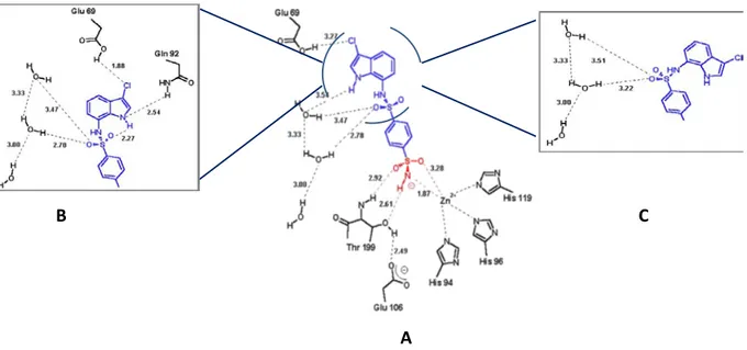
see in Figure 15, E7070 binds within the hCA II active site in three different conformations that
regards only the terminal, chloroindole fragment of the molecule, which is rather flexible and allows the three different spatial arrangements, whereas the benzenesulfonamide head binds unequivocally to the active site in one conformation, similarly to other sulfonamide CA inhibitors. About the known interaction between the zinc ion and the sulfonamide moiety the Zn–N bond is appreciably shortened in this complex as this distance is usually around 1.95–2.10 Å. This
shortening may be considered as a first factor favoring the high affinity of 7for hCA II. [76]
The benzenedisulfonamide moiety of 7lies in the hydrophobic part of the active site cleft, where it
makes van der Waals interactions with the side chains of Val 135, Phe 131, Leu 204, Pro 202, Trp 209, Val 121, Leu 198 and Thr 200. In conformation A (Fig. 2A), the chlorine atom is engaged in a
weak hydrogen bond with the COOH group of Glu69. In the second conformation of 7(Fig. 2B), the
NH group of the inhibitor is then involved in hydrogen bond (of 2.54 Å) with the -CONH2 group of
Gln92.The chlorine atom forms a shorter (1.88 Å) hydrogen bond with the -COOH group of Glu69.
Finally, the third conformation of 7 bound to hCA II (Fig. 2C) involves only a network of four
hydrogen bonds in which one oxygen atom of the secondary sulfonamide moiety and three water molecules participate. [76]
B
A
C
Figure 15. Complex between indisulam (7) and hCA II. A, B and C are the representation of the three conformation of
the chloroindole portion of the inhibitor 7 within the hCA II active site. [76]
Currently, 7is under clinical evaluation as antitumor agent in patients with colorectal cancer,
non-small cell lung cancer, and so on. [76]
Therefore, the crystal structures of selected well-known inhibitors in complex with druggable isoforms have furnished interesting information about the binding pose within catalytic site of druggable isoforms. Overall, the sulfonamide/sulfamate moiety establishes the network of contacts that control the binding recognition of CA. Additional interactions can address the isoform selectivity thus highlighting both the volume and shape of catalytic site cavity for each isoform.
Chapter 2
Results of the present thesis and Discussion
To date numerous small molecules have been claimed to inhibit metalloenzymes such as human
Carbonic Anhydrase (hCAs). Specifically, hCA inhibitors (hCAIs) are able to coordinate the Zn2+ ion
and establish additional interactions promoted by the aryl substituents in the region nearby the catalytic site. [69, 77]
At the medicinal chemistry laboratory of the University of Messina previous studies allowed the identification of a new class of hCAIs. Actually, a large series of isoquinolinesulfonamides have been synthesized and these derivatives were tested as inhibitors of hCA I, hCA II, hCA IX and hCA XIV.[1, 77-80]
Therefore, the data obtained demonstrated that most of these compounds are good inhibitors
with Ki in the range of nanomolar concetration. In particular, several synthesized sulfonamide
derivatives showed high inhibitory activity toward hCA II, hCA IX and hCA XIV. [1, 77]
13-22 23-31 N N R4 R3 R2 R1 R4 R3 R2 R1
Figure 16. hCA inhibitors Isoquinolinesulfonamides 13-31.
2.1 “In vivo” and “in silico” profiling of
isoquinolinesulfonamides 13-31
Our idea was to focus the interest on the development of new hCAIs able to produce neuroprotectant effects through a selective inhibition of hCA VII isoform, that is widely distributed in the brain. To achieve this objective, in the first step of this research project we decided to collect further biological data for the already synthesized isoquinolines 13-31. [1, 77-80]
Specifically, we determined the inhibitory effects towards hCA VII for compounds 13-18, for which
these data are not yet reported in literature. Considering that the activity of carbonic anhydrase can be associated to neurological disorders, such as epilepsy, linked to excitatory neurotransmission, we further explored in vivo effects. In fact, it is well known that neuronal excitability is related to γ-aminobutyric acid (GABA)ergic depolarization and the prolonged
activation of GABAA receptors can lead the imbalance of the ions. This imbalance can determine
the improvement of intracellular HCO3- ions that is consistently replenished by the activity of
carbonic anhydrase. For this reason the inhibition of CA represents a good strategy for the treatment of epilepsy.[81-83]
Several CA inhibitors such as acetazolamide (AAZ, 1) and topiramate (TPM, 7) have a long history
as anticonvulsants, given that hCA II and hCA VII are specific isoforms involved in GABA-mediated
neuronal excitation. So we herein hypothesize that the compounds 13-31 could represent an
innovative template for the development of new anticonvulsant agents.[11]
Following previously reported procedure, we re-synthesized compounds 13-31 and their
anticonvulsant effects were tested against audiogenic seizures in DBA/2 mice. [1, 77-80]
2.1.1 Synthesis of isoquinolinesulfonamide derivatives 13-31
The desired isoquinoline derivatives 13-31 were prepared via synthetic routes outlined in Schemes
3 and 4. We used the phenylenthylamine derivatives (32-34) as starting reagents, that were
converted into the two series of key intermediates 35-49 and 50a-51. Following the well-known
Pictet-Spengler approach, in the first step of the synthetic pathway the phenylethylammines 32-34
undergo a condensation with suitable aldehydes thus giving imines 35-49. [84, 85] The main feature
of this reaction is that gives good yields, reducing the reaction time and the some reaction work
without solvent. [86] In a parallel route the starting amines 32-34 reacted with the
4-(aminosulfonyl)benzoic acid and furnished the corresponding 4-methyl-N-phenethyl-benzamide
derivatives (50a-51) via N,N,N,N-Tetramethyl-O-(1H-benzotriazol-1-yl)uronium
hexafluorophosphate (HBTU) coupling in the presence of triethylamine (TEA).
i 32, R1 = R2 = OMe 33, R1 = OMe; R2 = H 34, R1 = R2 = H 35-49 50a, R1 = R2 = OMe 51, R1 = OMe; R2 = H ii N N H2 O N H R3 CHO R1 R2 R 3 R1 R2 SO2NH2 HO2C SO2NH2 R1 R2
Scheme 3. Reagents and conditions: i) HBTU, DMF, ET3N, rt, 2h; ii) MW: 5 min, 90°C, 200 Psi, 150 W.
In agreement with expected nucleophilicity of the amine, the imines 35-49 cyclized in the
corresponding isoquinolines 52-67 through microwave irradiation (MW, 5 min, 90°C, 200 Psi, 150
W) in the presence of trifluoroacetic acid (TFA). Successively, these intermediates reacted with a
large excess of sulfamide leading to the 3,4-dihydro-1H-isoquinoline-2-sulfonamide derivatives
13-18 and 23-31. 52-67 iv 35-49 13-15 R1 = R2 = OMe 16-18 R1 = OMe; R2 = H 23 R1 = R2 = R3 = H 24-31 R1= R2 = OMe R3 13, 16 C6H5 14, 17 4-Cl-C6H4 15, 18 4-NH2-C6H4 R3 24 H 25 CH3 26 C2H5 27 n-C3H7 28 c-C3H7 29 n-C4H9 30 31 iii R1 R2 R3 R2 R1 R 3 R3 R2 R1 SO2NH2 N H N N
Scheme 4. Reagents and conditions: iii) TFA MW: 5 min, 90 °C, 200 Psi, 150 W; iv) MW: 20 min, 100 °C, 200Psi, 150W.
By using the Bischler-Napieralski approach we prepared the desired isoquinolines 20-22 through a
multistep procedure. Initially, the phosphoryl chloride (POCl3) has been used to promote the
intramolecular electrophilic aromatic substitution allowing the cyclization of the intermediates
N-phenethyl-4-sulfamoyl-benzamides (50a,51). So, we obtained the corresponding
dihydroisoquinolines derivatives 68,69. Successively, the reagent NaBH4 was used to convert the
imine 68,69 in the corresponding secondary amine 19,21
(6,7-dimetossi-1-phenyl-1,2,3,4-tetrahydroisoquinoline and 6-metossi-1-phenyl-1,2,3,4-tetrehydroisoquinoline, respectively).
Finally, compounds 19,21 were treated with sulfamide to give the
3,4-dihydro-1H-isoquinoline-2-sulfonamides derivatives 20-22.
v 50a R1 = R2 = OMe 51 R1 = OMe, R2 = H 68 R69 R1 = R2 = OMe 1 = OMe, R2 = H 19 R1 = R2 = OMe 21 R1 = OMe, R2 = H 20 R1 = R2 = OMe 22 R1 = OMe, R2 = H vi iv N O N H N H N SO2NH2 R1 R2 R2 R1 SO2NH2 R2 R1 SO2NH2 R2 R1 SO2NH2 SO2NH2
Scheme 5. Reagents and conditions: v) POCl3, Δ, toluene, 2h; vi) NaBH4, CH3OH, 2h.
2.1.2 Biochemical screening and anticonvulsant effects
Table 2 reports the inhibitory effects of the isoquinolinesulfonamides 13-31 against hCA II and hCA
VII isoforms. The inhibitory constants were obtained using a Durrum-Gibson stop-flow
spectrophotometer that measures the hydration (or dehydration) of CO2 and then the release (or
removal) of protons. [87, 88]
The inhibition constant (Ki) values determined for compounds 13-31 were compared with the Ki
values of topiramate (TPM, 7) as reference standard showing both CA inhibition and
anticonvulsant properties (Topamax®). [71, 72] Table 2 summarizes the effective doses (ED50)
determined forcompounds 13-31 that were administered intraperitoneally 30 min before auditory
stimulation using DBA/2 mice, a strain susceptible to sound induced-seizures. The ED50 values
were determined for the inhibition of clonic and tonic seizures, these data were compared with
those measured for TPM.[78]
As you can see in Table 2, the monomethoxy compounds 16-18 are less efficacious inhibitors
against the hCA VII isoform than corresponding dimethoxy analogues 13-15, which proved to be
potent hCA VII inhibitors at nanomolar concentration (Ki < 8.2 nM). Moreover, compounds 13-15
proved to be inefficacious to inhibit hCA II (Ki >5000 nM). This unexpected isoform selectivity
might be considered an advantage for the development of CA inhibitors lacking off-target effects
related to activity toward ubiquitous isoforms such as hCA II. In particular, the compound 15,
which bears a 4-aminophenyl group at the C-1 position of the isoquinoline scaffold, displayed the highest selectivity for the hCA VII isoform (~2000-fold) over the hCA II isoform and showed relevant anticonvulsant effects in comparison with that shown by reference compound
topiramate 7.
The replacement of the optimized 4-aminophenyl substituent with the 4-benzenesulfonamide
moiety gave compounds 19-22. Similar to the results of previous SAR, we observed that the
dimethoxy isoquinolines derivatives 19 and 20 were more selective inhibitors of hCA VII than
monomethoxy analogues 21,22.[77] The “in vivo” screening revealed that compounds 19-22 were
less potent anticonvulsant when compared with the active inhibitor 15. The lacking of substituents
on benzene-fused ring as well as nitrogen atom of isoquinoline nucleus results in low selective hCA
VII inhibitors 23-24, which were active agents just in preventing tonic phase.


![Figure 12. Structure-based pharmacophore model generated using LigandScout [70] from the X-ray crystal structure of](https://thumb-eu.123doks.com/thumbv2/123dokorg/4584380.38843/29.892.276.681.336.696/figure-structure-based-pharmacophore-generated-ligandscout-crystal-structure.webp)

![Figure 14. Molecular dynamic/docking studies between topiramate (8) and hCA VA. [73, 74]](https://thumb-eu.123doks.com/thumbv2/123dokorg/4584380.38843/32.892.281.614.153.489/figure-molecular-dynamic-docking-studies-topiramate-hca-va.webp)