Case Report
Lipomatous Hypertrophy: An Accidental Finding in Heart
F. Petrelli
,
1,2L. Spagnoli,
1,2S. Aversa
,
1,2S. Tripodi,
2and C. Bellan
1,21Department of Medical Biotechnologies, University of Siena, Siena, Italy 2Pathology Unit, Azienda Ospedaliera Universitaria Senese, Siena, Italy Correspondence should be addressed to C. Bellan; [email protected] Received 8 May 2019; Accepted 27 June 2019; Published 14 July 2019 Academic Editor: Mark Li-cheng Wu
Copyright © 2019 F. Petrelli et al. This is an open access article distributed under the Creative Commons Attribution License, which permits unrestricted use, distribution, and reproduction in any medium, provided the original work is properly cited.
Lipomatous hypertrophy is an uncommon benign lesion of the atrium, generally asymptomatic, characterized by unencapsulated accumulation of adipose tissue entrapping cardiomyocytes. This pathology generally remains unnoticed and often emerges as an occasional finding. Here, we report two cases from our hospital including a review of the available literature.
1. Introduction
Lipomatous hypertrophy is a benign cardiac mass char-acterized by an unencapsulated accumulation of mature adipose tissue within the interatrial septum, rarely in the atrium wall [1]. As most patients with this condition remain asymptomatic, the majority of cases are generally picked up as incidental findings at the time of cardiac imaging, surgery, or necropsy [2, 3]. Some symptomatic cases, however, are described in literature [4, 5].
Here, we report two cases of lipomatous hypertrophy referred to our hospital, one after surgical removal and the other at autopsy.
2. Case Report
The first case was found incidentally on preoperative inves-tigation of a 73-year-old female patient with progressive coronary heart disease. The lesion, completely asymptomatic, was detected by transthoracic and transesophageal echocar-diography and initially appeared as an exophytic mass occu-pying the right atrial cavity, lacking the typical hourglass morphology and appearing suspicious for atrial myxoma. Despite the absence of symptoms related to the lesion, the surgeons decided to remove it anyway in order to promote blood overflow through the atrial cavity and prevent later obstruction. The patient was subsequently admitted to the Cardiac Surgery Department for planned coronary artery bypass surgery and mass resection. During surgery, the lesion
turned out to be also intraparietal but was totally resected. At macroscopic examination the lesion was 3 cm in diameter, yellowish, and slightly increased in consistency without a true capsule. Although surgery was successful, the patient died because of her illness.
The second case is a 73-year-old male patient with an incidental finding in autopsy of a lesion of 4 cm in diameter in the interatrial septum that appeared thickened. No clinical problems or suspicious symptoms had emerged from the clinical information gathered.
3. Results and Discussion
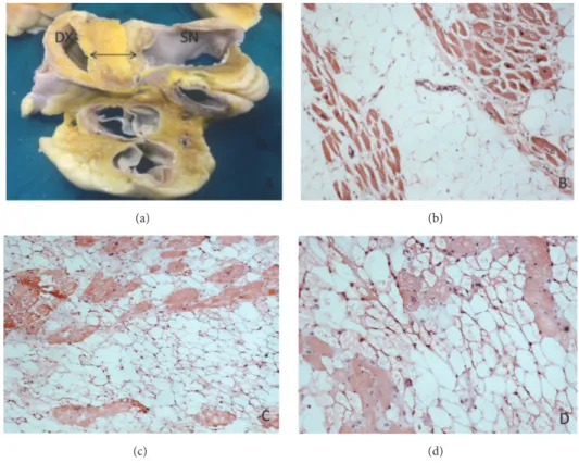
Both lesions, after fixation with 10% formalin solution, were sampled and embedded in paraffin to prepare 3-𝜇m-thick sections subsequently stained with haematoxylin-eosin. Histologically, both of the lesions demonstrated an unencapsulated accumulation of mature adipocytes with infiltrative growth pattern within hypertrophied cardiac myocytes. Lesion cells presented microvacuolated cytoplasm and centrally located nuclei, also exhibiting variation in shape and size, dispersed within the fat. Mitoses were absent (Figure 1). The pathologic differential diagnosis included cardiac myxoma, lipoma, and liposarcoma [6]. The diagnosis of myxoma was discarded due to the presence of fat and hypertrophied myocytes instead of the typical stellate or glob-ular myxoma cells, characterized by abundant eosinophilic cytoplasm, indistinct cell borders, oval nuclei with open chromatin, and indistinct nucleoli, and of myxoid ground
Hindawi
Case Reports in Pathology
Volume 2019, Article ID 8613724, 2 pages https://doi.org/10.1155/2019/8613724
2 Case Reports in Pathology
(a) (b)
(c) (d)
Figure 1: Macroscopic appearance of the lesion observed at autopsy (a) and histological overview showing extensive fat entrapping cardiomyocytes from atrial lesion of the first case (b) and from the interatrial septum of the second (c-d). Haematoxylin and Eosin 10X (b-c); Haematoxylin and Eosin 20X (d).
substance. Lipoma, unlike lipomatous hypertrophy, is an encapsulated lesion composed of only mature adipocytes and few, if any, myocytes. Finally, also well-differentiated liposarcoma could be excluded because it typically consists of lipoblasts containing large clear vacuoles and hyperchromatic indented nuclei [7].
Lipomatous hypertrophy represents a benign though rare condition that should always be considered in the differential diagnosis in case of an atrial mass detected during imaging, surgery, or necroscopy and definitively diagnosed by histo-logical examination.
Disclosure
The authors declare that the data cited in this report is avail-able in the references mentioned, accessed from PubMed.
Conflicts of Interest
The authors declare that they have no conflicts of interest regarding the publication of this paper.
References
[1] I. Nadra, D. Dawson, S. A. Schmitz, P. P. Punjabi, and P. Nihoy-annopoulos, “Lipomatous hypertrophy of the interatrial sep-tum: a commonly misdiagnosed mass often leading to unnec-essary cardiac surgery.,” Heart (British Cardiac Society), vol. 90, no. 12, p. e66, 2004.
[2] G. Bielicki, M. Lukaszewski, K. Kosiorowska, J. Jakubaszko, R. Nowicki, and M. Jasinski, “Lipomatous hypertrophy of the atrial septum - a benign heart anomaly causing unexpected surgical problems: A case report,” BMC Cardiovascular Disorders, vol. 18, no. 1, article no. 152, 2018.
[3] T. Strecker, M. Weyand, and A. Agaimy, “Lipomatous hypertro-phy of the atrial septum in a patient undergoing coronary artery bypass surgery,” Case Reports in Pathology, vol. 2016, Article ID 2080875, 4 pages, 2016.
[4] M. Bolognesi, “Lipomatous hypertrophy of the interatrial sep-tum and arrhythmia: A case report,” Journal of Integrative
Cardiology, vol. 2, no. 4, pp. 320-321, 2016.
[5] A. P. Burke, S. Litovsky, and R. Virmani, “Lipomatous hypertro-phy of the atrial septum presenting as a right atrial mass,” The
American Journal of Surgical Pathology, vol. 20, no. 6, pp. 678–
685, 1996.
[6] K. M. Lampropoulos, D. Kotsas, and T. Iliopoulos, “Lipomatous hypertrophy of interatrial septum,” BMJ Case Reports, 2012. [7] S. O’Connor, R. Recavarren, L. C. Nichols, and A. V.
Par-wani, “Lipomatous hypertrophy of the interatrial septum: an overview,” Archives of Pathology & Laboratory Medicine, vol. 130, no. 3, pp. 397–399, 2006.