UNIVERSITY OF CATANIA
Ph.D. School in
ONCOLOGY
Coordinator: Prof. Franca Stivala
XXIV Cycle
Ph.D. Thesis
TRANSCRIPTION FACTORS INVOLVED IN
THE GENESIS AND PROGRESSION OF
CANCER DIFFERENTLY MODULATED BY
TRANSFORMING GROWTH
FACTOR-BETA3 (TGF-3) IN PROSTATE CELL LINES
Silvia Caggia
I
Abstract
Transforming growth factor- (TGF-) is a member of a family of multifunctional cytokines that control different cellular processes including cell proliferation, morphogenesis, migration, extracellular matrix production, cytokine secretion, and apoptosis, as well as in normal physiological and disease processes. TGF- utilizes a multitude of intracellular signaling pathways in addition to Smads with actions that are dependent on circumstances, including dose, target cell type, and context. TGF- pathway has been implicated in cancer and has been recently considered as a putative therapeutic target. The aims of this study were to verify the effects of dose-dependent TGF-3 treatment in BPH-1 cell line, human benign prostate hyperplasia, and two prostate cancer cell lines, LNCaP, which is androgen-sensitive, and DU-145, which is androgen-non responsive, evaluating a correlation between p53 and YY1. Moreover, the expression of several parameters (PI3K, AKT, pAKT, PTEN, Bcl-2, Bax, PARP, Rb, pRb, cyclin A and iNOS) involved in both cell cycle progression and in apoptosis was evaluated through Western blot analysis on prostate cultures treated with 10 and 50 ng/ml of TGF-3 for 24 h. The production of nitric oxide (NO•) was
II
determined by Griess reagent and cell viability by MTT assay. The results of this research demonstrated profound differences in the responses of the BPH-1, LNCaP, and DU- 145 cell lines to TGF-3 stimulation. We believe that the findings could be important because of the clinical relevance that they may assume and the therapeutic implications for TGF- treatment of prostate cancer.
III
Acknowledgments
I want to thank those who have supported and encouraged me during my PhD, in particular Prof. V. Cardile, F. Stivala, M.C. Mazzarino, G. Malaponte, and M. Libra. I want also to thank my family for giving me the freedom and opportunity to pursue my own interests, all my friends, especially Michela, Genny, Marianna, Anna, Paola, Katya, and who made life in Catania enjoyable and sociable. I am grateful to the colleagues A. Graziano, G. Frasca, M. Coco and all who shared with me the joys and the difficulties in recent years between pipettes, flasks and cells. Finally, I wanted to remind all staff of the Department of Bio-medical Sciences, Physiology Section: I have welcomed and appreciated by their sympathy and kindness.
IV
Contents
Introduction ... 1
Prostate cancer ... 1
Transforming growth factor-beta3 (TGF-β3) ... 6
Apoptosis ... 12
Yin Yang 1 and p53 proteins ... 14
Inducible nitric oxide synthase (iNOS) ... 18
Phosphatidylinositol 3 kinase (PI3K) pathway ... 20
Bcl-2 and Bax ... 23
Poly(ADP-ribose) polymerase (PARP) ... 25
Cell cycle ... 27
Aims ... 30
Materials and methods ... 31
Cell cultures and treatments ... 31
Cell viability ... 32
Western blot analysis ... 32
YY1 silencing by small interfering RNA (siRNA) ... 34
Statistical analysis ... 35
Results ... 36
Discussion... 52
Conclusions and future work ... 63
1
Introduction
Prostate cancer
The prostate is a gland found only in men. It is just below the bladder and in front of the rectum. The site of the prostate varies with age. In younger men, it is the size of a walnut, but it can be much larger in older men. The tube that carries urine (urethra) runs through the centre of the prostate. The prostate contains cells that make some of the fluid (semen) that protects and nourishes the sperm.
http://www.myoptumhealth.com/portal/ADAM/item/Prostate+cancer The prostate begins to develop before birth and keeps on growing until a man reaches adulthood. Male hormones (androgens) cause this growth. If male hormone levels are low, the prostate gland will not grow to full size. In older men, though, the part of the prostate around the urethra may keep on growing. This causes BPH (benign prostatic hyperplasia) which can lead to problems passing urine because the
2
prostate can press on the urethra. BPH is a problem that often must be treated, but it is not cancer.
The prostate gland is composed of epithelial cells, which form two layers and stromal cells. There are three types of epithelial cells: secretory glandular cells, nonsecretory basal cells, and neuroendocrine cells [1]. The basal cells lack ARs (Androgen receptors), are androgen independent, and are thought to be stem cells for secretory epithelial cells [2]; the neuroendocrine cells may play a role in regulating the growth and function of the secretory cells [3]. The stroma of the prostate is composed of smooth muscle cells, fibroblasts, lymphocytes, and neuromuscular tissue embedded in an extracellular matrix. Evidence suggests that epithelial-stromal interactions play an important role in normal prostatic morphogenesis [4]. In normal tissue, such interactions are often paracrine with, for example, receptors for a particular growth factor present only on epithelial cells and production of the factor only by stromal cells. In cancer, some growth factor pathways become autocrine, enabling the epithelial cells, which express a growth factor and its receptor, to grow independently of stromal cells.
Prostate cancer is a form of cancer that develops in the prostate. Most prostate cancers are slow growing; however, there are cases of aggressive prostate cancers. Prostate cancer may cause pain, difficulty
3
in urinating, problems during sexual intercourse, or erectile dysfunction. Other symptoms can potentially develop during later stages of the disease. It tends to develop in men over the age of fifty and although it is one of the most prevalent types of cancer in men, many never have symptoms, undergo no therapy, and eventually die of other causes. This is because cancer of the prostate is, in most cases, slow-growing, symptom-free, and since men with the condition are older they often die of causes unrelated to the prostate cancer, such as heart/circulatory disease, pneumonia, other unconnected cancers, or old age. On the other hand, the more aggressive prostate cancers account for more cancer-related mortality than any other cancer except lung cancer [5].
http://www.myoptumhealth.com/portal/ADAM/item/Prostate+cancer The estimation by the International Agency for Research on Cancer revealed 679,000 new cases of and 221,000 deaths related to prostate cancer on a global level in 2002 [6] and more recently the National Cancer Institute estimated 240,890 new cases and 33,720 deaths from
4
prostate cancer in the United States in 2011. With an estimated 5-year prevalence of 2.3 million patients in the world, prostate cancer is a major global health problem.
The incidence of prostate cancer (CaP) increases with age [6], family history and race/ethnicity [6-8]. A study suggests that ~42% of the risk for CaP may be explained by heritable factors [9]. Several works based on epidemiological and genetic studies have proposed genes such as HPC1 [10], CAPB [11], BRCA1 and BRCA2 [12], as susceptibility genes for CaP. Chronic inflammation has been associated with increased risk in CaP [13]. The initiation, maintenance and pathology of the inflammatory response depend upon pro- and anti-inflammatory signals. Interleukin (IL)-1β, tumor necrosis factor (TNF)- and IL-10 are critical in the regulation of inflammation [14-17].
When the tumor is localized to the prostate, the relative cure rate by either radical prostatectomy or radiation therapy is high. However, if the cancer is detected at an advanced stage, when the disease has already spread outside the prostate, the prognosis for survival is dramatically decreased. The precise molecular mechanisms involved in prostate carcinogenesis remain uncertain, but obviously disruption in the balance of proliferation and apoptosis of epithelial cells is a key event. In this regard, a number of studies have focused on alterations
5
in transforming growth factor-beta (TGF- β) signaling during prostate carcinogenesis [18, 19].
Most human prostate cancer cell lines have been established from metastatic deposits, with the exception of PC-93 [20], grown from an AD (androgen-dependent) primary tumor. However, PC-93 and other widely used lines, including PC-3 [21], DU-145 [22], and TSU-PR1 [23], are all AI (androgen-independent); all lack ARs (with the possible exception of PC-93), PSA, and 5-reductase; and all produce poorly differentiated tumors if inoculated into nude mice. The paucity of cell lines that are AD has made studies of the progression of prostate cancer using human material very difficult. However, metastatic sublines of PC-3 have been developed by injection of cells into nude mice via different routes, especially orthotopically [24]. The LNCaP cell line, established from a metastatic deposit in lymph node [22], is the only human prostate cancer cell line that demonstrates androgen sensitivity but not androgen dependence. After its initial characterization [22], several laboratories found that this line was poorly tumorigenic in nude mice unless coinoculated with tissue-specific mesenchymal or stromal cells [25] or Matrigel [26], suggesting that extracellular matrix and paracrine-mediated growth factors play a role in prostate cancer growth and site-specific metastasis [27]. LNCaP cells grown in castrated mice that had
6
progressed to the AI state were cultured to obtain new cell lines. The C-4 LNCaP [28] line produces PSA and a factor that stimulates PSA production, and the C4-2 line metastasizes to lymph nodes and bone after subcutaneous or orthotopic inoculation [29, 30]. Another subline of LNCaP, LNCaP 104-R2, cultured in androgen-depleted medium for >100 passages, is stimulated by finasteride, causing some concern over the use of antiandrogens for the treatment of late-stage prostate cancer [31].
Transforming growth factor-beta3 (TGF-β3)
http://www.ncbi.nlm.nih.gov/Structure/
Transforming growth factor family members are multifunctional cytokines that control a diverse array of cellular processes including cell proliferation, morphogenesis, migration, extracellular matrix production, cytokine secretion, and apoptosis [32]. Members of the
7
transforming growth factor-β (TGF-β) superfamily, including TGF-βs, activins/inhibins, and bone morphogenetic proteins (BMPs), regulate a wide range of biological phenomena in metazoan organisms [33]. Activins and inhibins were originally identified as factors that regulate secretion of follicle stimulating hormone (FSH) from the pituitary gland and were later shown to regulate fundamental developmental processes such as mesoderm induction. BMPs induce bone and cartilage formation in ectopic tissues. Like activins, BMPs also play pivotal roles in various developmental events including the establishment of the dorso-ventral axis, neural induction, and organogenesis of kidneys and eyes. TGF-βs, the prototype of the superfamily, are potent growth inhibitors of various lineages of cells including epithelial, endothelial, and hematopoietic. Disruption of the signaling pathway of TGF-β is thus implicated in the initiation of tumors. Indeed, many cancer cells have lost responsiveness to TGF-β. TGF-β also induces production of extracellular matrices, cell migration, angiogenesis, and immunosuppression through which TGF-β may provide the environment that promotes tumor invasion. Therefore, elucidation of the signaling pathway of TGF-β is important in understanding the molecular basis of carcinogenesis [34].
Many studies have helped to determine which molecular and cellular mechanisms involving TGF-β1 play a causative role in tumorigenesis
8
[35–37]. In contrast to the TGF-β1 isoform, the role of the TGF-β3 isoform in tumorigenesis has been considerably less well studied, with assumptions regarding its role in tumorigenesis often being made based on observations reported for TGF-β1.
Regarding to the amino acid sequences, the three TGF-β isoforms share a high level of similarity between the active domains; TGF-β3 is 86% similar to that of TGF-β1 while it shares 91% similarity with that of TGF-β2. Despite homology in amino acid sequence, it is known that TGF-β3 differs significantly from TGF-β1 and -β2 in its detailed tertiary structure of the active domain. Nuclear Magnetic Resonance (NMR) data show that the alpha3 helical region of TGF-β1 is structurally ordered [38], while the alpha3 helical region of TGF-β3 is structurally disordered [39, 40]. This indicates that TGF-β3 can adopt a more flexible ‗‗open‘‘ state, which is observed in both the crystal structure of free TGF-β3 [41] and in its complex with TβRII [42]. One consequence of this difference in structural flexibility is that TGF-β1 may lock the receptor complex in a closed tight conformation, while TGF-β3 may allow a more open conformation of the receptor complex due to the greater flexibility of the TGF-β3 dimer. The implications of these observations are that the structure of the ligand/receptor complexes for TGF-β1 and -β3 may be significantly different and may engage the downstream signaling pathways in different ways, thus
9
leading to qualitatively and quantitatively different biological outcomes. TGF-β3 has been found to have an important role in normal developmental biology including systems such as the heart, lung and breast and to display isoform-specific biology at both the in vivo and
in vitro level. Understanding the unique biology of TGF-β3 is
important to understanding the role it may have in tumorigenesis. Data from experimental systems and human cancers clearly show that, in addition to the TGF-β ligands, the TGF-β receptors and their primary cytoplasmic signal transducers all play an important role in suppressing primary tumorigenesis in many organs [43, 44]. However, observations made in the later stages of disease suggest that increased TGF-β1 expression is required for disease progression [43, 45] indicating a duality for TGF-β1 in terms of tumorigenesis. It has since become clear that TGF-β1 maintains tissue homeostasis and prevents incipient tumors from progressing to a malignant phenotype by regulating not only cellular proliferation, differentiation, survival and adhesion, but also the cellular microenvironment. This duality in tumorigenesis established for TGF-β1 is now widely accepted for all TGF-β isoforms, but there is a paucity of functional data specifically relating to the role of TGF-β3 in disease progression. The few observations that have been reported for TGF-β3 expression in disease tissues have been limited by the low number of samples analyzed. All
10
TGF-β isoforms are expressed as latent complexes that have to be activated and, in order to propagate a signal, the activated ligand must complex with cell surface receptors [46–48]. It is difficult determine the amount of active ligand with the immunohistochemical techniques used in isolated observations. Often overlooked in the interpretation of these observations is the normal role of TGF-β3 expression in tissue homeostasis and response to injury. If a tumor is recognized as an insult to tissue homeostasis then elevated TGF-β3 levels could be interpreted as a tissue response to injury [49].
However, the complexity of the role of TGF-β in cancer biology involving aspects of tumor suppression and tumor progression requires a thorough understanding of the TGF-β function [50].
Several studies have demonstrated the involvement of the superfamily of TGF-β in numerous tumor types. Early in vitro studies found that normal melanocytes and some melanoma-derived cells express TGF-β [51], although melanoma cells have shown varying degrees of TGF-β resistance [52, 53]. In vivo studies using in situ hybridization analysis of primary melanomas revealed that TβRII was heterogeneously distributed when compared to benign melanocytic nevi, suggesting variable degrees of TGF-β resistance among melanoma cells within individual lesions [54]. Of the three TGF-β isoforms that were studied (TGF-β1, -β2 and -β3), TGF-β3 was the only isoform identified as
11
being consistently expressed in skin metastases at both the mRNA and protein level [54]. Two studies have reported contradictory levels of TGF-β3 expression in ovarian carcinoma biopsies. Bristow et al. [55] found that enhanced TGF-β1 and -β3 expression, as well as loss of expression of TβRI and TβRII, may contribute to ovarian carcinogenesis and/or tumor progression. However in a separate analysis of angiogenesis markers in biopsies of ovarian tumors from 40 patients, no change in the expression of TGF-β3 was found between normal and disease tissue [56]. In an analysis of 14 prostate adenocarcinomas using immunohistochemistry only three displayed β3 expression [57]. Other studies have reported the loss of TGF-β3 expression from basal epithelial cells of prostate carcinoma [58]. Gene expression analyses of epithelial tissues found TGF-β3 expression to be increased two-fold in one study [59], but down-regulated in another study [60]. A review of four independent datasets for prostate cancer in the Oncomine database revealed that TGF-3 expression was lower in diseased tissue compared with healthy tissue, indicating that elevated TGF-3 expression levels are associated with suppression of prostate cancer [61-64].
12
Apoptosis
http://biochemden.in/study-materials/immunology/what-is-apoptosis.html drugdiscoveryopinion.com/images/apoptosis.jpg
Transformation and malignant progression of prostate cancer is regulated by the inability of prostatic epithelial cells to undergo apoptosis rather than by increased cell proliferation [65]. Apoptosis is characterized by stereotypic morphological changes, evident in the nucleus where chromatin condenses to compact geometric figures accompanied by cytoplasmic shrinkage, phosphatidylserine (PS) exposure on the cell membrane, zeiosis and formation of apoptotic bodies [66, 67]. Apoptotic pathways can be generally grouped as ‗private‘ pathways and ‗common‘ pathways. The first pathway is induced by exogenous insults, such as tumor necrosis factor (TNF), ionizing or ultraviolet radiation, or growth factor- or androgen-withdrawal, and cell type involved that active distinct early cascades of molecular events. Activation of these biochemical events leading to
13
DNA fragmentation induced by caspase-mediated proteolytic events. The private pathways of apoptosis can be generally categorized into: (a) the extrinsic pathway that involves ligation of the cell surface death receptors such as Fas/CD95, TNF-R1, or TRAIL by their corresponding ligand leading to binding and activation of the death domain protein FADD and caspase-8; or (b) the intrinsic pathway, triggered by ionizing or ultraviolet radiation or by agents that elevate intracellular Ca2+ that causes alterations in the mitochondria leading to at least three different cell death pathways [67]. The first of these mitochondrial pathways involves classical apoptosis by cytochrome C release into the cytosol; the second involves necrotic programmed cell death mediated by release of reactive oxygen species; and the third involves the release of apoptosis inducing factor leading to paraptosis, which does not involve nucleosomal DNA fragmentation. Both androgen-dependent and -independent prostate cancer cells have an intact apoptotic machinery [68], and their resistance to apoptosis is due to alterations that block the apoptotic pathways at various levels. Many studies suggest that all kind of prostate cancer cell lines have different sensibility to Fas, TNF, or TRAIL induced apoptosis that leading to activation of caspase-8, -7, and -3 and release of cytochrome C. PC-3 and DU-145, androgen-independent prostate cancer cells, fail to initiate apoptosis upon androgen-withdrawal
14
owing to their inability to elevate intracellular Ca2+. The most described alterations that lead to inhibition of apoptosis are: over-expression of anti-apoptotic proteins Bcl-2 and BclXL, constitutive activation of pro-survival proteins such as Akt and NF-κB, loss or inactivation of tumor suppressors such as p53, PTEN, and Bin1[69].
Yin Yang 1 and p53 proteins
YY1 is a ubiquitous and multifunctional zinc-finger transcription factor (also known as d, NF-E1, UCRBP, and CF1) member of the Polycomb Group protein family, a group of homeobox gene receptors that play critical roles in hematopoiesis and cell cycle control. YY1 was initially cloned and characterized simultaneously by two independent groups, Shi et al. [70] and Park and Atchison [71] who were inspired by the original observation by Berns and Bohenzky [72] and Chang et al. [73]. While investigating the adenoassociated virus (AAV) P5 promoter region and its activation by E1A gene products, using systematic deletion analysis of the P5 promoter, Chang et al. [37] identified two elements associated with basal and E1A-induced P5 activity: (1) the R1–R2 region (P5-60 site), a tandem repeat sequence of 10 base pairs, and (2) a binding site for the major late transcription factor (MLTF). Both elements had a negative effect in the absence of E1A oncoprotein, but converted to transcriptional
15
activators in its presence. They theorized that the two trans-activators acted in concert to stimulate the P5 promoter and induce transcriptional activation in the presence of E1A. Noteworthy, simultaneous deletion of both elements reduced P5 promoter activity 25-fold, raising the possibility of the presence of the dual-acting transcriptional factor YY1 [73]. A purified YY1 genomic DNA probe was used in FISH analysis to map the location of the YY1 gene to the telomere region of human chromosome 14 at segment q32.2 [74]. The YY1 gene consists of five highly conserved exons encoding a protein of 414 aminoacids in length, and an estimated molecular weight of 44 kDa. However, due to the structure of the protein, SDS– polyacrylamide gel analysis reveals its weight to be 68 kDa [75]. The human YY1 gene produces eight different transcripts (a, b, c, d, e, f, g, and h) generated by alternative splicing, encoding eight different putative protein isoforms (three complete, three COOH-complete, and two partial). The functional significance of these isoforms remains elusive. There are two alternative promoters. Different transcripts differ by truncation of the 5‘ end, truncation of the 3‘ end, presence or absence of four cassette exons, and different boundaries on common exons due to variable splicing of an internal intron. The YY1 protein contains four C2H2-type zinc-finger motifs with two specific domains that characterize its function as an activator or repressor. Analysis of
16
GAL4 fusion protein revealed repression of transcription by the C-terminus domain (aa 298–397) [70, 75] using a chloramphenicol acetyl transferase (CAT)-based reporter system driven by a promoter rich in GAL-4-binding sites. Two other domains contributing to its repression include sequences within the zinc-finger motifs and a glycine-rich residue between amino acids 157 and 201. The N-terminus region (aa 43–53), however, acts as a potent activation domain [75, 76]. This region is followed by a glycine-rich domain and 11 consecutive histidine residues (aa 70–80). The role of this sequence remains elusive [75]. Inspired by its dual transcriptional activity, Shi et al. [75] named the protein ‗‗Yin Yang 1‘‘ from the Chinese ‗‗Yin‘‘, for repression and ‗‗Yang‘‘ for activation.
YY1 is involved in the transcriptional control of a large number of mammalian genes, approximately 10% of the total mammalian gene set [77]. These features suggest that YY1 might have an important role in cell biology, including cell cycle, control, embryogenesis, viral infection, programmed cell death, oncogenesis, but its role is quite controversial and also dependent on specific cell type.
Through interplay with various basal transcription factors and other transcriptional regulators, YY1 can exert wide activities at target promoters acting either as an activator, or a repressor, or an initiator binding protein [78, 79]. One feature, in fact, that distinguishes YY1
17
from other transcription factors is that this protein is not only able to initiate transcription but also to regulate it through the processes of activation and repression. Initial studies have shown a high number of target genes for YY1, whose products are important for cell proliferation and differentiation [80]. It was shown that YY1 is associated with tumor suppressor p53, which plays a crucial role in the cellular response to genotoxic stress and is involved in stopping the growth and apoptosis, depending of course on cellular conditions [81]. The inactivation of the p53 gene is a key event in the process of transforming normal cells into cancer cells in most human cancers [82]. p53 also has an essential role in cell cycle control [83], apoptosis [84], differentiation and development [85]. Each of these activities of p53 contributes to its ability to limit the cells tumorigenicity. This protein is a transcription factor that binds sequence-specifically to its binding site in the promoter of genes and activates transcription of the latter, but the regulation of the gene itself is still poorly understood. Yakoleva et al. [86] have shown that YY1 inhibits p53-activated transcription by binding to the p53 binding site, which contains the ACAT sequence. A protective role of apoptosis by YY1was suggested by studies done using siRNA or genetic mutations to target lymphoma cells [87]. In addition, studies done on mouse embryo fibroblast [88] or in cell lines of mouse oligodendrocytes [89] indicate that a decrease
18
in the levels of YY1 does not affect the levels of p53 or the apoptosis. Seligson et al. [90] showed an overexpression of YY1 in a considerable number of tumors and prostate intraepithelial neoplasia compared with normal tissue or benign prostatic hyperplasia.
Inducible nitric oxide synthase (iNOS)
Nitric oxide (NO•) is an uncharged molecule containing an unpaired electron that allow it to react with inorganic molecules (i.e. oxygen, superoxide or transition metals), structures in DNA, prosthetic groups (i.e. heme) or with proteins [91]. NO• biosynthesis is catalyzed by a family of enzymes called nitric oxide synthases (NOS). NOS are dimeric enzymes with each monomer composed of two distinct catalytic domains: NH2-terminal oxygenase domain and COOH-terminal reductase domain. N-COOH-terminal is the binding site for heme 5,6,7,8-tetrahydrobiopterin (BH4), oxygen and L-arginine, whereas NADPH, FMN and FAD bind on C-terminal [92]. Three distinct isoforms of the enzyme NOS have been reported; neuronal NOS (nNOS or NOSI), inducible (iNOS or NOSII) and endothelial NOS (eNOS or NOSIII). The three isoforms share about 50% sequence homology and are differentially regulated making the catalytic activity distinct for each isoform. The eNOS and nNOS isoforms are constitutive and calcium/calmodulin-dependent and generate NO• in
19
the picomolar-nanomolar range for short periods of time. The iNOS isoform is induced by cytokines and is not dependent upon calcium/ calmodulin for its enzymatic action. iNOS is expressed in essentially every cell type and can locally generate high output quantities of NO• at micromolar range for prolonged periods of time [92]. Current data suggest a dose-dependent relationship between NO• expression and tumor response. Modest NO• concentration seems have promalignant effects, while at high concentrations NO• acts as a potent anti-cancer agent, promoting apoptosis and inhibiting angiogenesis. During the last two decades, iNOS has been reported to be associated with several human malignant tumors including breast [93], brain [94], lung [95], prostate [96], colorectal [97] and pancreatic carcinomas [98], Kaposi‘s sarcoma [99] and melanoma [100]. High levels of NO• produced by iNOS may be useful for achieving direct apoptosis. Different studies have demonstrated that NO• may modulate tumor DNA repair mechanisms by up-regulating poly(ADP-ribose) polymerase (PARP) and the DNA-dependent protein kinase (DNAPK) [101]. NO• also induces the expression of MKP-1 leading to dephosphorylation of ERK, which is the initial and essential step that commits cancer cells to programmed cell death [102]. Apoptosis can also be promoted by NO• via down-regulation of expression of antiapoptotic protein survivin, as observed in human lung carcinoma cells [103].
20
NO• also up-regulates Fas expression in ovarian carcinoma cell lines through the specific inactivation of the transcription repressor yin-yang-1, which binds to the silencer region of the Fas promoter [104, 105].
Phosphatidylinositol 3 kinase (PI3K) pathway
The phosphatidylinositol 3 kinase (PI3K) pathway is highly involved in cancer because it mediates the transmission of growth factor signals from transmembrane receptor tyrosine kinases (RTKs), including epidermal growth factor receptor (EGFR), human epidermal growth factor receptor 2 (HER2), vascular endothelial growth factor receptor, and insulin-like growth factor receptor, to activate cellular growth and survival mechanisms [106, 107].
Members of the PI3K family of enzymes have the ability to phosphorylate the 3-hydroxyl group of phosphoinositides [108]. They are grouped into three classes according to their biochemical structure and the specific substrate. Class IA PI3Ks have been associated with malignant transformation in many different cancer types. They are heterodimers comprising a regulatory and catalytic subunit. The
PIK3R1, PIK3R2, and PIK3R3 genes encode a total of five splice
variants collectively known as the regulatory subunit, p85. The catalytic subunit of class IA PI3K is referred to as p110, and the three
21
isoforms are encoded by the PIK3CA, PIK3CB, and PIK3CD genes. The function of PI3K enzymes is primarily related to the p110 subunit. Within the class IA PI3K family, the p110-alpha subunit is involved in cellular growth signaling and is the isoform most often mutated or amplified in cancers [109]. In response to growth stimuli, RTKs activate PI3K, which phosphorylates phosphatidylinositol 4,5-bisphosphate (PIP2), converting it into phosphatidylinositol 3,4,5-trisphosphate (PIP3). PIP3 functions as a second messenger by recruiting AKT and phosphoinositide-dependent protein kinase (PDK)-1 to the plasma membrane, resulting in phosphorylation of AKT on Thr308. Once phosphorylated, further activation occurs by phosphorylation at Ser473 by the complex mTORC2 or DNA-PK. Activation of AKT results in the suppression of apoptosis induced by a number of stimuli including growth factor withdrawal, detachment of extracellular matrix, UV irradiation, cell cycle discordance and activation of FAS signalling [110–112]. Hyperactivated AKT has also
been shown to promote cell proliferation, cell growth and metabolism, resistance to hypoxia and migration [110, 113,114].
Phosphorylated AKT activates downstream targets that regulate cell survival, proliferation, cell growth, and protein translation. The molecular events described can be negatively regulated by phosphatase and tensin homolog (PTEN) protein, which
22
dephosphorylates PIP3. PTEN is a tumor suppressor gene located at human chromosome 10q23. It has a tyrosine phosphatase domain with dual-specificity protein-phosphatase and lipid-phosphatase activity in
vitro [115]. The lipid-phosphatase activity of PTEN is highly specific.
PTEN recognizes the substrate PIP3 and removes the D3 phosphate from the inositol ring. Work from many groups has established a strong foundation for the biological importance of the lipid phosphatase activity of PTEN and his gene is frequently deleted or mutated in a wide range of human tumors and cancer cell lines [116−118]. Loss of PTEN creates a state in which the PI3K pathway is constitutively active. This stimulates cell division, increases cell size and angiogenesis, and inhibits apoptosis[119].
23
In prostate cancer the alteration of PI3K/AKT pathway seems decisively contribute to the resistant phenotype and represent a failure in order to activate the apoptotic programme [120].
Bcl-2 and Bax
Apoptosis is regulated by a complicated series of interactions between Bcl-2 family proteins. This family of proteins conserves regions of Bcl-2 homology (BH): multiple regions (BH1-3 or BH1-4) or a single (BH3) region. These proteins interact and form hetero- or homo-dimers. Members of the Bcl-2 family are functionally classified into either pro-apoptotic or anti-apoptotic. Bcl-2, BclXL, Mcl-1, Bcl- 2A1 and Bcl-w are among the major members of the antiapoptotic Bcl-2 proteins discovered to date [121]. These proteins are structurally distinct from the pro-apoptotic Bcl-2 proteins because of the presence of all four BH domains (BH1–4) [121]. The pro-apoptotic Bcl-2 proteins are divided into the BH3-only proteins (Bim, Bad, Bid, Noxa and Puma), as well as the effector proteins (Bax and Bak), which contain only BH1–3 domains. However, structural alignment studies of globular Bcl-2 family proteins suggest that a BH4 motif could be present in the tertiary structure of both Bax and Bak as well [122]. The execution of the mitochondrial apoptotic machinery is a function of the ratio between the functionally antagonistic Bcl-2 proteins (such as
24
Bax/Bcl-2), with the fate of the cell being determined by the tilt in the ratio towards one or the other [123]. Bcl-2 is a known proto-oncogene whose overexpression results in malignant transformation in several cancers [124]. Interestingly, Bcl-2 is expressed in 70% of androgen-independent tumors [125].
Extensive studies have established Bcl-2 as a major player involved in prostate cancer progression and development of androgen-independence and metastasis [126]. Bcl-2 is localized to the outer mitochondrial membrane, ER and nuclear membrane [127] and can block the apoptosis-inducing release of cytochrome C and apoptosis inhibitory factor into the cytoplasm [128, 129]. In addition, Bcl-2 and BclXL may also suppress apoptosis in a cytochrome C-independent manner [130], perhaps because of their ability to inhibit cytotoxin-induced caspase-3 activity and subsequent poly (ADP-ribose) polymerase cleavage and lamin B1 degradation [131, 132]. Bcl-2 and BclXL have been found to be over-expressed in prostate cancer [133]. The functional consequence of their over-expression is suppression of apoptosis and not enhancement of cell proliferation. Over-expression of Bcl-2 and BclXL enables prostate cancer cells to resist apoptosis induced by androgen-withdrawal, physiological death inducers such as TRAIL, or chemotherapeutic agents.
25
Bax is a cytosolic monomer in viable cells but during apoptosis changes its conformation, integrates into the outer mitochondrial membrane and oligomerizes [134]. Although the mechanism is controversial, Bax and Bak oligomers are believed to provoke or contribute to the permeabilization of the outer mitochondrial membrane (PT), either by forming channels by themselves [135] or by interacting with components of the PT pore such as VDAC [136].
Poly(ADP-ribose) polymerase (PARP)
The poly(ADP-ribose) polymerase (PARP) superfamily in higher eukaryotes is composed of 17 members [137].The first PARP enzyme was discovered more that 40 years ago [138].
26
PARP-1 is the most abundant and most studied protein of the family; it is a nuclear protein whose zinc-finger DNA-binding domain localizes PARP-1 to the site of DNA damage. The residual PARP activity [~10%] is due to PARP-2 [139]. PARP-1 has a highly conserved structural and functional organization including: (1) an N-terminal double zinc-finger DNA-binding domain (DBD), (2) a nuclear localization signal, (3) a central automodification domain, and (4) a C-terminal catalytic domain. PARP-1‘s basal enzymatic activity is very low but is stimulated dramatically in the presence of a variety of allosteric activators, including damaged DNA, some undamaged DNA structures, nucleosomes, and a variety of protein-binding partners. PARP-1 also is involved in many cellular pathways including DNA replication [140, 141], transcription [142], chromatin remodelling [143]and cell death [144, 145].
Advances in apoptosis research have identified PARP as one of the intracellular ―death substrates‖, and have demonstrated that limited proteolysis of PARP by caspases is an early event or perhaps a prerequisite for the execution of programmed cell death in a variety of cells [146].
27
Cell cycle
The mammalian cell cycle is generally divided into four distinct phases: the G1, S, G2 and M phases.
Robbins “The pathological bases of disease” 6 ed. Piccin
Cell-cycle checkpoints ensure accurate chromosome replication and separation, thereby maintaining genetic stability. One checkpoint — the restriction point — occurs at mid-G1 phase, and the disruption of restriction-point regulation is a hallmark of cancer [147]. Progression through the restriction point is controlled by cyclins and cyclin-dependent kinases (CDKs) [148]. The cyclins were so named because of their cyclic expression during cell cycle. They perform multiple regulatory functions. First of all they bind with CDKs for their activation; some CDKs bind more than one cyclin. During the G1 phase an important target of CDKs is the retinoblastoma protein (Rb). Rb is codified by a gene localized on the short arm of chromosome
28
13. Rb exists in two different forms: phosphorylated or nonphosphorylated. It may be phosphorylated in various sites and its phosphorylation status changes during different phases of the cell cycle. Nonphosphorylated or hypophosphorylated forms of the protein block the cell cycle at the restriction point. In this form, Rb and its related proteins p170 and p130 (―pocket proteins‖) bind transcription factors, mainly members of the E2F (E2F1-3), and regulate the expression of genes necessary for progression to the S phase. During progression through the G1 phase, Rb is phosphorylated. This facilitates the release and activation of E2 promoter-binding–protein-dimerization partners (E2F-DP) transcription factors and permits progression to late G1 phase and completion of the cell cycle [149].
29
Cyclin A- and cyclin B-dependent kinases (CDK-2 and CDK-1) probably maintain Rb in its hyperphosphorylated form because Rb does not revert to the hypophosphorylated form until the end of mitosis [150]. When Rb is hyperphosphorylated, cells do not stop their cycle and their growth became uncontrolled [151]
30
Aims
In this research, in BPH-1 cell line, human benign prostate hyperplasia, and two prostate cancer cell lines, LNCaP, which is androgen-sensitive, and DU-145, which is androgen-non responsive, an attempt has been made to verify the effects of dose-dependent TGF-3 treatment on YY1 and p53 expression, to know the binding of YY1 to p53 and establish a correlation between p53 and YY1, and determine the expression of important intracellular signaling pathways, previously described, in TGF-3-treated prostate cell lines. The final purpose of this study was to explore how TGF-3 might affect prostate tumor progression.
31
Materials and methods
Cell cultures and treatments
Human benign prostate hyperplasia BPH-1, human prostate cancer androgen-non responsive DU-145 and androgen-responsive LNCaP cells were purchased from the American Type Culture Collection. The BPH-1 and LNCaP cell lines were grown in RPMI-1640 medium supplemented with 10% foetal calf serum, 1 mM L-glutamine and 10 μl/ml penicillin–streptomycin. Androgen-non responsive DU-145 human prostate cancer cells were maintained in Dulbecco‘s modified Eagle‘s medium (DMEM), containing 10% fetal calf serum, 1 mM L-glutamine, antibiotics (50 IU/ml penicillin and 50 μg/ml streptomycin) and 1% non essential amino acids. The cultures were incubated at 37° C in humidified 5% CO2/95% atmosphere and transferred to subcultures every 3 days following treatment with trypsin–EDTA. The cells were treated 1 day before they reached confluence. For experiments, 24 h before the cells were trypsinized, counted in a haemocytometer, and plated in 96 wells plates and in 100 mm Petri-dishes, depending on the type of experiments should be conducted. Each experimental prostate cell line was treated or not (untreated controls) with 10 or 50 ng/ml of TGF-β3 (GRF-10696; Immunological Sciences) for 24 h.
32
Cell viability
The MTT proliferation assay is based on the conversion by mitochondrial dehydrogenases of a substrate containing a tetrazolium ring into blue formazan, detectable spectrophotometrically. The level of blue formazan is then used as an indirect index of cell viability. Briefly, cell cultures (8 x 10³ cells/microwell) were set up in flat-bottomed 200 μl microplates, incubated at 37° C in a humidified atmosphere of 95% air/5% CO2 and after 24 h (60–70% confluence) treated with 10 and 50 ng/ml of TGF-β3 for 24 h. Four hours before the end of the culture period, 20 μl of 0.5% MTT in PBS was added to each microwell. After incubation with the reagent, the supernatant was removed and replaced with 100 μl of DMSO. The optical density of each sample was measured using a microplate spectrophotometer (Titertek Multiskan; DAS) at λ = 550 nm. Each sample was tested in quadruplicate (n = 12).
Western blot analysis
The expression of YY1, p53, PI3K, AKT, pAKT, PTEN, Bcl-2, Bax, PARP, Rb, pRb, cyclin A and iNOS was evaluated by Western blot analysis. Briefly, the untreated and TGF-β3-treated cell lines were washed twice with ice-cold PBS and collected with lysing buffer (M-PER® Mammalian Protein Extraction Reagent, Thermo scientific, PIERCE Biotechnology) with the addition of a protease inhibitor,
33
(complete, Mini, Protease Inhibitor Cocktail Tablets, Roche) according to the manifacture‘s protocol. Twenty micrograms of total protein, present in the supernatant, were loaded on each lane and separated by 4–12% Novex Bis–Tris gel electrophoresis (NuPAGE, Invitrogen). Proteins were then transferred to nitrocellulose membranes (Invitrogen) in a wet system. The transfer of proteins was verified by staining the nitrocellulose membranes with Ponceau S and the Novex Bis–Tris gel with Brillant blue R. Membranes were blocked in Tris buffered saline containing 0.01% Tween-20 (TBST) and 5% non-fat dry milk at room temperature for 1 hour. Rabbit polyclonal anti-YY1 (H-414; sc-1703, Santa Cruz Biotechnology, Santa Cruz, CA) (1:300 dilution), -p53 (FL-393; sc-6243, Santa Cruz Biotechnology, Santa Cruz, CA) (1:300 dilution), -PI3K (PI3-kinase p85a (B9) sc1637, Santa Cruz Biotechnology) (1:200 dilution), -PTEN (WH0005728M1, Sigma–Aldrich) (1:1000 dilution); -Bax (B3428, Sigma–Aldrich) (1:2000 dilution), -NOS2 (N-20, sc-651, Santa Cruz Biotechnology) (1:300 dilution), -cyclin A (06-138, Millipore) (1:200), -PARP (AB3583, Millipore) (1:200), rabbit monoclonal antiAKT (#9272, Cell Signalling) (1:1000 dilution), -pAKT (Ser473, 193H12; #4058, Cell Signalling) (1:1000 dilution), mouse monoclonal anti-Rb p110 (sc-102; Santa Cruz Biotechnology, Santa Cruz, CA,) (1:200), goat polyclonal anti-Bcl-2 (SAB2500154,
34
Sigma Aldrich) (1:500 dilution), -pRb (sc-16671; Santa Cruz Biotechnology, Santa Cruz, CA) (1:200), and rabbit polyclonal anti-actin (A2066; Sigma–Aldrich) (1:5000 dilution) antibodies were diluted in TBST/milk and membranes incubated at 4°C overnight. Antibodies were detected with horseradish peroxidase-conjugated secondary antibody using the enhanced chemiluminescence detection Supersignal West Pico chemiluminescent Substrate (Pierce Chemical Co., Rockford, IL). Bands were measured densitometrically and their relative density calculated based on the density of the actin bands in each sample. Values were expressed as arbitrary densitometric units (A.D.U.) corresponding to signal intensity.
YY1 silencing by small interfering RNA (siRNA)
To assess if YY1 activity is essential for p53 expression in prostate cell, we carried out target-specific YY1 knockdown using RNA interference techniques by YY1-specific siRNAs. The sequences of the siRNA constructs used for this study are as follows: GENE 1S1:
5‘-GAACUCACCUCCUGAUUA-U55-3‘; GENE 1AS1:
5‘-AUAAUCAGGAGGUGAGUU-C55-3‘) (Eurogentec). These
constructs were transfected into cells using the transfection METAFECTENE® SI (Cat. T100-1.0, Biontex) reagent, according to the manufacturer‘s protocol. Briefly, we seeded 2 ml of the
35
experimental prostate cancer cell lines suspension in complete cell culture medium at a density of 5 x 10⁵ cells/ml in 6-well plates. RNA and mix for transfection diluted according to the manufacturer‘s instructions were left to incubate for 24 h in presence and absence of TGF-β3 at the concentration of 10 and 50 ng/ml. At the end of this time, cells were collected and Western blot assay performed.
Determination of nitrite levels
Nitrite was determined by adding 100 μl of Griess reagent (1% sulphanylamide and 0.1% naphthyl-ethylenediamine dihydrochloride in 5% of hydrochloric acid) to 100 μl of samples. The optical density at λ = 550 nm was measured using a microtitre plate reader (Titertek Multiskan; DAS). Nitrite concentrations were calculated by comparison with respective optical densities of standard solutions of sodium nitrite prepared in medium.
Statistical analysis
Each experiment was repeated at least three times in triplicate and the mean ± SEM for each value was calculated. Statistical analysis of results [Student‘s t-test for paired and unpaired data; variance analysis (ANOVA)] was performed using the statistical software package SYSTAT, version 9 (Systat Inc., Evanston IL, USA). A difference was considered significant at P < 0.05.
36
Results
The three human prostate cancer cell lines, the cancer androgen dependent LNCaP, the cancer androgen-independent DU-145, and the benign hyperplasia BPH-1, were treated with 10 and 50 ng/ml of TGF-3 for 24 h. The results of the MTT assay indicated that TGF-3 has no effect on viability of LNCaP, because treatment of these cells with 10 and 50 ng/ml did not reduce their ability to metabolize tetrazolium salts (Fig. 1). On the contrary, TGF-3 increased and decreased the cell viability of BPH-1 and DU-145, respectively, particularly at 50 ng/ml.
*p < 0.05 compared to untreated BPH-1 (CTRL). °p < 0.05 compared to untreated DU-145 (CTRL) * ° ° Fig. 1 BPH-1 LNCaP DU-145 0 20 40 60 80 100 120 140 CTRL TGFβ-3 10 ng/ml TGFβ-3 50 ng/ml CTRL TGFβ-3 10 ng/ml TGFβ-3 50 ng/ml CTRL TGFβ-3 10 ng/ml TGFβ-3 50 ng/ml % v ia bi li ty
37
To determine whether the difference between the cell response to TGF-3 resulted from an alteration of downstream signal transduction pathways resulting from changes in expression of some proteins that modulate signalling, we examined, by Western blot the expression of a number of important intracellular signalling pathways in response to TGF-3 stimulation. As shown in Fig. 2A, Western blot analysis revealed a dose dependent increase in iNOS levels following 3-treatment in DU-145 cells. The data showed that untreated and TGF-3-treated BPH-1 did not express iNOS, untreated LNCaP produced higher and lower quantity of iNOS with respect to BPH-1 and DU-145, respectively, confirming the disparities in RNS generation in normal cells compared to cancer cells. After 24 h, iNOS levels in treated-DU-145 cells had increased around twofold at 10 ng/ml, with further increases (about fourfold) at 50 ng/ml. Compared to the untreated controls, TGF-3 did not modify the levels of iNOS in BPH-1- and LNCaP-treated cells both at 10 and 50 ng/ml. It may be noted that in prostate cultures NO• production has the same behaviour as the expression of iNOS (Fig. 2B).
38
*p < 0.05 with respect to untreated DU-145
Moreover, because Smad proteins transduce TGF- signals that regulate cell growth and differentiation, and YY1 has been identified as a transcription factor that positively or negatively regulates
iNOS 120 KDa
A)
B)
actin 42KDa Fig. 2 0 20 40 60 80 100 120 CTRL TGFβ-3 10 ng/ml TGFβ-3 50 ng/ml CTRL TGFβ-3 10 ng/ml TGFβ-3 50 ng/ml CTRL TGFβ-3 10 ng/ml TGFβ-3 50 ng/ml * * BPH-1 LNCaP DU-145 μM 0 0,2 0,4 0,6 0,8 1 CTRL TGFβ-3 10 ng/ml TGFβ-3 50 ng/ml CTRL TGFβ-3 10 ng/ml TGFβ-3 50 ng/ml CTRL TGFβ-3 10 ng/ml TGFβ-3 50 ng/ml A .D .U . BPH-1 LNCaP DU-145 BPH-1 LNCaP DU-145 * *39
transcription of many genes, as a novel Smad-interacting protein, the cells were analyzed for YY1, and p53 expression. The findings demonstrate that compared to the untreated cells in BPH-1 cells TGF-3-treatment resulted in dose-dependent increased expression of YY1 (threefold at 50 ng/ml) (Fig. 3) and decreased modification of p53 (twofold at 50 ng/ml) (Fig. 4); TGF-3 in LNCaP did not significantly modify the expression of YY1 (Fig. 3) and p53 (Fig. 4); it in DU-145 dose-dependently inhibited expression of YY1 (threefold at 50 ng/ml) in parallel increasing p53 (twofold) (Figs. 3 and 4).
*p < 0.05 compared to untreated BPH-1 (CTRL). °p < 0.05 with respect to untreated DU-145 (CTRL) Fig. 3 YY1 60 KDa actin 42 KDa BPH-1 LNCaP DU-145 BPH-1 LNCaP DU-145 0 0,2 0,4 0,6 0,8 1 1,2 CTRL TGFβ-3 10 ng/ml TGFβ-3 50 ng/ml CTRL TGFβ-3 10 ng/ml TGFβ-3 50 ng/ml CTRL TGFβ-3 10 ng/ml TGFβ-3 50 ng/ml A .D .U . * * ° °
40
*p < 0.05 compared to untreated BPH-1 (CTRL). °p < 0.05 with respect to untreated DU-145 (CTRL)
The protein extracts prepared from prostate cell lines were not transfected or were transfected with a scramble siRNA control or YY1 siRNA construct and analyzed with Western blot using polyclonal antibodies against YY1, p53, and -actin. As summarized in Fig. 5, Western blot analyses indicated up 85% reduction in the YY1 protein level in transiently transfected BPH-1 (where TGF-3-treatment resulted in dose-dependent increased expression of YY1), LNCaP and
0 0,2 0,4 0,6 0,8 1 1,2 1,4 1,6 CTRL TGFβ-3 10 ng/ml TGFβ-3 50 ng/ml CTRL TGFβ-3 10 ng/ml TGFβ-3 50 ng/ml CTRL TGFβ-3 10 ng/ml TGFβ-3 50 ng/ml A .D .U . Fig. 4 * * ° ° p53 53 KDa actin 42 KDa BPH-1 LNCaP DU-145 BPH-1 LNCaP DU-145
41
DU-145 cells, while control cells with no transfection and with transfection using another siRNA construct containing a scrambled sequence showed no change in the YY1 protein level. Two independent Western blots using -actin and p53 antibodies also confirmed the target-specific knockdown of YY1 by this siRNA construct (Fig. 5).
42
*p < 0.05 (YY1) compared to untreated BPH-1 (CTRL). >p < 0.05 (p53) compared to untreated BPH-1 (CTRL). (B) ♦p < 0.05 (YY1) compared to untreated LNCaP (CTRL). ■p < 0.05 (p53) compared to untreated LNCaP (CTRL). (C) °p < 0.05 (YY1) with respect to untreated DU-145. ●p < 0.05 (p53) with respect to untreated DU-145. Fig. 5 A) C) B) YY1 60 kDa p53 53 kDa actin 42 kDa YY1 60 kDa p53 53 kDa actin 42 kDa actin 42 kDa YY1 60 kDa p53 53 kDa
*
*
*
♦ ♦ ♦°
°
°
●
●
●
> > > ■ ■ ■43
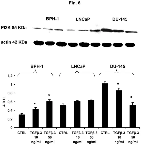
Several findings suggest a role of PI3K in TGF-3 signalling. TGF-3 can rapidly activate PI3K, as indicated by the phosphorylation of its downstream effector AKT [152]. Other studies suggest a negative regulation of the PI3K/AKT pathway by p53 through the transcriptional activation of PTEN [153]. PTEN antagonizes PI3K function by dephosphorylating phosphoinositol triphosphate (PIP3), resulting in the reduction in the phosphorylated AKT fraction and G1 arrest [154]. Thus, we determined the expression of PI3K, AKT, pAKT and PTEN in the experimental cultures. The results show that TGF-3 induced a dose-dependent increase of PI3K in BPH-1 (+100% at 50 ng/ml compared to the respective untreated controls), a not significant increase in LNCaP, and a dose-dependent decrease in DU- 145 (-80% at 50 ng/ml with respect to the untreated controls) (Fig. 6).
44
*p < 0.05 compared to untreated BPH-1 (CTRL). °p < 0.05 with respect to untreated DU-145 (CTRL)
The results of the levels of AKT and pAKT have been summarized in Fig. 7. Compared to untreated cells, there was an increase dose-dependent of AKT and pAKT in BPH-1; no change of AKT and pAKT in LNCaP; a decrease dose-dependent of AKT and pAKT in DU-145 after 24 h of TGF-3 treatment.
Fig. 6 PI3K 85 KDa actin 42 KDa BPH-1 LNCaP DU-145 BPH-1 LNCaP DU-145 * * ° ° 0 0,2 0,4 0,6 0,8 1 1,2 CTRL TGFβ-3 10 ng/ml TGFβ-3 50 ng/ml CTRL TGFβ-3 10 ng/ml TGFβ-3 50 ng/ml CTRL TGFβ-3 10 ng/ml TGFβ-3 50 ng/ml A .D .U .
45
*p < 0.05 compared to untreated BPH-1 (CTRL). °p < 0.05 with respect to untreated DU-145. ^p < 0.05 compared to untreated BPH-1 (CTRL). ♦p < 0.05 with respect to untreated DU-145 (CTRL)
In Fig. 8 the results of expression of PTEN have been reported demonstrating that the effects by TGF-3 on this pro-apoptotic protein had an opposite behaviour of PI3K/AKT/pAKT.
Fig. 7 actin 42 KDa BPH-1 LNCaP DU-145 pAKT 60 KDa AKT 60 KDa 0 0,2 0,4 0,6 0,8 1 1,2 CTRL TGFβ-3 10 ng/ml TGFβ-3 50 ng/ml CTRL TGFβ-3 10 ng/ml TGFβ-3 50 ng/ml CTRL TGFβ-3 10 ng/ml TGFβ-3 50 ng/ml A .D .U . AKT pAKT * * ^ ^ ° ° ♦ ♦ BPH-1 LNCaP DU-145
46
*p < 0.05 compared to untreated BPH-1 (CTRL). °p < 0.05 with respect to untreated DU-145(CTRL)
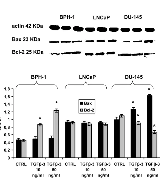
It has been demonstrated in vitro that nitric oxide (NO•) can trigger apoptosis in mesangial cells and tubular cells, as well as other cell types by mechanisms involving the up-regulation of p53, Bax and ceramide generation [155]. It has been suggested that long lasting production of NO• acts as a pro-apoptotic modulator by activating the caspase family of proteases through the release of mitochondrial cytochrome c into the cytosol, upregulation of p53 expression, activation of c-Jun NH2-terminal kinase-stress-activated protein
Fig. 8 BPH-1 LNCaP DU-145 actin 42 KDa PTEN 37 KDa 0 0,2 0,4 0,6 0,8 1 1,2 1,4 1,6 1,8 CTRL TGFβ-3 10 ng/ml TGFβ-3 50 ng/ml CTRL TGFβ-3 10 ng/ml TGFβ-3 50 ng/ml CTRL TGFβ-3 10 ng/ml TGFβ-3 50 ng/ml A .D .U . BPH-1 LNCaP DU-145 * * ° °
47
kinase (JNK/SAPK), and altering the expression of apoptosis-associated proteins including the Bcl-2 family of proteins [156]. Therefore, we determined the effect of TGF-3 on Bax, and Bcl-2. The Bax/Bcl-2 ratio is a relevant relationship since it is generally recognized that maintenance of an appropriate Bax/Bcl- 2 balance in cells prevents apoptosis [157]. Fig. 9 shows that compared to untreated respective controls the cellular level of Bax was unchanged after 24 h of TGF-3 treatment in BPH-1 and LNCaP, but it increased about 40% and further increased to 70% in TGF-3-treated DU-145, respectively. Bcl-2 did not exhibit modification in LNCaP by TGF-3 at the two used concentrations; it showed dose-dependent decrease in BPH-1 and increase in DU- 145. Thus, the TGF-3 treatment on BPH-1 and DU-145 caused the relative Bax/Bcl-2 ratio to change from the normalized pretreatment ratio of 1/1 to about 1.5/1 and 2/1, respectively.
48
*p < 0.05, compared to untreated BPH-1 (CTRL). ^p < 0.05 compared to untreated DU-145 (CTRL). °p < 0.05 with respect to untreated DU-145 (CTRL)
The results of the levels of Rb and pRb have been summarized in Fig. 10. Compared to untreated cells, there was an increase dose-dependent of Rb and pRb in BPH-1; in LNCaP there was no change of Rb and pRb; a decrease dose-dependent of pRb and an increase of Rb in DU-145 after 24 h of TGF-3 treatment (Fig. 10).
Fig. 9 BPH-1 LNCaP DU-145 actin 42 KDa Bax 23 KDa Bcl-2 25 KDa * * ° ° ^ ^ BPH-1 LNCaP DU-145 0 0,2 0,4 0,6 0,8 1 1,2 1,4 1,6 1,8 CTRL TGFβ-3 10 ng/ml TGFβ-3 50 ng/ml CTRL TGFβ-3 10 ng/ml TGFβ-3 50 ng/ml CTRL TGFβ-3 10 ng/ml TGFβ-3 50 ng/ml A .D .U . Bax Bcl-2
49
*p < 0.05 compared to untreated BPH-1 (CTRL). ●p < 0.05 with respect to untreated BPH-1 (CTRL). >p < 0.05 compared to untreated DU-145 (CTRL). °p < 0.05 with respect to untreated DU-145 (CTRL)
Because cyclin A is a sensor of cell division signals, it was important to determine whether its expression correlated with pRb expression. A correlation plot (Fig. 11) demonstrated a significant positive relationship. Figure 11 shows a decrease dose-dependent in BPH-1; a significant increase in DU-145; in LNCaP there was no modification in the expression of cyclin A (Fig. 11).
50 Fig. 11 BPH-1 LNCaP DU-145 actin 42 KDa 0 0,2 0,4 0,6 0,8 1 1,2 1,4 CTRL TGFβ-3 10ng/ml TGFβ-3 50ng/ml CTRL TGFβ-3 10ng/ml TGFβ-3 50ng/ml CTRL TGFβ-3 10ng/ml TGFβ-3 50ng/ml A .D .U . * * ° ° cyclin A 58 KDa BPH-1 LNCaP DU-145
*p < 0.05 compared to untreated BPH-1 (CTRL). °p < 0.05 with respect to untreated DU-145 (CTRL)
Since several forms of cancer are more dependent on PARP than regular cells and PARP is inactivated by caspase cleavage, PARP levels were determined. Compared with the untreated cells, there was an increase of PARP expression in BPH-1 cell line, a significant decrease in DU-145 cells, while in LNCaP no modification was detected (Fig. 12).
51
52
Discussion
Work over recent years has shed light on the molecular mechanisms underlying cancer. From the study of the pathways that regulate tumorigenesis and progression, a numerous amount of novel putative therapeutic targets have emerged, among them the TGF- pathway. The purpose of this study was to understand how TGF-3 may influence the progression of prostate cancer and to determine whether TGF-3 influence the expression of YY1 and p53. The results of this research showed that cancer androgen-dependent LNCaP, cancer androgen-independent DU-145, and benign hyperplasia BPH-1 cells answer in different manner to the treatment with 10 and 50 ng/ml of TGF-3 for 24 h. TGF-3 differently and dose-dependently influenced cell viability, YY1, p53, PI3K, AKT, pAKT, PTEN, Bcl-2, Bax, PARP, Rb, pRb, cyclin A and iNOS expression, and NO• production of three cell lines. TGF-3 demonstrated to be a potent anti-proliferative or pro-proliferative factor depending on the cell type, and it can act as an inducer of apoptosis. To date, up to 33 TGF--related genes have been identified in mammalian genomes as the result of genome sequencing project [152]. It utilizes a multitude of intracellular signalling pathways. The type III TGF-receptor (TGFR3) has recently surfaced as a tumor suppressor within the
53
prostate [158, 159]. In a study on differential gene expression of transforming growth factors alpha and beta, epidermal growth factor, keratinocyte growth factor, and their receptors in fetal and adult human prostatic tissues and cancer cell lines, Authors demonstrated that human BPH-1 cell lines and human prostate cancer cell lines (LNCaP, DU-145, PC-3) express mRNA transcripts for TGF-3 [160]. On stimulation from ligand transfer, TGFR2 binds and phosphorylates the type I receptor (TGFR1), which in turn phosphorylates either the Smad2 or Smad3 transcription factor. Phosphorylation of Smad2/3 promotes binding to Smad4. The whole complex is then translocated into the nucleus where it regulates the transcription of myriad genes related to growth inhibition, production of the extracellular matrix, and apoptosis, among others [161]. Additionally, TGF- binding to its receptors activates many non-canonical signalling pathways including PI3K, MAP kinase, and small GTPase pathways [162]. Several findings suggest a role in TGF- signalling, which can rapidly activate PI3K, as indicated by the phosphorylation of its downstream effector of AKT [152]. PI3K have been linked to an extraordinarily diverse group of cellular functions, including cell growth, proliferation, differentiation, motility, survival and intracellular trafficking. Many of these functions relate to the
54
ability of class I PI3K to activate protein kinase B (PKB, AKT) as in the PI3K/AKT/mTOR pathway. AKT/PKB is a serine/threonine protein kinase that plays a key role in multiple cellular processes such as glucose metabolism, cell proliferation, apoptosis, transcription and cell migration. The PI3K/mTOR pathway can be activated by overproduction of growth factors or chemokines, loss of INPP4B or PTEN expression, or by mutations in growth factor receptors, Ras, PTEN, or PI3K itself. Activation of this pathway contributes to cell growth, cell cycle entry, cell survival, and cell motility, all important aspects of tumorigenesis. A major role for PI3K pathway activation in human tumors has been more recently established following both the positional cloning of the PTEN tumor suppressor gene, and the discovery that the PTEN protein product was a lipid phosphatase that antagonizes PI3K function and consequently inhibits downstream signalling through AKT [163]. Thus, TGF-3 appears to have a greater role in cancer development and progression, and it is more than just an accessory protein. YY1 is a multifunctional protein which can act as a transcriptional repressor or activator through combinatorial interaction with other transcription factors, coactivators, corepressors and chromatin-remodeling complex. High levels of YY1 inhibit the expression of p53 target genes after DNA damage [164]. Inhibition of YY1 expression has been shown to modulate the cellular
55
sensitivity to p53-mediated apoptosis in response to DNA damage [165]. The numerous evidences that YY1 overexpression may promote p53 degradation or inhibit its transcriptional activity may give further insight into YY1‘s role in cancer development. The p53 tumor suppressor protein, also nicknamed the ‗‗guardian of the genome‘‘, is a transcription factor that protects cells against a range of physiological stresses, such as oncogene activation, radiation, mitotic stress, ribosomal stress and chemical insults. These events lead to signals that are relayed to p53 which gets activated, and through a complex network of interactions gets located to the nucleus. There it turns on a program of transcription or repression of myriad genes that induce growth arrest, repair, apoptosis, senescence or altered metabolism; apoptosis is also thought to be induced by direct interactions of cytoplasmic p53 with mitochondria associated proteins. Due to its role as such a central hub, it is no surprise that cells that lack p53 are prone to tumors and these tumors are characterized by significant genetic abnormalities. In unstressed mammalian cells, p53 has a short half-life and is normally maintained at low levels by continuous ubiquitylation catalyzed by Mdm2 [166], COP1 (constitutively photomorphogenic 1) [167], and Pirh2 (p53-induced protein with a RING-H2 domain) [168], and subsequent degradation by 26S proteasome. In this study, we showed the role of TGF-3 in