Funding: this work was supported by grants from Banca del Piemonte to UR, PRIN 2006 Ministero Italiano della Ricerca Scientifica e Tecnologica to UR and ID, Regione Piemonte to UR, Gruppo di Sostegno DBA Italia to UR, DBA Foundation to ID, and Telethon Italia Grant GGP07242 to ID.
We thank the Daniella Maria Arturi Foundation for supporting communication among DBA researchers. Manuscript received on May 27, 2009. Revised version arrived on July 26, 2009. Manuscript accepted on August 5, 2009.
Correspondence: Ugo Ramenghi, MD Associate Professor of Pediatrics, Hematology Unit, Pediatric Department, University of Torino Piazza Polonia 94, 10126 Torino, Italy.
E-mail: [email protected]
Background
Diamond-Blackfan anemia is a rare, pure red blood cell aplasia of childhood due to an intrinsic defect in erythropoietic progenitors. About 40% of patients display various mal-formations. Anemia is corrected by steroid treatment in more than 50% of cases; non-responders need chronic transfusions or stem cell transplantation. Defects in the RPS19 gene, encoding the ribosomal protein S19, are the main known cause of Diamond-Blackfan anemia and account for more than 25% of cases. Mutations in RPS24, RPS17, and
RPL35A described in a minority of patients show that Diamond-Blackfan anemia is a
dis-order of ribosome biogenesis. Two new genes (RPL5, RPL11), encoding for ribosomal pro-teins of the large subunit, have been reported to be involved in a considerable percentage of patients.
Design and Methods
In this genotype-phenotype analysis we screened the coding sequence and intron-exon boundaries of RPS14, RPS16, RPS24, RPL5, RPL11, and RPL35A in 92 Italian patients with Diamond-Blackfan anemia who were negative for RPS19 mutations.
Results
About 20% of the patients screened had mutations in RPL5 or RPL11, and only 1.6% in
RPS24. All but three mutations that we report here are new mutations. No mutations were
found in RPS14, RPS16, or RPL35A. Remarkably, we observed a higher percentage of somatic malformations in patients with RPL5 and RPL11 mutations. A close association was evident between RPL5 mutations and craniofacial malformations, and between hand malformations and RPL11 mutations.
Conclusions
Mutations in four ribosomal proteins account for around 50% of all cases of Diamond-Blackfan anemia in Italian patients. Genotype-phenotype data suggest that mutation screening should begin with RPL5 and RPL11 in patients with Diamond-Blackfan anemia with malformations.
Key words: red cells, bone marrow failure, anemia, DBA, ribosomal proteins.
Citation: Quarello P, Garelli E, Carando A, Brusco A, Calabrese R, Dufour C, Longoni D, Misuraca A, Vinti L, Aspesi A, Biondini L, Loreni F, Dianzani I, and Ramenghi U. Diamond-Blackfan ane-mia: genotype-phenotype correlations in Italian patients with RPL5 and RPL11 mutations Haematologica. 2010; 95:206-213. doi:10.3324/haematol.2009.011783
©2010 Ferrata Storti Foundation. This is an open-access paper.
Diamond-Blackfan anemia: genotype-phenotype correlations in Italian patients
with
RPL5 and RPL11 mutations
Paola Quarello,1Emanuela Garelli,1 Adriana Carando,1Alfredo Brusco,2Roberto Calabrese,1Carlo Dufour,3 Daniela Longoni,4 Aldo Misuraca,5 Luciana Vinti,6 Anna Aspesi,7 Laura Biondini,8 Fabrizio Loreni,8 Irma Dianzani,7 and Ugo Ramenghi1
1 Hematology Unit, Pediatric Department, University of Torino; 2Department of Genetics, Biology and Biochemistry, University of Torino; 3Pediatric Hematology, IRCCS Gaslini, Genova; 4Pediatric Department, University of Milano Bicocca, Monza; 5 Onco-Hematology, Pausillipon Hospital, Napoli; 6Pediatric Oncology and Hematology, IRCCS San Matteo, Pavia; 7Department of Medical Sciences, University of Eastern Piedmont, Novara, and 8Department of Biology, University 'Tor Vergata', Roma, Italy
Introduction
Diamond-Blackfan anemia (DBA, MIM#105650) is a rare inherited congenital bone-marrow-failure syndrome characterized by normochromic macrocytic anemia typi-cally presenting in infancy or early childhood. The bone marrow is normocellular, but erythroid precursors are absent or their numbers markedly decreased, because their progenitors are unable to differentiate and are prone to apoptosis. Laboratory findings such as increased mean corpuscular volume, high erythrocyte adenosine deami-nase activity (eADA), and elevated hemoglobin F after 6 months of age are observed in most patients; an increase in eADA may be the only manifestation.1The congenital anomalies, mainly involving the head, upper limbs, heart and urogenital system, found in more than one-third of patients reflect the fact that DBA is a broad disorder of development.2 The range of severity of such malforma-tions is wide, even within the same family.
The genetic basis of DBA is heterogeneous. Approximately 40% of patients have mutations in one of the genes for ribosomal proteins (RP): RPS7, RPS17,
RPS19, RPS24, RPL5, RPL11, or RPL35A. These genes
encode for RP of either the small or the large ribosomal subunit.3-7 The identification of the role of another RP gene, RPS14, in the pathogenesis of an acquired myelodysplastic disease, the 5q- syndrome, has recently attracted great interest, since it extends the spectrum of ribosomal diseases.8
A clear genotype-phenotype correlation is not apparent in patients with RPS19 mutations, and identical mutations have been found in patients with a wide range of clinical presentations, even within the same family. No informa-tion of genotype-phenotype correlainforma-tions is so far available for patients with RPS24, RPS17, or RPL35A mutations, because the number of subjects studied are too small.3-5 On the other hand a correlation between RPL11 or RPL5 mutations and hand malformations and cleft lip and/or palate, respectively, has been reported.6,7
Here we report the results of screening for six RP genes (RPS14, RPS16, RPS24, RPL5, RPL11, and RPL35A) in 92 unrelated Italian patients who were negative for RPS19 mutations, and a genotype-phenotype analysis for the
RPL5 and RPL11 genes.
Design and Methods
Patients
One hundred twenty-eight unrelated DBA families were stud-ied. Fourteen had more than one clinically affected individual. The diagnosis of DBA was always based on normochromic, often macrocytic anemia, reticulocytopenia, erythroid bone marrow aplasia or hypoplasia, and, in some patients, congenital malforma-tions and elevated eADA. We excluded short stature because it was difficult to evaluate in the context of severe anemia, iron over-load and chronic corticosteroid use.
Informed consent was obtained from all patients and/or their family members participating in the study. RPS19 mutations were found in 36/128 (28%) unrelated DBA patients using both sequencing and multiplex ligation-dependent probe analysis (MLPA): these data have already been reported.9,10Specifically, 33
RPS19 mutations were identified by sequencing whereas three
heterozygous RPS19 deletions missed by sequencing were found using the MLPA technique.10
Molecular analysis of RP genes
Genomic DNA from 92 unrelated Italian DBA probands nega-tive for RPS19 mutations was isolated from peripheral blood leukocytes using a commercial kit (Gentra Systems, Inc., Minneapolis, MN, USA). We analyzed RPS24, RPL5, RPL11 and
RPL35A because mutations in these genes have been reported in
the literature.3,4,6 We screened RPS16 because it is involved in
binding of initiation factor eIF-2 to the 40S subunit.11The RPS14
gene was screened because it has an important role in erythroid proliferation and maturation.8This gene, together with others, is
deleted in the 5q- syndrome, an acquired myelodysplastic syn-drome which is very different from DBA, but considered a riboso-mal disease.8
We analyzed these six RP genes by direct sequencing of the cod-ing exons and intron-exon boundaries. The primer sequences are available on request. Polymerase chain reaction (PCR) products were purified with the QIAquick purification kit (QIAGEN GmbH, D-40724 Hilden, Germany), and sequenced on both strands with an ABI PRISM BigDye Terminator kit (Applied Biosystems, Foster City, CA, USA) on an Applied Biosystems 3100 DNA Sequencer (Applied Biosystems). When sequence changes were found, independent PCR products were sequenced to con-firm the mutations.
Subsequently, we sequenced DNA samples from available fam-ily members to determine whether the mutation co-segregated with the DBA phenotype within the pedigree. To determine whether these sequence changes were polymorphic variations, we sequenced DNA samples from 100 Italian control individuals, and verified that none was reported in the Single Nucleotide Polymorphism database (dbSNP at www.ncbi.nlm.nih.gov/SNP) or in the Ensembl database (www.ensembl.org). To verify whether the
RPS24 missense mutation (p.Asn124Ser) was a polymorphism we
performed ScrFI enzymatic digestion on samples from 150 Italian control individuals.
The nomenclature used to describe the sequences is in accor-dance with the Human Genome Variation Society recommenda-tions (http://www.hgvs.org).
To ascertain whether the two RPL5 mutations detected in patient 2 (Table 1) were on the same allele a test based on NlaIII restriction enzyme digestion was set up. The splice site mutations detected in patients 10 and 12 (Table 1) were analyzed with a Splice Site Prediction website (BDGP, Berkeley Drosophila Genome Project, http://www.fruitfly.org).
Cell culture and transfection
Human embryonic kidney (HEK) 293T cells (ATCC #CRL-11268) were cultured in Dulbecco’s modified essential medium supplemented with 10% fetal bovine serum, 100 U/mL penicillin and 100 µg/mL streptomycin at 37°C with 5% CO2. For
transfec-tion, RPS24 cDNA was reverse transcribed and amplified from total RNA using SuperScript® III (Invitrogen, San Diego, CA, USA). Both variants 1 and 2 (NM_033022 e NM_001026) were obtained and inserted in a pcDNA3.1+ vector (Invitrogen) that contained the neomycin resistance gene and was modified to express a Flag tag at the C-terminus. Site-directed mutagenesis for mutant c.371A>G (p.Asn124Ser) was carried out using PfuTurbo® DNA Polymerase (Stratagene, La Jolla, CA, USA) and the follow-ing primers: forward tgcaaaggccagtgttggtgctg-3’, reverse
5’-cagcaccaacactggcctttgca-3’. A PCR-based strategy was applied to obtain mutant 64_66delCAA, using Platinum®Pfx DNA Polymerase (Invitrogen) and the primers, forward 5’-Phos-atggt-cattgatgtccttcaccc-3’, reverse 5’-tttcctctgaagtagtcggttggt-3’. All constructs were verified by DNA sequencing. About 2 µg of each plasmid and 20 µL of lipofectamine transfection reagent (Invitrogen, Lipofectamine plus 2000) were used for transfection of HEK293 cells plated at 90% confluence. After 24 h cells were analyzed.
RPS24 protein analysis
To prepare total extracts, cells were washed twice with phos-phate-buffered saline (150 mM NaCl, 2.7 mM KCl, 8 mM Na2PO4
and 1.4 mM KH2PO4) and treated with lysis buffer (150 mM NaCl,
50 mM Tris–HCl (pH 7.5), 1% NP40, 0.5% sodium deoxycholate, 0.1% sodium dodecylsulfate [SDS], aprotinin 1 mg/mL, leupeptins 1 mg/mL, pepstatin A 1 mg/mL, phenylmethylsulfonylfluoride 100 mg/mL). After 1 min of incubation on ice, the extract was
cen-trifuged for 10 min at maximum speed in a microcentrifuge at 4°C. For fractionation of the extract, cells were lysed in 300 µL of 10 mM Tris HCl (pH 7.5), 10 mM NaCl, 3 mM MgCl2, 0.05% NP40
with protease inhibitors and centrifuged for 5 min at 1000 g. To prepare the nuclear fraction, the pellet was resuspended in 1.2 mL of 10 mM Tris HCl (pH 7.9), 10 mM NaCl, 5 mM MgCl2, 0.3 M
sucrose and layered onto a 2 mL cushion of 0.6 M sucrose. After 10 min centrifugation at 1,000g at 4°C, the pellet was dissolved in SDS–polyacrylamide gel electrophoresis (PAGE) loading buffer as a nuclear fraction. The supernatant of the first centrifugation at 1000 g was either used as cytoplasmic fraction or further purified. To prepare the ribosomal fraction the cytoplasmic extract was lay-ered onto 1 mL of 15% sucrose, 30 mM Tris–HCl (pH 7.5), 100 mM NaCl, 10 mM MgCl2and centrifuged in a Beckman type 70.1
rotor for 90 min at 100,000g. The pellet (ribosomal fraction) was resuspended directly in SDS–PAGE loading buffer. The super-natant (free cytoplasmic proteins) was precipitated with 10% trichloroacetate and the pellet, washed with acetone, was
resus-Table 1.RPL5 mutations.
Patient Malformation eADA Response to Status at Mutation Exon/ DNA Predicted Inheritance
(gender) status first steroid last intron mutation effect on
treatment follow-up protein
1 (F) None I R SD Missense Exon 1 c.3G>C p.Met1? n.a.
2 (F) Atrial septal defect, I NR TD Nonsense/ Exon 6 c.[678C>A; 680T>G] p.[Tyr226X; n.a.
flat thenar eminence, missense Ile227Arg]
short stature
3 (F) Cleft lip, cleft palate, I R SD Deletion Exon 3 c.134_138delACACA p.Asn45ThrfsX66 De novo
short stature (*) (m,f,s normal
sequence) 4 (M) Myelomeningocele, n.a. n.a. FU Deletion Exon 3 c.169_172delAACA p.Asn57GlufsX12 De novo
cleft palate, (*) (m,f normal
facial dysmorphism sequence)
5 (F) Flat thenar eminence, n.a. n.a. FU Deletion Exon 3 c.169_172delAACA p.Asn57GlufsX12 Sporadic
grouped carpal bones, (*) (f,b normal
short stature sequence)
6 (M) Micrognathia, palpebral n.a. n.a. FU Deletion Exon 3 c.183_184delTT p.Ile61MetfsX51 De novo
ptosis, cleft palate, (m,f normal
triphalangeal thumb, sequence)
brachydactyly, learning difficulties, aortic valve stenosis, short stature
7 (F) Cleft palate, I R REMtrt Deletion Exon 5 c.336delG p.Arg112SerfsX14 De novo
micrognathia, (m,f,b normal
triphalangeal thumb sequence)
8 (M) Facial dysmorphisms, I R SD Insertion Exon 2 c.39_40insT p.Lys14X n.a.
strabism
9 (F) Cleft palate, persistent n.a. n.a. n.a. Insertion Exon 6 c.692_693insT p.Thr232AsnfsX50 De novo
foramen ovale (m,f normal
sequence) 10 (M) Palpebral ptosis n.a. NR TD Splice site Intron 1 c.3+3G>C p.? Familial (m) 11 (M) Short stature n.a. NR TD Splice site Intron 3 c.189+1G>A del ex3 fs n.a. 12 (F) Cleft palate, micrognathia I no therapy REMs Splice site Intron 4 c.324+5G>T del ex4 in frame n.a.
flat thenar eminence, persistent foramen ovale, facial dysmorphisms
pended in SDS–PAGE loading buffer. For western analysis, pro-teins were separated on a 12% SDS polyacrylamide gel, trans-ferred to a nitrocellulose Protran membrane (Schleicher and Schuell), and incubated with the following primary antibodies and antisera: mouse monoclonal anti-RPS19,12mouse anti-Flag (Sigma,
F3165), rabbit NPT II (Upstate, 06-747), and mouse anti-GAPDH (Millipore MAB374). Primary antibodies were revealed using horseradish peroxidase-conjugated goat anti-rabbit antibody (Jackson Immunoresearch) and a chemiluminescence detection system (Ablot Plus, Euroclone). Quantitation analyses were per-formed using the LAS3000 Image System (Fuji) and ImageQuant software (GE Healthcare).
Genotype-phenotype correlations and statistical analysis
Genotype-phenotype correlations were evaluated in the whole group of patients. Differences between two independent samples were checked with Student’s t-test or the Mann-Whitney test as appropriate, whereas the Kruskal-Wallis test was used to assess the differences between more than two groups. Associations between categorical variables were assessed with Fisher’s exact test or with odds ratio and 95% confidence interval (95% CI). For categorical variables with more than two categories simple logis-tic regression was used to calculate the odds ratio and 95% CI.
All tests were two-sided and P values less than 0.05 were
con-sidered statistically significant. Data were analyzed with the SPSS 16 software (SPSS Inc., Chicago, IL, USA).
Results
RPL5
We identified RPL5 sequence changes in 12 out of the 92 probands (Table 1). These included seven deletions or insertions of one to five nucleotides causing a frameshift, three donor splice-site mutations, one missense and one nonsense mutation. All mutations but one were new mutations not yet described in the literature and were found in one patient only. The four-nucleotide deletion in exon 3 (c.169_172delAACA) detected in two unrelated patients has already been reported.6,7 De novo sequence changes were identified in six probands; one patient (n. 10, Table 1) inherited the RPL5 mutation (c.3+3G>C) from his healthy mother, who had normal hemoglobin level, mean corpuscular volume and eADA. Parental DNA was not available for the other five patients.
A five-nucleotide deletion in exon 3 (c.134_138delACACA) was found in a female patient (n. 3, Table 1), who also carried a RPS24 missense mutation
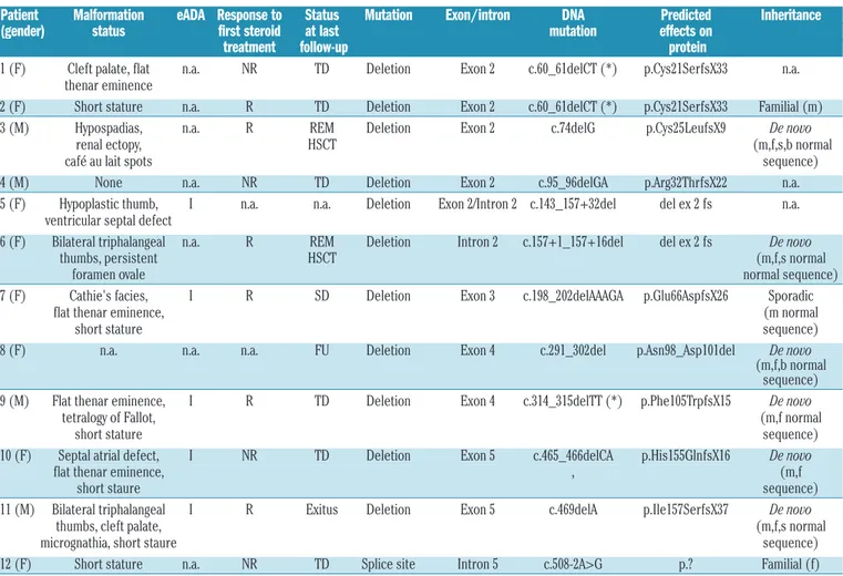
Table 2. RPL11 mutations.
Patient Malformation eADA Response to Status Mutation Exon/intron DNA Predicted Inheritance
(gender) status first steroid at last mutation effects on
treatment follow-up protein
1 (F) Cleft palate, flat n.a. NR TD Deletion Exon 2 c.60_61delCT (*) p.Cys21SerfsX33 n.a. thenar eminence
2 (F) Short stature n.a. R TD Deletion Exon 2 c.60_61delCT (*) p.Cys21SerfsX33 Familial (m)
3 (M) Hypospadias, n.a. R REM Deletion Exon 2 c.74delG p.Cys25LeufsX9 De novo
renal ectopy, HSCT (m,f,s,b normal
café au lait spots sequence)
4 (M) None n.a. NR TD Deletion Exon 2 c.95_96delGA p.Arg32ThrfsX22 n.a.
5 (F) Hypoplastic thumb, I n.a. n.a. Deletion Exon 2/Intron 2 c.143_157+32del del ex 2 fs n.a. ventricular septal defect
6 (F) Bilateral triphalangeal n.a. R REM Deletion Intron 2 c.157+1_157+16del del ex 2 fs De novo
thumbs, persistent HSCT (m,f,s normal
foramen ovale normal sequence)
7 (F) Cathie's facies, I R SD Deletion Exon 3 c.198_202delAAAGA p.Glu66AspfsX26 Sporadic
flat thenar eminence, (m normal
short stature sequence)
8 (F) n.a. n.a. n.a. FU Deletion Exon 4 c.291_302del p.Asn98_Asp101del De novo
(m,f,b normal sequence) 9 (M) Flat thenar eminence, I R TD Deletion Exon 4 c.314_315delTT (*) p.Phe105TrpfsX15 De novo
tetralogy of Fallot, (m,f normal
short stature sequence)
10 (F) Septal atrial defect, I NR TD Deletion Exon 5 c.465_466delCA p.His155GlnfsX16 De novo
flat thenar eminence, , (m,f
short staure sequence)
11 (M) Bilateral triphalangeal I R Exitus Deletion Exon 5 c.469delA p.Ile157SerfsX37 De novo
thumbs, cleft palate, (m,f,s normal
micrognathia, short staure sequence)
12 (F) Short stature n.a. NR TD Splice site Intron 5 c.508-2A>G p.? Familial (f)
F: female; M: male; I: increased value; n.a.: not available; R: responsive to steroid treatment; NR: not responsive to steroid treatment; SD: steroid dependent; TD: transfusion dependent; FU: lost at last follow-up; REM HSCT: remission after hematopoietic stem cell transplantation, f: father, m: mother, s: sister, b: brother. (*): mutation previously reported.
in exon 4 causing a substitution of an Asparagine with a Serine at codon 124 (c.371A>G; p.Asn124Ser). This patient is steroid-dependent, has a cleft palate, short stature and increased eADA. The RPL5 mutation was de
novo, while the RPS24 mutation was found in her healthy
father and unaffected sister (Figure 1; Table 1, n. 3). Patient n. 2 (Table 1) had two mutations on the same allele. The first was a nonsense mutation causing a prema-ture stop codon (p.Tyr226X). The second was a missense mutation involving the adjacent codon 227 (p.Ile227Arg), and caused the substitution of an Isoleucine with an Arginine. Parental DNA was not available for molecular analysis.
The splice site mutations detected in patients n. 10 (c.3+3G>C; Table 1) and n. 12 (c.324+5G>T; Table 1) mod-ified the splice site strength score from 0.97 and 0.98 to 0.32 and 0.23, respectively (BDGP, http://www.fruitfly.org). These data suggested splice site suppression in both mutants. We could not perform expression analysis because fresh blood samples from these patients were not available.
RPL11
We identified sequence changes in 12 patients (Table 2). Most mutations (11/12, 92%) were 1-47 nucleotide dele-tions. An acceptor splice-site mutation was also observed. Nine mutations are described here for the first time; two, including one present in two unrelated probands, had been reported by Gazda et al.6Parental DNA was available for nine probands; seven had de novo mutations. Two probands (n. 2 and n. 12, Table 2) inherited the mutation from their apparently healthy mother and affected father, respectively. This father reported mild anemia during infancy associated with short stature, increased mean cor-puscular volume and high eADA; he has never needed any treatment, whereas his daughter is transfusion-dependent, and does not respond to steroids.
RPS24
We found two new heterozygous changes in RPS24 (2/92). A deletion of three nucleotides in exon 2 (c.64_66delCAA), resulting in the loss of the highly con-served glutamine 22, was identified in a patient without somatic malformations and in clinical remission at last fol-low-up. This mutation was not found in other family members, nor has it been reported as a polymorphism (www.ncbi.nlm.nih.gov/SNP; www.ensembl.org).
A missense mutation (p.Asn124Ser) in exon 4 was found in a patient who also carried a five-nucleotide dele-tion (c.134_138delACACA) in RPL5 as described above (Figure 1; Table 1 n. 3). This missense mutation was car-ried by two healthy family members (Figure 1; Table 1, n. 3), but has never been reported as a polymorphism nor was it found in 300 Italian control chromosomes.
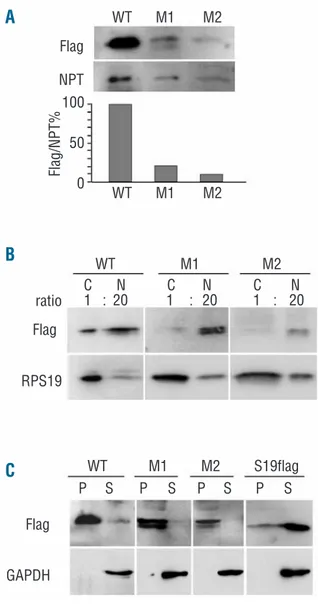
Neither RPS24 mutation predicted a dramatic alteration of the gene product, differently from RPL5 and RPL11 mutations. Thus, we decided to study the properties of RPS24 proteins encoded by the mutated genes, using an approach similar to that which we previously used to study RPS19 missense mutations.12 We prepared cDNA
(pcDNA3.1). RPS24 pre-mRNA is alternatively spliced into three different mRNA isoforms that encode proteins with a slightly different C terminus (one or three addition-al amino acids).3 We prepared cDNA encoding the two major RPS24 variants (1 and 2) but, since in a preliminary analysis they showed identical behaviors (data not shown), we focused our attention on one protein variant (variant 1). DNA constructs encoding for: (i) wild type RPS24 (WT), (ii) RPS24 with codon 22 deletion (M1), and (iii) RPS24 with the p.Asn124Ser mutation (M2) were used in transient transfection experiments into HEK293 cells. After transfection, cell extracts were analyzed by western blotting to measure protein levels or further purified to investigate the subcellular localization and the capacity to assemble into the ribosome. The levels of WT, M1 and M2 RPS24 were normalized for the amount of neomycin phosphotransferase II expressed by the pcDNA3.1 vector. Assuming that the different RPS24 cDNA have the same transcriptional and translational activity, the results can be considered proportional to the stability of the different proteins. As shown in Figure 2A the levels of both M1 and M2 are clearly lower than that of WT RPS24. This indi-cates that both mutations affect protein stability. Consistent with the instability of the mutated RPS24, a possible degradation product appeared sometimes on the western blot as a faster-migrating band of variable intensi-ty. Next, we separated nuclear and cytoplasmic fractions from the extract of the transfected cells. Western blot analysis, illustrated in Figure 2B, showed that mutated RPS24 (M1 and M2) accumulated into the nucleus more evidently than did the WT RPS24. Finally, to verify the capacity of the mutated RPS24 to be incorporated into the ribosome, we further fractionated cytoplasmic extracts through ultracentrifugation on a sucrose cushion. After 2 h of centrifugation all ribosomes and ribosomal subunits were found in the pellet whereas free cytoplasmic pro-teins could be recovered from the supernatant. As an addi-tional control for this experiment, we transfected a plas-mid expressing an RPS19 construct (RPS19flag) with the Flag epitope at the N terminus. The RPS19flag fusion pro-tein was previously shown to assemble poorly into the Figure 1.Pedigree of a DBA patient carrying mutations in both the
RPL5 and RPS24 genes.
No anemia eADA normal value No malformations
No anemia eADA normal value No malformations
No anemia eADA normal value No malformations
Sterold dependent anemia eADA increased value Cleft palate, short stature
RPS24 gene: p.Asn124Ser RPS24 gene: p.Asn124Ser RPL5 gene: c.134_138delACACA
II-2 II-1
I-2 I-1
Western blot analysis, presented in Figure 2C, con-firmed that RPS19flag was mostly in the free cytoplasmic fraction. In contrast, both the WT and the mutated RPS24 (M1 and M2) appeared to be mainly associated with the ribosome. This result indicates that, although the mutated RPS24 are less stable and accumulate into the nucleus, a small fraction of them is incorporated into the ribosome and exported into the cytoplasm. The conclusion of our analyses is that the two mutations here analyzed alter some properties of RPS24 in a similar way. However, a fraction of the mutated RPS24 was able to associate with the ribosome.
RPS14, RPS16 and RPL35A
No mutations were found in RPS14, RPS16, or RPL35A.
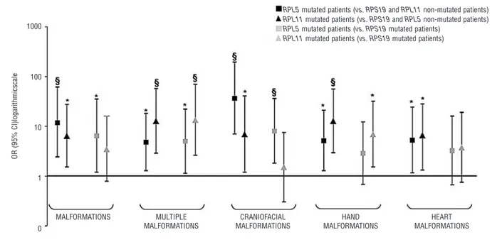
Genotype-phenotype correlations
The clinical features of patients with RPL5 and RPL11 mutations are summarized in Tables 1 and 2. The results of the genotype-phenotype statistical analysis performed by logistic regression are presented in Figure 3.
Most of the patients with RPL5 (83%) and RPL11 (73%) mutations had physical malformations. Patients with
RPS19 mutations and patients without mutations in any
of these three genes showed lower percentages of mations (43% and 29%). Specifically, the risk of malfor-mation was 12-fold higher in RPL5-mutated patients than in patients with no mutations in RPL5, RPL11 or RPS19 and 7-fold higher in RPL5-mutated patients than in RPS19-mutated patients. Similarly, RPL11-RPS19-mutated patients had a 6-fold higher and 3.5-fold higher risk of somatic anomalies compared to RPL5, RPL11 and RPS19 non-mutated and
RPS19-mutated patients. Moreover, patients with either RPL5 or RPL11 mutations more frequently had multiple
malformations than did RPL5, RPL11 and RPS19 non-mutated and RPS19-non-mutated patients.
We also found that craniofacial abnormalities were closely associated with RPL5 mutations: RPL5-mutated patients had a 37-fold and an 8-fold higher risk of cranio-facial malformations than RPL5, RPL11 and RPS19 non-mutated and RPS19-non-mutated patients, respectively.
RPL11-mutated probands had a higher risk of craniofacial
malformation than RPL5, RPL11 and RPS19 non-mutated patients only. Interestingly, cleft lip and/or palate was observed in 8/111 clinically evaluable patients. None of these eight had a mutation in RPS19, whereas all had mutations in RPL5 or RPL11.
A significant association between hand malformations and RPL11 mutations was also observed: RPL11-mutated patients had a 13-fold and a 7-fold higher risk of hand mal-formation compared to RPL5, RPL11 and RPS19 non-mutated and RPS19-non-mutated patients, respectively. A sim-ilar, though weaker, association was found in RPL5-mutat-ed patients comparRPL5-mutat-ed to RPL5, RPL11 and RPS19 non-mutated patients; no difference was observed between
RPS19 and RPL5-mutated patients.
The risk of cardiac malformations was 5-fold and 6-fold higher in RPL5 and RPL11-mutated patients compared to
RPL5, RPL11 and RPS19 non-mutated patients only. We
did not find a correlation between mutational status and response to first steroid treatment or status at last follow-up.
Discussion
For about 10 years, RPS19 seemed to be the only gene involved in the pathogenesis of DBA and mutations in this gene accounted for 25% of cases.9In the last 3 years, how-ever, heterozygous mutations in several genes encoding ribosomal proteins of either the small or the large
riboso-A
B
C
Figure 2. Transient transfection of RPS24 constructs. (A) Total extracts from HEK293 cells transfected with plasmids coding for RPS24 WT (WT) and mutated forms of the protein (M1, M2) were separated by SDS-PAGE and transferred on nitrocellulose mem-brane. The blots were decorated with Flag (Flag) and anti-neomycin phosphotransferase II (NPT) antibodies. Quantitation of the signals is reported in the lower part as the ratio of Flag/NPT; (B) Total extracts from transfected cells (as in A) were separated into nuclear (N) and cytoplasmic (C) fractions. The loading ratio between cytoplasmic and nuclear extracts was 1:20 (number of cells). Western blotting was performed as in A with anti-Flag (Flag) and anti-RPS19 (RPS19). (C) Cytoplasmic extracts from HEK293 cells transfected as in A were fractionated by ultracentrifugation on a sucrose cushion as indicated in the Design and Methods sec-tion. Pellets, which include ribosome and ribosomal subunits (P) and trichloroacetate-precipitated supernatants, which include free cytoplasmic proteins (S) were analyzed by western blot with anti-Flag (anti-Flag) and anti-glyceraldehyde phosphate dehydrogenase (GAPDH) antibodies. GAPDH Flag Flag Flag Flag/NPT% NPT RPS19 ratio WT M1 M2 S19flag WT M1 M2 WT M1 M2 100 50 0 WT M1 M2 C N C N C N 1 : 20 1 : 20 1 : 20 P S P S P S P S
mal subunit have been reported.3-7Specifically, Gazda et al. found mutations in RPL5 and RPL11 in about 7% and 5%, respectively, of DBA patients,6while Cmejla et al. showed a higher frequency of RPL5 (21%) and RPL11 (7%) muta-tions among Czech patients.7
We report here the results of our screening of RPS24,
RPL5 and RPL11 in 92 Italian patients who were negative
for RPS19 mutations. No mutations were found in RPS14,
RPS16, and RPL35A.
Twenty-eight percent of our patients (36/128) showed a mutation in RPS19,9,10 9.3% (12/128) in RPL5, 9.3% (12/128) in RPL11, and only 1.6% (2/128) in RPS24. This frequency of RPL11 mutations was higher than that found in other screening studies. As far as concern RPL5 muta-tions, the frequency in our study was similar to that found by Gazda et al. but lower than in Czech patients.6,7These differences may be due to the populations studied. All but three mutations found in these genes had never been pre-viously described. Interestingly, we found a double het-erozygote for two RP genes. This patient, a steroid-dependent female with cleft palate, had a de novo five-nucleotide deletion in RPL5 exon 3 (c.134_138delACACA) and a substitution in RPS24 (p.Asn124Ser). The latter was inherited from her healthy father, and also carried by a healthy sister (Figure 1). Several points support the hypothesis that the RPL5 mutation is pathogenic: it is a de
novo frameshift mutation and the malformation
pheno-type of this patient is typical of that of patients with a
RPL5 mutation. There is, however, controversy on the role
of the missense RPS24 p.Asn124Ser: this mutation is car-ried by healthy relatives and Asparagine 124 is a
non-con-served amino acid. However, a silent phenotype has already been described in DBA patients with RPS24 muta-tions.3p.Asn124Ser was not found in 300 Italian normal chromosomes and has never been reported as a single nucleotide polymorphism.
To ascertain whether p.Asn124Ser is a mutation or a silent polymorphism we checked whether this mutant was able to reach the nucleolus and be included in active ribosomes. Our studies show that though the mutant is rather unstable as compared with the wild type form, it is able to reach the nucleolus and a small fraction is even able to associate with the ribosome. Altogether, our stud-ies support the following model: the bulk of the DBA phe-notype is most probably due to the RPL5 frameshift muta-tion, whereas the RPS24 missense mutation is a pheno-type modifier.
Interestingly, similar functional data were found for the in frame RPS24 mutation: however, in this case, the fact that this is a de novo mutation is a strong indication that this deletion has a role in the pathogenesis of the disease.
Overall, our data suggest that all DBA patients, even those with a recognized mutation in RP genes, should be screened for other DBA genes to ascertain whether a sec-ond mutation is present. A possible digenic effect may explain the variable expressivity and incomplete pene-trance in members of the same family.
Incomplete penetrance was shown for RPL5 and RPL11 mutations in two families of our cohort (n. 10, Table 1; n. 2, Table 2), as previously described for RPS19.13All RPS19 mutations described have been found in heterozygosis, suggesting haploinsufficiency due to a loss-of-function
Figure 3.Malformation status of patients with RPL5 and RPL11 mutations. Associations between malformation status and RP gene muta-tions were assessed with odds ratio (OR) and 95% CI calculated from logistic regression; OR are drawn on a logarithmic scale. RPL5 and
MALFORMATIONS 1000 100 10 1 0 OR (95% CI)logarithmicscale MULTIPLE MALFORMATIONS CRANIOFACIAL MALFORMATIONS HAND MALFORMATIONS
RPL5 mutated patients (vs. RPS19 and RPL11 non-mutated patients) RPL11 mutated patients (vs. RPS19 and RPL5 non-mutated patients) RPL5 mutated patients (vs. RPS19 mutated patients)
RPL11 mutated patients (vs. RPS19 mutated patients)
HEART MALFORMATIONS
effect.9Similarly, most RPL5 and RPL11 mutations cause a premature termination, suggesting that haploinsufficiency is the cause of the disease.
Gazda et al. reported that RPL5 and RPL11 mutations are more frequently associated with physical malformations than are RPS19 mutations. RPL5 mutations seem to cause a more severe malformation status, including craniofacial, thumb, and heart anomalies, compared to mutations in
RPS19.6Remarkably, Gazda et al. reported a close associa-tion between RPL5 and cleft lip and/or palate. Mutaassocia-tions in RPL11 were predominantly associated with thumb abnormalities.6Similarly, Cmejla et al. observed that all ten Czech DBA patients with either an RPL5 or an RPL11 mutation had one or more physical malformations; specif-ically, hand anomalies were always present and most patients were born small for gestational age.7 Cleft lip and/or palate was not observed in RPL5-mutated and
RPL11-mutated patients.
We observed a high percentage of multiple somatic mal-formations in patients with RPL5 and RPL11 mutations. Close associations between RPL5 mutations and craniofa-cial malformations and between hand malformations and
RPL11 mutations were also evident. However, unlike
Gazda et al., we observed cleft lip and/or palate in both
RPL5- and RPL11-mutated DBA patients, and can thus rule
out a unique association between RPL5 mutations and this type of malformation.
In conclusion, we report a high frequency of RPL5 (9.3%) and RPL11 (9.3%) mutations in our DBA cohort. Mutations in four ribosomal proteins account for around 50% of all cases of DBA in Italian patients. Genotype-phe-notype data suggest that mutation screening should begin with RPL5 and RPL11 in DBA patients with a malforma-tion, and specifically in those who have craniofacial aber-rations, such as cleft palate, or hand abnormalities.
Authorship and Disclosures
PQ performed genotype-phenotype correlation analyses and drafted the manuscript with UR and ID, who were involved in the conception of the study. UR is the author taking primary responsibility for the paper. EG and AC per-formed sequencing analyses. AB critically revised the paper and was responsible for important intellectual content. RC was responsible for the statistical analysis. CD, DL, AM and LV contributed clinical data. AA performed cloning. LB and FL performed RPS24 functional experiments. All authors contributed to the revision of the manuscript.
The authors declare no potential conflicts of interest.
References
1. Willig TN, Niemeyer CM, Leblanc T, Tiemann C, Robert A, Budde J, et al. Identification of new prognosis factors from the clinical and epidemiologic analy-sis of a registry of 229 Diamond-Blackfan anemia patients. DBA group of Societe d'Hematologie et d’Immunologie Pe-diatrique (SHIP), Gesellshaft fur Pa-diatrische Onkologie und Hamatologie (GPOH), and the European Society for Pediatric Hematology and Immunology (ESPHI). Pediatr Res. 1999;46(5):553-61. 2. Vlachos A, Ball S, Dahl N, Alter BP, Sheth S,
Ramenghi U, et al. Diagnosing and treating Diamond Blackfan anaemia: results of an international clinical consensus conference. Br J Haematol. 2008;142(6):859-76. 3. Gazda HT, Grabowska A, Merida-Long LB,
Latawiec E, Schneider HE, Lipton JM, et al. Ribosomal protein S24 gene is mutated in Diamond-Blackfan anemia. Am J Hum Genet. 2006;79(6):1110-8.
4. Farrar JE, Nater M, Caywood E, McDevitt MA, Kowalski J, Takemoto CM, et al. Abnormalities of the large ribosomal
sub-unit protein, Rpl35a, in Diamond-Blackfan anemia. Blood. 2008;112(5):1582-92. 5. Cmejla R, Cmejlova J, Handrkova H, Petrak
J, Pospisilova D. Ribosomal protein S17 gene (RPS17) is mutated in Diamond-Blackfan anemia. Hum Mutat. 2007;28(12): 1178-82.
6. Gazda HT, Sheen MR, Vlachos A, Choesmel V, O'Donohue MF, Schneider H, et al. Ribosomal protein L5 and l11 muta-tions are associated with cleft palate and abnormal thumbs in Diamond-Blackfan anemia patients. Am J Hum Genet. 2008; 83(6):769-80.
7. Cmejla R, Cmejlova J, Handrkova H, Petrak J, Petrtylova K, Mihal V, et al. Identification of mutations in the ribosomal protein L5 (RPL5) and ribosomal protein L11 (RPL11) genes in Czech patients with Diamond-Blackfan anemia. Hum Mutat. 2009;30(3): 321-7.
8. Ebert BL, Pretz J, Bosco J, Chang CY, Tamayo P, Galili N, et al. Identification of RPS14 as a 5q- syndrome gene by RNA interference screen. Nature. 2008; 451(7176): 335-9. 9. Campagnoli MF, Ramenghi U, Armiraglio
M, Quarello P, Garelli E, Carando A, et al. RPS19 mutations in patients with
Dia-mond-Blackfan anemia. Hum Mutat. 2008; 29(7):911-20.
10. Quarello P, Garelli E, Brusco A, Carando A, Pappi P, Barberis M, et al. Multiplex liga-tion-dependent probe amplification enhances molecular diagnosis of Diamond-Blackfan anemia due to RPS19 deficiency. Haematologica. 2008;93(11):1748-50. 11. Bommer UA, Stahl J, Henske A, Lutsch G,
Bielka H. Identification of proteins of the 40 S ribosomal subunit involved in interac-tion with initiainterac-tion factor eIF-2 in the qua-ternary initiation complex by means of monospecific antibodies. FEBS Lett. 1988; 233(1):114-8.
12. Angelini M, Cannata S, Mercaldo V, Gibello L, Santoro C, Dianzani I, et al. Missense mutations associated with Diamond-Blackfan anemia affect the assembly of ribosomal protein S19 into the ribosome. Hum Mol Genet. 2007;16(14): 1720-7. 13. Willig TN, Draptchinskaia N, Dianzani I,
Ball S, Niemeyer C, Ramenghi U, et al. Mutations in ribosomal protein S19 gene and Diamond Blackfan anemia: wide varia-tions in phenotypic expression. Blood. 1999;94(12):4294-306.