INTRODUCTION
Very and extremely preterm infants suffer from severe diseases associated with premature birth, including bronchopulmonary dysplasia (BPD), periventricular leukomalacia (PVL), necrotizing enterocolitis (NEC), patent ductus arteriosus (PDA), sepsis and retinopathy of prematurity (ROP). During the 90s, the universal introduction of antenatal steroids and surfactant replacement as standard therapies for the prevention and treatment of neonatal respiratory distress syndrome (RDS) in the neonatal intensive care units (NICUs) has dramatically changed the natural history of diseases affecting prematurely born infants.
Indeed, together with a reduction in the severity of neonatal RDS, the sequelae of perinatal lung and brain injury profoundly changed: The old BPD and cystic PVL were replaced by newly emerging diseases, the so-called “new BPD” and “noncystic, diffuse PVL”, respectively. These “new” sequelae differ from the old ones in severity (in general are less severe), pathogenesis, pathological features and clinical presentation.[1-6] In general, focal injury/necrosis and the consequent fibrosis/astrogliosis, the main components of old BPD and cystic PVL, appear to be milder and to contribute to a lesser extent to the pathogenesis of new BPD and noncystic PVL. Conversely, tissue simplification
Address for correspondence:
Dr. Alessandro Borghesi,
Fondazione IRCCS Policlinico San Matteo, Piazzale Golgi n. 19, 27100, Pavia, Italy. E-mail: [email protected]
and developmental arrest (larger and fewer alveoli in the lungs and hypomyelination with defective white matter development and neuronal abnormalities in the brain) are the key and predominant components of new BPD and of the diffuse, noncystic form of PVL.[3,6]
While surfactant replacement and prenatal steroid proved revolutionary in changing the destiny of premature infants during the 90s, no preventive strategy is currently available to reduce the incidence of these emerging diseases, and the prevalence of all complications of prematurity has reached a steady state across the last decade [Table 1]. Overall, the sequelae of prematurity still represent a burden for neonatal medicine and global health.
Stem Cell Therapy for Neonatal Diseases Associated with
Preterm Birth
Alessandro Borghesi, Claudia Cova, Diego Gazzolo1, Mauro Stronati
Neonatal Intensive Care Unit and Laboratory of Neonatal Immunology, Fondazione IRCCS Policlinico San Matteo, Pavia, 1Neonatal Intensive Care Unit,
Azienda Ospedaliera Santi Antonio e Biagio e Cesare Arrigo, Alessandria, Italy
ABSTRACT
In the last decades, the prevention and treatment of neonatal respiratory distress syndrome with antenatal steroids and surfactant replacement allowed the survival of infants born at extremely low gestational ages. These extremely preterm infants are highly vulnerable to the detrimental effects of oxidative stress and infection, and are prone to develop lung and brain diseases that eventually evolve in severe sequelae: The so-called new bronchopulmonary dysplasia (BPD) and the noncystic, diffuse form of periventricular leukomalacia (PVL). Tissue simplification and developmental arrest (larger and fewer alveoli and hypomyelination in the lungs and brain, respectively) appears to be the hallmark of these emerging sequelae, while fibrosis is usually mild and contributes to a lesser extent to their pathogenesis. New data suggest that loss of stem/ progenitor cell populations in the developing brain and lungs may underlie tissue simplification. These observations constitute the basis for the application of stem cell-based protocols following extremely preterm birth. Transplantation of different cell types (including, but not limited to, mesenchymal stromal cells, endothelial progenitor cells, human amnion epithelial cells) could be beneficial in preterm infants for the prevention and/or treatment of BPD, PVL and other major sequelae of prematurity. However, before this new knowledge can be translated into clinical practice, several issues still need to be addressed in preclinical in vitro and in vivo models.
Key words:
Bronchopulmonary dysplasia, bronchopulmonary, endothelial, EPC, mesenchymal, MSC, newborn, periventricular leukomalacia, preterm, progenitor cells, periventricular leukomalacia, stem cells
R
eview
A
Rticle
›››
Access this article online
Quick Response Code:
Website:
www.jcnonweb.com
DOI:
Therefore, the research and development of new tools for the prevention of the sequelae resulting from preterm birth are imperative.
Common mechanisms of pathogenesis in different diseases associated with prematurity
The extremely preterm infant as a whole is exposed to an array of risk factors that exert different pathogenic effects on different organs, depending, at least in part, on the sensitivity of the target tissues.
Sequelae of preterm birth often result from exposure to factors causing injury during the days immediately preceding birth and/or during the first two to three weeks of life. These pre-, peri- and post-natal factors occur when organ expansion and development of architectural complexity reach their maximum.
Risk factors for BPD and brain injury ultimately contribute to tissue damage by two main mechanisms: Ischemia/ reperfusion and infection/inflammation, both promoting the production of free oxygen radicals causative of oxidative stress. In addition, tissue damage is enhanced by deprivation of maternal/placental protective growth factors and molecules (e.g., estrogens) and by a (probably) weak genetic predisposition.[7,8] The final effect of these pathogenic mechanisms is the development, at a variable extent, of scarring and fibrosis, and, most importantly, tissue simplification and developmental arrest [Figure 1]. Although an understanding of the mechanisms underlying tissue simplification is still incomplete, it is admitted that exposure to free radicals (reactive oxygen and nitrogen species: ROS and RNS, respectively) is detrimental to stem and progenitor cells, while their terminally differentiated counterparts are more resistant to oxidative damage.[6,9-11] Therefore, the combination of risk factors cited above, including oxidative stress, genetic predisposition and deprivation of maternal/placental molecules, may contribute to tissue simplification by directly damaging stem/progenitor cells [Figure 1].
Data emerging from the literature support this hypothesis (see next sections) and open new paths of study for the upcoming years.
STem Cell DePleTION IN PVl
Cystic PVL is characterized by focal necrosis deep inside the periventricular white matter, evolving in micro- or macrocyst formation and well correlates with cerebral palsy. However, nowadays, the predominant component of PVL is a more diffuse, noncystic, damage in the central cerebral white matter with associated secondary decreased cerebral
cortical gray matter volume, which better correlates with cognitive/behavioral deficits. This latter is a cell-specific injury that appears to be caused by preferential death of premyelinating oligodendrocytes (preoligodendrocytes) during a developmental window of vulnerability.[6,12] Preoligodendrocytes are abundant in the brain of preterm (but not term) infants and progressively differentiate into mature oligodendrocytes between 28 and 40 weeks gestational age to form the myelin sheath. Both ischemia and infection contribute to the production of ROS and RNS, at least in part through the activation of microglia. It is known that preoligodendrocytes, but not mature oligodendrocytes, are highly vulnerable to oxidative stress, and are preferentially lost upon exposure to pathogenic factors associated with prematurity. These considerations support the notion that progenitor cell depletion (preoligodendrocyte depletion), occurring during the process of myelin formation, might result in developmental arrest and tissue simplification (hypomyelination in the central cerebral white matter and secondary neuronal abnormalities), the hallmarks of diffuse PVL [Figure 2].
Table 1: Incidence of major diseases associated with preterm birth in a population of very low birth weight infants (<1500 g)
2000 2005 2010
Necrotizing enterocolitis (%) 4.9 5.3 5.1 Bronchopulmonary dysplasia (%) 27.3 26.3 23.5
IVH grades III-IV (%) 5.8 6 5.4
Periventricular leukomalacia (%) 3 2.8 2.8 Number of involved centers 20,000 30,000 44,000 The number indicates the percentage of very low birth weight infants who developed the disease. Data are extrapolated from the Vermont Oxford Network database
Figure 1: Common mechanisms of pathogenesis in diverse diseases of prematurity Ischemia/reperfusion Infection/inflammation -ROS/RNS -Oxidative stress Tissue damage Loss of stem/ progenitor cells Deprivation of maternal factors
Cell therapies aiming at preventing brain injury associated with preterm birth should therefore focus on protecting the cerebral stem/progenitor cells, and in particular preoligodendrocytes, and promote endogenous repair.
STem Cell lOSS IN BPD: INITIAl
eVIDeNCe
BPD affects up to 43% of infants with birth weight less than 1500 g.[3] It is characterized by fibrosis and arrested alveolar and lung vascular growth, resulting in chronic need of oxygen supplementation until late in infancy, and disturbances in lung function and neurodevelopment in older children and adults.[1,3]
Alveolar simplification is predominant over fibrosis in the “new BPD” compared with the old BPD, and changes in ventilation or respiratory support in order to minimize lung injury have not, or only slightly, changed the incidence of BPD.[13-16] These observations suggest that, beyond ventilation-induced injury, specific developmental pathways or cell populations are disrupted in infants developing BPD in the post-surfactant era. Initial evidence supports the hypothesis that, at the cellular level, stem/progenitor cell depletion is a major pathogenic mechanism of lung tissue simplification and arrest in alveolar development in infants with BPD.
eNDOThelIAl PROgeNITOR
Cell DePleTION
Experimental and clinical studies have been performed in support of the view that loss of endothelial progenitors could be a mechanism underlying the abnormal vascular growth in BPD.
The term “EPC” encompasses a wide range of cell populations with angiogenic properties, some of which (the endothelial colony-forming cells, ECFC) are of endothelial origin and are able to differentiate, in vitro and in vivo, into endothelial cells and to give rise to functional vessels.[17]
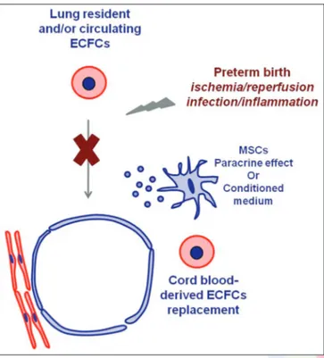
Other cell populations, among which the triple-positive CD34 + CD133 + VEGFR-2 + cells, have also been tagged as EPCs, due to their angiogenic capacity; however, the latter are of hematopoietic origin and they are not able to undergo endothelial differentiation in vitro and in vivo, but probably contribute to vascularization as bystander cells or by orchestrating endothelial cell growth and differentiation.[18-20] In a mouse model of BPD, Balasubramaniam et al. found that exposure to hyperoxia decreases pulmonary vascular density, simplifies distal lung structure and reduces EPCs of hematopoietic origin in blood, bone marrow and lungs, suggesting a role of mobilization, recruitment and engraftment of these cells to the lung in maintaining or repairing lung structure.[21] However, these findings are not consistent with human studies, in which this heterogeneous population (identified by flow cytometry with the expression of CD34, CD133, VEGFR-2, CD45 and CD144 surface markers in different combinations) do not correlate with the development of BPD, neither at birth nor at some days after birth.[22-24] Conversely, cord blood circulating ECFCs (i.e., EPCs of angiogenic origin, enumerated as number of colonies developing from cord blood mononuclear cells), have been shown, in two independent studies, to be lower in cord blood from infants who later develop BPD compared with infants without BPD.[22,25] Although both studies suggest that loss of circulating ECFCs may be implicated in the pathogenesis of lung vascular disruption typical of BPD, none of the two studies provide a mechanistic link. Interestingly, unpublished experimental data by Alphonse et al., recently reported in two reviews, demonstrate that lung-resident ECFCs from rats exposed to hyperoxia are functionally impaired (have compromised proliferative, clonogenic and in vitro vessel-forming potential), suggesting that oxygen-induced depletion of lung-resident ECFCs may be a possible mechanism underlying arrested lung vascular growth in BPD.[26,27] Several in vitro studies demonstrated that ECFCs are highly sensitive to oxidative stress, a major contributor to the pathogenesis of BPD thus indirectly sustaining the idea that their disruption under oxidative conditions (hyperoxia and infection/inflammation) may be linked to the pathogenesis of BPD.[9-11] Further human and animal studies are needed to definitely establish a link between loss of lung-resident and/or circulating ECFCs and the development of BPD in preterm infants. A possible working hypothesis for these studies is schematically explained in Figures 3 and 4.
meSeNChymAl STROmAl Cell
DePleTION
MSCs are cells of mesenchymal origin able to differentiate into mesoderm-derived cells.[28] MSCs display a wide variety of properties, including immunomodulatory and regenerative capacity as well as the ability to provide paracrine Figure 2: Mechanisms of brain injury in diffuse periventricular
leukomalacia. Mesenchymal stem cell-based treatments may protect the developing brain or stimulate the endogenous repair
antiapoptotic, proangiogenic and antiscarring (antifibrotic) stimuli.[29-32] In addition, they have been demonstrated to be able to protect lung epithelial cells against acute lung injury by restituting alveolar cell bioenergetics through the transfer of mitochondria to lung epithelial cells.[33]
The hypothesis that the loss of resident MSCs in the lung stroma may contribute to the development of BPD is supported by one experimental study. In a rat model of BPD, van Haaften et al. demonstrated that after 2 weeks of exposure to hyperoxia, the histological pattern of alveolar simplification was associated with a reduced number of circulating and lung-resident MSCs.[34] Popova et al. more recently collected the tracheal aspirates from 56 ventilated preterm infants for isolation of MSCs; in their study, the presence of MSCs in the tracheal aspirate increased the adjusted odds of BPD by nearly 22 times.[35] Of note, the studied cells were able to acquire a myofibroblast phenotype that was different from the human bone marrow-derived MSCs that do not undergo myofibroblastic differentiation in response to transforming growth factor b1; the difference in the studied cell populations may account for contrasting results between the two studies.[26]
In addition, the possible role of circulating bone marrow-derived MSCs homing to the lung, under omeostatic or developmental conditions and/or after lung injury, is currently unknown.
Altogether, these observations suggest that depletion of circulating or resident ECFCs and/or lung stroma resident MSCs may contribute, at least in part, to altered endothelial growth, abnormal epithelial–stromal interactions, defective elastogenesis and extracellular matrix remodeling, and may account for increased vulnerability of epithelial cells to acute injury thus contributing to subsequent alveolar simplification, the hallmark of new BPD. These findings may
Figure 4: Working hypothesis on the role of endothelial colony-forming cell depletion in preterm infants developing bronchopulmonary dysplasia
Figure 3: Possible interventions to prevent bronchopulmonary dysplasia (BPD) based on the hypothesis that lung stem/progenitor cells and/or endothelial colony-forming cell (ECFCs) are depleted in infants developing BPD. Administration of mesenchymal stem cells or their conditioned medium or ECFC replacement may prevent lung injury
provide a rationale basis for ECFC or MSC administration to protect the developing lung following preterm birth.
Animal models for cell therapy in neonatal lung and brain injury
The main goals of a cell-based therapy for the treatment or prevention of neonatal diseases associated with preterm birth include (i) replacement of depleted stem/ progenitor cells, (ii) reduction of inflammation and fibrosis and (iii) protection of cells (both mature cells and resident stem/progenitor cells) from injury associated with infection, ischemia and oxidative stress.
BPD and PVL are complex and multifactorial diseases, and a combination of the above-mentioned goals could be favorable to achieve protection and optimal lung and brain development. Here, we will only focus on some issues regarding the most promising cell-based approaches, and the most likely to be used in clinical trials, without comprehensively describing all the possible cell-based treatment options available for neonatal diseases.
mSC‑BASeD TReATmeNTS
MSCs exhibit numerous properties, making them suitable for cell therapy in BPD and PVL. Their regenerative potential and immunomodulatory properties have been previously exploited in other MSC-based clinical protocols, including allogenic hematopoietic stem cell transplantation, to reduce the incidence or severity of acute graft–versus– host disease.[29-31,36-39]
The use of MSCs has several advantages over the use of other stem/progenitor cell populations:
• MSCs have been previously used in clinical protocols and have been demonstrated to be safe;
• Their biological behavior (at least for bone marrow-derived MSCs) has been extensively investigated in in vitro and animal models;
• MSCs display multiple beneficial properties, including immunomodulatory capacity, antifibrosis activity, anti-apoptotic and growth-promoting activity and ability to restore the bioenergetic balance through transfer of mitochondria to lung epithelial cells; • MSCs have already been demonstrated to be effective
in animal models of neonatal diseases, including BPD and hypoxic–ischemic encephalopathy, and their administration could have a “multiorgan” beneficial effect not limited to the lungs and the brain.
In experimental models of BPD, intratracheal, intraperitoneal or intravenous (systemic) administration of bone marrow-derived MSC improved alveolar, airway and vascular structure, attenuated inflammation, decreased
fibrosis, ameliorated right heart function and improved exercise capacity.[34,40,41] Importantly, the beneficial therapeutic actions of MSCs appear to be mediated through paracrine mechanisms and immunomodulatory effects, rather than through cell engraftment.[42] Consistently, administration of the conditioned medium from MSC cultures prevents lung injury [Figure 3].[43]
Whether stem and progenitor cell therapy could be beneficial in attenuating the complications of PVL is still an unanswered question. Animal models are rather focused on the use of MSCs in the recovery process after brain injury following birth asphyxia or stroke [Figure 2].[44-49] Whether the same cell populations and routes of administration for hypoxic–ischemic encephalopathy could be used in PVL still needs to explored with animal models.
ePC‑BASeD TReATmeNTS
The rationale for using angiogenic cells (either EPCs of hematopoietic origin or true endothelial progenitors, namely ECFCs) for the prevention of BPD is based on the following observations:
• Lung vascular growth is disrupted in infants with BPD • Low numbers of ECFC in cord blood correlate with
subsequent development of BPD, suggesting a role for ECFC depletion in the pathogenesis of lung vascular disruption.
In an oxygen-induced BPD mouse model, Balasubramaniam et al. showed restoration of the alveolar structure and vessel density in mice treated with bone marrow myeloid progenitor cell population (bone marrow-derived angiogenic cells), a novel myeloid cell population with angiogenic properties.[50]
In addition, interesting unpublished preliminary data by Alphonse et al. show that intrajugular administration of human umbilical cord-derived ECFCs (i.e., EPCs of endothelial origin) improved lung function, preserved alveolar development, attenuated pulmonary hypertension (improved pulmonary artery acceleration times and decreased right ventricular hypertrophy) in hyperoxia-exposed mouse pups, suggesting that human umbilical cord-derived ECFCs may offer a new therapeutic option for BPD by replacing depleted ECFCs or by restoring their function [Figure 3].[26,27]
human amnion epithelial cell‑based treatments
Given their availability from human amniotic membranes, hAECs could be a good candidate for cell replacement. In the developing lung, hAEC have been shown to prevent ventilation-induced injury and injury related to infection in ovine models, mainly via modulation of the inflammatory
response rather than by engraftment. Further studies will clarify the therapeutic potential of hAEC in BPD.[51,52]
Unsolved issues: Which infants? Which cells? how many? Which route? When (Repair or prevention)?
Before stem and progenitor cell therapy becomes a true option for very and extremely preterm infants, the following issues should be resolved:
• The accurate definition of infants at higher risk of sequelae of prematurity, representing the best candidates for treatment and/or prevention;
• The right timing for cell administration (and the definition of the goal: Repair or prevention?);
• The most suitable cell population targeted to specific diseases and/or clinical conditions;
• The best source of stem cell populations (e.g., for MSCs: Adult bone marrow or adipose tissue, umbilical cord, cord blood, placenta or amniotic fluid) and their safety, as addressed by in vitro and in vivo nontumorigenicity and genetic stability upon expansion and transplantation;
• The exact number of cells to be administered and the best route of administration, possibly depending on the clinical picture and the target organs in individual infants.
CONClUSIVe RemARkS
Neonatal diseases associated with preterm birth still represent a burden for global health and families. The search for new tools to minimize the risk of complications associated with prematurity is urgent. Stem cell technology offers new possibilities to prevent or cure these invalidating diseases. Further understanding of the developmental biology in physiological and pathological conditions, of the multiple mechanisms underlying disruption of normal organ development after very/extremely preterm birth, and of the complex biology of stem cells, will allow the performance of targeted studies and the design of appropriate preventive and treatment protocols.
methods for locating and selecting data
Articles were searched on Pubmed using the following terms: (“bronchopulmonary dysplasia” (Mesh Term) AND (“Mesenchymal stromal cells” (MeSH Terms)); (“bronchopulmonary dysplasia” (Mesh Term) AND (“endothelial” (MeSH Terms)); (“bronchopulmonary dysplasia” (Mesh Term) AND (“Stem cells”(MeSH Terms)).
ACkNOWleDgmeNTS
The authors would like to thank Micol Angelini, Maria Antonietta Avanzini, Lina Bollani, Rita Campanelli, Francesca Garofoli, Elisa Lenta, Stefania Longo, Rita Maccario, Melissa Mantelli, Roberta Maragliano, Margherita Massa, Iolanda Mazzucchelli, Vittorio
Rosti (all from Fondazione IRCCS Policlinico San Matteo, Pavia, Italy), Francesca Novara (Università degli Studi di Pavia, Italy), Antonio WD Gavilanes and Luc JI Zimmermann (Maastricht University, The Netherlands). Although not directly involved in the writing of the present manuscript, the persons listed above substantially contributed to the thinking over the topics discussed here, with their critical and constructive discussions.
ReFeReNCeS
1. Bancalari EH, Jobe AH. The respiratory course of extremely preterm infants: A dilemma for diagnosis and terminology. J Pediatr 2012;161:585-8.
2. Jobe AH, Bancalari E. Bronchopulmonary dysplasia. Am J Respir Crit Care Med 2001;163:1723-9.
3. Bancalari E, Claure N, Sosenko IR. Bronchopulmonary dysplasia: Changes in pathogenesis, epidemiology and definition. Semin Neonatol 2003;8:63-71.
4. Hamrick SE, Miller SP, Leonard C, Glidden DV, Goldstein R, Ramaswamy V, et al. Trends in severe brain injury and neurodevelopmental outcome in premature newborn infants: The role of cystic periventricular leukomalacia. J Pediatr 2004;145:593-9. 5. Huppi PS. Immature white matter lesions in the premature infant.
J Pediatr 2004;145:575-8.
6. Volpe JJ. Cerebral white matter injury of the premature infant-more common than you think. Pediatrics 2003;112:176-80.
7. McCurnin DC, Pierce RA, Willis BC, Chang LY, Yoder BA, Yuhanna IS, et al. Postnatal estradiol up-regulates lung nitric oxide synthases and improves lung function in bronchopulmonary dysplasia. Am J Respir Crit Care Med 2009;179:492-500.
8. Lavoie PM, Pham C, Jang KL. Heritability of bronchopulmonary dysplasia, defined according to the consensus statement of the national institutes of health. Pediatrics 2008;122:479-85.
9. Baker CD, Ryan SL, Ingram DA, Seedorf GJ, Abman SH, Balasubramaniam V. Endothelial colony-forming cells from preterm infants are increased and more susceptible to hyperoxia. Am J Respir Crit Care Med 2009;180:454-61.
10. Ingram DA, Krier TR, Mead LE, McGuire C, Prater DN, Bhavsar J,
et al. Clonogenic endothelial progenitor cells are sensitive to oxidative
stress. Stem Cells 2007;25:297-304.
11. Fujinaga H, Baker CD, Ryan SL, Markham NE, Seedorf GJ, Balasubramaniam V, et al. Hyperoxia disrupts vascular endothelial growth factor-nitric oxide signaling and decreases growth of endothelial colony-forming cells from preterm infants. Am J Physiol Lung Cell Mol Physiol 2009;297:L1160-9.
12. Khwaja O, Volpe JJ. Pathogenesis of cerebral white matter injury of prematurity. Arch Dis Child Fetal Neonatal Ed 2008;93:F153-61. 13. Finer NN, Carlo WA, Walsh MC, Rich W, Gantz MG, Laptook AR,
et al. Early CPAP versus surfactant in extremely preterm infants.
N Engl J Med 2010;362:1970-9.
14. Morley CJ. CPAP and low oxygen saturation for very preterm babies? N Engl J Med 2010;362:2024-6.
15. Morley CJ, Davis PG, Doyle LW, Brion LP, Hascoet JM, Carlin JB. Nasal CPAP or intubation at birth for very preterm infants. N Engl J Med 2008;358:700-8.
16. Vaucher YE, Peralta-Carcelen M, Finer NN, Carlo WA, Gantz MG, Walsh MC, et al. Neurodevelopmental outcomes in the early CPAP and pulse oximetry trial. N Engl J Med 2012;367:2495-504. 17. Ingram DA, Mead LE, Tanaka H, Meade V, Fenoglio A, Mortell K,
et al. Identification of a novel hierarchy of endothelial progenitor
cells using human peripheral and umbilical cord blood. Blood 2004;104:2752-60.
18. Case J, Mead LE, Bessler WK, Prater D, White HA, Saadatzadeh MR,
et al. Human CD34+AC133+VEGFR-2+cells are not endothelial
progenitor cells but distinct, primitive hematopoietic progenitors. Exp Hematol 2007;35:1109-18.
19. Hirschi KK, Ingram DA, Yoder MC. Assessing identity, phenotype, and fate of endothelial progenitor cells. Arterioscler Thromb Vasc Biol 2008;28:1584-95.
20. Prater DN, Case J, Ingram DA, Yoder MC. Working hypothesis to redefine endothelial progenitor cells. Leukemia 2007;21:1141-9. 21. Balasubramaniam V, Mervis CF, Maxey AM, Markham NE,
Abman SH. Hyperoxia reduces bone marrow, circulating, and lung endothelial progenitor cells in the developing lung: Implications for the pathogenesis of bronchopulmonary dysplasia. Am J Physiol Lung Cell Mol Physiol 2007;292:L1073-84.
22. Borghesi A, Massa M, Campanelli R, Bollani L, Tzialla C, Figar TA,
et al. Circulating endothelial progenitor cells in preterm infants
with bronchopulmonary dysplasia. Am J Respir Crit Care Med 2009;180:540-6.
23. Paviotti G, Fadini GP, Boscaro E, Agostini C, Avogaro A, Chiandetti L,
et al. Endothelial progenitor cells, bronchopulmonary dysplasia and
other short-term outcomes of extremely preterm birth. Early Hum Dev 2011;87:461-5.
24. Safranow K, Kotowski M, Lewandowska J, Machalinska A, Dziedziejko V, Czajka R, et al. Circulating endothelial progenitor cells in premature infants: Is there an association with premature birth complications? J Perinat Med 2012;40:455-62.
25. Baker CD, Balasubramaniam V, Mourani PM, Sontag MK, Black CP, Ryan BS, et al. Cord blood angiogenic progenitor cells are decreased in bronchopulmonary dysplasia. Eur Respir J 2012;40:1516-22. 26. O’Reilly M, Thebaud B. Cell-based strategies to reconstitute lung
function in infants with severe bronchopulmonary dysplasia. Clin Perinatol 2012;39:703-25.
27. Alphonse RS, Rajabali S, Thebaud B. Lung injury in preterm neonates: The role and therapeutic potential of stem cells. Antioxid Redox Signal 2012;17:1013-40.
28. Dominici M, Le Blanc K, Mueller I, Slaper-Cortenbach I, Marini F, Krause D, et al. Minimal criteria for defining multipotent mesenchymal stromal cells. The International Society for Cellular Therapy position statement. Cytotherapy 2006;8:315-7.
29. Bernardo ME, Pagliara D, Locatelli F. Mesenchymal stromal cell therapy: A revolution in Regenerative Medicine? Bone Marrow Transplant 2012;47:164-71.
30. Le Blanc K. Mesenchymal stromal cells: Tissue repair and immune modulation. Cytotherapy 2006;8:559-61.
31. Avanzini MA, Bernardo ME, Cometa AM, Perotti C, Zaffaroni N, Novara F, et al. Generation of mesenchymal stromal cells in the presence of platelet lysate: A phenotypic and functional comparison of umbilical cord blood- and bone marrow-derived progenitors. Haematologica 2009;94:1649-60.
32. Singer NG, Caplan AI. Mesenchymal stem cells: Mechanisms of inflammation. Annu Rev Pathol 2011;6:457-78.
33. Islam MN, Das SR, Emin MT, Wei M, Sun L, Westphalen K, et al. Mitochondrial transfer from bone-marrow-derived stromal cells to pulmonary alveoli protects against acute lung injury. Nat Med 2012;18:759-65.
34. van Haaften T, Byrne R, Bonnet S, Rochefort GY, Akabutu J, Bouchentouf M, et al. Airway delivery of mesenchymal stem cells prevents arrested alveolar growth in neonatal lung injury in rats. Am J Respir Crit Care Med 2009;180:1131-42.
35. Popova AP, Bozyk PD, Bentley JK, Linn MJ, Goldsmith AM, Schumacher RE, et al. Isolation of tracheal aspirate mesenchymal stromal cells predicts bronchopulmonary dysplasia. Pediatrics 2010;126:e1127-33.
36. Le Blanc K, Frassoni F, Ball L, Locatelli F, Roelofs H, Lewis I,
severe, acute graft-versus-host disease: A phase II study. Lancet 2008;371:1579-86.
37. Matthay MA, Thompson BT, Read EJ, McKenna DH, Jr., Liu KD, Calfee CS, et al. Therapeutic potential of mesenchymal stem cells for severe acute lung injury. Chest 2010;138:965-72.
38. Salem HK, Thiemermann C. Mesenchymal stromal cells: Current understanding and clinical status. Stem Cells 2010;28:585-96. 39. Sensebe L, Bourin P. Mesenchymal stem cells for therapeutic
purposes. Transplantation 2009;87:S49-53.
40. Aslam M, Baveja R, Liang OD, Fernandez-Gonzalez A, Lee C, Mitsialis SA, et al. Bone marrow stromal cells attenuate lung injury in a murine model of neonatal chronic lung disease. Am J Respir Crit Care Med 2009;180:1122-30.
41. Chang YS, Oh W, Choi SJ, Sung DK, Kim SY, Choi EY, et al. Human umbilical cord blood-derived mesenchymal stem cells attenuate hyperoxia-induced lung injury in neonatal rats. 2009;18:869-86. 42. Pierro M, Ionescu L, Montemurro T, Vadivel A, Weissmann G,
Oudit G, et al. Short-term, long-term and paracrine effect of human umbilical cord-derived stem cells in lung injury prevention and repair in experimental bronchopulmonary dysplasia. Thorax 2012 [In Press].
43. Ionescu L, Byrne RN, van Haaften T, Vadivel A, Alphonse RS, Rey-Parra GJ, et al. Stem cell conditioned medium improves acute lung injury in mice: In vivo evidence for stem cell paracrine action. Am J Physiol Lung Cell Mol Physiol 2012;303:L967-77.
44. van Velthoven CT, Kavelaars A, van Bel F, Heijnen CJ. Mesenchymal stem cell transplantation changes the gene expression profile of the neonatal ischemic brain. Brain Behav Immun 2011;25:1342-8. 45. van Velthoven CT, Kavelaars A, van Bel F, Heijnen CJ. Nasal
administration of stem cells: A promising novel route to treat neonatal ischemic brain damage. Pediatr Res 2010;68:419-22. 46. van Velthoven CT, Kavelaars A, van Bel F, Heijnen CJ. Repeated
mesenchymal stem cell treatment after neonatal hypoxia-ischemia has distinct effects on formation and maturation of new neurons and oligodendrocytes leading to restoration of damage, corticospinal motor tract activity, and sensorimotor function. J Neurosci 2010;30:9603-11.
47. van Velthoven CT, Kavelaars A, van Bel F, Heijnen CJ. Mesenchymal stem cell treatment after neonatal hypoxic-ischemic brain injury improves behavioral outcome and induces neuronal and oligodendrocyte regeneration. Brain Behav Immun 2010;24:387-93. 48. van Velthoven CT, Kavelaars A, van Bel F, Heijnen CJ. Regeneration
of the ischemic brain by engineered stem cells: Fuelling endogenous repair processes. Brain Res Rev 2009;61:1-13.
49. Kim ES, Ahn SY, Im GH, Sung DK, Park YR, Choi SH, et al. Human umbilical cord blood-derived mesenchymal stem cell transplantation attenuates severe brain injury by permanent middle cerebral artery occlusion in newborn rats. Pediatr Res 2012;72:277-84.
50. Balasubramaniam V, Ryan SL, Seedorf GJ, Roth EV, Heumann TR, Yoder MC, et al. Bone marrow-derived angiogenic cells restore lung alveolar and vascular structure after neonatal hyperoxia in infant mice. Am J Physiol Lung Cell Mol Physiol 2010;298:L315-23. 51. Vosdoganes P, Hodges RJ, Lim R, Westover AJ, Acharya RY,
Wallace EM, et al. Human amnion epithelial cells as a treatment for inflammation-induced fetal lung injury in sheep. Am J Obstet Gynecol 2011;205:156 e26-33.
52. Hodges RJ, Jenkin G, Hooper SB, Allison B, Lim R, Dickinson H, et al. Human amnion epithelial cells reduce ventilation-induced preterm lung injury in fetal sheep. Am J Obstet Gynecol 2012;206:448 e8-15.
How to cite this article: Borghesi A, Cova C, Gazzolo D, Stronati M. Stem cell therapy for neonatal diseases associated with preterm birth. J Clin Neonatol 2013;2:1-7.
Source of Support: Associazione ONLUS – Aiutami a crescere (www.aiutamiacrescere.it). Conflict of Interest: None declared.