For Peer Review Only
Effects of dietary resveratrol on the sleep-wake cycle in the non-human primate grey mouse lemur (Microcebus
murinus)
Journal: Chronobiology International Manuscript ID: LCBI-2011-0207.R3
Manuscript Type: Original Reports Date Submitted by the Author: n/a
Complete List of Authors: Pifferi, Fabien; UMR CNRS MNHN 7179, Rahman, Anisur; UMR CNRS MNHN 7179, Languille, Solene; UMR CNRS MNHN 7179,
Auffret, Alexandra; University Aix-Marseille, CIC-CPCET UMR 6193
Babiloni, Claudio; University of Foggia, Department of Biomedical Sciences Blin, Olivier; University Aix-Marseille, CIC-CPCET UMR 6193
Lamberty, Yves; UCB Pharma,
Richardson, Jill; GlaxoSmithKline, R&D China U.K. Group Aujard, F.; UMR CNRS MNHN 7179,
Keywords: Circadian Rhythms, Electroencephalography, Microcebus murinus, Resveratrol, Sleep, Metabolism
For Peer Review Only
Effects of dietary resveratrol on the sleep-wake cycle in the non-human primate grey mouse lemur (Microcebus murinus)
Pifferi F1, Rahman A1, Languille S1, Auffret A2, Babiloni C5,6, Blin O2, Lamberty Y3, Richardson J.C.4, Aujard F1
1
UMR CNRS-MNHN 7179, Brunoy, France
2
CIC-CPCET, UMR 6193 University Aix-Marseille, France
3
UCB Pharma, Braine l’Alleud, Belgium
4
GlaxoSmithKline, R&D China U.K. Group, Stevenage, United Kingdom
5
Department of Biomedical Sciences, University of Foggia, Foggia, Italy
6
Department of Imaging, San Raffaele Cassino, Italy
Part of the Pharmacog consortium
Running title: Dietary resveratrol and sleep-wake rhythms Corresponding author: Dr. Fabienne Aujard – email: [email protected]
The research leading to the present results has received funding from the European Community's Seventh Framework Programme (FP7/2007-2013) for the Innovative Medicine Initiative under Grant Agreement n°115009 (prediction of cognitive properties of new drug candidates for neurodegenerative diseases in early clinical development, Pharmacog) for the study of electroencephalographic (EEG) rhythms in lemurs during the sleep-wake cycle. For further information on the PharmaCog project, please refer to www.alzheimer-europe.org. 3 4 5 6 7 8 9 10 11 12 13 14 15 16 17 18 19 20 21 22 23 24 25 26 27 28 29 30 31 32 33 34 35 36 37 38 39 40 41 42 43 44 45 46 47 48 49 50 51 52 53 54 55 56
For Peer Review Only
Abstract
Converging evidence shows that the non-human primate grey mouse lemur (Microcebus murinus) is ideal for the study of the ageing process and for testing the effects of new therapies and dietary interventions on age-associated pathologies. One such dietary supplement is resveratrol (RSV), a dietary polyphenolic compound with several positive effects on metabolic functions and longevity (Languille et al., 2011; Pifferi et al., 2011). However, little is known about the effect of RSV on the lemur sleep-wake cycle, which reflects mammalian brain function and health. In the present study, we investigated this effect by comparing sleep-wake cycles in adult lemurs based on electroencephalographic (EEG) rhythms. The effect of short-term RSV supplementation on the sleep-wake cycle of mouse lemurs was evaluated in entrained conditions (long-day photoperiods, light:dark 14:10). After three weeks of RSV supplementation, animals exhibited a significantly increased proportion of active wake, occurring mainly during the resting phase of the sleep-wake cycle (+163%). The increase in active wake with RSV supplementation was accompanied by a significant reduction of both paradoxical sleep (-95%) and slow-wave sleep (-38%). These changes mainly occurred during the resting phase of the sleep-wake cycle (RSV supplementation induced negligible changes in active wake during the active phase of the sleep-wake cycle). The present data suggest that RSV may be a potent regulator of sleep-wake rhythms and could be of major interest in the study of sleep perturbations associated with ageing and neuropathology.
Keywords: circadian rhythms, electroencephalography, Microcebus murinus, resveratrol,
sleep, metabolism. 3 4 5 6 7 8 9 10 11 12 13 14 15 16 17 18 19 20 21 22 23 24 25 26 27 28 29 30 31 32 33 34 35 36 37 38 39 40 41 42 43 44 45 46 47 48 49 50 51 52 53 54 55 56
For Peer Review Only
Introduction
There is growing evidence that close relationships exist between circadian rhythms and metabolism (Ramsey and Bass, 2011). In particular, disturbances of the biological clock predispose rodents to metabolic disorders such as dyslipidaemia, insulin resistance and obesity (Duez and Staels, 2008; Dallman and Weaver, 2010). Recent studies suggest that relationships between metabolism and circadian rhythms could be driven by changes in the expression of clock genes. For example, it has been demonstrated that the administration of bezafibrate, a hypolipidaemic PPARalpha ligand, led to a phase advance of the endogenous clock and changed the circadian expression of clock genes such as per2, Bmal1 and Rev-ERBα in mice (Shirai et al., 2007). Caloric restriction, a regime known to prolong lifespan in various species (Canto et al., 2009), also affects circadian rhythms (Challet et al., 1998 ; Giroud et al., 2008), likely via the activation of sirtuin 1 (SIRT1). SIRT1 is a NAD(+)-dependent deacetylase that directly binds to the CLOCK/BMAL1 complex to regulate the expression of clock genes (Asher et al., 2008; Nakahata et al. 2008, 2009; Ramsey et al., 2009). The beneficial effects exerted by caloric restriction could be mediated through the resetting of the circadian clock, leading to synchrony in metabolism and physiology (Froy et al. 2010).
Considering the premises described above, the sleep-wake cycle is an important parameter in the study of relationships existing among diet, energy metabolism and circadian rhythms. Sleep regulation is driven by both circadian rhythms and energy homeostasis (Ramsey and Bass, 2011; Huang et al., 2011) and is considered a marker of mammalian brain function and health (Huang et al., 2011; McCoy and Strecker, 2011). In 3 4 5 6 7 8 9 10 11 12 13 14 15 16 17 18 19 20 21 22 23 24 25 26 27 28 29 30 31 32 33 34 35 36 37 38 39 40 41 42 43 44 45 46 47 48 49 50 51 52 53 54 55 56
For Peer Review Only
rodents, fasting induced a strong reduction in total sleep duration (Guesdon, 2005; Alvarenga et al., 2005). Compared to controls, rats under dietary caloric restriction (-40% for 8 weeks) showed an increase in wake time and decreases in slow-wave sleep (SWS) and paradoxical sleep (PS) during the light period (Alvarenga et al., 2005) as well as a decrease of the PS/SWS ratio, reflecting a state of energy depletion (Guesdon 2005). Based on the data described above, it has been speculated that one of the functions of sleep is to protect the body against the deleterious effects of free radicals produced by a high metabolic rate during waking hours (Guesdon et al, 2005). Free radicals are highly reactive oxidant molecules that can affect cell membranes and induce cell aging and death. During PS, lower metabolic rate and slightly lower brain temperature may provide an opportunity to renew the enzymes affected by free radicals. If this hypothesis is true, then nutrients with anti-oxidant effects are expected to affect the regulation of the sleep-wake cycle and the metabolic/thermoregulatory activity that coincides with aging.
On this basis, the present study was aimed at the investigation of the relationships between circadian rhythms and energy metabolism through the monitoring of sleep during a metabolic challenge induced by resveratrol (RSV) supplementation. RSV is a dietary polyphenolic compound present in a variety of foods, including vegetables, fruits, seeds, tea and wine, with several documented positive effects on metabolic functions and longevity (Barger et al., 2008) as well as protective effects on cardiovascular and neurodegenerative diseases and cancer (Baur, 2010; Sun et al., 2010). Although the mechanisms by which RSV exerts such a wide range of beneficial effects on these diseases have not yet been clearly elucidated, a number of studies have reported that this 3 4 5 6 7 8 9 10 11 12 13 14 15 16 17 18 19 20 21 22 23 24 25 26 27 28 29 30 31 32 33 34 35 36 37 38 39 40 41 42 43 44 45 46 47 48 49 50 51 52 53 54 55 56
For Peer Review Only
compound possesses antioxidant and anti-inflammatory properties (Xia et al., 2010; Zhang et al., 2010). One of the proposed mechanisms to explain the role of RSV on age-related pathologies is the activation of SIRT1 (Das et al., 2010). RSV is known to increase SIRT1 activity in mammals and to improve mitochondrial function in rodents (Lagouge et al., 2006). In primates, short-term RSV supplementation reduces body mass gain and resting metabolic rate (Dal-Pan et al., 2010). In addition to its effects on energy metabolism, we recently demonstrated that RSV was able to modify the endogenous period in non-human primates (Pifferi et al., 2011). In this previous study, grey mouse lemurs supplemented with RSV for 15 days exhibited a shortening of their endogenous free-running period compared to the control. It had been previously demonstrated that RSV regulates circadian clock genes in cultured Rat-1 fibroblast cells (Oike et al., 2008). A concentration of 100 µM RSV was shown to increase the amplitude of oscillation of clock genes Per1, Per2 and Bmal1. These results suggest that RSV might act as a regulator of circadian clocks.
Based on these results, we hypothesised that changes in the temporal organisation of sleep-wake rhythms could be expected in response to RSV supplementation in the diet. The mechanism of the action of RSV could either take place through the stimulation of the sirtuins pathway or through its antioxidant effect (possibly also via sirtuins) by lowering the production of free radicals and thereby lowering the requirement for sleep (particularly PS) without deleterious effects. The aim of the present study was to evaluate the impact of a metabolic challenge induced by RSV dietary supplementation on circadian rhythms by considering the impact of this supplementation on sleep/wake 3 4 5 6 7 8 9 10 11 12 13 14 15 16 17 18 19 20 21 22 23 24 25 26 27 28 29 30 31 32 33 34 35 36 37 38 39 40 41 42 43 44 45 46 47 48 49 50 51 52 53 54 55 56
For Peer Review Only
architecture regulated by both the circadian system and energy homeostasis. Toward that end, we focused on the impact of a short-term (three-week) RSV dietary supplementation on the expression of sleep-wake rhythms in a non-human primate, the grey mouse lemur (Microcebus murinus). Grey mouse lemurs are small nocturnal primates originating from Madagascar with very marked circadian rhythms (Aujard et al., 2006) and represent a model of primary interest for the study of biological rhythms. Grey mouse lemurs are nocturnal, with high levels of activity during the active period and almost complete rest during the resting period. In the present study, sleep-wake rhythms have been investigated by electroencephalography in adult mouse lemurs. To our knowledge, the present work is the first to assess the impact of RSV on sleep.
Materials and Methods
Animals and housing conditions
Ten male grey mouse lemurs (Microcebus murinus) born in the laboratory breeding colony of UMR 7179 (CNRS/MNHN, France, license approval No. A91.114.1) were utilised in this study. The mean age of the animals at the beginning of the experiment was 37.4 ± 3.5 and 33.4 ± 2.2 months for CTL and RSV groups, respectively. Throughout the experiments, animals were housed in individual cages, provided with branches and a wooden nest in which temperature and humidity were controlled and maintained constant (ambient temperature = 24–26 °C, relative humidity = 55%) and given food and water ad libitum. The animals’ diet included fresh banana (393 kJ/100 g) and a homemade milky mixture containing baby cereals, eggs and milk (435 kJ/100 g).
3 4 5 6 7 8 9 10 11 12 13 14 15 16 17 18 19 20 21 22 23 24 25 26 27 28 29 30 31 32 33 34 35 36 37 38 39 40 41 42 43 44 45 46 47 48 49 50 51 52 53 54 55 56
For Peer Review Only
All experiments were performed in accordance with the Principles of Laboratory Animal Care (National Institutes of Health publication 86-23, revised 1985) and the European Communities Council Directive (86/609/EEC). The research was conducted under authorisation n° 91–305 from the Direction Départementale des Services Vétérinaires de l'Essonne and the Internal Review Board of the UMR 7179. All experiments were conducted under personal license (authorisation n° 91–460, issued 5 June, 2009) delivered by the Ministry of Education and Science. The present experimental protocol is conform to international ethical standards as outlined in Portaluppi et al. (2010).
Experimental design
During the first week of the experiment, the five animals of the RSV group received the control diet (30 g/d of their usual food, no RSV), and EEG/EMG activities were continuously recorded to obtain reference “baseline” data on the sleep-wake cycle. This condition was named RSV T0. During the three following weeks, these animals received their usual diet supplemented with RSV (Trans-resveratrol, Sequoia Research, UK; 30 g/d of their usual mixture + 200 mg/kg/d of resveratrol). The dose was defined based on previous studies by our research group (Pifferi et al., 2011; Dal-Pan et al., 2010). The last week of RSV supplementation (RSV T3) was used to record EEG/EMG activities for the evaluation of the effects of RSV on the sleep-wake cycle.
The five animals of the CTL group received the control diet during the four-week experiment (30 g/d of their usual food). The EEG/EMG activities were monitored during the first (CTL T0 condition) and the fourth (CTL T3 condition) weeks of the experiment. 3 4 5 6 7 8 9 10 11 12 13 14 15 16 17 18 19 20 21 22 23 24 25 26 27 28 29 30 31 32 33 34 35 36 37 38 39 40 41 42 43 44 45 46 47 48 49 50 51 52 53 54 55 56
For Peer Review Only
The experiments were performed during the long-day photoperiod (light:dark 14:10). During the experiments, the feeding time was randomly varied to avoid any rhythmic entrainment resulting from a regular feeding time.
The body weight of the animals was systematically measured during the experiment. No statistically significant variations in body weight were observed after 3 weeks of RSV treatment (101.6 ± 6.4 g) compared to the RSV T0 condition (95.2 ± 5.5 g) or between the CTL T0 (97.0 ± 4.6 g) and CTL T3 (103.8 ± 5.1 g) conditions.
EEG sleep-wake rhythm recording
Recordings of sleep-wake rhythms were obtained by wireless telemetry. A small EEG/EMG transmitter weighing 2.5 g (PhysioTel F20-EET, DataScience Co. Ltd., Minnesota, USA) was implanted into the visceral cavity under ketamine anaesthesia (Imalgene, 100 mg/kg ip). The transmitter allowed for the simultaneous recording of one EEG channel and one EMG channel (1-500 Hz sampling rate). The electrode wires were subcutaneously led from the abdomen to the skull. For EEG recoding, leads were sealed to the skull using dental cement in the region of the anterior frontal cortex according to the stereotaxic atlas of the mouse lemur brain (Bons et al., 1998). For EMG recording, a pair of bipolar electrodes was sutured in the neck muscles with non-absorbable polyamide suture. After surgery, animals were returned to their home cage and were allowed to recover for 15 days before the beginning of the experiments. Total recovery was checked by a visual inspection of the complete healing of the surgical incisions and by the verification of a stable daily pattern of body temperature and locomotor activity 3 4 5 6 7 8 9 10 11 12 13 14 15 16 17 18 19 20 21 22 23 24 25 26 27 28 29 30 31 32 33 34 35 36 37 38 39 40 41 42 43 44 45 46 47 48 49 50 51 52 53 54 55 56
For Peer Review Only
variation. A receiving plate (RPC-1, Data Science Co Ltd., Minnesota, USA) located in the cage enabled EEG and EMG data recording.
Data analysis
Data were acquired with Dataquest Lab Pro v. 3.0 (Data Science Co. Ltd., Minnesota, USA) and computed with Neuroscore software v. 2.1 (Data Science Co. Ltd., Minnesota, USA). EEG and EMG signals were acquired during five consecutive days within the first week of treatment (CTL T0 and RSV T0) and during the last week of the CTL condition and RSV supplementation (CTL T3 and RSV T3). Sleep was scored manually in 10-sec intervals according to the following stages as described by Rechtschaffen and Kales in 1968 and amended in 2007 by the American Academy of Sleep Medicine (Iber et al., 2007): slow-wave sleep (SWS), rapid-eye movement sleep or paradoxical sleep (PS), quiet wake (QW) and active wake (AW). Artifacts (Ar) were also taken into account. To better understand the specific effects of RSV on sleep-wake rhythms, we also performed a separate scoring of active (dark) and resting (light) phases. Proportions of sleep and wake stages are expressed as a percentage of recording time. The number and the mean length (in seconds) of bouts of the different stages were also determined. A bout was defined as a period of time spent in a particular stage.
Statistical analysis
Sleep-wake parameters (% of recording time, number of bouts, bout length) were averaged from the five consecutive days of recording for each condition. Statistical analysis was performed by an analysis of variance (ANOVA) with a post-hoc 3 4 5 6 7 8 9 10 11 12 13 14 15 16 17 18 19 20 21 22 23 24 25 26 27 28 29 30 31 32 33 34 35 36 37 38 39 40 41 42 43 44 45 46 47 48 49 50 51 52 53 54 55 56
For Peer Review Only
Bonferroni’s multiple comparison test. Specifically, an ANOVA “within group” design in the RSV animals (N=5) tested the effects of RSV supplementation (in a comparison between RSV T0 and RSV T3) on sleep-wake parameters (% of recording time, number of sleep bouts, sleep bout length) and body temperature (the mean body temperature of each animal for five consecutive days, light phase and dark phase) as dependent variables. A control ANOVA “within group” design in the CTL animals (N=5) tested the effect of time (in a comparison between CTL T0 and CTL T3) on the dependent variables. Another ANOVA design tested the control hypothesis of no differences in the variables described between CTL and RSV lemurs at the baseline condition (T0).
Differences were considered significant at p<0.05. All values are the mean ± SEM of the different parameters. In the following sections, proportions of the different stages of sleep and wake are expressed as a percentage of the recording time, the number of sleep bouts and the mean bout length (in sec).
Results
Sleep-wake scoring during whole recordings The CTL T0 condition (Figure 1)
During the five consecutive days of EEG recording under CTL T0 conditions, SWS accounted for 35.9 ± 1.5 % of the total recording time, whereas PS, AW and QW represented 7.2 ± 0.4, 52.4 ± 3.6 and 4.0 ± 0.5 % of the total recording time, respectively.
The effect of time on CTL animals (Figure 1)
3 4 5 6 7 8 9 10 11 12 13 14 15 16 17 18 19 20 21 22 23 24 25 26 27 28 29 30 31 32 33 34 35 36 37 38 39 40 41 42 43 44 45 46 47 48 49 50 51 52 53 54 55 56
For Peer Review Only
After a three-week experiment, CTL T3 animals exhibited a significant decrease of AW (-18% compared to CTL T0 animals, p<0.01) with increased SWS (+20 % compared to CTL T0 animals, p<0.001). The decreased AW proportion was associated with a significant decrease of AW bout duration (-30 %, p<0.001) without significant changes in the number of bouts.
The effect of RSV supplementation (Figure 1)
During the five consecutive days of EEG recording under RSV T0 conditions, slow-wave sleep (SWS) represented 39.3 ± 1.7 % of the total recording time, whereas PS, AW and QW represented 6.7 ± 0.4, 49.5 ± 2.4 and 4.0 ± 0.6 % of the total recording time, respectively. No differences were observed between CTL T0 and RSV T0 animals, confirming the robustness of our reference data. Three weeks of RSV supplementation led to a significant decrease of SWS (-33 %, p<0.001) and PS (-95 %, p<0.01) as well as to a significant increase of AW (+45 %, p<0.001) compared to RSV T0 conditions. The effects of RSV supplementation were the opposite of the effects of time measured in CTL animals, which provoked a significant increase of SWS and decrease of AW. The level of QW remained significantly unchanged with RSV supplementation compared to RSV T0. The effect of RSV on the proportion of AW was accompanied by a significant increase in the duration of AW bouts (+17%, p<0.001), but the total number remained unchanged. Conversely, changes in the proportion of SWS, PS and QW with RSV supplementation were accompanied by a significant decrease in the number of bouts (-50 %, p<0.001; -93 %, p<0.01 and -70 %, p<0.001, respectively) without any significant change in bout duration. 3 4 5 6 7 8 9 10 11 12 13 14 15 16 17 18 19 20 21 22 23 24 25 26 27 28 29 30 31 32 33 34 35 36 37 38 39 40 41 42 43 44 45 46 47 48 49 50 51 52 53 54 55 56
For Peer Review Only
Sleep-wake scoring during the resting phase
The CTL T0 condition and the effect of time (Figure 2)
During the resting phase, CTL T0 animals exhibited 23.6 ± 2.0 % of AW and 6.2 ± 0.7 % of QW, whereas SWS and PS represented 57.7 ± 1.9 % and 12.1 ± 0.7 %, respectively, of the recorded resting phase. After three weeks of experiments, no significant changes during the resting phase were observed between CTL T0 and CTL T3 conditions. Thus, the time of observation during recordings (between CTL T0 and CTL T3 conditions) has no effect during the resting phase.
The effects of RSV supplementation (Figure 2)
During the resting phase, RSV T0 animals exhibited 23.5 ± 1.3 % of AW and 5.2 ± 0.6 % of QW, whereas SWS and PS represented 60.6 ± 1.0 % and 10.5 ± 0.4 %, respectively, of the recorded resting phase. In contrast to the effect of time, RSV supplementation induced significant changes in resting phase sleep-wake rhythms. After three weeks of RSV supplementation, changes in sleep-wake stages followed the same trend as during whole recordings. We measured a strong increase in AW (+163%, p<0.001) accompanied by a 38% decrease of SWS (p<0.001) and an almost complete disappearance of PS (-95% compared to the RSV T0 condition, p<0.001). In contrast to what was observed during whole recordings, resting phase QW was significantly decreased by RSV treatment (-86%, p<0.05). Similar to what was observed during whole recordings, changes with RSV supplementation were accompanied by a significant decrease in the number of PS bouts (-93 %, p<0.001), SWS bouts (-55%, p<0.001) and QW bouts (-87%, p<0.001). The 3 4 5 6 7 8 9 10 11 12 13 14 15 16 17 18 19 20 21 22 23 24 25 26 27 28 29 30 31 32 33 34 35 36 37 38 39 40 41 42 43 44 45 46 47 48 49 50 51 52 53 54 55 56
For Peer Review Only
duration of AW bouts significantly increased with RSV treatment (+135%, p<0.001), whereas the number of bouts remained unchanged.
Sleep-wake scoring during the active phase
The CTL T0 condition and the effect of time (Figure 3)
During the active phase, CTL T0 animals exhibited 91.9 ± 2.4 % of AW and 0.9 ± 0.4 % of QW, whereas SWS and PS represented 6.2 ± 2.1 % and 0.5 ± 0.3 %, respectively, of the active phase recording time. After three weeks of experiments, CTL T3 animals exhibited a significant decrease of AW time (-16 % compared to CTL T0, p<0.001). Changes in AW were accompanied by a significant increase in the proportion of SWS (+125% compared to CTL T0, p<0.001), which reached 14.1% of the total recording time. Thus, the effect of time observed during the whole recording on the decrease of AW and the increase of SWS mainly occurs during the active phase. Similar effects on bouts were observed with a decrease of AW bout duration (-50%, p<0.01), and increases of SWS were explained by the increased number of bouts (+53%, p<0.01).
The effects of RSV supplementation (Figure 3)
During the active phase, RSV T0 animals exhibited 88.4 ± 1.9 % of AW and 2.4 ± 1.1 % of QW, whereas SWS and PS represented 7.1 ± 0.7 % and 1.1 ± 0.4 %, respectively, of the total score. After three weeks of RSV, no significant changes occurred in the proportions of the different sleep and wake parameters. However, RSV supplementation led to an elimination of the effect of time.
3 4 5 6 7 8 9 10 11 12 13 14 15 16 17 18 19 20 21 22 23 24 25 26 27 28 29 30 31 32 33 34 35 36 37 38 39 40 41 42 43 44 45 46 47 48 49 50 51 52 53 54 55 56
For Peer Review Only
Hypnograms
24h Hypnograms of one animal representative of each condition (CTL T0, CTL T3, RSV T0 and RSV T3) are reported in Figure 4. This representation illustrates that sleep is highly fragmented under CTL T0, CTL T3 and RSV T0 conditions, with regular periods of arousal throughout the diurnal resting phase. The differences between CTL T0 and CTL T3 were also visible in this figure, the number of sleep boots during the active phase being clearly higher after 3 weeks of experiments, and particularly from 17:00 to 00:00. Conversely, in RSV T3 condition, animals exhibited higher active wake from 17:00 to 00:00 compared to CTL T3 animals. The higher level of activity during the resting phase in RSV T3 condition compared to RSV T0 was remarkable with this representation. The hypnograms show that the AW was particularly increased in RSV T3 condition compared to RSV T0 during the last part of the resting period, specifically from 11:00 to 17:00.
Body temperatures
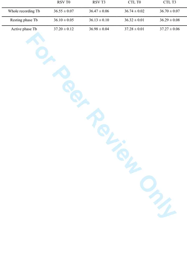
Body temperatures (Tb) during whole recording, resting phase and active phase (the mean body temperature of five consecutive days, expressed in °C) are reported in Table 1. The mean body temperatures during whole recording, light phase and dark active phase remained significantly unchanged with RSV treatment compared to baseline conditions (RSV T0). No significant difference was observed between the CTL T0 and CTL T3 conditions either. Discussion 3 4 5 6 7 8 9 10 11 12 13 14 15 16 17 18 19 20 21 22 23 24 25 26 27 28 29 30 31 32 33 34 35 36 37 38 39 40 41 42 43 44 45 46 47 48 49 50 51 52 53 54 55 56
For Peer Review Only
In the present study, we assessed the role of RSV dietary supplementation on the sleep-wake rhythms of adult mouse lemurs. Toward that end, the polysomnographic recording of sleep was performed for the first time in this nocturnal primate by EEG and EMG. The present work is therefore the first to describe the sleep-wake rhythms in the grey mouse lemur. RSV T0 and CTL animals exhibited approximately 55% wakefulness (50% of AW + 4% of QW) and approximately 45% sleep (40% of SWS + 7% of PS). Sleep mainly occurred during the resting phase, with 71% of recording time (60% of SWS + 11% of PS). Conversely, activity represented close to 90% of the total recording time during the active phase, and sleep represented less than 8% of this period. These proportions are very similar to what has previously been observed in other non-human primates. In a study of sleep patterns in macaques (Macaca mulatta) housed under long-day photoperiods, Hsieh et al. (2008) reported that wake represented 54% of a total of 24 h recording time, whereas sleep represented 46% of total recording time (35% of SWS + 11% of PS). During the dark resting period of macaques, sleep represented 89% of the total recording time (70% of SWS and 19% of PS), whereas during the light active phase, wake represented 75% of total recording time, and sleep represented 25% (with 20% of SWS and 5% of PS). The sleep patterns observed in macaques are very similar to our own measurements in mouse lemurs. The main difference between the two species concerns the amount of sleep during the resting phase, which is higher in macaques (89%) than in mouse lemurs (71%). This difference may result from the shorter period of the resting phase in macaques (8 h) compared to mouse lemurs (14 h). Another important difference between mouse lemurs and macaques is the level of fragmentation of sleep. Sleep is highly fragmented in mouse lemurs, judging by the high number of QW and 3 4 5 6 7 8 9 10 11 12 13 14 15 16 17 18 19 20 21 22 23 24 25 26 27 28 29 30 31 32 33 34 35 36 37 38 39 40 41 42 43 44 45 46 47 48 49 50 51 52 53 54 55 56
For Peer Review Only
wake bouts (approximately 135 during the resting phase, Figure 2). However, wake bouts (active + quiet) represented less than 2 h during the resting period, which spans 14 h when animals are maintained in a long-day photoperiod. As a final remark, the fragmentation of sleep, especially during the last quarter of the resting period, is confirmed by hypnogram observation (Figure 4). Indeed CTL T0, CTL T3 and RSV T0 animals exhibited a high level of sleep fragmentation during the light period, what can be considered a particular feature of this primate species. Indeed, according to Hsieh et al. (2008) Macaques have a consolidated sleep period of around 8h (from 22:00 to 6:00) with brief arousals throughout the night. These arousals seem to be less important in Macaques compared to what we observed in Mouse lemurs. In addition, it is important to note that mouse lemurs generally sleep in groups (Perret, 1998). In the present study, we measured sleep in isolated animals, which could have a significant effect on the fragmentation of sleep. Furthermore, experiments were conducted under a 14:10 light:dark photoperiod, which may also influence sleep parameters. Further experiments will be required to monitor sleep under other conditions, such as during nest-sharing or under other light:dark cycles.
After three weeks of RSV supplementation, animals exhibited a significantly increased proportion of AW, occurring mainly during the resting phase of the animals (+163%). The increase of AW with RSV supplementation was accompanied by a significant reduction of both PS (-95%) and SWS (-38%). These changes mainly occurred during the resting phase because no significant changes were observed with RSV supplementation during the active phase. Sleep disturbances in RSV-supplemented animals were not 3 4 5 6 7 8 9 10 11 12 13 14 15 16 17 18 19 20 21 22 23 24 25 26 27 28 29 30 31 32 33 34 35 36 37 38 39 40 41 42 43 44 45 46 47 48 49 50 51 52 53 54 55 56
For Peer Review Only
accompanied by significant changes in body temperature or body weight throughout the experiment.
Under RSV supplementation, increased AW proportions occurred mainly during the resting phase through a lengthening of the AW bout duration, whereas the number of bouts remained unchanged. This higher AW level led to a decrease of SWS and, in particular, a decreased number of SWS bouts. The effects of RSV were mainly observed during the resting phase, and increased AW bout duration impacted the number of sleep bouts without increasing sleep fragmentation. Conversely, the effect of time (observed in CTL animals) was mainly observed during the active phase, with a decreased level of activity and increased sleep, which supports the impact of RSV observed in RSV-supplemented animals. After three weeks of experiments, the animals in our study explore their environment less, which may lead to lower locomotor activity and may explain why animals sleep more. The increased level of AW under RSV supplementation was higher in the second half of the resting phase during which animals are more active. This finding is in accordance with the shortening of the endogenous period in RSV-supplemented animals demonstrated in a previous study (Pifferi et al., 2011) and corresponds to a significant reduction of subjective day duration, which could explain the above-described observation.
These results suggest that metabolic and nutritional factors may act as modulators of sleep, which in turn could act on the central processes responsible for modulating energy reserves and metabolism. Such a regulation could be managed by the amount of SWS and 3 4 5 6 7 8 9 10 11 12 13 14 15 16 17 18 19 20 21 22 23 24 25 26 27 28 29 30 31 32 33 34 35 36 37 38 39 40 41 42 43 44 45 46 47 48 49 50 51 52 53 54 55 56
For Peer Review Only
PS. It has been suggested that sleep, through SWS and PS parameters and the PS/SWS ratio, is a means with which to express the metabolic status of the organism (Guesdon et al., 2005). Energy homeostasis could be adjusted as a function of the way that sleep develops (total sleep duration as well as the PS/SWS ratio), with SWS and PS being differentially implicated in either energy or protein metabolism. Some researchers suggest the existence of a link between SWS and energy metabolism (in particular, the metabolism of glucose and lipids) and between PS and protein metabolism. Thus, the need in SWS and PS may depend highly on the metabolic status of the organism (Guesdon et al., 2005). Through its action on SIRT1 and peroxisome-proliferator-activated receptor gamma coactivator 1-alpha (PGC1-α), RSV improves mitochondrial function and energy metabolism (in particular, by decreasing fat mass) (Lagouge et al., 2006), modifications that may also lead to major changes in sleep regulation and explain the differences observed under RSV supplementation
Another hypothesis to explain the effect of RSV on the decreased amount of sleep, particularly PS, in mouse lemurs involves its antioxidant function. The production of free radicals increases when metabolic rate is high, mainly during waking. These highly reactive molecules can affect cell membranes and eventually lead to cell death. Some suggest that one of the various roles of sleep is to protect the body against the deleterious effects of free radicals produced during waking (Guesdon et al., 2005). During PS, metabolic rate and slightly lower brain temperature may provide an opportunity to renew the enzymes affected by free radicals. RSV, which is a potent antioxidant molecule 3 4 5 6 7 8 9 10 11 12 13 14 15 16 17 18 19 20 21 22 23 24 25 26 27 28 29 30 31 32 33 34 35 36 37 38 39 40 41 42 43 44 45 46 47 48 49 50 51 52 53 54 55 56
For Peer Review Only
(reviewed in Xia et al., 2010; Zhang et al., 2010), contributes to a decrease in the production of free radicals and may lead to lower requirements for sleep.
The present work demonstrates that RSV may have a potent effect on circadian rhythms by modifying the regulation of sleep-wake rhythms. In a recent study, we demonstrated that the free-running period of mouse lemurs was shortened by short-term RSV supplementation (Pifferi et al., 2011), confirming that this nutrient can be a powerful regulator of biological rhythms. The potential effect of RSV on circadian rhythm regulation had been previously demonstrated in cultured rat cells (Oike et al., 2008) in which RSV was able to regulate the expression of the circadian clock genes Per1, Per2 and Bmal1. It can be hypothesised that RSV affects circadian rhythms through the SIRT1/PGC1α (Sirtuin 1/peroxisome proliferators-activated receptor gamma-coactivator-1α) pathway, and some studies have already found evidence in favour of this hypothesis (Lagouge et al., 2006). The modulation of SIRT1 and the PGC1-α regulation by RSV could thus account for the impact of RSV dietary supplementation on circadian rhythms and sleep regulation. However, although most of the literature describes an antioxidant effect of RSV, this effect seems to be dependent on the time of administration (when administered by the i.p. route); RSV exhibits antioxidant effects when administered during the dark period and pro-oxidant effects during the light period (Gadacha et al., 2008).
Conclusions
This study describes the features of the sleep-wake cycle in adult grey mouse lemurs. 3 4 5 6 7 8 9 10 11 12 13 14 15 16 17 18 19 20 21 22 23 24 25 26 27 28 29 30 31 32 33 34 35 36 37 38 39 40 41 42 43 44 45 46 47 48 49 50 51 52 53 54 55 56
For Peer Review Only
approximately 45% sleep. Sleep mainly occurred during the light phase, accounting for 71% of the recording time. Conversely, activity represented close to 90% of the total recording time during the dark phase; sleep represented less than 8% of this period. This study also showed the effects of short-term (three-week) RSV supplementation on the sleep-wake cycle in adult lemurs. After RSV supplementation, the lemurs exhibited a significantly increased proportion of AW time, occurring mainly during the resting phase of the sleep-wake cycle (+163%). The rise of AW with RSV supplementation was accompanied by a significant reduction of both PS (-95%) and SWS (-38%). These changes mainly occurred during the resting phase of the sleep-wake cycle (RSV supplementation induced negligible changes in AW during the active phase of the sleep-wake cycle). The results of this study suggest that RSV might act as a potent regulator of sleep-wake rhythms. RSV supplementation might thus represent a new and promising non-pharmacological treatment of sleep-wake rhythm perturbations associated with normal or pathological ageing such as Alzheimer’s disease (David et al., 2010).
Acknowledgements
Recognition is due to Dr. Marina Bentivoglio, Dr. Giuseppe Bertini, Dr. Paolo Fabene and Dr. Philippe Lestaevel for their help in EEG/EMG recording methods and sleep scoring. Special recognition is also due to Dr. Marina Bentivoglio for her help in proofreading and improving the manuscript. The authors acknowledge the participation in this project of Dr. Martine Perret, director of the laboratory of Mécanismes Adaptatifs et Evolution, UMR 7179, Centre National de la Recherche Scientifique/Muséum National d'Histoire Naturelle, Brunoy, France.
3 4 5 6 7 8 9 10 11 12 13 14 15 16 17 18 19 20 21 22 23 24 25 26 27 28 29 30 31 32 33 34 35 36 37 38 39 40 41 42 43 44 45 46 47 48 49 50 51 52 53 54 55 56
For Peer Review Only
Declaration of Interest
The co-authors declare no conflicts of interest and no financial or personal relationships with other people or organisations that could inappropriately influence the present work.
References
Alvarenga TA, Andersen ML, Papale LA, Antunes IB, Tufik S. (2005). Influence of long-term food restriction on sleep pattern in male rats. Brain Res. 1057:49-56.
Aujard F, Cayetanot F, Bentivoglio M, Perret M. (2006). Age-related effects on the biological clock and its behavioral output in a primate. Chronobiol Int. 23:451-60.
Asher G, Gatfield D, Stratmann M, Reinke H, Dibner C, Kreppel F, Mostoslavsky R, Alt FW, Schibler U. (2008). SIRT1 regulates circadian clock gene expression through PER2 deacetylation. Cell. 134:317–328.
Barger JL, Kayo T, Pugh TD, Prolla TA, Weindruch R. (2008). Short-term consumption of a resveratrol-containing nutraceutical mixture mimics gene expression of long-term caloric restriction in mouse heart. Exp Gerontol. 43:859-66.
Baur JA. (2010). Resveratrol, sirtuins, and the promise of a DR mimetic. Mech Ageing Dev. 131:261-9.
Bons N, Silhol S, Barbié V, Mestre-Francés N, Albe-Fessard D. (1998). Brain Res Bull. 46:1-173. A stereotaxic atlas of the grey lesser mouse lemur brain (Microcebus murinus).
Cantó C, Auwerx J. (2009). Caloric restriction, SIRT1 and longevity. Trends Endocrinol 3 4 5 6 7 8 9 10 11 12 13 14 15 16 17 18 19 20 21 22 23 24 25 26 27 28 29 30 31 32 33 34 35 36 37 38 39 40 41 42 43 44 45 46 47 48 49 50 51 52 53 54 55 56
For Peer Review Only
Challet E, Solberg LC, Turek FW. (1998). Entrainment in calorie-restricted mice: conflicting zeitgebers and free-running conditions. Am J Physiol. 274:R1751-61.
Dallmann R, Weaver DR. (2010). Altered body mass regulation in male mPeriod mutant mice on high-fat diet. Chronobiol Int. 27:1317-28.
Dal-Pan A, Blanc S, Aujard F. (2010). Resveratrol suppresses body mass gain in a seasonal non-human primate model of obesity. BMC Physiol. 22;10:11.
Das DK, Mukherjee S, Ray D. (2010). Resveratrol and red wine, healthy heart and longevity. Heart Fail Rev. 15:467-77
David R, Zeitzer J, Friedman L, Noda A, O'Hara R, Robert P, Yesavage JA. (2010). Non-pharmacologic management of sleep disturbance in Alzheimer's disease. J Nutr Health Aging. Mar;14:203-6.
Duez H, Staels B. (2008). Rev-erb alpha gives a time cue to metabolism. FEBS Lett. 582:19-25.
Froy O, Miskin R. (2010). Effect of feeding regimens on circadian rhythms: implications for aging and longevity. Aging (Albany NY). 2:7-27.
Gadacha W, Ben-Attia M, Bonnefont-Rousselot D, Aouani E, Ghanem-Boughanmi N, Touitou Y. (2008). Resveratrol opposite effects on rat tissue lipoperoxidation: pro-oxidant during day-time and antipro-oxidant at night.. Redox Rep. 14:154-8.
3 4 5 6 7 8 9 10 11 12 13 14 15 16 17 18 19 20 21 22 23 24 25 26 27 28 29 30 31 32 33 34 35 36 37 38 39 40 41 42 43 44 45 46 47 48 49 50 51 52 53 54 55 56
For Peer Review Only
Giroud S, Blanc S, Aujard F, Bertrand F, Gilbert C, Perret M. (2008). Chronic food shortage and seasonal modulations of daily torpor and locomotor activity in the gray mouse lemur (Microcebus murinus). Am J Physiol Regul Integr Comp Physiol. 294:R1958-67
Guesdon B, Minet-Ringet J, Tomé DG, Even PC. (2005). Restriction-refeeding of calories and protein induces changes to slow wave and PS that parallel changes in body lipid and protein levels in rats. Behav Brain Res. 164:156-64.
Hsieh KC, Robinson EL, Fuller CA. (2008). Sleep architecture in unrestrained rhesus monkeys (Macaca mulatta) synchronized to 24-hour light-dark cycles. Sleep. 31:1239-50.
Huang W, Ramsey KM, Marcheva B, Bass J. (2011). Circadian rhythms, sleep, and metabolism. J Clin Invest. 121:2133-41.
Iber, C., Ancoli-Israel, S., Chesson, A. L. and Quan, S. F. (2007). The AASM Manual for the Scoring of Sleep and Associated Events: Rules, Terminology, and Technical Specifications, 1st edn. American Academy of Sleep Medicine, Westchester, IL.
Lagouge M, Argmann C, Gerhart-Hines Z, Meziane H, Lerin C, Daussin F, Messadeq N, Milne J, Lambert P, Elliott P, Geny B, Laakso M, Puigserver P, Auwerx J. (2006). Resveratrol improves mitochondrial function and protects against metabolic disease by activating SIRT1 and PGC-1alpha. Cell.127:1109-22.
Languille S, Blanc S, Blin O, Canale CI, Dal-Pan A, Devau G, Dhenain M, Dorieux O, Epelbaum J, Gomez D, Hardy I, Henry PY, Irving EA, Marchal J, Mestre-Francés N, 3 4 5 6 7 8 9 10 11 12 13 14 15 16 17 18 19 20 21 22 23 24 25 26 27 28 29 30 31 32 33 34 35 36 37 38 39 40 41 42 43 44 45 46 47 48 49 50 51 52 53 54 55 56
For Peer Review Only
Aujard F. (2012). The grey mouse lemur: A non-human primate model for ageing studies. Ageing Res Rev. 11:150-62.
McCoy JG, Strecker RE. (2011). The cognitive cost of sleep lost. Neurobiol Learn Mem. 96:564-82.
Nakahata Y, Kaluzova M, Grimaldi B, Sahar S, Hirayama J, Chen D, Guarente LP, Sassone-Corsi P. (2008). The NAD+-dependent deacetylase SIRT1 modulates CLOCK-mediated chromatin remodeling and circadian control. Cell. 134:329-40.
Nakahata Y, Sahar S, Astarita G, Kaluzova M, Sassone-Corsi P. (2009). Circadian control of the NAD+ salvage pathway by CLOCK-SIRT1. Science. 324:654–657.
Oike H, Kobori M. (2008). Resveratrol regulates circadian clock genes in Rat-1 fibroblast cells. Biosci Biotechnol Biochem. 72:3038-40.
Perret M. (1998). Energetic Advantage of Nest-Sharing in a Solitary Primate, the Lesser Mouse Lemur (Microcebus murinus). Journal of Mammalogy. 79:1093-1102
Pifferi F, Dal-Pan A, Menaker M, Aujard F. (2011). Resveratrol dietary supplementation shortens the free-running circadian period and decreases body temperature in a prosimian primate. J Biol Rhythms. 26:271-5.
Portaluppi F, Smolensky M, Touitou Y. (2010). Ethics and methods for biological rhythm research on animals and human beings. Chronobiol. Int. 27:1911-1929.
3 4 5 6 7 8 9 10 11 12 13 14 15 16 17 18 19 20 21 22 23 24 25 26 27 28 29 30 31 32 33 34 35 36 37 38 39 40 41 42 43 44 45 46 47 48 49 50 51 52 53 54 55 56
For Peer Review Only
Ramsey KM, Yoshino J, Brace CS, Abrassart D, Kobayashi Y, Marcheva B, Hong HK, Chong JL, Buhr ED, Lee C, Takahashi JS, Imai S, Bass J. (2009). Circadian clock feedback cycle through NAMPT-mediated NAD+ biosynthesis. Science. 324:651–654.
Ramsey KM, Bass J. (2011). Circadian Clocks in Fuel Harvesting and Energy Homeostasis. Cold Spring Harb Symp Quant Biol. Advance online publication. doi: 10.1101/sqb.2011.76.010546
Rutter J, Reick M, McKnight SL. (2002). Metabolism and the control of circadian rhythms. Annu Rev Biochem. 71:307-31.
Shirai H, Oishi K, Kudo T, Shibata S, Ishida N. (2007). PPARalpha is a potential therapeutic target of drugs to treat circadian rhythm sleep disorders. Biochem Biophys Res Commun. 357:679-82.
Sun AY, Wang Q, Simonyi A, Sun GY. (2010). Resveratrol as a Therapeutic Agent for Neurodegenerative Diseases. Mol Neurobiol. 41:375-83.
Xia EQ, Deng GF, Guo YJ, Li HB. (2010). Biological activities of polyphenols from grapes. Int J Mol Sci. 4:622-46.
Zhang F, Liu J, Shi JS. (2010). Anti-inflammatory activities of resveratrol in the brain: Role of resveratrol in microglial activation. Eur J Pharmacol. 25:1-7
3 4 5 6 7 8 9 10 11 12 13 14 15 16 17 18 19 20 21 22 23 24 25 26 27 28 29 30 31 32 33 34 35 36 37 38 39 40 41 42 43 44 45 46 47 48 49 50 51 52 53 54 55 56
For Peer Review Only
W AW PS SWS Ar 0 20 40 60 80 CTL T0 CTL T3 *** ** % o f re c o rd in g t im e W AW PS SWS Ar 0 20 40 60 80 RSV T0 RSV T3 *** *** ** % o f re c o rd in g t im e W AW PS SWS Ar 0 100 200 300 400 500 CTL T0 CTL T3 N u m b e r o f b o u ts B W AW PS SWS Ar 0 100 200 300 400 500 RSV T0 RSV T3 *** *** ** N u m b e r o f b o u ts W AW PS SWS Ar 0 100 200 300 400 CTL T0 CTL T3 *** M e a n b o u t le n g th ( m in ) C W AW PS SWS Ar 0 100 200 300 400 RSV T0 RSV T3 *** M e a n b o u t le n g th ( m in ) Figure 1Sleep/wake parameters during the whole recording time in CTL T0/CTL T3 and RSV T0/RSV T3 animals. (A) Sleep/wake stages (% of recording time, mean ± SEM), (B) the number of bouts (mean ± SEM) and (C) bout length duration (min, mean ± SEM). W: Wake; AW: Active Wake; PS: Paradoxical Sleep; SWS: Slow-wave Sleep; Ar: Artefact. CTL: Control; RSV: Resveratrol. Differences were considered significant with p<0.05 (*), p<0.01 3 4 5 6 7 8 9 10 11 12 13 14 15 16 17 18 19 20 21 22 23 24 25 26 27 28 29 30 31 32 33 34 35 36 37 38 39 40 41 42 43 44 45 46 47 48 49 50 51 52 53 54 55 56
For Peer Review Only
W AW PS SWS Ar 0 20 40 60 80 CTL T0 CTL T3 A % o f re c o rd in g t im e W AW PS SWS Ar 0 20 40 60 80 RSV T0 RSV T3 * *** *** *** *** % o f re c o rd in g t im e W AW PS SWS Ar 0 100 200 300 400 CTL T0 CTL T3 N u m b e r o f b o u ts B W AW PS SWS Ar 0 100 200 300 400 RSV T0 RSV T3 *** *** *** N u m b e r o f b o u ts W AW PS SWS Ar 0 50 100 150 200 250 CTL T0 CTL T3 M e a n b o u t le n g th ( m in ) C W AW PS SWS Ar 0 50 100 150 200 250 RSV T0 RSV T3 *** M e a n b o u t le n g th ( m in ) Figure 2Sleep/wake parameters during the resting phase in CTL T0/CTL T3 and RSV T0/RSV T3 animals. (A) Sleep/wake stages (% of recording time, mean ± SEM), (B) the number of bouts (mean ± SEM) and (C) bout length duration (min, mean ± SEM). W: Wake; AW: Active Wake; PS: Paradoxical Sleep; SWS: Slow-wave Sleep; Ar: Artefact. CTL: Control; RSV: Resveratrol. Differences were considered significant with p<0.05 (*), p<0.01 (**) 3 4 5 6 7 8 9 10 11 12 13 14 15 16 17 18 19 20 21 22 23 24 25 26 27 28 29 30 31 32 33 34 35 36 37 38 39 40 41 42 43 44 45 46 47 48 49 50 51 52 53 54 55 56
For Peer Review Only
W AW PS SWS Ar 0 20 40 60 80 100 CTL T0 CTL T3 *** *** A % o f re c o rd in g t im e W AW PS SWS Ar 0 20 40 60 80 100 RSV T0 RSV T3 % o f re c o rd in g t im e W AW PS SWS Ar 0 50 100 150 CTL T0 CTL T3 *** * N u m b e r o f b o u ts B W AW PS SWS Ar 0 50 100 150 RSV T0 RSV T3 N u m b e r o f b o u ts W AW PS SWS Ar 0 500 1000 1500 2000 CTL T0 CTL T3 ** M e a n b o u t le n g th ( m in ) C W AW PS SWS Ar 0 500 1000 1500 2000 RSV T0 RSV T3 M e a n b o u t le n g th ( m in ) Figure 3Sleep/Wake parameters during the active phase in CTL T0/CTL T3 and RSV T0/RSV T3 animals. (A) Sleep/wake stages (in % of recording time, mean ± SEM), (B) the number of bouts (mean ± SEM) and (C) bout length duration (in min, mean ± SEM). W: Wake; AW: Active Wake; PS: Paradoxical Sleep; SWS: Slow-wave Sleep; Ar: Artefact. CTL: Control; RSV: Resveratrol. Differences were considered significant with p<0.05 (*), 3 4 5 6 7 8 9 10 11 12 13 14 15 16 17 18 19 20 21 22 23 24 25 26 27 28 29 30 31 32 33 34 35 36 37 38 39 40 41 42 43 44 45 46 47 48 49 50 51 52 53 54 55 56
For Peer Review Only
Figure 4
Typical hypnograms recorded during 24 h under RSV T0, RSV T3, CTL T0 and CTL T3 conditions by electroencephalography. QW: Quiet Wake; AW: Active Wake; PS: Paradoxical Sleep; SWS: Slow-wave Sleep; Ar: Artefact. CTL: Control; RSV: Resveratrol. Animals were maintained under 14:10 Light:Dark photoperiod (light turned on at 03:00 and turned off at 17:00).
Light on CTL T3 RSV T0 RSV T3 QW AW PS SWS Light off Ar QW AW PS SWS Ar QW AW PS SWS Ar QW AW PS SWS 3 4 5 6 7 8 9 10 11 12 13 14 15 16 17 18 19 20 21 22 23 24 25 26 27 28 29 30 31 32 33 34 35 36 37 38 39 40 41 42 43 44 45 46 47 48 49 50 51 52 53 54 55 56
For Peer Review Only
Table 1. Mean body temperatures (Tb) during whole recording, light phase and dark phase.
RSV T0 RSV T3 CTL T0 CTL T3 Whole recording Tb 36.55 ± 0.07 36.47 ± 0.06 36.74 ± 0.02 36.70 ± 0.07 Resting phase Tb 36.10 ± 0.05 36.13 ± 0.10 36.32 ± 0.01 36.29 ± 0.08 Active phase Tb 37.20 ± 0.12 36.98 ± 0.04 37.28 ± 0.01 37.27 ± 0.06 3 4 5 6 7 8 9 10 11 12 13 14 15 16 17 18 19 20 21 22 23 24 25 26 27 28 29 30 31 32 33 34 35 36 37 38 39 40 41 42 43 44 45 46 47 48 49 50 51 52 53 54 55 56