C A S E R E P O R T
Open Access
An unusual case of acute respiratory failure in
a patient with pulmonary veins stenosis late
after catheter ablation of atrial fibrillation: a
case report and the review of the literature
Savino Spadaro
1*, Sara Saturni
2, Delia Cadorin
1, Maria V. Colamussi
1, Matteo Bertini
3, Roberto Galeotti
4,
Riccardo Cappato
5, Franco Ravenna
2and Carlo A. Volta
1Abstract
Background: Atrial fibrillation (AF) can be treated with percutaneous catheter ablation procedures into the left atrium. Pulmonary veins stenosis (PV) stenosis is a severe complication of this procedure.
Case presentation: we report a case of late hemoptysis secondary to severe PV stenosis in a man who underwent AF ablation 9 months before onset of symptoms. He presented four episodes of bleeding and developed an acute respiratory failure (ARF). Parameters of respiratory mechanics and medical investigation did not show any abnormalities. Only computed tomography (CT) angiography showed stenosis of 3 out of 4 native PVs. PV balloon dilatation in all affected PVs and a stent was implanted in 1 of the 3 PVs with full restoration of respiratory function during 1 year follow-up.
Conclusion: PV stenosis may be the underlying cause of recurrent haemoptysis after AF ablation in the presence of normal respiratory parameters. This diagnosis can be confirmed by means of CT angiography and magnetic resonance imaging can provide accurate localization of stenosis.
Keywords: Acute respiratory failure, Haemoptysis, Pulmonary veins stenosis, Atrial fibrillation, Catheters ablation Background
Massive haemoptysis is characterized by a relevant bron-chial hemorrhage, usually quantified in 100–1000 ml in about 24 hours, and it is potentially life-threatening, representing a medical emergency [1]. When a patient presents haemoptysis the main clinical problems explored are infections, tumors, bronchiectasis, malformations, vas-culitis, coagulative defects [1]. Haemoptysis as a complica-tion due to cardiac ablacomplica-tion for atrial fibrillacomplica-tion (AF) is not even take into consideration [2]. However, AF is the most common clinically important cardiac arrhythmia oc-curring in 1–2 % of European population. Over 6 million Europeans suffer from this arrhythmia, which is increasing in frequency as the population ages [3, 4].
Usually, the first approach to AF is medical therapy or electric cardioversion. Catheter ablation strategies should be reserved for patients with AF, which remains symptom-atic despite optimal medical therapy, or for paroxysmal AF in young patients with severe symptoms [4].
Percutaneous catheter ablation procedures involve the application of radiofrequency energy into the left atrium. Specifically, the technique consists of the elec-trical isolation of pulmonary veins (PV) from left atrium by encircling their ostium [3, 5]. Initially, almost 15 years ago, the electrical isolation of PV was per-formed applying radiofrequency energy, very close to the ostium, but 7–8 years ago the majority of centers began avoiding this technique and radiofrequency energy is now applied outside the ostium of PV thus reducing the risk of pulmonary vein stenosis (PVS) in long-term follow-up. Indeed, nowadays PVS after AF ablation is considered a rare complication. In general,
* Correspondence:[email protected]
1Department of Morphology, Surgery and Experimental Medicine, Section of
Anesthesia and Intensive care, University of Ferrara, Via Aldo Moro, 8, Ferrara 44121, Italy
Full list of author information is available at the end of the article
© 2015 Spadaro et al. Open Access This article is distributed under the terms of the Creative Commons Attribution 4.0 International License (http://creativecommons.org/licenses/by/4.0/), which permits unrestricted use, distribution, and reproduction in any medium, provided you give appropriate credit to the original author(s) and the source, provide a link to the Creative Commons license, and indicate if changes were made. The Creative Commons Public Domain Dedication waiver (http://creativecommons.org/publicdomain/zero/1.0/) applies to the data made available in this article, unless otherwise stated.
the complications of this procedure performed with standard catheters include cardiac tamponade, systemic embolism, phrenic and vagus nerve lesion, atrial-esophageal fistula, pulmonary dysfunction and bleeding deriving from the anticoagulation required. PVS is a rare and potentially severe complication of this proced-ure [5] which occurs in 0.4 % to 1 % of patients, even in experienced centers [2, 3].
The clinical presentation, investigation, management, and outcome of this disease have not been completely explained.
This report describes a case of severe pulmonary vein stenosis associated with major haemoptysis and ARF requiring intensive treatment.
Case presentation
A 40-year-old male, heavy smoker, occupational expos-ure to inhaling substances (his work consisted in floor tiling), was admitted to our intensive care unit on December 2013 because of major haemoptysis thus causing respiratory failure.
The patient’s medical history included hypertension and AF which began on December 2011, with no re-sponse to antiarrhythmic medications. In September 2012, the patient underwent catheter ablation for AF in another hospital. The procedure was conducted as con-ventionally with irrigated tip radiofrequency electrode. In particular, circumferential ablation about 1 cm away from PV orifice for all PVs was performed with max-imum delivered power 35 watts at pre-set maxmax-imum temperature setting 38 °C. Since July 2013 he referred the onset of dyspnea and asthenia with several episodes
of spontaneous haemoptysis. In October he was admitted to a medical ward. Complete blood count, coagulation study, arterial blood gases, bacterial and fungal cultures, virologic test, autoimmunity study, electrocardiogram, chest radiography and Positron Emission Tomography were carried out, but did not show any abnormalities. An echocardiogram performed on September 2013, showed a normal dimension and normal biventricular systolic func-tion (estimated Ejecfunc-tion Fracfunc-tion >70 %) and pulmonary artery pressure was estimated about 25–30 mmHg. Reumatological and Otolaryngological examinations were normal. He had no fever, chills, night sweats or weight loss. A thoraco-abdominal Computed Tomography study, performed in October 2013, documented the presence of mediastinal lymphoadenomegalia.
At the beginning of the flexible bronchial endos-copy, which was aimed at perform transbronchial fine needle aspiration of the node station 4R, 4 L and 7 (in order to exclude a proliferative pathology), the patient had hemorrhaged (about 400 ml in 30 minutes). The bleeding started immediately spreading from tracheo-bronchial mucosa. However, the tracheo-bronchial tree anatomy was normal, and no evident hemorrhage source was found. Bleeding was copious and persistent, thus mak-ing the collection of a sample impossible and orotra-cheal intubation was required. Then, an angiography of the bronchial arteries was performed, but it did not reveal any active source of bleeding. However, a select-ive embolization was performed because of convoluted aspect of the bronchial arteries. Following the proced-ure, the patient was transferred to our intensive care unit in order to treat the respiratory failure.
Subsequently, he presented four more episodes of sud-den major bleeding with acute desaturation. At the be-ginning, the oxygenation improved few hours after the bleeding events. Then the patient became severely hyp-oxic since the PaO2/FIO2= 118 with a positive end
expira-tory pressure (PEEP) of 10 cmH2O. The pulmonary
shunt fraction, calculated with the Automatic Lung Parameter Estimator system (ALPE Essential, Mermaid Care A/S, Nr. Sundby, Denmark) was about 16 %. A protective lung ventilation was established. The patient was paralyzed and ventilated in Volume controlled mode with a VT of 5.7 ml/Kg and a PEEP of 10 cmH2O. Chest
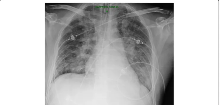
X-ray showing patchy bilateral infiltrates (Fig. 1). In order to improve oxygenation, the PEEP level was rise to 15 cmH2O and the shunt fraction decreased to 12 %.
According to our protocol, we decided to prone him; the oxygenation further ameliorate since the shunt fraction decreased to 7 %. On the opposite, the static compliance of the respiratory system (Crs,st), equal to 52 ml/ cmH2O, did not change by rising PEEP and by placing
the patient in prone position. Cst, rs was calculated as VT/(plateau pressure– PEEPtot), where Plateau pressure
and PEEPtot are the pressure registered after an end in-spiratory/end expiratory occlusion of at least 5 sec, respectively. After 24 h of prone position, without further benefit in terms of oxygenation, the patient returned in supine position and the curarization was stopped. The shunt fraction was about 14 % with a PEEP of 10 cmH2O.
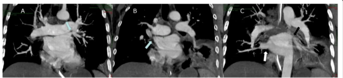
The evident relationship between bleeding events and hypoxia imposed the re-analysis of the medical history of the patient. This helped us to notice the temporal cor-relation with the cardiac ablation. Indeed the latter could be responsible for severe complications, such as the PVS. Hence, to clarify this aspect, another Chest CT angiography was performed, at the seventh day of hospitalization. This showed the concentric severe sten-osis of the right superior pulmonary vein (Fig. 2a) and the 50 % stenosis of both left pulmonary veins (Fig. 2b) while the right inferior vein appeared ectatic (Fig. 2c).
The patient still intubated was then transferred to the Specialist Cardiologic Centre in order to perform bal-loon angioplasty with stent placement of the stenotic veins since the medical therapy was not able to reverse the ARF. One week after the procedure, the patient was successfully extubated.
Discussion
PVS is a relatively rare condition, which can be both con-genital or acquired. The concon-genital form is linked almost completely to congenital heart diseases and inadequate embryological cardiovascular connections, and evidences in childhood [6]. In adults, the pathology is even rarer, but approximately 15 years ago it appears in literature as a consequence of catheter ablation strategies for arrhyth-mias [6, 7], arising a bimodal age distribution [6].
The most common causes of acquired PVS in adult patients are radiofrequency ablation procedures for AF treatment. However, the underlying molecular mecha-nisms remain poorly defined, probably involving scar-ring, contraction of the venous wall and peri-adventitial inflammation or collagen deposition as a result of ther-mal injury, which may compromise or even occlude the lumen of PV [3, 5].
PVS is asymptomatic in most cases, especially when only one vein is involved. When clinically evident, the presentation varies depending several factors including: 1) the number of PV involved; 2) the severity of the lesions; 3) the response of the entire pulmonary vascula-ture to the lesion; 4) the time course of stenosis; 5) the clinical setting; and 6) the presence and extent of collat-eral vessels [3].
Patients with more extensive and severe involve-ment may present respiratory symptoms like dyspnea, orthopnea, cough, chest pain, recurrent pulmonary in-fections. Rarely, does an X-ray show bronchitis or pneumonia [3, 5].
Haemoptysis, the main symptom described in our case report, is relatively infrequent [3, 7]. Its etiopathogenesis has not yet been clarified, but the increase in venous
Fig. 2 a Computed tomography angiography A-P projection showed the concentric severe stenosis of the left superior pulmonary vein (arrow). b CT angiography A-P projection showed 50 % stenosis of both right superior pulmonary (arrow). c CT angiography A-P projection showed ectatic right inferior vein (white arrow) and stenosis of left inferior pulmonary vein (black arrow)
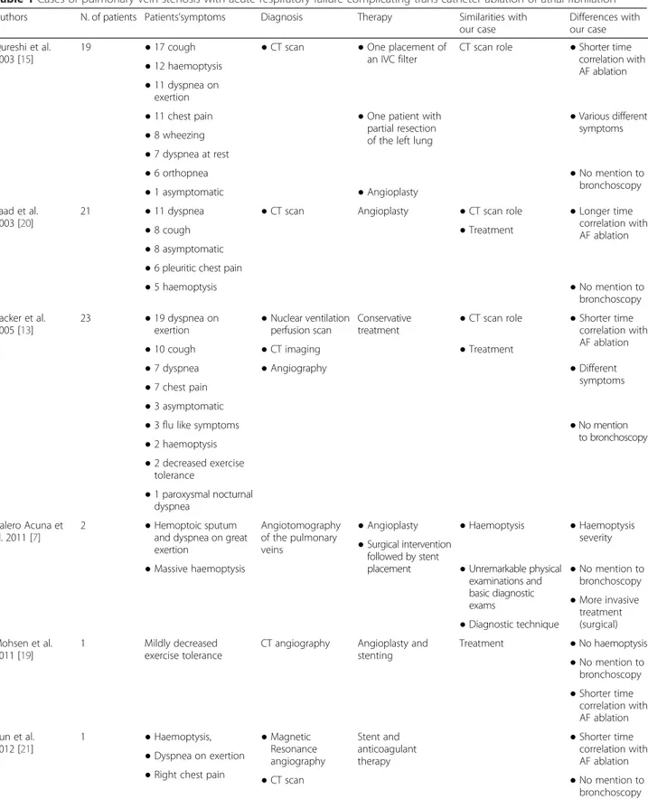
Table 1 Cases of pulmonary vein stenosis with acute respiratory failure complicating trans catheter ablation of atrial fibrillation
Authors N. of patients Patients’symptoms Diagnosis Therapy Similarities with our case
Differences with our case Qureshi et al.
2003 [15]
19 ● 17 cough ● CT scan ● One placement of an IVC filter
CT scan role ● Shorter time correlation with AF ablation ● 12 haemoptysis
● 11 dyspnea on exertion
● 11 chest pain ● One patient with partial resection of the left lung
● Various different symptoms ● 8 wheezing ● 7 dyspnea at rest ● 6 orthopnea ● No mention to bronchoscopy ● 1 asymptomatic ● Angioplasty Saad et al. 2003 [20]
21 ● 11 dyspnea ● CT scan Angioplasty ● CT scan role ● Longer time correlation with AF ablation ● 8 cough ● Treatment
● 8 asymptomatic ● 6 pleuritic chest pain
● 5 haemoptysis ● No mention to bronchoscopy Packer et al. 2005 [13] 23 ● 19 dyspnea on exertion ● Nuclear ventilation perfusion scan Conservative treatment
● CT scan role ● Shorter time correlation with AF ablation ● 10 cough ● CT imaging ● Treatment
● 7 dyspnea ● Angiography ● Different symptoms ● 7 chest pain
● 3 asymptomatic
● 3 flu like symptoms ● No mention to bronchoscopy ● 2 haemoptysis ● 2 decreased exercise tolerance ● 1 paroxysmal nocturnal dyspnea Calero Acuna et al. 2011 [7] 2 ● Hemoptoic sputum and dyspnea on great exertion
Angiotomography of the pulmonary veins
● Angioplasty ● Haemoptysis ● Haemoptysis severity ● Surgical intervention
followed by stent placement
● Massive haemoptysis ● Unremarkable physical examinations and basic diagnostic exams ● No mention to bronchoscopy ● More invasive treatment (surgical) ● Diagnostic technique Mohsen et al. 2011 [19] 1 Mildly decreased exercise tolerance
CT angiography Angioplasty and stenting Treatment ● No haemoptysis ● No mention to bronchoscopy ● Shorter time correlation with AF ablation Yun et al. 2012 [21] 1 ● Haemoptysis, ● Magnetic Resonance angiography Stent and anticoagulant therapy ● Shorter time correlation with AF ablation ● Dyspnea on exertion
● Right chest pain ● CT scan ● No mention to bronchoscopy
pressure in the pre-stenotic zone could explain the lung tissue congestion and the risk of bleeding in this area [8].
Interestingly, our patient presented a severe hypoxemia requiring prone positioning [9]. This acute hypoxemic respiratory failure was interpreted as a consequence of repeated bleeding at the mucosal level capable of flooding the alveoli. It is true that the presence of blood in the lungs can be impetus for respiratory infections able to severely decrease oxygenation, such as pneumonia. How-ever, we did not isolate bacteria in the bronchial aspirates or in the blood cultures; and the procalcitonin value was very low (0.04 ng/ml; n.v. <0.05). Further, although strictly speaking the acute respiratory failure of this patient could have been classifies as moderate ARDS, the lungs were not as stiff as in ARDS patients, since the values of static compliance were higher than those re-ported for patients with ARDS (33 ml/cmH2O) and
even for patients ventilated for other diseases (44 ml/ cmH2O) [10]. Hence, we thought that the principal
diagnosis should have been different from the first that was hypothesized. Hemorrhagic pulmonary vas-culitis was then considered in the differential diagno-sis and a second CT angiography became mandatory.
The diagnosis of PVS may not be simple to identify, given the variability of clinical presentation and the atyp-ical findings. Presence of dyspnea or cough after catheter ablation in the region of the PV should raise suspicions of PVS [3, 5]. Our case report suggests that even an ARF associated with nearly “normal” data of respiratory me-chanics should imply a CT angiography and, to a lesser extent, Magnetic Resonance (MR) [11, 12] in order to ex-clude PVS. Indeed common radiological imaging (Chest X-ray or CT) is often irrelevant [7, 13]. Consequently, echocardiography should be routinely performed after AF ablation, because of the good feasibility, and the overall benefits in a population with increased risk of PVS devel-opment [6, 14]. Instead, CT angiography is a valid diag-nostic tool, even if the resolution sometimes is not excellent, reducing the possibility to evaluate the stenosis degree [6, 15]. Furthermore, bronchoscopy has a major diagnostic role, especially when bleeding is the most rele-vant symptom, as underlined by international guidelines, which indicate the endoscopic evaluation as the most
important instrument to investigate haemoptysis, after CT scans [16]. Nevertheless, there are cases in which bronchial endoscopy is not even taken into account, probably because of different clinical presentation (Table 1). However, the bronchoscopy value is particu-larly significant in differential diagnosis, in case of a normal CT, if the patient is high risk for lung carcin-oma, or if symptoms continue [16].
PVS can be treated acutely with balloon dilatation of the PV, although the long-term outcome is uncertain [5]. Treatment options are currently limited, and re-stenosis after PV intervention has been described and considered relatively frequent [17]. Hence patients with PVS need careful follow-up because of the risk of recurrence, which occurs in 50 % of patients within 1 year [6].
Prevention of PVS is mainly related to placing the ab-lation site from inside to outside the orifice of the PV and reducing the ablation temperature and energy. This strategy should decrease the risk of PVS down to less than 1 % [18, 19].
Conclusions
Clinicians should be aware of the possibility of PVS in pa-tients who underwent ablation procedure for AF and those which present haemoptysis and acute hypoxemic re-spiratory failure. Hence pulmonary venous hypertension should be considered among the causes of ARF. Presence of dyspnea, cough, haemoptysis and acute respiratory fail-ure after catheter ablation in the region of the PV should raise a high suspicion of PVS and a CT angiography should be performed since it can provide accurate location and evaluation of the extent of stenosis.
Consent section
Written informed consent was obtained from the patient for publication of this case report and any accompanying images. A copy of the written consent is available for review by the Editor of this journal.
Abbreviations
ARDS:Acute respiratory distress syndrome; ARF: Acute respiratory failure; AF: Atrial fibrillation; CT: Computed tomography; PEEP: Positive end expiratory pressure; PV: Pulmonary veins; PVS: Pulmonary veins stenosis.
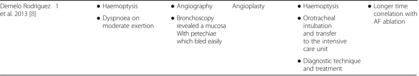
Table 1 Cases of pulmonary vein stenosis with acute respiratory failure complicating trans catheter ablation of atrial fibrillation (Continued)
Demelo Rodriguez et al. 2013 [8]
1 ● Haemoptysis ● Angiography Angioplasty ● Haemoptysis ● Longer time correlation with AF ablation ● Dyspnoea on
moderate exertion ● Bronchoscopyrevealed a mucosa With petechiae which bled easily
● Orotracheal intubation and transfer to the intensive care unit ● Diagnostic technique and treatment
Competing interests
None of the authors has any conflict of interest to declare.
Authors’ contributions
SS, MVC and CAV conceived of the idea for the report and wrote the draft. MB, SAS wrote the first draft. FR, RC and RG revised the manuscript. DC, SS helped to draft the manuscript. All authors read and approved the final manuscript.
Acknowledgements
We thank Dr Angela Ernan for the English editing revision. This work was supported by University of Ferrara
Author details
1
Department of Morphology, Surgery and Experimental Medicine, Section of Anesthesia and Intensive care, University of Ferrara, Via Aldo Moro, 8, Ferrara 44121, Italy.2Department of Morphology, Surgery and Experimental Medicine, Respiratory Medicine, S.Anna Hospital, Ferrara, Italy.3Department
of Cardiology, S. Anna Hospital, University of Ferrara, Ferrara, Italy.
4Department of Morphology, Surgery and Experimental Medicine, Vascular
and interventional radiology Unit. S. Anna Hospital, Ferrara, Italy.5Centro di Ricerca Aritmologia Clinica ed Elettrofisiologia, Milano, Rozzano, Italy.
Received: 18 December 2014 Accepted: 12 October 2015
References
1. Larici AR, Franchi P, Occhipinti M, Contegiacomo A, Del Ciello A, Calandriello L, et al. Diagnosis and management of hemoptysis. Diagn Interv Radiol. 2014;20(4):299–309.
2. Gupta A, Perera T, Ganesan A, Sullivan T, Lau DH, Roberts-Thomson KC, et al. Complications of catheter ablation of atrial fibrillation: a systematic review. Circ Arrhythm Electrophysiol. 2013;6(6):1082–8.
3. Holmes Jr DR, Monahan KH, Packer D. Pulmonary vein stenosis complicating ablation for atrial fibrillation: clinical spectrum and interventional considerations. JACC Cardiovasc Interv. 2009;2(4):267–76.
4. European Heart Rhythm Association, European Association for Cardio-Thoracic Surgery, Camm AJ, Kirchhof P, Lip GY, Schotten U, et al. Guidelines for the management of atrial fibrillation: the Task Force for the Management of Atrial Fibrillation of the European Society of Cardiology (ESC). Eur Heart J. 2010;31(19):2369–429.
5. Robbins IM, Colvin EV, Doyle TP, Kemp WE, Loyd JE, McMahon WS, et al. Pulmonary vein stenosis after catheter ablation of atrial fibrillation. Circulation. 1998;98(17):1769–75.
6. Latson LA, Prieto LR. Congenital and acquired pulmonary vein stenosis. Circulation. 2007;115:103–8.
7. Calero Acuña C, Elías HT. Haemotopsis as a first sign of pulmonary venous stenosis secondary to radiofrequency ablation of atrial fibrillation. Arch Bronconeumol. 2011;47:162–3.
8. Demelo-Rodríguez P, Del Toro-Cervera J, Andrés-Del OB. Haemoptysis and pulmonary vein stenosis after ablation for atrial fibrillation: pathophysiology and therapeutic options. Arch Bronconeumol. 2013;49(8):366–7.
9. Esan A, Hess DR, Raoof S, George L, Sessler CN. Severe hypoxemic respiratory failure: part 1–ventilatory strategies. Chest. 2010;137(5):1203–16.
10. Raoof S, Goulet K, Esan A, Hess DR, Sessler CN. Severe hypoxemic respiratory failure: part 2—non ventilatory strategies. Chest. 2010;137(6):1437–48. 11. Dong J, Vasamreddy CR, Jayam V, Dalal D, Dickfeld T, Eldadah Z, et al.
Incidence and predictors of pulmonary vein stenosis following catheter ablation of atrial fibrillation using the anatomic pulmonary vein ablation approach: results from paired magnetic resonance imaging. J Cardiovasc Electrophysiol. 2005;16(8):845–52.
12. Douglas Jr JS, Holmes Jr DR, Kereiakes DJ, Grines CL, Block E, Ghazzal ZM, et al. Cilostazol for Restenosis Trial (CREST) Investigators. Coronary stent restenosis in patients treated with cilostazol. Circulation. 2005;112(18):2826–32.
13. Packer DL, Keelan P, Munger TM, Breen JF, Asirvatham S, Peterson LA, et al. Clinical presentation, investigation, and management of pulmonary vein stenosis complicating ablation for atrial fibrillation. Circulation. 2005;111(5):546–54. 14. Minich LL, Tani LY, Breinholt JP, Tuohy AM, Shaddy RE. Complete follow-up
echocardiograms are needed to detect stenosis of normally connecting pulmonary veins. Echocardiography. 2001;18:589–92.
15. Qureshi AM, Prieto LR, Latson LA, Lane GK, Mesia CI, Radvansky P, et al. Transcatheter angioplasty for acquired pulmonary vein stenosis after radiofrequency ablation. Circulation. 2003;108:1336–42.
16. Du Rand IA, Blaikley J, Booton R, Chaudhuri N, Gupta V, Khalid S, et al. British Thoracic Society guideline for diagnostic flexible bronchoscopy in adults: accredited by NICE. Thorax. 2013;68(1):i1–i44.
17. Pürerfellner H, Aichinger J, Martinek M, Nesser HJ, Cihal R, Gschwendtner M, et al. Incidence, management, and outcome in significant pulmonary vein stenosis complicating ablation for atrial fibrillation. Am J Cardiol. 2004;93(11):1428–31. A10.
18. Bertaglia E, Zoppo F, Tondo C, Colella A, Mantovan R, Senatore G, et al. Early complications of pulmonary vein catheter ablation for atrial fibrillation: a multicenter prospective registry on procedural safety. Heart Rhythm. 2007;4:1265–71.
19. Takahashi A, Kuwahara T, Takahashi Y. Complications in the catheter ablation of atrial fibrillation: incidence and management. Circ J. 2009;73:221–6. 20. Mohsen A, Mazhari R, Reiner J. Multimodality imaging to diagnose pulmonary
vein stenosis following atrial fibrillation ablation. J Invasive Cardiol. 2011;23(9):E214–5.
21. Saad EB, Marrouche NF, Saad CP, Ha E, Bash D, White RD, et al. Pulmonary vein stenosis after catheter ablation of atrial fibrillation: emergence of a new clinical syndrome. Ann Intern Med. 2003;138(8):634–8.
Submit your next manuscript to BioMed Central and take full advantage of:
• Convenient online submission
• Thorough peer review
• No space constraints or color figure charges
• Immediate publication on acceptance
• Inclusion in PubMed, CAS, Scopus and Google Scholar
• Research which is freely available for redistribution
Submit your manuscript at www.biomedcentral.com/submit