Abstract. MicroRNA (miRNA or miR) therapeutics in cancer
are based on targeting or mimicking miRNAs involved in cancer
onset, progression, angiogenesis, epithelial-mesenchymal
transition and metastasis. Several studies conclusively have
demonstrated that miRNAs are deeply involved in tumor onset
and progression, either behaving as tumor-promoting miRNAs
(oncomiRNAs and metastamiRNAs) or as tumor suppressor
miRNAs. This review focuses on the most promising examples
potentially leading to the development of anticancer,
miRNA-based therapeutic protocols. The inhibition of miRNA activity
can be readily achieved by the use of miRNA inhibitors and
oligomers, including RNA, DNA and DNA analogues (miRNA
antisense therapy), small molecule inhibitors, miRNA sponges
or through miRNA masking. On the contrary, the
enhance-ment of miRNA function (miRNA replaceenhance-ment therapy) can
be achieved by the use of modified miRNA mimetics, such
as plasmid or lentiviral vectors carrying miRNA sequences.
Combination strategies have been recently developed based on
the observation that i) the combined administration of different
antagomiR molecules induces greater antitumor effects and
ii) some anti-miR molecules can sensitize drug-resistant tumor
cell lines to therapeutic drugs. In this review, we discuss two
additional issues: i) the combination of miRNA replacement
therapy with drug administration and ii) the combination of
antagomiR and miRNA replacement therapy. One of the solid
results emerging from different independent studies is that
miRNA replacement therapy can enhance the antitumor effects
of the antitumor drugs. The second important conclusion of the
reviewed studies is that the combination of anti-miRNA and
miRNA replacement strategies may lead to excellent results, in
terms of antitumor effects.
Contents
1. Introduction
2. Tumor suppressor miRNAs
3. OncomiRNAs and metastamiRNAs
4. Mimicking tumor suppressor miRNAs in miRNA
replacement therapy
5. Targeting oncomiRNAs
6. MicroRNAs and epithelial-mesenchymal transition
7. MicroRNAs and neoangiogenesis
8. Selected examples of miRNA therapeutics: mimicking
miR-124
9. Selected examples of miRNA therapeutics: mimicking
miR-93
10. Selected examples of anti-miRNA therapeutics: targeting
miR-221/222
11. Combined treatments: targeting multiple miRNAs
12. Combined treatments: co-administration of antitumor
drugs and miRNA therapeutic agents
13. Combining miRNA replacement strategies with
anti-miRNAs and siRNA molecules
14. Conclusion
Introduction
MicroRNAs (miRNAs or miRs) are a family of small
(19-25 nucleotides in length) non-coding RNAs that have a key
role in the regulation of gene expression through the inhibition
or the reduction of protein synthesis following mRNA
comple-mentary sequence base pairing (1-4). A single or multiple
mRNAs can be targeted at the 3' untranslated region (3'UTR),
Targeting oncomiRNAs and mimicking tumor suppressor
miRNAs: Νew trends in the development of miRNA
therapeutic strategies in oncology (Review)
ROBERTO GAMBARI
1, ELEONORA BROGNARA
1, DEMETRIOS A. SPANDIDOS
2and ENRICA FABBRI
1 1Department of Life Sciences and Biotechnology and
Biotechnology Center, Ferrara University, Ferrara, Italy;
2
Laboratory of Clinical Virology, University of Crete School of Medicine, Heraklion, Crete, Greece
Received March 9, 2016; Accepted April 29, 2016
DOI: 10.3892/ijo.2016.3503
Correspondence to: Professor Roberto Gambari, Department of
Life Sciences and Biotechnology and Biotechnology Center, Ferrara University, Via Fossato di Mortara 74, I-44121 Ferrara, ItalyE-mail: [email protected]
Abbreviations: miRNAs or miRs, microRNAs; PNA, peptide nucleic
acids; CTCs, circulating tumor cells; EMT, epithelial-mesenchymal transition; MET, mesenchymal-epithelial transition; CML, chronic myelogenous leukemia; CRC, colorectal carcinomaKey words: microRNAs, peptide nucleic acids, miRNA replacement
therapy, antagomiR, epithelial-mesenchymal transition, metastasis, miR-124coding sequence (CDS) or 5' untranslated region (5'UTR)
sequence, and it is calculated that >60% of human mRNAs
are recognized by miRNAs (1-4). The miRNA/mRNA
interaction occurs at the level of the RNA-induced silencing
complex (RISC) and causes translational repression or mRNA
degradation, depending on the degree of complementarity
with target mRNA sequences (5-8). Since their discovery and
first characterization, the number of human miRNAs
identi-fied and deposited in the miRBase databases (miRBase v.21,
www.mirbase.org) has increaed (it is >2,500) (9-16) and the
research studies on miRNAs have confirmed the very high
complexity of the networks constituted by miRNAs and RNA
targets (17-22).
Alterations in miRNA expression have been demonstrated
to be associated with different human pathologies, and guided
alterations of specific miRNAs have been suggested as novel
approaches for the development of innovative therapeutic
protocols (23,24). Studies have conclusively demonstrated that
miRNAs are deeply involved in tumor onset and progression,
either behaving as tumor-promoting miRNAs (oncomiRNAs
and metastamiRNAs) or as tumor suppressor miRNAs (25,26).
In general, miRNAs able to promote cancer target mRNAs
coding for tumor suppressor proteins, whereas miRNAs
exhibiting tumor suppressor properties usually target mRNAs
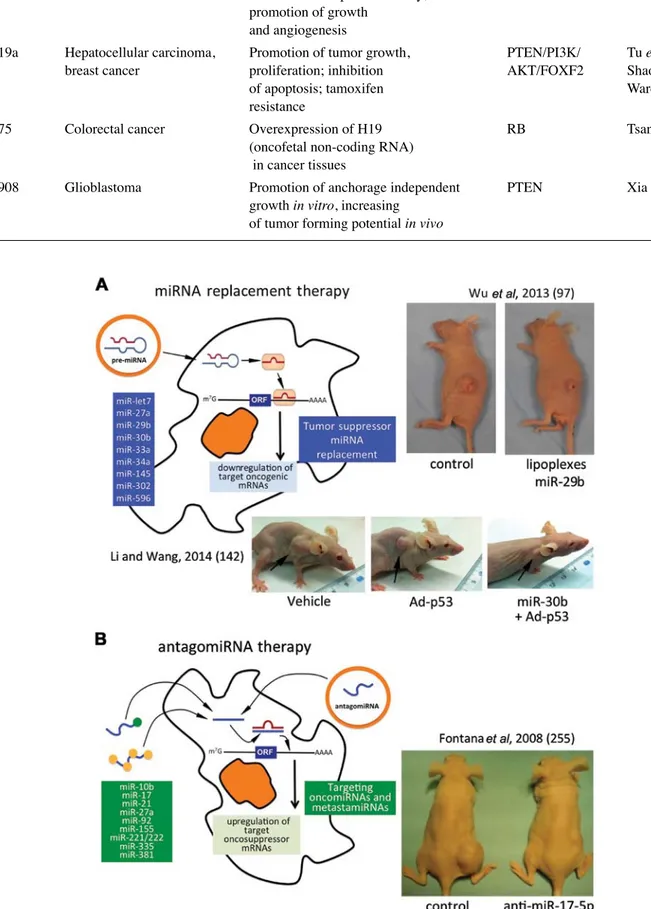
coding oncoproteins (see the scheme depicted in Fig. 1A).
This has a very important implication in diagnosis and/or
prognosis, including the recent discovery that the pattern of
circulating cell-free miRNAs in serum allows us to perform
molecular analyses on these non-invasive liquid biopsies with
deep diagnostic and prognostic implications. This research
field has confirmed that cancer-specific miRNAs are present
in extracellular body fluids, and may play a very important role
in the crosstalk between cancer cells and surrounding normal
cells (27-32).
Interestingly, the evidence of the presence of miRNAs
in serum, plasma and saliva supports their potential as an
additional set of biomarkers for cancer. The extracellular
miRNAs are protected by exosome-like structures, small
intraluminal vesicles shed from a variety of cells (including
cancer cells), with a biogenesis connected with endosomal
sorting complex required for transport machinery in
multive-sicular bodies (29). For instance, miR-141 and miR-221/222
are predicted biomarkers in liquid biopsies from patients with
colon cancer (33,34).
On the other hand, tumor-associated miRNAs are
suit-able targets for intervention therapeutics, as previously
reported (35-44) and summarized in Fig. 1B. The inhibition of
miRNA activity can be readily achieved by the use of miRNA
inhibitors and oligomers, including RNA, DNA and DNA
analogues (miRNA antisense therapy) (45-47), small molecule
inhibitors, locked nucleic acids (LNAs) (48-53), peptide
nucleic acids (PNAs) (54-57), morpholinos (58-60), miRNA
sponges (61-67), mowers (68) or through miRNA masking
that inhibits miRNA function by masking the miRNA binding
site of a target mRNA using a modified single-stranded RNA
complementary to the target sequence (69-75). On the contrary,
the enhancement of miRNA function (miRNA replacement
therapy) can be achieved by the use of modified miRNA
mimetics, either synthetic, or produced by plasmid or lentiviral
vectors carrying miRNA sequences (76-81).
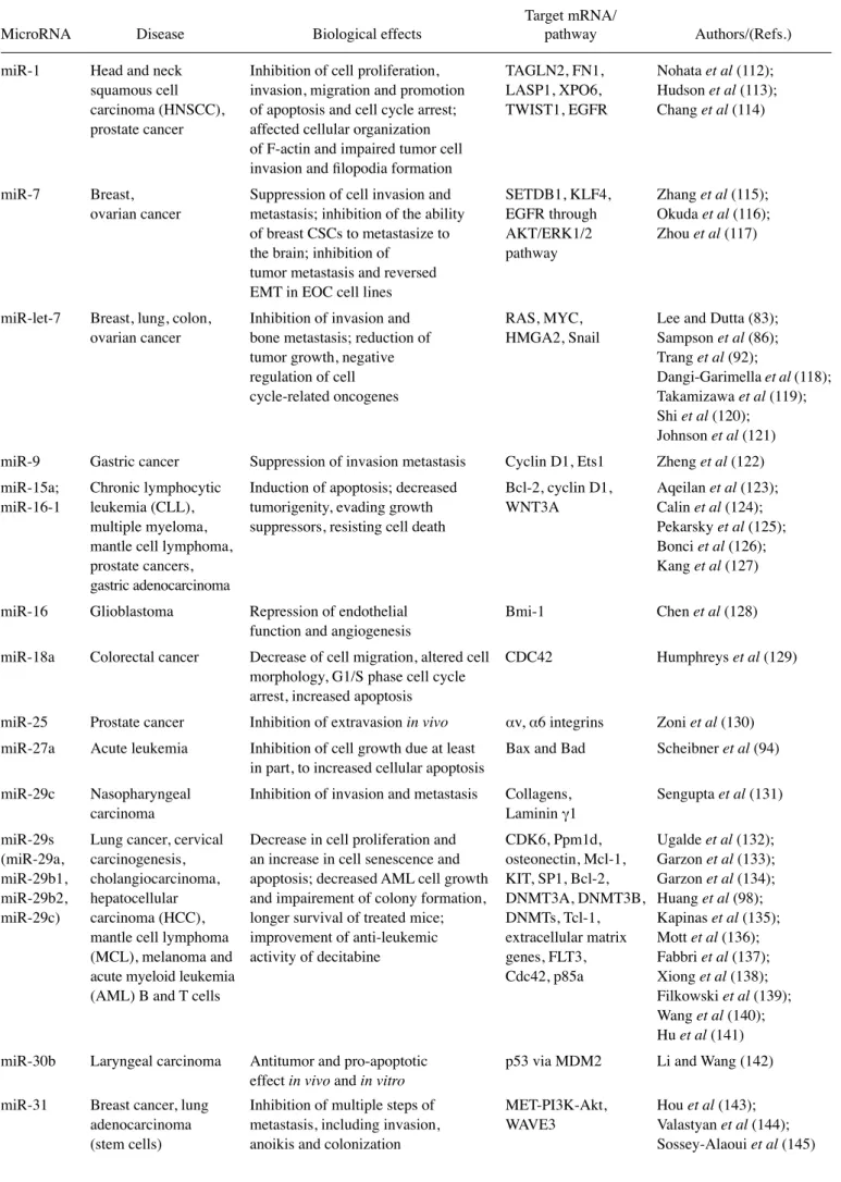
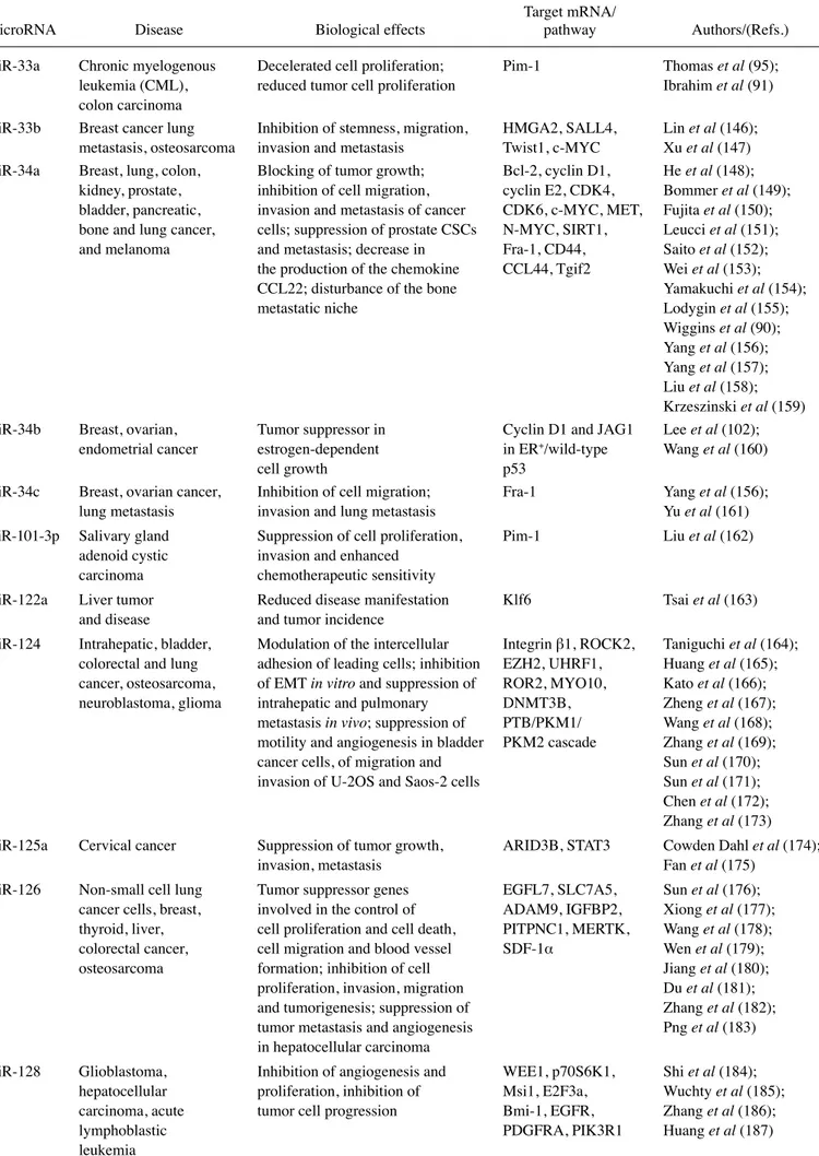
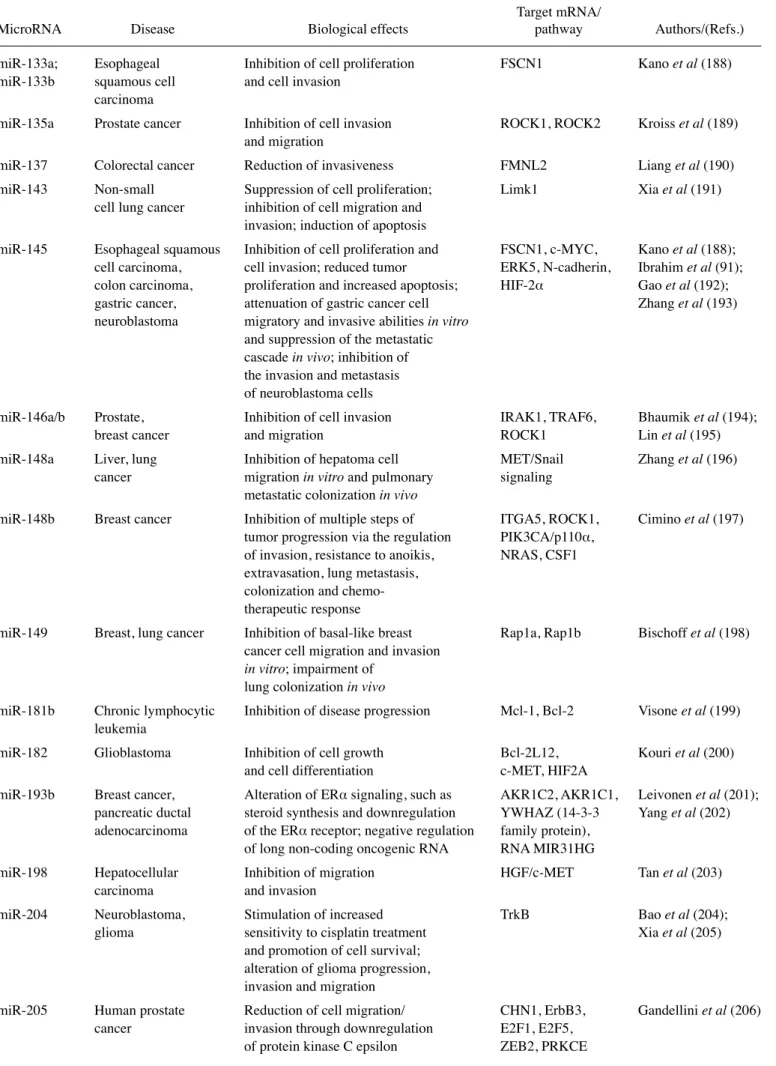
2. Tumor suppressor miRNAs
Several miRNAs exhibit onco-suppressor properties by
targeting mRNAs coding oncoproteins (82-105). Therefore,
these onco-suppressor miRNAs have been found to be often
downregulated in tumors. For instance, Fernandez et al (106)
recently described the intriguing tumor suppressor activity
of miR-340, showing the miR-340-mediated inhibition of
multiple negative regulators of p27, a protein involved in
apoptosis and cell cycle progression. These interactions with
oncoprotein-coding mRNA targets determine the inhibition of
cell cycle progression, the induction of apoptosis and growth
inhibition. The miR-340-mediated downregulation of three
post-transcriptional regulators [Pumilio RNA-binding family
member (PUM)1, PUM2 and S-phase kinase-associated
protein 2 (SkP2)] correlates with the upregulation of p27.
PUM1 and PUM2 inhibit p27 at the translational level, by
rendering the p27 transcript available to interact with two
oncomiRs (miR-221 and miR-222), while the oncoprotein
SkP2 inhibits the CDk inhibitor at the post-translational level
by triggering the proteasomal degradation of p27, showing that
miR-340 affected not only the synthesis but also the decay
Figure 1. (A) Scheme outlining the ability of miRNAs to promote cancer and metastasis (green arrowed line) or to suppress mRNAs coding oncop-roteins (red line). (B) Examples of proposed approaches for the development of therapeutic protocols to modulate the biological activity of miRNAs involved in cancer. The objectives of these molecular interventions are the downregulation of oncomiRNAs and metastamiRNAs (orange arrow) or the upregulation/mimicking of onco-suppressor miRNAs (green arrow). Modified from Ghelani et al (3).of p27. Moreover their data confirm the recent identification
of transcripts encoding several pro-invasive proteins such as
c-Met, implicated in breast cancer cell migration, RhoA and
Rock1, implicated in the control of the migration and invasion
of osteosarcoma cells, and E-cadherin mRNA, involved in the
miR-340-induced loss of intercellular adhesion (106 and refs
within).
Recently, miR-18a was demonstrated to play a protective
role in colorectal carcinoma (CRC) by inhibiting the
prolifera-tion, invasion and migration of CRC cells by directly targeting
the TBP-like 1 (TBPL1) gene. The onco-suppressor activity
of miR-18a in CRC tissues and cell lines was supported by
the finding that the content of this mRNA is markedly lower
in tumor cells with respect to normal control tissues and
cells (107). In addition Xishan et al (108) found that miR-320a
acts as a novel tumor suppressor gene in chronic myelogenous
leukemia (CML) and can decrease the migratory, invasive,
proliferative and apoptotic behavior of CML cells, as well
as epithelial-mesenchymal transition (EMT), by attenuating
the expression of the BCR/ABL oncogene. Furthermore
Zhao et al (109) demonstrated that miR-449a functions as a
tumor suppressor in neuroblastoma by inducing cell
differen-tiation and cell cycle arrest. Finally, kalinowski et al (110) and
Gu et al (111) demonstrated the significant role of miR-7 in
cancer which functions by directly targeting and inhibiting key
oncogenic signaling molecules involved in cell cycle
progres-sion, proliferation, invasion and metastasis. A partial list of
onco-suppressor miRNAs is presented in Table I.
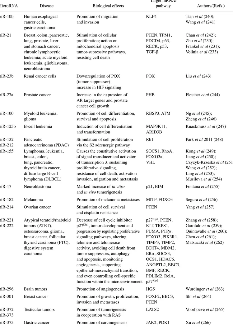
3. OncomiRNAs and metastamiRNAs
miRNAs can act as oncogenes and have been demonstrated to
play a causal role in the onset and progression of human cancer
(oncomiRNAs) (224-233). Recent findings have nevertheless
identified a subclass of miRNAs whose expression is highly
associated with the acquisition of metastatic phenotypes and are
referred to as miRs endowed with either metastasis-promoting
or tumor suppressor inhibitory activities (213,234,235).
Recent data have revealed that miR-25 may act as an
onco-miRNA in osteosarcoma, negatively regulating the
protein expression of the cell cycle inhibitor, p27. In agreement
with this hypothesis restoring the p27 level in
miR-25-over-expressing cells was shown to reverse the enhancing effect
of miR-25 on Saos-2 and U2OS cell proliferation (236). In
addition a recent study published by Siu et al (237), describes
miR-96 as a potential target of therapeutics for metastatic
prostate cancer, demonstrating the enhanced effects in cellular
growth and invasiveness of miR-96 in cell lines (AC1, AC3
and SC1) derived from prostate-specific, Pten/Tp53 double
knockout mice and confirmed in tissue samples from prostate
cancer patients. miR-96 acts as an oncomiR and metastamiR
through TGF-
β/mTOR signaling, promoting bone
metas-tasis and contributing to a reduced survival rate in prostate
cancer (237). Furthermore Xia et al (238) demonstrated that
the overexpression of miR-1908 significantly decreased the
expression of PTEN in glioblastoma cells, one of the most
frequently mutated tumor suppressors in human cancer,
resulting in an increase in proliferation, migration and
inva-sion. Finally Sachdeva et al (239), found that miR-182 targets
multiple genes in lung metastasis and regulates intravasation,
thus increasing the number of circulating tumor cells (CTCs).
Only the simultaneous restoration of miR-182 target genes
decreased the number of metastases in vivo, demonstrating that
a single miRNA can regulate the metastasis of primary tumors
in vivo by the coordinated regulation of multiple genes. Selected
examples of oncomiRNAs and metastamiRNAs are presented
in Tables II and III. All these miRNAs act by inhibiting tumor
suppressor pathways.
4. Mimicking tumor suppressor miRNAs in miRNA
replacement therapy
Using the development of anticancer therapies as a
represen-tative field of investigation, the therapeutic strategy based on
miRNA replacement is targeted to pathological cells which
downregulate onco-suppressor miRNAs playing a role in
controlling the expression of mRNAs encoding key
onco-proteins. The downregulation of these oncogene-targeting
miRNAs is clearly the key step for oncogene upregulation
leading to tumor onset and progression. Table IV presents
selected examples of miRNA replacement therapy in cancer
research and treatment (90-92,94-97,99).
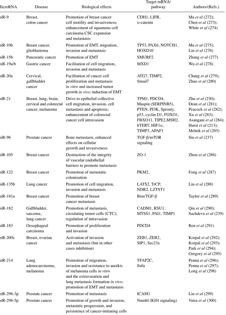
As a first representative example, Fig. 2A presents the
major results obtained by Wu et al (97), who reported that the
in vivo restoration of miR-29b may represent an option for lung
cancer treatment. To demonstrate the efficacy of this strategy,
they developed a cationic lipoplexes (LPs)-based carrier
that efficiently delivered miR-29b both in vitro and in vivo.
LPs containing miR-29b (LP-miR-29b) efficiently delivered
miR-29b to A549 cells and reduced the expression of the key
target, CDk6. In a xenograft murine model, in which LPs
efficiently accumulated at tumor sites, the systemic delivery of
LP-miR-29b increased miR-29b expression in tumors,
down-regulated CDk6 mRNA expression in tumors and, as shown
in the upper panels of Fig. 2A, significantly inhibited tumor
growth.
A second example of miRNA replacement therapy has
been published by Glover et al (304), who reported that
miR-7-5p (miR-7) reduces cell proliferation in vitro and induces
G1 cell cycle arrest. The systemic miR-7 administration with
delivery vesicles reduced adrenocortical carcinoma (ACC)
xenograft growth originating from both ACC cell lines and
primary ACC cells. As far as the potential mechanisms of
action, miR-7 was demonstrated to target Raf-1 proto-oncogene
serine/threonine kinase (RAF1). Additionally, miR-7 therapy
in vivo led to the inhibition of cyclin dependent kinase 1
(CDk1) (304). Two other methods have also been used to
successfully deliver miR-7 in vivo to treat cancer. In a study by
Babae et al (305), a miR-7 mimic was systemically delivered
using clinically viable, biodegradable, targeted polyamide
nanoparticles. This strategy led to the successful inhibition of
tumor growth and vascularisation in a glioblastoma xenograft
model system. In an earlier study, Wang et al (306) was able to
inhibit glioma xenograft growth and metastasis using a plasmid
based miR-7 vector systemically delivered by encapsulation in
a cationic liposome formulation.
Moreover, Cortez et al (307) revealed a novel function of
miR-200c, a member of the miR-200 family, in regulating
intracellular reactive oxygen species signaling. They used a
lung cancer xenograft model to demonstrate the therapeutic
Table I. miRNAs exhibiting tumor suppressor functions.
Target mRNA/
MicroRNA Disease Biological effects pathway Authors/(Refs.) miR-1 Head and neck Inhibition of cell proliferation, TAGLN2, FN1, Nohata et al (112);
squamous cell invasion, migration and promotion LASP1, XPO6, Hudson et al (113); carcinoma (HNSCC), of apoptosis and cell cycle arrest; TWIST1, EGFR Chang et al (114) prostate cancer affected cellular organization
of F-actin and impaired tumor cell invasion and filopodia formation
miR-7 Breast, Suppression of cell invasion and SETDB1, kLF4, Zhang et al (115); ovarian cancer metastasis; inhibition of the ability EGFR through Okuda et al (116); of breast CSCs to metastasize to AkT/ERk1/2 Zhou et al (117) the brain; inhibition of pathway
tumor metastasis and reversed EMT in EOC cell lines
miR-let-7 Breast, lung, colon, Inhibition of invasion and RAS, MYC, Lee and Dutta (83); ovarian cancer bone metastasis; reduction of HMGA2, Snail Sampson et al (86);
tumor growth, negative Trang et al (92);
regulation of cell Dangi-Garimella et al (118); cycle-related oncogenes Takamizawa et al (119);
Shi et al (120);
Johnson et al (121)
miR-9 Gastric cancer Suppression of invasion metastasis Cyclin D1, Ets1 Zheng et al (122) miR-15a; Chronic lymphocytic Induction of apoptosis; decreased Bcl-2, cyclin D1, Aqeilan et al (123); miR-16-1 leukemia (CLL), tumorigenity, evading growth WNT3A Calin et al (124);
multiple myeloma, suppressors, resisting cell death Pekarsky et al (125); mantle cell lymphoma, Bonci et al (126); prostate cancers, kang et al (127) gastric adenocarcinoma
miR-16 Glioblastoma Repression of endothelial Bmi-1 Chen et al (128) function and angiogenesis
miR-18a Colorectal cancer Decrease of cell migration, altered cell CDC42 Humphreys et al (129) morphology, G1/S phase cell cycle
arrest, increased apoptosis
miR-25 Prostate cancer Inhibition of extravasion in vivo αv, α6 integrins Zoni et al (130) miR-27a Acute leukemia Inhibition of cell growth due at least Bax and Bad Scheibner et al (94)
in part, to increased cellular apoptosis
miR-29c Nasopharyngeal Inhibition of invasion and metastasis Collagens, Sengupta et al (131)
carcinoma Laminin γ1
miR-29s Lung cancer, cervical Decrease in cell proliferation and CDk6, Ppm1d, Ugalde et al (132); (miR-29a, carcinogenesis, an increase in cell senescence and osteonectin, Mcl-1, Garzon et al (133); miR-29b1, cholangiocarcinoma, apoptosis; decreased AML cell growth kIT, SP1, Bcl-2, Garzon et al (134); miR-29b2, hepatocellular and impairement of colony formation, DNMT3A, DNMT3B, Huang et al (98); miR-29c) carcinoma (HCC), longer survival of treated mice; DNMTs, Tcl-1, kapinas et al (135);
mantle cell lymphoma improvement of anti-leukemic extracellular matrix Mott et al (136); (MCL), melanoma and activity of decitabine genes, FLT3, Fabbri et al (137); acute myeloid leukemia Cdc42, p85a Xiong et al (138); (AML) B and T cells Filkowski et al (139);
Wang et al (140);
Hu et al (141)
miR-30b Laryngeal carcinoma Antitumor and pro-apoptotic p53 via MDM2 Li and Wang (142) effect in vivo and in vitro
miR-31 Breast cancer, lung Inhibition of multiple steps of MET-PI3k-Akt, Hou et al (143); adenocarcinoma metastasis, including invasion, WAVE3 Valastyan et al (144); (stem cells) anoikis and colonization Sossey-Alaoui et al (145)
Table I. Continued.
Target mRNA/
MicroRNA Disease Biological effects pathway Authors/(Refs.) miR-33a Chronic myelogenous Decelerated cell proliferation; Pim-1 Thomas et al (95);
leukemia (CML), reduced tumor cell proliferation Ibrahim et al (91) colon carcinoma
miR-33b Breast cancer lung Inhibition of stemness, migration, HMGA2, SALL4, Lin et al (146); metastasis, osteosarcoma invasion and metastasis Twist1, c-MYC Xu et al (147) miR-34a Breast, lung, colon, Blocking of tumor growth; Bcl-2, cyclin D1, He et al (148);
kidney, prostate, inhibition of cell migration, cyclin E2, CDk4, Bommer et al (149); bladder, pancreatic, invasion and metastasis of cancer CDk6, c-MYC, MET, Fujita et al (150); bone and lung cancer, cells; suppression of prostate CSCs N-MYC, SIRT1, Leucci et al (151); and melanoma and metastasis; decrease in Fra-1, CD44, Saito et al (152);
the production of the chemokine CCL44, Tgif2 Wei et al (153); CCL22; disturbance of the bone Yamakuchi et al (154); metastatic niche Lodygin et al (155);
Wiggins et al (90);
Yang et al (156);
Yang et al (157);
Liu et al (158);
krzeszinski et al (159)
miR-34b Breast, ovarian, Tumor suppressor in Cyclin D1 and JAG1 Lee et al (102);
endometrial cancer estrogen-dependent in ER+/wild-type Wang et al (160)
cell growth p53
miR-34c Breast, ovarian cancer, Inhibition of cell migration; Fra-1 Yang et al (156); lung metastasis invasion and lung metastasis Yu et al (161) miR-101-3p Salivary gland Suppression of cell proliferation, Pim-1 Liu et al (162)
adenoid cystic invasion and enhanced carcinoma chemotherapeutic sensitivity
miR-122a Liver tumor Reduced disease manifestation klf6 Tsai et al (163) and disease and tumor incidence
miR-124 Intrahepatic, bladder, Modulation of the intercellular Integrin β1, ROCk2, Taniguchi et al (164); colorectal and lung adhesion of leading cells; inhibition EZH2, UHRF1, Huang et al (165); cancer, osteosarcoma, of EMT in vitro and suppression of ROR2, MYO10, kato et al (166); neuroblastoma, glioma intrahepatic and pulmonary DNMT3B, Zheng et al (167); metastasis in vivo; suppression of PTB/PkM1/ Wang et al (168);
motility and angiogenesis in bladder PkM2 cascade Zhang et al (169); cancer cells, of migration and Sun et al (170); invasion of U-2OS and Saos-2 cells Sun et al (171);
Chen et al (172);
Zhang et al (173)
miR-125a Cervical cancer Suppression of tumor growth, ARID3B, STAT3 Cowden Dahl et al (174); invasion, metastasis Fan et al (175)
miR-126 Non-small cell lung Tumor suppressor genes EGFL7, SLC7A5, Sun et al (176); cancer cells, breast, involved in the control of ADAM9, IGFBP2, Xiong et al (177); thyroid, liver, cell proliferation and cell death, PITPNC1, MERTk, Wang et al (178); colorectal cancer, cell migration and blood vessel SDF-1α Wen et al (179);
osteosarcoma formation; inhibition of cell Jiang et al (180); proliferation, invasion, migration Du et al (181); and tumorigenesis; suppression of Zhang et al (182); tumor metastasis and angiogenesis Png et al (183) in hepatocellular carcinoma
miR-128 Glioblastoma, Inhibition of angiogenesis and WEE1, p70S6k1, Shi et al (184); hepatocellular proliferation, inhibition of Msi1, E2F3a, Wuchty et al (185); carcinoma, acute tumor cell progression Bmi-1, EGFR, Zhang et al (186); lymphoblastic PDGFRA, PIk3R1 Huang et al (187) leukemia
Table I. Continued.
Target mRNA/
MicroRNA Disease Biological effects pathway Authors/(Refs.) miR-133a; Esophageal Inhibition of cell proliferation FSCN1 kano et al (188) miR-133b squamous cell and cell invasion
carcinoma
miR-135a Prostate cancer Inhibition of cell invasion ROCk1, ROCk2 kroiss et al (189) and migration
miR-137 Colorectal cancer Reduction of invasiveness FMNL2 Liang et al (190) miR-143 Non-small Suppression of cell proliferation; Limk1 Xia et al (191)
cell lung cancer inhibition of cell migration and invasion; induction of apoptosis
miR-145 Esophageal squamous Inhibition of cell proliferation and FSCN1, c-MYC, kano et al (188); cell carcinoma, cell invasion; reduced tumor ERk5, N-cadherin, Ibrahim et al (91); colon carcinoma, proliferation and increased apoptosis; HIF-2α Gao et al (192);
gastric cancer, attenuation of gastric cancer cell Zhang et al (193) neuroblastoma migratory and invasive abilities in vitro
and suppression of the metastatic cascade in vivo; inhibition of
the invasion and metastasis of neuroblastoma cells
miR-146a/b Prostate, Inhibition of cell invasion IRAk1, TRAF6, Bhaumik et al (194); breast cancer and migration ROCk1 Lin et al (195) miR-148a Liver, lung Inhibition of hepatoma cell MET/Snail Zhang et al (196) cancer migration in vitro and pulmonary signaling
metastatic colonization in vivo
miR-148b Breast cancer Inhibition of multiple steps of ITGA5, ROCk1, Cimino et al (197) tumor progression via the regulation PIk3CA/p110α,
of invasion, resistance to anoikis, NRAS, CSF1 extravasation, lung metastasis,
colonization and chemo-therapeutic response
miR-149 Breast, lung cancer Inhibition of basal-like breast Rap1a, Rap1b Bischoff et al (198) cancer cell migration and invasion
in vitro; impairment of
lung colonization in vivo
miR-181b Chronic lymphocytic Inhibition of disease progression Mcl-1, Bcl-2 Visone et al (199) leukemia
miR-182 Glioblastoma Inhibition of cell growth Bcl-2L12, kouri et al (200) and cell differentiation c-MET, HIF2A
miR-193b Breast cancer, Alteration of ERα signaling, such as AkR1C2, AkR1C1, Leivonen et al (201); pancreatic ductal steroid synthesis and downregulation YWHAZ (14-3-3 Yang et al (202) adenocarcinoma of the ERα receptor; negative regulation family protein),
of long non-coding oncogenic RNA RNA MIR31HG
miR-198 Hepatocellular Inhibition of migration HGF/c-MET Tan et al (203) carcinoma and invasion
miR-204 Neuroblastoma, Stimulation of increased TrkB Bao et al (204); glioma sensitivity to cisplatin treatment Xia et al (205)
and promotion of cell survival; alteration of glioma progression, invasion and migration
miR-205 Human prostate Reduction of cell migration/ CHN1, ErbB3, Gandellini et al (206) cancer invasion through downregulation E2F1, E2F5,
potential of the systemic delivery of miR-200c to enhance
radiosensitivity in lung cancer. The results obtained suggest
that the antitumor effects of miR-200c result partially from
its regulation of the oxidative stress response; they further
suggested that miR-200c, in combination with radiation, may
represent an effective therapeutic strategy in the future.
Recently, Wu et al (308) reported that the expression of
miR-708-5p suppressed lung cancer invasion and metastasis
in vitro and in vivo. In particular, it induces apoptosis and
suppresses cell migration by inhibiting the cytoplasmic
local-ization of p21, and also weakens the stem cell-like properties
of lung cancer cells. In their study, they present the systemic
delivery of the PEI/miR-708-5p complexes for miRNA
replace-ment therapy in a mouse model of lung cancer, demonstrating
an efficient antitumor activity with no side-effects.
5. Targeting oncomiRNAs
The effects of therapeutic molecules against miRNAs have
been the object of very recent studies, in part summarized in
Table V (309-316). Of course, the endpoint of the treatment
of target cells with molecules against selected miRNAs is
the alteration of miRNA-regulated genes. As a first example,
Wagenaar et al (317) developed potent and specific
single-stranded oligonucleotide inhibitors of miR-21 and used them
to verify dependency on miR-21 in a panel of liver cancer cell
lines. Treatment with anti-miR-21, but not with a mismatch
control anti-miRNA, resulted in the significant derepression
of direct targets of miR-21 and led to the loss of viability in
the majority of HCC cell lines tested. The robust induction
of caspase activity, apoptosis and necrosis was noted in the
Table I. Continued.
Target mRNA/
MicroRNA Disease Biological effects pathway Authors/(Refs.) miR-206 Breast cancer Inhibition of cell invasion MET Chen et al (207)
and migration
miR-214 Colorectal cancer, Suppression of cell migration FGFR1 Chen et al (208) liver metastasis and invasion in vitro; inhibition
of liver metastasis of colorectal cancer cells in vivo
miR-218 Gastric cancer Suppression of tumor metastases ROBO1 Tie et al (209) miR-296-5p Prostate cancer Reduction of growth invasion HMGA1 Wei et al (210)
and progression
miR-302 Breast cancer Sensitization of radioresistant breast AkT1, RAD52 Liang et al (99) cancer cells to ionizing radiation
miR-302b Hepatocellular Suppression of cell proliferation EGFR Wang et al (211) carcinoma
miR-335 Breast cancer Inhibition of cell invasion, SOX4, PTPRN2, Tavazoie et al (212); migration and metastasis MERTk, TNC Hurst et al (213) miR-383 Medulloblastoma Control of cell growth PRDX3 Li et al (214) miR-449 Gastric cancer, non- Inhibition of cell proliferation, GMNN, MET, Bou kheir et al (215)
small cell lung cancer inhibition of migration and invasion CCNE2, SIRT1 Luo et al (216) miR-493 Colon, lung cancer Inhibition of the settlement of IGFR, E2F1, Okamoto et al (217);
metastasized colon cancer cells Mkk7 Gu et al (218); in the liver; promotion of the death Sakai et al (219) of colon cancer cells; suppression
of tumor growth, invasion and metastasis in lungs
miR-504 Hypopharyngeal Inhibition of cancer cells CDk6 kikkawa et al (220) squamous cell proliferation
carcinoma
miR- Breast cancer Inhibition of cell invasion in vitro RELA, keklikoglou et al (221) 520c/373 and the cell intravasation in vivo TGFBR2
miR-545 Pancreatic ductal Inhibition of cell growth RIG-1, CDk4 Song et al (222); adenocarcinoma, and proliferation Bowen et al (223) lung cancer cells
miR-596 Oral squamous cell Growth inhibition LGALS3BP Endo et al (96) carcinoma (OSCC)
Table II. miRNAs exhibiting oncogenic functions.
Target mRNA/
MicroRNA Disease Biological effects pathway Authors/(Refs.) miR-10b Human esophageal Promotion of migration kLF4 Tian et al (240);
cancer cells, and invasion Wang et al (241) gastric carcinoma
miR-21 Breast, colon, pancreatic, Stimulation of cellular PTEN, TPM1, Chan et al (242); lung, prostate, liver proliferation; action on PDCD4, p63, Zhu et al (230); and stomach cancer, mitochondrial apoptosis RECk, p53, Frankel et al (231); chronic lymphocytic tumor-supressive pathways, TGF-β Volinia et al (233)
leukemia; acute myeloid resisting cell death leukaemia, glioblastoma,
neuroblastoma
miR-23b Renal cancer cells Downregulation of POX POX Liu et al (243) (tumor suppressor),
increase in HIF signaling
miR-27a Prostate cancer Increase in the expression of PHB Fletcher et al (244) AR target genes and prostate
cancer cell growth
miR-100 Myeloid leukemia, Promotion of cell differentiation, RBSP3, ATM Ng et al (245); glioma survival and apoptosis Zheng et al (246) miR-125b B-cell leukemia Induction of cell differentiation MAP3k11, knackmuss et al (247)
and transformation ARID3B
miR-132 Pancreatic Stimulation of cell proliferation Rb1 Park et al 2011 (248) miR-212 adenocarcinoma (PDAC) via the β2 adrenergic pathway
miR-155 Lymphoma, leukemia, Causes the constitutive activation SOCS1, RhoA, kong et al (249); breast, colon, of signal transducer and activator FOXO3a, Jiang et al (250);
lung, pancreatic, of transcription 3, sustaining VHL Czyzyk-krzeska et al (251); thyroid brain cancer, proliferative signaling, Wang et al (252);
diffuse large B-cell resistance of cell death, activation Ling et al (253); lymphoma (DLBCL) invasion, migration and metastasis Musilova et al (254) miR-17 Neuroblastoma Marked increase of in vitro p21, BIM Fontana et al (255)
and in vivo tumorigenesis
miR-182 Melanoma Promotion of melanoma metastases MITF, FOXO3 Segura et al (256) miR-214 Ovarian cancer Stimulation of cell survival PTEN Yang et al (257)
and cisplatin resistance
miR-221 Atypical teratoid/rhabdoid Decrease of cell cycle inhibitor p27kip1, PTEN, Zhang et al (258);
miR-222 tumors (ATRT), p27kip1, tumor development and kIT, TRPS1, Garofalo et al (259);
osteosarcoma, glioma, progression by regulating proliferative PUMA, PTPµ, Quintavalle et al (260); breast cancer, follicular signaling pathways, altering FOXO3, PIk3R1, Chen et al (261); thyroid carcinoma (FTC), telomere and telomerase TIMP3, TIMP2, Matsuzaki et al (262) digestive system activity, avoiding cell death from DDIT4, MDM2,
carcinoma tumor suppressors, autophagy ERα, SOCS3, and apoptosis, monitoring OCS1, HDAC6, angiogenesis, supporting ANGPTL2, BBC3, epithelial-mesenchymal transition, BMF, RECk, and even controlling cell-specific PDLIM2, RelA, function within the microenvironment p57kip2
miR-296 Brain tumors Promotion of angiogenesis HGS Wurdinger et al (263) miR-301 Breast cancer Promotion of growth, proliferation, FOXF2, BBC3, Shi et al (264)
invasion and metastases PTEN
miR-372 Testicular tumors Promotion of tumorigenesis LATS2 Voorhoeve et al (265) miR-373 in cooperation with RAS
Table II. Continued.
Target mRNA/
MicroRNA Disease Biological effects pathway Authors/(Refs.) miR-378 Breast carcinoma Ehnancement of cell survival; Sufu, Fus-1 Lee et al (267)
reduction of caspase-3 activity; promotion of growth
and angiogenesis
miR-519a Hepatocellular carcinoma, Promotion of tumor growth, PTEN/PI3k/ Tu et al (268); breast cancer proliferation; inhibition AkT/FOXF2 Shao et al (269);
of apoptosis; tamoxifen Ward et al (270)
resistance
miR-675 Colorectal cancer Overexpression of H19 RB Tsang et al (271) (oncofetal non-coding RNA)
in cancer tissues
miR-1908 Glioblastoma Promotion of anchorage independent PTEN Xia et al (238) growth in vitro, increasing
of tumor forming potential in vivo
Figure 2. (A) miRNA replacement therapy: partial list of tumor suppressor miRNAs (in the blue box) and selected examples of the in vivo restoration of miR-29b (97) and of miR-30b (142), leading to the inhibition of tumor cell growth. (B) Targeting oncomiRNAs and metastamiRNAs with antagomiRNAs: partial list of onco/ metastamiRNAs and a selected example of the antitumor effects of antagomiR-17-5p (255).
Table III. miRNAs promoting metastasis.
Target mRNA/
MicroRNA Disease Biological effects pathway Authors/(Refs.) miR-9 Breast, Promotion of breast cancer CDH1, LIFR, Ma et al (272);
colon cancer cell motility and invasiveness; α-catenin Chen et al (273);
enhancement of squamous cell White et al (274) carcinoma CSC expansion
and metastasis
miR-10b Breast cancer, Promotion of EMT, migration, TP53, PAX6, NOTCH1, Ma et al (275);
glioblastoma invasion and metastasis HOXD10 Lin et al (276) miR-15b Pancreatic cancer Promotion of EMT SMURF2 Zhang et al (277) miR-19a/b Gastric cancer Facilitation of cell migration, MXD1 Wu et al (278)
invasion and metastasis
miR-20a Cervical, Facilitation of cancer cell ATG7, TIMP2, Chang et al (279); gallbladder proliferation and metastasis Smad7 Zhao et al (280) cancer in vitro and increased tumor
growth in vivo; induction of EMT
miR-21 Breast, lung, brain, Drive to epithelial collective TPM1, PDCD4, Zhu et al (230); cervical and colorectal cell migration, invasion, cell Maspin (SERPINB5), Dean et al (281); cancer, melanoma metastasis and apoptosis; PTEN, PI3k, Sprouty, Peacock et al (282);
enhancement of colorectal p53, cyclin D1, FOXO1, Xu et al (283); cancer cell intravasion FBXO11, TIPE2,MSH2, Asangani et al (284);
hTERT, HIF1α, Hurst et al (213);
TIMP3, APAF1 Melnik et al (285) miR-96 Prostate cancer Bone metastasis, enhanced TGF-β/mTOR Siu et al (237)
effects on cellular signaling growth and invasiveness
miR-105 Breast cancer Destruction of the integrity ZO-1 Zhou et al (286) of vascular endothelial
barriers to promote metastasis
miR-122 Breast cancer Promotion of metastatic PkM2, Fong et al (287)
colonization
miR-135b Lung cancer Promotion of cell migration, LATS2, TrCP, Lin et al (288) invasion and metastasis NDR2, LZTST1
miR-181a Breast cancer Promotion of breast Bim/TGF-β Taylor et al (289)
cancer metastasis
miR-182 Gallbladder, Promotion of metastasis, CADM1, RSU1, Qiu et al (290); sarcoma, circulating tumor cells (CTC); MTSS1, PAI1, TIMP1 Sachdeva et al (239) lung cancer regulation of intravasion
miR-183 Oesophageal Promotion of proliferation PDCD4 Ren et al (291) carcinoma and invasion
miR-200s Breast, ovarian Activation of invasion ZEB1, ZEB2, korpal et al (292); cancer and metastasis (but in other SIP1, Sec23a korpal et al (293); cases inhibition) Park et al (294);
Gregory et al (295)
miR-214 Lung Promotion of migration, TFAP2C, Penna et al (296); adenocarcinoma, invasion and resistance to anoikis Sufu Penna et al (297); melanoma of melanoma cells in vitro Long et al (298)
and the extravasation and lung metastasis formation in vivo; promotion of EMT and metastasis
miR-296-3p Prostate cancer Promotion of metastasis ICAM1 Liu et al (299) miR-296-5p Prostate cancer Promotion of growth and invasion, Numbl (klf4 signaling) Vaira et al (300)
metastatic progression, and persistence of cancer-initiating cells
anti-miR-21-treated HCC cells. Furthermore, the ablation of
miR-21 activity resulted in the inhibition of HCC cell
migra-tion and in the suppression of clonogenic growth (317).
In another study, using PNAs as anti-miRNA molecules,
Fabani et al (318) targeted miR-155, demonstrating the
deregu-lation of mRNA Bat5, Sfp1 and Jarid2. In our laboratory,
Brognara et al analyzed the effects of PNAs targeting miR-221
on breast cancer cells (319). In order to maximize uptake in
target cells, a polyarginine-peptide (R8) was conjugated,
gener-ating an anti-miR-221 PNA displaying very high affinity for
RNA and efficient uptake within target cells without the need
for transfection reagents. Targeting miR-221 with this PNA
molecule resulted in i) a specific decrease in the hybridization
levels of miR-221 measured by RT-qPCR, ii) the upregulation of
Table III. Continued.
Target mRNA/
MicroRNA Disease Biological effects pathway Authors/(Refs.) miR-362-5p Hepatocellular Promotion of cell proliferation, CYLD Ni et al (301)
carcinoma migration, invasion in vitro; and tumor growth and metastasis in vivo
miR-373 Breast cancer Drives EMT and metastasis TXNIP Chen et al (302) miR-520c Fibrosarcoma, Promotion of migration MT1-MMP Lu et al (303)
benign prostatic and metastasis hyperplasia,
glioblastoma
Table IV. miRNA replacement therapy of cancer: selected examples.
Tumor type miRNA target Modulated mRNA Effects following miR treatement Authors/(Refs.) Lung cancer miR-34a Repression of c-Met, Bcl-2; Block of tumor growth Wiggins et al (90)
partial repression of CDk4
Colon carcinoma miR-33a Pim-1 Reduced tumor proliferation Ibrahim et al (91) Colon carcinoma miR-145 c-Myc and ERk5 Reduced tumor proliferation Ibrahim et al (91)
and increased apoptosis
Lung cancer miR-let7 Negative regulation of Reduction of tumor growth Trang et al (92) the cell cycle oncogenes
RAS, MYC and HMGA2
Acute leukemia miR-27a Bax and Bad Inhibition of cell growth due, Scheibner et al (94) at least in part, to increased
cellular apoptosis
CML cells miR-33a Pim-1 Decelerated cell proliferation Thomas et al (95) Oral squamous cell miR-596 LGALS3BP Growth inhibition Endo et al (96) carcinoma (OSCC)
Non-small cell lung miR-29b CDk6, DNMT3B, Inhibition of Wu et al (97) adenocarcinomas, MCL-1 tumorigenicity in vivo
A549 cells
Acute myeloid miR-29b Downregulation of Decreased AML cell growth and Huang et al (98) leukemia DNMTs, CDk6, impairement of colony formation;
SP1, kIT and FLT3 longer survival of treated mice; improvement of antileukemic activity of decitabine
Laryngeal miR-30b p53 via MDM2 Antitumor and pro-apoptotic Li and Wang (142) carcinoma effect in vivo and in vitro
Breast cancer miR-302 AkT1 and RAD52 Sensitized radioresistant breast Liang et al (99) cancer cells to ionizing radiation
p27
kip1mRNA and protein expression, measured by RT-qPCR
and western blot analysis, respectively. As regards the in vivo
effects of anti-miRNA therapy, Yan et al (320) addressed the
potential effects of PNA-anti-miR-21 in vivo on the growth of
breast cancer cells. In their experiments, MCF-7 cells treated
with PNA-anti-miR-21 or PNA-control were subcutaneously
injected into female nude mice and detectable tumor masses
were observed in few mice in the MCF/PNA-anti-miR-21
group, while much larger tumors were detected in all mice in
the MCF/PNA-control group. Both tumor weight and number
showed that MCF/PNA-control cells formed larger tumors
more rapidly than MCF/PNA-anti-miR-21 cells in nude mice.
As a final example, Cheng et al (57) demonstrated that the PNA
anti-miRs with a peptide with a low pH-induced
transmem-brane structure (pHLIP) target the tumor microenvironment,
transport anti-miRs across plasma membranes under acidic
conditions, such as those found in solid tumors and effectively
inhibit the miR-155 oncomiR in a mouse model of lymphoma.
6. MicroRNAs and epithelial-mesenchymal transition
EMT is a powerful process in tumor invasion, metastasis
and tumorigenesis, and describes the molecular
reprogram-ming and phenotypic changes that are characterized by a
transition from polarized immotile epithelial cells to motile
mesenchymal cells (Fig. 3). This process is characterized by
the loss of polarity and cell-cell contacts by the differentiated
epithelial cells, with deep alterations occurring at the level of
tight junctions and desmosomes. The breach of the basement
membrane is a following step, leading to the invasion of blood
and/or lymphatic vessels by these mesenchymal differentiated
cancer cells, which at the end of the process, causes
migra-tion, often accompanied by drug resistance (Fig. 3). It is now
well-known that several miRNAs are important regulators of
EMT. Some of these are miR-7, miR-17/20, miR-22, miR-30,
miR-200 and its family members. Most of these miRNAs
potentiate EMT, while some well-characterized miRNAs
play a suppressive role in EMT. For instance, the metastasis
suppressor role of the miR-200 members is strongly
associ-ated with the inhibition of EMT. This is well described in the
published review by Zhang and Ma (321), and in the studies
by Zaravinos et al (322) and kiesslich et al (323), showing the
most recent advances regarding the influence of miRNAs in
EMT and the regulatory effects they exert on major signaling
pathways in various types of cancer (Fig. 3). In Caski cervical
cancer cells, the oncomiR-155 acts as a tumor suppressor and
suppresses EGF-induced EMT, decreasing
migration/inva-sion capacities, inhibiting cell proliferation and enhancing the
chemosensitivity to DDP in humans (324). Chang et al (279)
demonstrated that the overexpression of miR-20a in gallbladder
carcinoma cells induced EMT and promoted metastasis via the
direct inhibition of Smad7, correlating this miRNA with local
invasion, distant metastasis and a poor prognosis in patients
with gallbladder carcinoma.
In the ovarian surface epithelium, EMT is considered the
key regulator of the post-ovulatory repair process and it can
be triggered by a range of environmental stimuli. The
aber-rant expression of the miR-200 family (miR-200a, miR-200b,
miR-200c, miR-141 and miR-429) in ovarian cancer, and its
involvement in the initiation and progression of ovarian cancer
have been well demonstrated. The miR-200 family members
seem to be strongly associated with EMT and to have a
Table V. AntagomiR-based miRNA targeting therapy of cancer: selected examples.
Cells/tissues miRNA target Modulated mRNA Effects following antagomiR treatment Authors/(Refs.) Neuroblastoma miR-17 p21, BIM Strongly increase of in vitro and Fontana et al (255) in vivo tumorigenesis
Human miR-27a FOXO3a Suppression of U87 growth Ge et al (309) glioblastoma in vitro and in vivo
Malignat miR-335 Daam1 Growth arrest, cell apoptosis, Shu et al (310) astrocytoma cells invasion repression and marked
regression of astrocytoma xenografts
Cutaneous miR-155 CDC73 Decreased cell viability, increased Rather et al (311) squamous cell apoptosis, and marked regression
carcinoma (SCC) of xenografts in nude mice
Neuroblastoma miR-92 Dkk3 Increases release of the tumor suppressor Haug et al (312) Dickkopf-3 (Dkk3), a secreted protein
of the Dkk family of Wnt regulators
Glioma miR-381 LRRC4 Decreased cell proliferation and tumor growth Tang et al (313) Breast cancer miR-10b Hoxd10 Suppression of formation of lung metastases Ma et al (314) Prostate cancer miR-221/miR-222 p27 Reduction of tumor growth Mercatelli et al (315) Pancreatic cancer miR-221/miR-21 SOCS6, SMAD7, Modulation of tumorigenesis, Zhao et al (316)
CDk6, kLF12, metastasis, and chemotherapy MAPk10 resistance in stem-like cells