R E S E A R C H
Open Access
Autophagy drives osteogenic
differentiation of human gingival
mesenchymal stem cells
Chiara Vidoni
1, Alessandra Ferraresi
1, Eleonora Secomandi
1, Letizia Vallino
1, Chiara Gardin
2, Barbara Zavan
2,3,
Carmen Mortellaro
4and Ciro Isidoro
1*Abstract
Background/aim: Autophagy is a macromolecular degradation process playing a pivotal role in the maintenance
of stem-like features and in the morpho-functional remodeling of the tissues undergoing differentiation. In this
work we investigated the involvement of autophagy in the osteogenic differentiation of mesenchymal stem cells
originated from human gingiva (HGMSC). METHODS: To promote the osteogenic differentiation of HGMSCs we
employed resveratrol, a nutraceutical known to modulate autophagy and cell differentiation, together with
osteoblastic inductive factors. Osteoblastic differentiation and autophagy were monitored through western blotting
and immunofluorescence staining of specific markers.
Results: We show that HGMSCs can differentiate into osteoblasts when cultured in the presence of appropriate
factors and that resveratrol accelerates this process by up-regulating autophagy. The prolonged incubation with
dexamethasone,
β-glycerophosphate and ascorbic acid induced the osteogenic differentiation of HGMSCc with
increased expression of autophagy markers. Resveratrol (1
μM) alone elicited a less marked osteogenic
differentiation yet it greatly induced autophagy and, when added to the osteogenic differentiation factors, it
provoked a synergistic effect. Resveratrol and osteogenic inductive factors synergistically induced the
AMPK-BECLIN-1 pro-autophagic pathway in differentiating HGMSCs, that was thereafter downregulated in osteoblastic
differentiated cells. Pharmacologic inhibition of BECLIN-1-dependent autophagy precluded the osteogenic
differentiation of HGMSCs.
Conclusions: Autophagy modulation is instrumental for osteoblastic differentiation of HGMSCs. The present
findings can be translated into the regenerative cell therapy of maxillary / mandibular bone defects.
Keywords: AMPK, BECLIN-1, Phytotherapy, Osteoblast, Resveratrol
Background
Bone resorption, bone wound healing and
osteo-integra-tion of implants remain major clinical challenges in
ortho-pedics and dentistry. An attractive solution is exploiting
the regenerative potential of Mesenchymal Stem Cells
(MSCs) isolated from adult tissues that could differentiate
into osteoblasts and chondrocytes [
1
–
3
]. In this context,
interest recently arose for MSCs from the lamina propria
of the gingiva (GMSCs), that represents an easily
accessible source from which MSCs can be isolated with
minimally invasive techniques [
4
–
6
]. GMSCs can be
prop-agated in vitro for long-time while maintaining a stable
phenotype and can be induced to differentiate into the
osteogenic lineage employing a variety of substances,
in-cluding herbal-derived polyphenols [
7
–
11
].
Recently, interest arose for the potential of resveratrol
(RV, trans 3,5,4′ trihydroxy-stylbene), a naturally
occur-ring polyphenol, to prevent and cure bone loss-related
dis-eases [
12
,
13
]. RV shows anti-inflammatory [
14
] and
anti-osteoclastic activities [
15
,
16
] while showing osteoblastic
differentiation promoting activities on MSCs [
17
–
21
].
© The Author(s). 2019 Open Access This article is distributed under the terms of the Creative Commons Attribution 4.0 International License (http://creativecommons.org/licenses/by/4.0/), which permits unrestricted use, distribution, and reproduction in any medium, provided you give appropriate credit to the original author(s) and the source, provide a link to the Creative Commons license, and indicate if changes were made. The Creative Commons Public Domain Dedication waiver (http://creativecommons.org/publicdomain/zero/1.0/) applies to the data made available in this article, unless otherwise stated.
* Correspondence:[email protected]
1Laboratory of Molecular Pathology, Department of Health Sciences,
Università del Piemonte Orientale“A. Avogadro”, Via P. Solaroli 17, 28100 Novara, Italy
However, the osteogenic response to RV has not been
tested yet in human GMSCs (HGMSCs).
Stem cell differentiation implies a
morpho-func-tional remodeling of the cell that is accomplished
through dynamic and coordinated processes of
macro-molecular degradation and synthesis along with
tran-scriptional and epigenetic reprogramming [
22
–
24
].
Macromolecular degradation in stem cells undergoing
differentiation occurs via macro-autophagy (now on
simply autophagy), which consists in the entrapment
of cellular components such as organelles, membranes
and cytosolic proteins within a double-membrane
vesicle (the autophagosome) that will eventually fuse
with lysosomes to form an autolysosome wherein the
substrates will be degraded to completion [
24
,
25
].
Autophagy
is
a
stress-response
and
homeostatic
process that plays a pivotal role in bone homeostasis
[
26
]. However, whether and how autophagy is
impli-cated in the osteogenic differentiation of MSCs
re-mains to be elucidated yet. Here, we have investigated
the functional role and the regulation of autophagy
during the osteogenic differentiation of HGMSCs
using RV as an inducer of autophagy [
27
] and of
osteogenic differentiation of MSCs [
18
] at the same
time. We show that RV synergizes with osteogenic
in-ductive factors to accelerate the osteogenic
differenti-ation of HGMSCs and that this effect is strictly
dependent on the modulation of autophagy.
Methods
Isolation of human gingival mesenchymal stem cells
Human Gingival Mesenchymal Stem Cells (HGMSCs)
were isolated from gingival tissue samples of adult
healthy patients undergoing orthodontic surgery
proce-dures. Each subject gave written informed consent, in
accordance with the Helsinki Declaration, before their
inclusion in the study. The Ethical Committee of Padova
Hospital (Padova, Italy) approved the research protocol.
After collection, gingival biopsies were briefly washed
with Phosphate Buffered Saline (PBS; EuroClone, Milan,
Italy), minced, then enzymatically digested with a
solu-tion of 3 mg/mL collagenase type I (Sigma-Aldrich, Saint
Louis, MO, USA) and 4 mg/mL dispase (Sigma-Aldrich)
in PBS for 2 h at 37 °C, as described elsewhere [
28
].
Once digested, the solution was filtered through 70 mm
Falcon strainers (Becton & Dickinson, Franklin Lakes,
NJ). The isolated cells were then cultured with
Dulbec-co’s Modified Eagle’s Medium (DMEM) high glucose
(EuroClone), supplemented with 10% Fetal Bovine
Serum (FBS; EuroClone), and 1% penicillin/streptomycin
(P/S; EuroClone). Culture medium was refreshed twice a
week. At 80–90% confluence, cells were detached with
trypsin-EDTA solution (Sigma-Aldrich) and passaged
repeatedly.
Characterization of HGMSCs by flow cytometry
Adherent cells at passage 3 were dissociated and
resus-pended in flow cytometry staining buffer (R&D Systems,
Minneapolis, MN, USA) at a final cell concentration of
1 × 10
6cells/mL. For surface markers characterization,
the following fluorescent monoclonal mouse anti-human
antibodies were used: CD73 APC (eBioscience™, Thermo
Fisher Scientific, San Diego, CA, USA), CD90 BV510
(BD Biosciences, San Jose, CA, USA), CD105
PE-Cya-nine7 (eBioscience™), CD14 PE (eBioscience™), CD34
APC-eFluor 780 (eBioscienceTM), and CD45 Pacific
Orange
(Thermo
Fisher
Scientific),
as
published
elsewhere [
29
]. Cells were washed twice with 2 mL of
flow cytometry staining buffer and resuspended in
500
μL of flow cytometry staining buffer. Fluorescence
was evaluated by flow cytometry in Attune NxT flow
cytometer (Thermo Fisher Scientific). Data were
ana-lyzed using Attune NxT software (Thermo Fisher
Scientific).
Cell culture and reagents
HGMSCs were cultivated under standard conditions
(37 °C, 95% air: 5% CO
2v/v) in
α-Minimum Essential
Medium Eagle (α-MEM, Cod. M8042, Sigma-Aldrich, St.
Luis, MO, USA) supplemented with 10%
heat-inacti-vated Fetal Bovine Serum (FBS, cod. ECS0180L;
Euro-clone S.p.A., Milan, Italy), 2 mM L-glutamine (cod.
G7513, Sigma-Aldrich) and 1% w/v of
Penicillin/Strepto-mycin (cod. P0781, Sigma-Aldrich). For osteogenic
dif-ferentiation, cells were incubated up to 21 days in
α-MEM supplemented with 50
μg/mL of L-ascorbic acid
2-phosphate (Cod. 49,752, Sigma-Aldrich), 100 nM of
dexamethasone (Cod. D1756, Sigma-Aldrich), and 10
mM of
β-glycerophosphate (Cod. G9422, Sigma-Aldrich)
(referred to as
‘differentiation medium’) [
30
].
Differenti-ation medium was replaced twice or thrice a week by
adding all previous reagents. Resveratrol (RV, Cod.
R5010, Sigma-Aldrich) was added to the standard or
differentiation medium as indicated. Where reported,
5
μM spautin-1 (Sp1, Cod. SML0440, Sigma-Aldrich)
was added to the culture medium.
HGMSCs were seeded on 35 mm Petri dishes at
80.000 cells per dish or on sterile glass coverslips at
10.000 cells per dish for western blot and
immunofluor-escence analysis, respectively. For histochemical staining
with Alizarin Red Staining, the cells were cultured on
24-well plates at 20.000 cells per well and let adhere 24
h before treatments.
Antibodies
The following primary antibodies were employed for
western blotting and immunofluorescence techniques:
rabbit monoclonal anti-RUNX2 (Cod. 12,556, Cell
Sig-naling Technology Inc., Danvers, MA, USA), rabbit
polyclonal anti-collagen Type 1 alpha 1 (Cod. NB600–
408, Novus Biological Centennial, USA), mouse
monoclo-nal anti-osteopontin (Cod. MA5–17180, Thermo Fisher
Scientific Inc., Waltham, MA, USA), mouse monoclonal
anti-osteocalcin (Cod. sc-74,495, Santa Cruz
Biotechnol-ogy Inc., Dallas, TX, USA), mouse monoclonal
anti-beclin-1 (Cod. 612,112, BD Biosciences, San Jose, CA,
USA), rabbit monoclonal anti-phospho-beclin-1 (Ser93)
(Cod. 14,717, Cell Signaling Technology Inc.), rabbit
monoclonal anti-PI3 Kinase Class III (Vps34, Cod. 4263,
Cell Signaling Technology Inc.), rabbit polyclonal
anti-LC3B (Cod. L7543, Sigma-Aldrich), mouse monoclonal
anti-β-tubulin (Cod. T5293, Sigma-Aldrich Corp.), mouse
monoclonal anti-
β-actin (Cod. A5441, Sigma-Aldrich),
mouse monoclonal anti-LAMP1 (Cod. 555,798, Becton,
Dickinson and Company, New Jersey, NJ, USA), rabbit
polyclonal anti-AMPKα (Cod. 2532, Cell Signaling
Tech-nology Inc.) and rabbit monoclonal anti-phospho-AMPKα
(Thr172) (Cod. 2535, Cell Signaling Technology Inc.).
Western blotting
HGMSCs were homogenized in RIPA buffer (0.5%
deoxy-cholate, 1% NP-40, 0.1% Sodium Dodecyl Sulfate in PBS
solution) supplemented with protease inhibitor cocktail
and phosphatase inhibitors (Na
3VO
4and NaF). Proteins
were determined by Bradford assay, denatured with 5X
Loading buffer at 95 °C for 10 min and fractionated by
SDS-PAGE at different acrylamide percentage (15, 12.5,
8% or 6%) according to the m.w. of the target protein.
Mo-lecular weight markers were PageRuler Prestained Protein
Ladder (Cod. 26,616, Thermo Fisher Scientific Inc.) for 15
and 12.5% gels, and Spectra Multicolor High Range
Pro-tein Ladder (cod. 26,625, Thermo Fisher Scientific Inc.)
for 8 and 6% gels. After PAGE, the proteins were blotted
onto
PVDF
membranes
(cod.
162–0177, Bio-Rad,
Hercules, CA, USA). Membranes were blocked with 5%
non-fat milk (cod. 68,514–61-4, SERVA Electrophoresis
GmbH, Heidelberg, Germany) containing 0.2% Tween-20
for 1 h at room temperature (RT), incubated with specific
primary antibody overnight at 4 °C and, thereafter, with
the secondary HRP-conjugated antibody for at least 1 h at
RT.
β-tubulin and β-actin were used as homogenate
protein loading control. Membranes were developed with
the enhanced chemiluminescence method (ECL, cod.
NEL103E001 EA; PerkinElmer Inc., Waltham, MA, USA).
The relative band intensity was acquired with the
Versa-DOC Imaging System apparatus (Bio-Rad) and quantified
by Quantity One 4.5.0 software (Bio-Rad). At least three
independent replicates per each western blot were
performed.
Immunofluorescence
HGMSCs plated on sterile coverslips were incubated as
indicated. At the end, the cells were washed with PBS,
fixed with ice-cold 100% methanol and permeabilized
with 0.2% Triton X-100 in PBS for 10 min. Then, cells
were incubated with the specific primary antibodies
overnight at 4 °C. The following day, the coverslips were
washed with 0.1% Triton X-100 in PBS and incubated
for 1 h at RT with goat-anti-rabbit IgG Alexa Fluor™ plus
488 (Cod. A32731, Thermo Fisher Scientific Inc.) or
goat-anti-mouse IgG Alexa Fluor™ plus 555 (Cod.
A32727, Thermo Fisher Scientific Inc.) secondary
anti-bodies, as appropriate. Nuclei were stained with DAPI
(4′,6-diamidino-2-phenylindole, cod. 32,670,
Sigma-Al-drich Corp.). Thereafter, coverslips were mounted onto
glasses using slow-FADE anti-FADe reagent (Cod.
S36936, Life Technologies Ltd) and fluorescence images
were acquired with the Leica DMI6000 fluorescence
microscope (Leica Microsystems AG, Wetzlad, DE). The
fluorescence intensity was measured by Image-J 1.48v
software (
http://imagej.nih.gov/ij/
) and indicated as
IntDen, for either single channel and co-labelling (red +
green = yellow). IntDen (Integrated Density) refers to the
average value of fluorescence in a selected area
normal-ized to the number of cells. At least three slides were
prepared for each experimental condition and
fluores-cence in up to 100–200 cells in total present in six to
ten microscopic fields randomly chosen was quantified.
Images shown are representative of at least three
separ-ate experiments.
Assessment of osteogenic differentiation
The presence of calcium deposition in the extracellular
matrix, a sign of osteoblastic activity, was detected by
Alizarin Red S staining [
31
]. At the end of incubation in
medium containing or not the osteogenic differentiation
factors and/or resveratrol, the cell culture was washed
with PBS, fixed in 10% formaldehyde for 30 min at room
temperature (RT), rinsed twice with deionized water and
stained with 40 mM Alizarin Red S (Cod. TMS-008-C,
Sigma-Aldrich), pH 4.1, for 45 min at RT. Then, the
cultures were washed four times with deionized water to
remove non-specifically bound stain. After drying,
stained monolayers were observed and imaged under the
phase microscope. The area of calcium deposits,
indi-cated as Calcium Deposition (% Area), was calculated
using the Image-J 1.48v software.
Statistics
All experiments were performed at least three times,
separately. Data in histograms are shown as average ±
S.D. GraphPad Prism was employed (GraphPad Software
Inc.) for statistical analysis. Statistical significance of the
data was given by one-way ANOVA analysis of variance
followed by Tukey’s test. Differences were considered
significant for *p < 0.05; **p < 0.01; ***p < 0.001.
Results
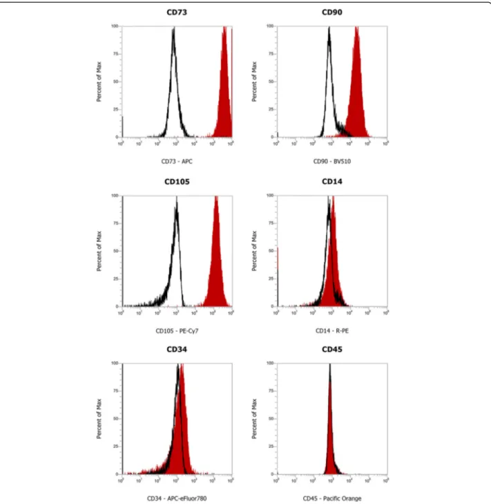
Characterization of human gingival mesenchymal stem
cells
The HGMSCs isolated from the gingival samples were
characterized according to their surface protein
expres-sion by flow cytometry. As shown in Fig.
1
, the cells
were found positive for the established MSCs-specific
surface markers CD73, CD90, and CD105 [
32
]. Flow
cy-tometry immunophenotyping also revealed the negativity
to CD14, CD34, and CD45, confirming the absence of
hematopoietic cells in the isolated stem cells population.
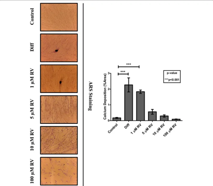
Resveratrol promotes the osteogenic differentiation of
human gingival mesenchymal stem cells
To determine the optimal conditions for osteogenic
differ-entiation by RV, HGMSCs were incubated for up to 21
days with RV in concentration ranging from 1 to 100
μM.
As positive control, the cells were incubated in a medium
Fig. 1 Isolation and characterization of Human Gingival Mesenchymal Stem Cells. Characterization of cell surface markers in HGMSCs at passage 3 by flow cytometry. The stem cells isolated from the gingival biopsies are positive to CD73, CD90, and CD105 MSCs-specific markers, and negative to CD14, CD34, and CD45 hematopoietic markers
supplemented with the osteogenic differentiation factors
dexamethasone,
β-glycerophosphate and ascorbic acid
(from now on, referred as
‘differentiation medium’). To
monitor the mineralization associated with osteogenic
dif-ferentiation of HGMSCs the cultures were stained with
Alizarin Red S to detect the calcium deposits in the
extra-cellular space. The mineralization was clearly detectable
after a minimum of 7–14 days culture in differentiation
medium (not shown). Representative images in Fig.
2
(and
relative quantification) show that the osteogenic
differenti-ation promoted by RV is maximal at 1
μM and declines
when concentration raises up to 100
μM, which turned out
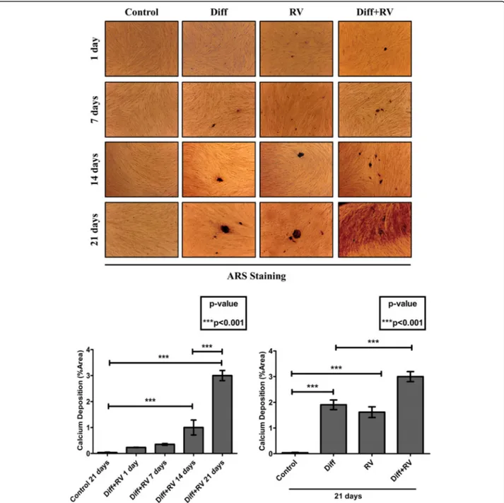
to be toxic. When 1
μM RV was added to the
differenti-ation medium, osteogenic differentidifferenti-ation of HGMSCs (as
mirrored by the mineralization of the extracellular matrix)
was accelerated, indicating a synergism between RV and
osteogenic differentiation factors (Fig.
3
). To characterize
at molecular level this effect, we analyzed the expression of
signaling and structural protein markers of the osteogenic
differentiation [
33
]. Western blotting showed that RUNX2,
the transcription factor of osteocalcin (OCN) and of other
genes associated with osteoblast differentiation, [
34
,
35
]
was upregulated in the HGMSCs cultured in the
differenti-ation medium or in the presence of RV alone (though to a
lower extent in the latter case) (Fig.
4
). Interestingly, when
RV was added to the differentiation medium, the
expres-sion of RUNX2 was further induced compared to the
cul-ture conditions in either the differentiation medium or RV
Fig. 2 Resveratrol promotes the osteoblastic differentiation of Human Gingival Mesenchymal Stem Cells. Adherent HGMSCs were cultured for 21 days in control medium supplemented or not with resveratrol (RV) at the indicated concentration or in differentiation medium (Diff) containing the three osteoblastic inductive factors dexamethasone,β- glycerophosphate and ascorbic acid. At the end, the cultures were processed for Alizarin Red S staining of extracellular calcium deposits. The stained area was quantified using the ImageJ software
alone (Fig.
4
). The combined expression of collagen 1
(COL1A1) and of osteopontin (OPN) is suggestive of
differ-entiation of MSCs toward the osteogenic line, while OCN
synthesis is switched on in osteoblasts and its expression is
therefore proofing that osteoblast differentiation indeed
oc-curred [
33
]. Compared to the osteogenic inductive factors,
RV alone elicited a slight increase in the expression of these
markers (Fig.
4
). However, when RV was added to the
osteogenic differentiation medium it greatly stimulated the
expression of these three proteins in a synergistic manner
with the osteogenic factors (Fig.
4
). To prove further the
ac-quisition of an osteogenic phenotype by HGMSCs under
these culture conditions, we performed the
immunofluores-cence co-staining of the above markers. Representative
im-ages and their quantification are shown in Fig.
5
. The data
confirm that after 21 days of treatment with 1
μM RV alone
the expression of these markers was weakly induced, while
the addition of RV to the osteogenic differentiation medium
Fig. 3 Resveratrol synergizes with osteogenic inductive factors to accelerate osteoblastic differentiation of Human Gingival Mesenchymal Stem Cells. Adherent HGMSCs were cultured for 1 to 21 days in control medium or in differentiation medium (Diff) supplemented or not with 1μM resveratrol (RV). At the end, the cultures were processed for Alizarin Red S staining of extracellular calcium deposits. Quantification of stained area in the time-course is reported in the histogram
synergistically augmented their expression in the cells.
Noteworthy, RV greatly stimulated the nuclear
transloca-tion of RUNX2 (Fig.
5
a).
Osteogenic differentiation of human gingival mesenchymal
stem cells associates with induction of autophagy
To see if autophagy is involved in the osteogenic
differentiation of HGMSCs we first performed the
immunofluorescence staining of autophagic vacuoles with
antibodies specific to LC3, a lipidated protein specifically
associated with the membranes of the autophagosomes,
and to LAMP1, an integral protein of the lysosomal
mem-branes [
36
]. The co-labeling marks the autolysosome and
is indicative of the effective fusion of autophagosomes with
lysosomes. Representative images taken at day 1 and day
21 are shown in Fig.
6
. ImageJ quantification of co-labeled
Fig. 4 Expression of osteoblastic differentiation markers in Human Gingival Mesenchymal Stem Cells. Adherent HGMSCs were cultured for 21 days in control medium or in differentiation medium (Diff) supplemented or not with 1μM resveratrol (RV). At the end, cell homogenates were processed for western blotting analysis of the expression of the osteoblastic transcription factor RUNX-2 and of the osteogenic differentiation markers COL1A1, OPN and OCN. Densitometry of the specific bands (average ± S.D.) of three independent experiments is shown in the histograms
vesicles (autolysosomes) indicated that the autophagy flux
was greatly and promptly (since day 1) stimulated by RV,
while it was initially (at day 1) downregulated and later (at
day 21) induced by the osteogenic differentiation factors.
Remarkably, the formation and accumulation of
autolyso-somes were greatly stimulated in the cells cultured in
dif-ferentiation medium supplemented with RV (Fig.
6
). As a
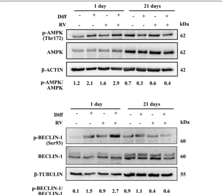
further proof of the induction of autophagy during
HGMSCs
differentiation,
we
analyzed
the
AMPK-BECLIN-1 pathway, as primary candidate of the signaling
pathway triggered by RV [
37
]. It was found that under
osteogenic inductive culture conditions AMPK was active
and, particularly, BECLIN-1 was synergistically activated
by the combination of RV and osteogenic differentiation
Fig. 5 Immunofluorescence staining of osteoblastic differentiation markers in Human Gingival Mesenchymal Stem Cells. HGMSCs were plated on sterile coverslips, let adhere and cultured for 21 days in control medium or in differentiation medium (Diff) supplemented or not with 1μM resveratrol (RV). At the end, the coverslips were fixed and processed for immunofluorescence staining of the osteogenic differentiation markers. Fluorescence staining was quantified with the ImageJ software
factors (Fig.
7
). It is to be noted that by day 21, when the
culture reached confluency and osteoblastic differentiation
almost reached completion, the AMPK-BECLIN-1
path-way was switched off (Fig.
7
).
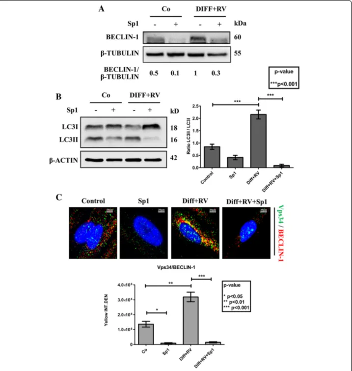
Inhibition of BECLIN-1-depedent autophagy impairs
osteogenic differentiation of HGMSCs by resveratrol
Finally, we investigated whether autophagy was actively
involved in the differentiation process or it was just an
accompanying epiphenomenon. To this end, we
moni-tored the occurrence of osteogenic differentiation in
HGMSCs cultivated in the presence of spautin-1, a
po-tent inhibitor of autophagy that promotes the
prote-asome-mediated degradation of BECLIN-1 [
38
]. We first
determined the appropriate concentration of spautin-1
that could inhibit chronically (for 21 days) autophagy
with no toxic side effect on HGMSCs cell viability (data
not shown). 5
μM Spautin-1 effectively depleted the cell
Fig. 6 Osteoblastic differentiation of Human Gingival Mesenchymal Stem Cells associates with induction of autophagy. HGMSCs were plated on sterile coverslips, let adhere and cultured for 1 to 21 days in control medium or in differentiation medium (Diff) supplemented or not with 1μM resveratrol (RV). At the end, the coverslips were fixed and processed for immunofluorescence staining of the autophagy markers LC3 (marker of autophagosomes) and LAMP1 (marker of lysosomes). Fluorescence staining was quantified with the ImageJ software. Integrated fluorescence intensity of co-labeled area (yellow) was calculated and reported in histograms
of BECLIN-1 (Fig.
8
a), resulting in a strong inhibition of
autophagy as shown by the impaired conversion of
LC3-I into LC3-LC3-ILC3-I (Fig.
8
b). This was further confirmed by
the lack of interaction between BECLIN-1 and Vps34
(aka PI3KC3) in the cells cultivated in RV-supplemented
differentiation medium in the presence of spautin-1, as
shown by immunofluorescence co-staining (Fig.
8
c).
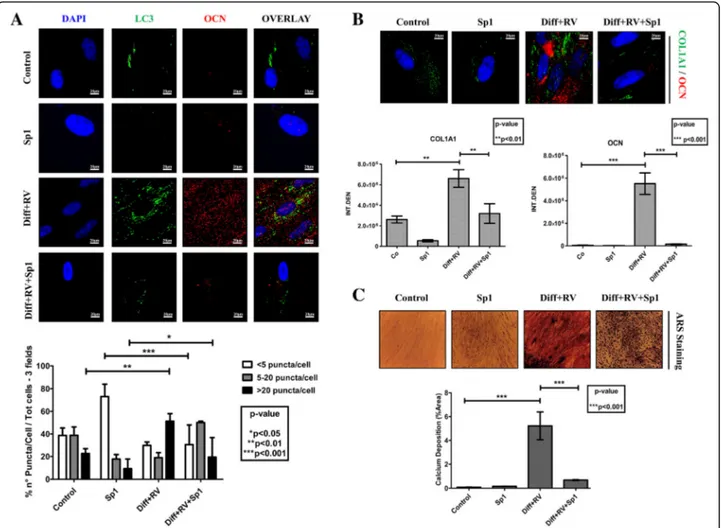
Next, we searched for the mechanistic link between
au-tophagy and osteogenic differentiation. The chronic
in-cubation in differentiation medium supplemented with
RV led to the synthesis and accumulation of OCN in the
cells with upregulated autophagy, as indicated by the
presence of LC3-positive dots (Fig.
9
a). However, in the
parallel cultures co-treated with spautin-1 the number of
LC3-positive dots per cell was greatly reduced to the
level in controls and the OCN staining was faintly visible
(Fig.
9
a). Similarly, intense fluorescent staining of OCN
and COL1A1 was apparent in the cells cultivated in
RV-supplemented differentiation medium, while it was no
apparent in the parallel culture co-treated with Sp-1 (Fig.
9
b). From a functional point of view, spautin-1 inhibition
of autophagy resulted in impaired mineralization of the
extracellular matrix, as indicated by the lack of
Ali-zarin Red-positive deposits of calcium (Fig.
9
c).
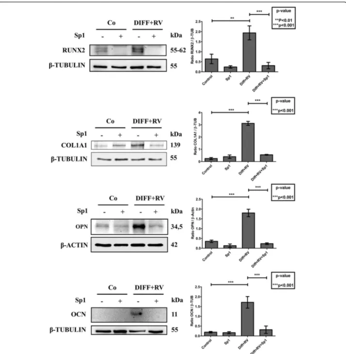
Add-itionally, western blotting of RUNX2, COL1A1, OPN
and OCN showed that the expression of these
Fig. 7 Osteoblastic differentiation of Human Gingival Mesenchymal Stem Cells associates with modulation of the AMPK-BECLIN-1 autophagy signaling pathway. HGMSCs were cultured for 1 to 21 days in control medium or in differentiation medium (Diff) supplemented or not with 1μM resveratrol (RV). At the end, cell homogenates were processed for western blotting analysis of the expression of activated (phosphorylated) AMPK and BECLIN-1, two signaling proteins that govern autophagy. Densitometry (arbitrary units) is included. Similar data were reproduced in another independent experiment
Fig. 8 Spautin-1 abrogates induction of BECLIN-1-dependent autophagy in Human Gingival Mesenchymal Stem Cells cultivated in osteoblastic differentiation condition. HGMSCs were cultured for 21 days in control medium or in differentiation medium supplemented with 1μM resveratrol (Diff + RV) in the absence or in the presence of spautin-1 (Sp1). At the end, cell homogenates were processed for western blotting analysis of the expression of a BECLIN-1 (target of spautin-1) and of b LC3 (marker of autophagosome). Densitometry (arbitrary units) of the specific bands is included. Data were reproduced in three independent experiments. The ratio LC3-II/LC3-I is assumed as an index of autophagosome and autolysosome accumulation in the cell. c HGMSCs were plated on sterile coverslips, let adhere and cultured for 21 days in control medium or in differentiation medium supplemented with 1μM resveratrol (Diff + RV) in the absence or the presence of spautin-1 (Sp1). At the end, the coverslips were fixed and processed for immunofluorescence staining of the autophagy interactome markers Vps34 (PI3KC3) and BECLIN-1. Fluorescence staining was quantified with the ImageJ software. Integrated fluorescence intensity of co-labeled area (yellow) was calculated and reported in histograms. Data from three coverslips per condition reproduced in three separate experiments
markers of osteogenic differentiation was completely
prevented in the cultures exposed to Sp-1 despite the
concomitant presence in the medium of RV and
osteogenic inductive factors (Fig.
10
). From these data
we may conclude that autophagy is functionally linked
to osteogenic differentiation of HGMSCs.
Discussion
Autophagy is the major pathway for the cellular bulk
degradation associated with the remodeling of cellular
structures during differentiation of MSCs [
23
,
24
].
During cellular differentiation, the autophagy pathway
is finely tuned to meet the metabolic needs associated
with the morpho-functional changes [
22
]. Two
princi-pal signaling pathways converging on the ULK1
com-plex control autophagy: the mTORC1 pathway, with
inhibitory function, and the AMPK pathway, with
acti-vating function [
39
]. The mTORC1 pathway is
posi-tively triggered by growth factors and availability of
nutrients, the AMPK pathway is activated when there is
a lack of ATP production because of lack of nutrients
or mitochondrial poisoning [
40
]. The AMPK-ULK1
pathway was shown to positively regulate
autophagy-dependent mitochondrial homeostasis in embryonic
stem cells, contributing to stemness properties [
41
].
Consistently, autophagy plays a role in maintaining the
stemness properties and is modulated during stem cell
differentiation [
42
]. In this work, we analyzed the
con-tribution of autophagy in the osteoblastic
differenti-ation of HGMSCs induced by RV or the osteoblastic
inductive factors dexamethasone,
β-glycerophosphate
and
ascorbic
acid
or
their
combination.
We
Fig. 9 Spautin-1 prevents the autophagy-associated osteoblastic differentiation of Human Gingival Mesenchymal Stem cells. a HGMSCs were plated on sterile coverslips, let adhere and cultured for 21 days in control medium or in differentiation medium supplemented with 1μM resveratrol (Diff + RV) in the absence or the presence of spautin-1 (Sp1). The coverslips were then fixed and processed for immunofluorescence staining of the autophagy marker LC3 and of the osteoblastic differentiation marker OCN. As an index of autophagy in the cells, LC3 puncta were quantified (as per the guidelines 36). b HGMSCs were plated and treated as in a and the coverslips processed for immunofluorescence staining of the osteoblastic differentiation markers OCN and COL1A1. Integrated fluorescence intensity quantified with the ImageJ software is shown in the histograms. c HGMSCs cells were plated on plastic and treated as in a and at the end the cultures were processed for Alizarin Red S staining of extracellular calcium deposits. The stained area was quantified using the ImageJ software
demonstrate that RV induces the osteoblastic
differenti-ation of HGMSCs when used at 1
μM, while it is toxic
at concentrations above 10
μM. Our results are in
agreement with similar studies conducted on human
bone-derived MSCs [
17
] and human embryonic stem
cells [
18
]. Compared to the osteogenic inductive
fac-tors, RV elicited a less pronounced differentiation by
day 21. It is likely that prolonging the incubation with
RV would eventually attain a full osteoblastic
differenti-ation. RV induced the expression and promoted the
nu-clear translocation of the osteogenic transcription
factor RUNX2. Remarkably, RV synergized with the
osteogenic inductive factors accelerating the
osteo-blastic differentiation of HGMSCs, as indicated by
Fig. 10 Spautin-1 prevents the expression of osteoblastic differentiation markers in Human Gingival Mesenchymal Stem cells. HGMSCs were cultured for 21 days in control medium or in differentiation medium supplemented with 1μM resveratrol (Diff + RV) in the absence or in the presence of spautin-1 (Sp1). At the end, cell homogenates were processed for western blotting analysis of the expression of the osteoblastic transcription factor RUNX-2 and of the osteogenic differentiation markers COL1A1, OPN and OCN. Densitometry of the specific bands (average ± S.D.) of three independent experiments is shown in the histograms
anticipation and increased mineralization of the
extra-cellular matrix. Signs of osteoblastic differentiation
such as OCN synthesis and extracellular calcium
de-posits became detectable after 7 days of culture.
Osteoblastic differentiation of HGMSCs was strictly
dependent on BECLIN-1 dependent autophagy, as
dem-onstrated by the observation that it was prevented by
spautin-1 induced depletion of BECLIN-1 and
conse-quent inhibition of autophagy. It is likely that RV
accel-erated the osteoblastic differentiation of HGMSCs
cultivated in the osteogenic differentiation medium
be-cause of its strong stimulation of autophagy. RV has
been shown to induce autophagy in mouse embryonic
stem cells via activation of the AMPK/ULK1 pathway,
and this correlated with enhanced pluripotency of the
cells [
37
]. RV mimics a situation of energy restriction
and activates the AMPK pathway regardless of the
pres-ence of nutrients, leading to activation of autophagy
[
27
]. Activation of AMPK bypasses the block by
mTORC1 and triggers autophagy through direct
acti-vating phosphorylation of ULK1 and of BECLIN-1 [
43
,
44
]. Under metabolic stress conditions, the parallel
acti-vation of mTOR and of AMPK allows the coordinated
and contemporary protein degradation and protein
syn-thesis processes, with the former providing the amino
acids needed for the latter [
45
]. We observed that
AMPK was transitorily activated in HGMSCs
undergo-ing cell differentiation and it was down-regulated when
osteoblastic differentiation was achieved. This same
pattern
was
paralleled
by
phosphorylated
Ser93
BECLIN-1, which is operated by AMPK [
44
].
Modula-tion of AMPK activaModula-tion drives osteoblast
differenti-ation: it is induced during early differentiation and its
silencing or inhibition causes bone loss, yet its
constitu-tive activation prevents full differentiation [
46
]. This
modulation was paralleled by modulation of autophagy,
suggesting that down-regulation of AMPK-dependent
autophagy could favor glycolysis, which is necessary in
the late stages of differentiation [
46
]. Interestingly,
similar findings were reported in the myoblast to
myo-tube differentiation of muscle satellite cells, which are
regarded as stem-like cells [
47
]. Thus, down-regulation
of the AMPK-BECLIN-1 pathway is consistent with the
progressive downregulation of autophagy to basal levels
once that full differentiation is achieved when it is no
more requested the degradation of redundant or
un-wanted cell components.
Conclusions
In summary, here, we provide for the first time the
evidence that RV and osteogenic inductive factors
syner-gize to induce the osteoblastic differentiation of HGMSCs
and that this process relies on modulation of autophagy.
Human gingiva represents an abundant and easily
accessible source of MSCs. The possibility of inducing the
differentiation of HGMSCs in an osteogenic sense in vitro
can be translated into the regenerative cell therapy of
maxillary / mandibular bone defects [
5
,
6
,
48
]. Inductive
factors such as RV and autophagy modulators can be
incorporated into scaffold nanostructures containing
HGMSCs and be implanted in situ for repairing and
reconstructive purposes) [
49
].
Abbreviations
COL1A1:Collagen 1α1; HGMSC: Human gingival mesenchymal stem cell; OCN: Osteocalcin; OPN: Osteopontin; RV: Resveratrol; Sp1: Spautin-1
Acknowledgements
This research had the financial support of the Università del Piemonte Orientale. The Microscope fluorescence imaging facility was donated by Comoli, Ferrari & SpA (Novara, Italy). C.V. was supported with a fellowship funded by the Associazione per la Ricerca Medica Ippocrate-Rhazi (Novara, Italy).
Authors’ contributions
CI and CM conceived and coordinated the study; CV and CI designed the experiments; CG and BZ isolated and characterized the HGMSCs; CV, AF, ES and LV performed the experiments; CV and AF drafted the manuscript; CI revised and finalized the manuscript. All authors read and approved the final manuscript.
Author’s information
Chiara Vidoni received her PhD degree in Medical Sciences and Biotechnology at Università del Piemonte Orientale (Novara, Italy) in 2017. She performed her PhD studies under the mentorship of Prof. Ciro Isidoro in the Laboratory of Molecular Pathology. She completed her Master’s degree in Medical Biotechnologies at Università del Piemonte Orientale in 2012. She received her Bachelor’s degree in Biotechnologies at Università del Piemonte Orientale in 2009. From 2017, she is postdoctoral fellow in Prof. Isidoro’s Laboratory. Her current research focused on the role and regulation of autophagy in neurodegenerative diseases, cancer and regenerative medicine. She has co-authored ten articles published in peer-reviewed journals. Alessandra Ferraresi is a post-doctoral fellow at Università del Piemonte Orientale in Prof. Isidoro’s lab. She received her PhD in Medical Sciences and Biotechnology at Università del Piemonte Orientale (Novara, Italy) in 2019. She completed her Master’s degree in Pharmaceutical Biotechnologies with honors at Alma Mater Studiorum-Università di Bologna in 2014. She received her Bachelor’s degree in Biotechnologies at Università di Parma in 2011. Her current research focused on the role and regulation of autophagy in cancer, particularly in cancer cell migration and resistance to metabolic stress. She has co-authored nine articles published in peer-reviewed journals. Eleonora Secomandi is a PhD student in Medical Sciences and
Biotechnology at Università del Piemonte Orientale (Novara, Italy), working under the mentorship of Prof. Ciro Isidoro in the Laboratory of Molecular Pathology. She completed the Master’s degree in Medical Biotechnologies with honors at Università del Piemonte Orientale di Novara in 2018. She received her Bachelor’s degree in Biological Sciences at Università del Piemonte Orientale in 2016. Her current research focused on cancer biology, protein synthesis and regulation of autophagy in cancer.
She has co-authored four articles published in peer-reviewed journals. Letizia Vallino is a master student in Biomedical and Biomolecular Biology at Università del Piemonte Orientale in Alessandria (Italy). In 2018 to present, she works in Laboratory of Molecular Patology under the supervision of Prof. Ciro Isidoro. She received her Bachelor’s degree in Biology Sciences at Università del Piemonte Orientale in Vercelli (Italy) in 2017. Her current research focused on cancer biology, particularly cancer-related autophagy, cancer metabolism and epigenetic control. She has co-authored one article published in peer-reviewed journals.
Chiara Gardin is a post doc student working under the mentorship of Prof. Barbara Zavan in the Laboratory of Tissue Engineering and Regenerative Medicine at Università of Ferrara (Ferrara, Italy). She completed her PhD working under the mentorship of Prof. Giorgio Valle in the laboratory of Molecular Sciences at University of Padova (Padova). She completed her
master’s degree in Biological Sciences with honors at University of Padova. Her current research focused on the role Mesenchymal stem cell as tool on tissue regeneration.
She has co-authored seven articles published in peer-reviewed journals. Barbara Zavan received her PhD on Tissue Engineering and Regenerative Medicine at University of Padova (Padova, Italy) and her doctoral degree (summa cum laude) in Biological Sciences from the University of Padova (Padova, Italy). Currently, he is Associate Professor of Medical Sciences Tecnology at the school of Medicine of University of Ferrara (Ferrara, Italy). He is Visiting Professor at the University of Murcia, Faculty of Medicine (Murcia, Spain). Her research seeks to understand how changes in stem cell activity impact tissue homeostasis and repair throughout life and to identify systemic molecules or surfaced modification are responsible for regulation of regenerative potential. She has a multidisciplinary research group focused on both high quality fundamental science and translation for human health. She has active collaborations with bioengineers, chemists and clinicians in order to study stem cell-niche interactions in vitro.
Carmen Mortellaro obtained the degree in Medicine and Surgery and the Specialization Diploma in Odontostomatology and Orthognathology from the University of Turin. She is currently Full Professor of Stomatological Diseases at the School of Medicine and Surgery of the University of Piemonte Orientale and Director of the Orthodontic-Surgical Operative Unit for the diagnosis and therapy of craniofacial skeletal alterations and oral sur-gery. She teaches diseases of the oral cavity, oral surgery, dental materials, or-thodontics, pedodontics, clinical semeiotics of the craniofacial district, exodontic surgery and anatomy. She is President Anthec (Academy of Non Transfusional Hemo-Components). She has authored 250 publications in sci-entific journals and international conference proceedings and peer reviewer of some international journals, including Indian Journal of Dentistry and Jour-nal of Oral Pathology & Medicine. She is a member of the Editorial Board of the Craniofacial Journal.
Ciro Isidoro (www.isidorolab.com) received his doctoral degree (summa cum laude) in Biological Sciences from the University of Torino (Italy) and his doctoral degree (summa cum laude) in Medicine and Surgery from the University of Piemonte Orientale (Novara, Italy). Currently, he is Professor of Pathology and of Experimental Oncology at the school of Medicine of University of Piemonte Orientale (Novara, Italy). He is Visiting Professor at the Department of Cell Biology of the University of Oklahoma Collage of Medicine (Oklahoma City, United States). He is Professeur Honoraire at the Faculté de Medecine et de Phamacie de l’Université de Franche-Comté (Besancon, France). He is member of the scientific board of the“Centre for Integrative Cancer Research” of the Georgia Tech Institute, University of Georgia (Atlanta, US). He is Co-Editor in Chief of the J. of Traditional and Complementary Medicine, and Associate Editor of Autophagy, BMC Cancer, Molecular Carcinogenesis, J. of Ovarian Research, Genes and Cancers, J. of Molecular Signaling, and other journals. His fields of expertise include lyso-some biogenesis and function, autophagy regulation in cancer and in neuro-degenerative diseases, cell death pathways, mechanisms of action of natural products, epigenetic regulation of autophagy and of cell death.
Funding
This research had the financial support of the Università del Piemonte Orientale.
Availability of data and materials
All data generated or analysed during this study are included in this research article.
Ethics approval and consent to participate Include a statement on ethics approval and consent.
Consent for publication Not applicable.
Competing interests
The authors declare that they have no competing interests.
Author details
1Laboratory of Molecular Pathology, Department of Health Sciences,
Università del Piemonte Orientale“A. Avogadro”, Via P. Solaroli 17, 28100 Novara, Italy.2Maria Cecilia Hospital, GVM Care & Research, via Corriera 1,
48033, Cotignola, Ravenna, Italy.3Medical Sciences Department, University of
Ferrara, Via Fossato di Mortara, 70 Ferrara, Italy.4Oral Surgery Unit,
Department of Medical Science, Università del Piemonte Orientale“A. Avogadro”, Novara, Italy.
Received: 16 May 2019 Accepted: 5 August 2019
References
1. Griffin M, Iqbal SA, Bayat A. Exploring the application of mesenchymal stem cells in bone repair and regeneration. J Bone Joint Surg Br. 2011;93(4):427–34.https:// doi.org/10.1302/0301-620X.93B4.25249Review. PubMed PMID: 21464477. 2. Pagni G, Kaigler D, Rasperini G, Avila-Ortiz G, Bartel R, Giannobile WV. Bone
repair cells for craniofacial regeneration. Adv Drug Deliv Rev. 2012;64(12): 1310–9.https://doi.org/10.1016/j.addr.2012.03.005Epub 2012 Mar 10. Review. PubMed PMID: 22433781; PubMed Central PMCID: PMC3383887. 3. Grässel S, Lorenz J. Tissue-engineering strategies to repair chondral and
osteochondral tissue in osteoarthritis: use of mesenchymal stem cells. Curr Rheumatol Rep. 2014;16(10):452.https://doi.org/10.1007/s11926-014-0452-5
Review. PubMed PMID: 25182680; PubMed Central PMCID: PMC4182613. 4. Wang F, Yu M, Yan X, Wen Y, Zeng Q, Yue W, Yang P, Pei X. Gingiva-derived
mesenchymal stem cell-mediated therapeutic approach for bone tissue regeneration. Stem Cells Dev. 2011;20(12):2093–102.https://doi.org/10.1089/ scd.2010.0523Epub 2011 Apr 27. PubMed PMID: 21361847.
5. Zhao N, Wu Z, Qin L, Guo Z, Li D. Characteristics and tissue regeneration properties of gingiva-derived mesenchymal stem cells. Crit Rev Eukaryot Gene Expr. 2015;25(2):135–44 Review. PubMed PMID: 26080607.
6. Fawzy El-Sayed KM, Dörfer CE. Gingival mesenchymal stem/progenitor cells: a unique tissue engineering gem. Stem Cells Int. 2016;2016:7154327.https:// doi.org/10.1155/2016/7154327Epub 2016 May 29. Review. PubMed PMID: 27313628; PubMed Central PMCID: PMC4903147.
7. Xu D, Xu L, Zhou C, Lee WY, Wu T, Cui L, Li G. Salvianolic acid B promotes osteogenesis of human mesenchymal stem cells through activating ERK signaling pathway. Int J Biochem Cell Biol. 2014;51:1–9.https://doi.org/10.1 016/j.biocel.2014.03.005Epub 2014 Mar 19. PubMed PMID: 24657587. 8. Du L, Yang P, Ge S. Isolation and characterization of human gingiva-derived
mesenchymal stem cells using limiting dilution method. J Dent Sci. 2016; 11(3):304–14.https://doi.org/10.1016/j.jds.2016.03.010Epub 2016 May 6. PubMed PMID: 30894989; PubMed Central PMCID: PMC6395297. 9. Chen Y, Liu H. The differentiation potential of gingival mesenchymal stem
cells induced by apical tooth germ cell-conditioned medium. Mol Med Rep. 2016;14(4):3565–72.https://doi.org/10.3892/mmr.2016.5726Epub 2016 Sep 6. PubMed PMID: 27600358; PubMed Central PMCID: PMC5042793. 10. Ha DH, Yong CS, Kim JO, Jeong JH, Park JB. Effects of tacrolimus on
morphology, proliferation and differentiation of mesenchymal stem cells derived from gingiva tissue. Mol Med Rep. 2016;14(1):69–76.https://doi. org/10.3892/mmr.2016.5217Epub 2016 May 6. PubMed PMID: 27177273; PubMed Central PMCID: PMC4918528.
11. Murugan Girija D, Y Ranga Rao S, Kalachaveedu M, Subbarayan R. Osteogenic differentiation of human gingival mesenchymal stem cells by Aristolochia bracteolata supplementation through enhanced Runx2 expression. J Cell Physiol. 2017;232(7):1591–5.https://doi.org/10.1002/jcp.25 835Epub 2017 Feb 21. PubMed PMID: 28150858.
12. Mobasheri A, Shakibaei M. Osteogenic effects of resveratrol in vitro: potential for the prevention and treatment of osteoporosis. Ann N Y Acad Sci. 2013;1290:59–66.https://doi.org/10.1111/nyas.12145Review. PubMed PMID: 23855466.
13. Murgia D, Mauceri R, Campisi G, De Caro V. Advance on Resveratrol Application in Bone Regeneration: Progress and Perspectives for Use in Oral and Maxillofacial Surgery. Biomolecules. 2019;9(3).https://doi.org/10.3390/ biom9030094Review. PubMed PMID: 30857241; PubMed Central PMCID: PMC6468380.
14. Rafe T, Shawon PA, Salem L, Chowdhury NI, Kabir F, Zahur SMB, Akther R, Noor HB, Mohib MM, Sagor MAT. Preventive role of Resveratrol against inflammatory cytokines and related diseases. Curr Pharm Des. 2019.https:// doi.org/10.2174/1381612825666190410153307[Epub ahead of print] PubMed PMID: 30968773.
15. He X, Andersson G, Lindgren U, Li Y. Resveratrol prevents RANKL-induced osteoclast differentiation of murine osteoclast progenitor RAW 264.7 cells through inhibition of ROS production. Biochem Biophys Res Commun.
2010;401(3):356–62.https://doi.org/10.1016/j.bbrc.2010.09.053Epub 2010 Sep 17. PubMed PMID:20851107.
16. Matsuda Y, Minagawa T, Okui T, Yamazaki K. Resveratrol suppresses the alveolar bone resorption induced by artificial trauma from occlusion in mice. Oral Dis. 2018;24(3):412–21.https://doi.org/10.1111/odi.12785
Epub 2017 Oct 23. PubMed PMID:28944599.
17. Dai Z, Li Y, Quarles LD, Song T, Pan W, Zhou H, Xiao Z. Resveratrol enhances proliferation and osteoblastic differentiation in human mesenchymal stem cells via ER-dependent ERK1/2 activation. Phytomedicine. 2007;14(12):806– 14 Epub 2007 Aug 8. PubMed PMID: 17689939.
18. Tseng PC, Hou SM, Chen RJ, Peng HW, Hsieh CF, Kuo ML, Yen ML. Resveratrol promotes osteogenesis of human mesenchymal stem cells by upregulating RUNX2 gene expression via the SIRT1/FOXO3A axis. J Bone Miner Res. 2011;26(10):2552–63.https://doi.org/10.1002/jbmr.460
PubMed PMID: 21713995.
19. Shakibaei M, Shayan P, Busch F, Aldinger C, Buhrmann C, Lueders C, Mobasheri A. Resveratrol mediated modulation of Sirt-1/Runx2 promotes osteogenic differentiation of mesenchymal stem cells: potential role of Runx2 deacetylation. PLoS One. 2012;7(4):e35712.https://doi.org/10.1371/ journal.pone.0035712Epub 2012 Apr 23. PubMed PMID: 22539994; PubMed Central PMCID: PMC3335081.
20. Casarin RC, Casati MZ, Pimentel SP, Cirano FR, Algayer M, Pires PR, Ghiraldini B, Duarte PM, Ribeiro FV. Resveratrol improves bone repair by modulation of bone morphogenetic proteins and osteopontin gene expression in rats. Int J Oral Maxillofac Surg. 2014;43(7):900–6.https://doi.org/10.1016/j.ijom.2 014.01.009Epub 2014 Feb 13. PubMed PMID: 24530035.
21. Ornstrup MJ, Harsløf T, Sørensen L, Stenkjær L, Langdahl BL, Pedersen SB. Resveratrol increases osteoblast differentiation in vitro independently of inflammation. Calcif Tissue Int. 2016;99(2):155–63.https://doi.org/10.1007/ s00223-016-0130-xEpub 2016 Mar 21. PubMed PMID: 27000750. 22. Fortini P, Iorio E, Dogliotti E, Isidoro C. Coordinated metabolic changes and
modulation of autophagy during Myogenesis. Front Physiol. 2016;7:237.
https://doi.org/10.3389/fphys.2016.00237eCollection 2016. PubMed PMID: 27378945; PubMed Central PMCID: PMC4909729.
23. Wang S, Xia P, Rehm M, Fan Z. Autophagy and cell reprogramming. Cell Mol Life Sci. 2015;72(9):1699–713.https://doi.org/10.1007/s00018-014-1829-3
Epub 2015 Jan 9. Review. PubMed PMID: 25572296.
24. Boya P, Codogno P, Rodriguez-Muela N. Autophagy in stem cells: repair, remodelling and metabolic reprogramming. Development. 2018;145(4).
https://doi.org/10.1242/dev.146506Review. PubMed PMID: 29483129. 25. Dikic I, Elazar Z. Mechanism and medical implications of mammalian
autophagy. Nat Rev Mol Cell Biol. 2018;19(6):349–64.https://doi.org/10.1038/ s41580-018-0003-4Review. PubMed PMID: 29618831.
26. Jaber FA, Khan NM, Ansari MY, Al-Adlaan AA, Hussein NJ, Safadi FF. Autophagy plays an essential role in bone homeostasis. J Cell Physiol. 2019; 234(8):12105–15.https://doi.org/10.1002/jcp.27071Epub 2019 Feb 28. Review. PubMed PMID: 30820954.
27. Ferraresi A, Titone R, Follo C, Castiglioni A, Chiorino G, Dhanasekaran DN, Isidoro C. The protein restriction mimetic resveratrol is an autophagy inducer stronger than amino acid starvation in ovarian cancer cells. Mol Carcinog. 2017;56(12):2681–91.https://doi.org/10.1002/mc.22711Epub 2017 Sep 7. PubMed PMID: 28856729.
28. Ge S, Mrozik KM, Menicanin D, Gronthos S, Bartold PM. Isolation and characterization of mesenchymal stem cell-like cells from healthy and inflamed gingival tissue: potential use for clinical therapy. Regen Med. 2012; 7(6):819–32.https://doi.org/10.2217/rme.12.61PubMed PMID: 23164082. 29. Gardin C, Ferroni L, Bellin G, Rubini G, Barosio S, Zavan B. Therapeutic
Potential of Autologous Adipose-Derived Stem Cells for the Treatment of Liver Disease. Int J Mol Sci. 2018;19(12).https://doi.org/10.3390/ijms19124 064PubMed PMID: 30558283; PubMed Central PMCID: PMC6321531. 30. Chevallier N, Anagnostou F, Zilber S, Bodivit G, Maurin S, Barrault A, Bierling
P, Hernigou P, Layrolle P, Rouard H. Osteoblastic differentiation of human mesenchymal stem cells with platelet lysate. Biomaterials. 2010;31(2):270–8.
https://doi.org/10.1016/j.biomaterials.2009.09.043Epub 2009 Sep 26. PubMed PMID: 19783038.
31. Puchtler H, Meloan SN, Terry MS. On the history and mechanism of alizarin and alizarin red S stains for calcium. J Histochem Cytochem. 1969;17(2):110– 24 PubMed PMID: 4179464.
32. Dominici M, Le Blanc K, Mueller I, Slaper-Cortenbach I, Marini F, Krause D, Deans R, Keating A, Prockop DJ, Horwitz E. Minimal criteria for defining multipotent mesenchymal stromal cells. The International Society for
Cellular Therapy position statement. Cytotherapy. 2006;8(4):315–7 PubMed PMID: 16923606.
33. Long MW. Osteogenesis and bone-marrow-derived cells. Blood Cells Mol Dis. 2001;27(3):677–90 Review. PubMed PMID: 11482883.
34. Ducy P, Karsenty G. Two distinct osteoblast-specific cis-acting elements control expression of a mouse osteocalcin gene. Mol Cell Biol. 1995;15(4): 1858–69 PubMed PMID: 7891679; PubMed Central PMCID: PMC230411. 35. Lee KE, Seymen F, Ko J, Yildirim M, Tuna EB, Gencay K, Kim JW. RUNX2
mutations in cleidocranial dysplasia. Genet Mol Res. 2013;12(4):4567–74.
https://doi.org/10.4238/2013.October.15.5PubMed PMID: 24222232. 36. Klionsky DJ, Abdelmohsen K, Abe A, Abedin MJ, Abeliovich H, Acevedo Arozena
A, et al. Guidelines for the use and interpretation of assays for monitoring autophagy (3rd edition). Autophagy. 2016;12(1):1–222 PubMed PMID: 26799652. 37. Suvorova II, Knyazeva AR, Petukhov AV, Aksenov ND, Pospelov VA.
Resveratrol enhances pluripotency of mouse embryonic stem cells by activating AMPK/Ulk1 pathway. Cell Death Dis. 2019;5:61.https://doi.org/1 0.1038/s41420-019-0137-yeCollection 2019. PubMed PMID: 30729040; PubMed Central PMCID: PMC6361884.
38. Liu J, Xia H, Kim M, Xu L, Li Y, Zhang L, Cai Y, Norberg HV, Zhang T, Furuya T, Jin M, Zhu Z, Wang H, Yu J, Li Y, Hao Y, Choi A, Ke H, Ma D, Yuan J. Beclin1 controls the levels of p53 by regulating the deubiquitination activity of USP10 and USP13. Cell. 2011;147(1):223–34.https://doi.org/10.1016/j.cell.2 011.08.037PubMed PMID: 21962518; PubMed Central PMCID: PMC3441147. 39. Kim J, Kundu M, Viollet B, Guan KL. AMPK and mTOR regulate autophagy
through direct phosphorylation of Ulk1. Nat Cell Biol. 2011;13(2):132–41.
https://doi.org/10.1038/ncb2152Epub 2011 Jan 23. PubMed PMID: 21258367; PubMed Central PMCID: PMC3987946.
40. Ravikumar B, Sarkar S, Davies JE, Futter M, Garcia-Arencibia M, Green-Thompson ZW, Jimenez-Sanchez M, Korolchuk VI, Lichtenberg M, Luo S, Massey DC, Menzies FM, Moreau K, Narayanan U, Renna M, Siddiqi FH, Underwood BR, Winslow AR, Rubinsztein DC. Regulation of mammalian autophagy in physiology and pathophysiology. Physiol Rev. 2010;90(4): 1383–435.https://doi.org/10.1152/physrev.00030.2009Review. PubMed PMID: 20959619.
41. Gong J, Gu H, Zhao L, Wang L, Liu P, Wang F, Xu H, Zhao T.
Phosphorylation of ULK1 by AMPK is essential for mouse embryonic stem cell self-renewal and pluripotency. Cell Death Dis. 2018;9(2):38.https://doi. org/10.1038/s41419-017-0054-zPubMed PMID: 29348566; PubMed Central PMCID: PMC5833692.
42. Sotthibundhu A, Promjuntuek W, Liu M, Shen S, Noisa P. Roles of autophagy in controlling stem cell identity: a perspective of self-renewal and differentiation. Cell Tissue Res. 2018;374(2):205–16.https://doi.org/10.1 007/s00441-018-2829-7Epub 2018 Apr 25. Review. PubMed PMID: 29696372.
43. Egan DF, Shackelford DB, Mihaylova MM, Gelino S, Kohnz RA, Mair W, Vasquez DS, Joshi A, Gwinn DM, Taylor R, Asara JM, Fitzpatrick J, Dillin A, Viollet B, Kundu M, Hansen M, Shaw RJ. Phosphorylation of ULK1 (hATG1) by AMP-activated protein kinase connects energy sensing to mitophagy. Science. 2011;331(6016):456–61.https://doi.org/10.1126/science.1196371
Epub 2010 Dec 23. PubMed PMID: 21205641; PubMed Central PMCID: PMC3030664.
44. Kim J, Kim YC, Fang C, Russell RC, Kim JH, Fan W, Liu R, Zhong Q, Guan KL. Differential regulation of distinct Vps34 complexes by AMPK in nutrient stress and autophagy. Cell. 2013;152(1–2):290–303.https://doi.org/10.1016/j. cell.2012.12.016PubMed PMID: 23332761; PubMed Central PMCID: PMC3587159.
45. Follo C, Vidoni C, Morani F, Ferraresi A, Seca C, Isidoro C. Amino acid response by Halofuginone in Cancer cells triggers autophagy through proteasome degradation of mTOR. Cell Commun Signal. 2019;17(1):39.
https://doi.org/10.1186/s12964-019-0354-2PubMed PMID: 31046771; PubMed Central PMCID: PMC6498594.
46. Xi G, Rosen CJ, Clemmons DR. IGF-I and IGFBP-2 stimulate AMPK activation and autophagy, which are required for osteoblast differentiation.
Endocrinology. 2016;157(1):268–81.https://doi.org/10.1210/en.2015-1690Epub 2015 Nov 10. PubMed PMID: 26556533; PubMed Central PMCID: PMC4701891. 47. Fortini P, Ferretti C, Iorio E, Cagnin M, Garribba L, Pietraforte D, Falchi M,
Pascucci B, Baccarini S, Morani F, Phadngam S, De Luca G, Isidoro C, Dogliotti E. The fine tuning of metabolism, autophagy and differentiation during in vitro myogenesis. Cell Death Dis. 2016;7:e2168.https://doi.org/10.1 038/cddis.2016.50PubMed PMID: 27031965; PubMed Central PMCID: PMC4823951.
48. Shakoori P, Zhang Q, Le AD. Applications of mesenchymal stem cells in Oral and craniofacial regeneration. Oral Maxillofac Surg Clin North Am. 2017; 29(1):19–25.https://doi.org/10.1016/j.coms.2016.08.009Review. PubMed PMID: 27890225.
49. Kamath MS, Ahmed SS, Dhanasekaran M, Santosh SW. Polycaprolactone scaffold engineered for sustained release of resveratrol: therapeutic enhancement in bone tissue engineering. Int J Nanomedicine. 2014;9:183– 95.https://doi.org/10.2147/IJN.S49460Epub 2013 Dec 23. PubMed PMID: 24399875; PubMed Central PMCID: PMC3875521.
Publisher
’s Note
Springer Nature remains neutral with regard to jurisdictional claims in published maps and institutional affiliations.