Relevance of BRAF
V600E
Mutation Testing Versus
RAS Point Mutations and RET/PTC Rearrangements
Evaluation in the Diagnosis of Thyroid Cancer
Martina Rossi,1,2Mattia Buratto,1Federico Tagliati,1Roberta Rossi,1,2Sabrina Lupo,1,2Giorgio Trasforini,2Giovanni Lanza,3Paola Franceschetti,2Stefania Bruni,2 Ettore degli Uberti,1,2 and Maria Chiara Zatelli1,2
Background: A molecular profile including BRAF and RAS mutations as well as RET/PTC rearrangement
evaluation has been proposed to provide an accurate presurgical assessment of thyroid nodules and to reduce the
number of unnecessary diagnostic surgeries, sparing patients’ health and saving healthcare resources. However,
the application of such molecular analyses may provide different results among different centers and
popula-tions in real-life settings. Our aims were to evaluate the diagnostic utility of assessing the presence of BRAF and
RAS mutations and RET/PTC1 and RET/PTC3 rearrangements in all cytological categories in an Italian group of
thyroid nodule patients assessed prospectively, and to understand whether and which mutation testing might be
helpful in cytologically indeterminate nodules.
Methods: A total of 911 patients were submitted to ultrasound and fine-needle aspiration biopsy examination.
Cytological evaluation was performed in parallel with molecular testing and compared to pathological results in
940 thyroid nodules, including 140 indeterminate lesions.
Results: BRAF mutation testing provided the best contribution to cancer diagnosis, allowing the disease to be
detected at an early stage, and identifying indeterminate nodules in which diagnostic lobectomy could be
spared. On the contrary, RAS and RET/PTC analysis did not further increase diagnostic sensitivity for thyroid
cancer. In addition, we found RET/PTC rearrangements in benign lesions, indicating that this molecular marker
might not be useful for the detection of thyroid cancer.
Conclusion: BRAF
V600Emutation analysis is superior to RAS point mutations and evaluation of RET/PTC
rearrangements in the diagnosis of thyroid cancer, even in indeterminate lesions.
Introduction
T
he diagnostic and therapeutic approachto thyroid cancer has been highly debated in recent years. Ultra-sound (US), cytology, and molecular profiling (by mRNA gene expression platforms, protein immunocytochemistry, miRNA panels, and screening for somatic mutations. in-cluding BRAFV600E and RAS mutations, as well as RET/ PTC1, RET/PTC3, PAX8/PPARc, TK, and ALK rearrange-ments) have been employed in order to provide the most accurate presurgical assessment of thyroid nodules with the aim of increasing the sensitivity for cancer detection and avoiding surgery for lesions erroneously identified as ma-lignant (1–3). The availability of presurgical information can improve preoperative risk stratification and often influences the extent of surgery (4–7). The revised American ThyroidAssociation (ATA) guidelines indicate that thyroid cancer should be treated according to risk stratification, assessed based on disease stage (8). The provided evidence indicates that treatment needs to be tailored according to the risk of recurrence, suggesting that a more conservative attitude, avoiding radioiodine ablation, may be indicated for patients with very low risk of recurrence (9,10). As a consequence, early diagnosis is crucial in order to detect the disease at an early stage and to guide the patient to a less aggressive treatment, thereby avoiding unnecessary risks for the pa-tient’s health and saving healthcare resources (11,12). The main diagnostic tool is fine-needle aspiration biopsy (FNAB). However, FNAB cannot provide a definitive diagnosis in cases with nondiagnostic (ND) or indeterminate cytology. The latter may represent a malignant lesion in *20% of the cases, which are not accurately predictable by US risk factors
1
Section of Endocrinology and Internal Medicine, Department of Medical Sciences, University of Ferrara, Ferrara, Italy. 2
Endocrinology Unit, Azienda Ospedaliero-Universitaria di Ferrara, Ferrara, Italy. 3
Section of Pathology and Biomolecular Diagnostics, Department of Morphology, Surgery and Experimental Medicine, University of Ferrara, Ferrara, Italy.
ª Mary Ann Liebert, Inc. DOI: 10.1089/thy.2014.0338
and thus lead to the need for diagnostic surgery (13). The preoperative use of molecular markers is still highly debated because, among other reason, the incidence of mutations in the different categories outlined in the Bethesda System for Reporting Thyroid Cytopathology (BSRTC) (14) is still un-known. To date, the ATA guidelines suggest considering molecular testing only to refine a cytological indeterminate result (8). Moreover, genetic, environmental, and clinical background may profoundly impact the incidence of muta-tions, and hence there is a need to explore the applicability of molecular testing of thyroid nodules in different populations in the clinical setting. The aim of our study was to evalu-ate the diagnostic utility of assessing the presence of three previously employed thyroid cancer molecular markers— BRAF and RAS mutations, and RET/PTC1 and RET/PTC3 rearrangements—in FNAB material from all cytological ca-tegories in a real-life context involving a group of Italian thyroid nodule patients in order to improve patient manage-ment and surgical treatmanage-ment. In addition, we aimed to assess mutation incidence in each Bethesda category and to under-stand whether and which mutation testing might be helpful in indeterminate nodules.
We therefore assessed the feasibility of obtaining reli-able results from FNAB material for the search for these molecular markers (BRAFV600E, RAS mutations, and RET/ PTC rearrangements) in daily clinical practice employing previously reported methods with slight modifications.
Materials and Methods Subjects
From January 2007 to July 2013, 6500 thyroid nodules from 5800 patients underwent FNAB at the Section of En-docrinology of the University of Ferrara. Among these, 940 FNAB specimens from 911 consecutive patients, displaying at least two clinical and/or US characteristics of suspected malignancy, prospectively underwent evaluation for somatic mutations, including BRAFV600E and RAS point mutations and RET/PTC1 and RET/PTC3 rearrangements, partially overlapping the approach described previously by Nikiforov et al. (15). Patients gave written informed consent for mo-lecular analysis and data collection.
Medical and US examination
All 911 patients recruited in this study were submitted to a careful US examination by a single operator (S.L.) during routine medical care. The collected US features included nodule size (< or > 1 cm), structure (solid, mixed, or cystic), echogenicity (iso-, hypo-, or hyperechoic), presence or ab-sence of micro calcifications, and margins. In addition, the patients’ clinical information regarding age, sex, family history of thyroid cancer, or history of previous external beam radiation exposure was collected.
FNAB procedures
All 940 US-guided FNAB procedures were performed by two experienced endocrinologists (G.T and P.F.) using a standardized protocol, as previously described (16). Cytolo-gical evaluation was performed in parallel with molecular testing. All FNAB results were categorized according to the BSRTC (14), including class III (atypia of undetermined
significance/follicular lesion of undetermined significance [AUS/FLUS]), IV (follicular neoplasm or suspicious for a follicular neoplasm [FN]), and V (suspicious of malignancy [SM]) categories in the group of indeterminate lesions.
DNA and RNA isolation
FNAB material from a needle pass through the nodule was used for cytology, and a second pass was collected in 5 mL of RNA Later solution (Resnova) for molecular analysis. Genomic DNA for BRAF and RAS somatic mutation analysis was obtained as previously described (16,17). Total RNA isolation for RET/PTC1 and RET/PTC3 rearrangements evaluation was performed by centrifuging 2 mL of FNAB sample for 5 minutes at 5000g, and the pellet was then sus-pended in 350 lL of RLT Lysis Buffer (Qiagen). Later, the samples were processed in the QIAcube instrument (Qiagen) using the RNeasy micro kit (Qiagen) according to manu-facturer’s protocol, obtaining 30 lL of purified total RNA. Samples were then processed as described below. All sam-ples displaying a genetic variation were tested in a second assay by a different technician.
BRAF and RAS mutation analysis
BRAFV600E mutation analysis was performed as previ-ously described (16,17), employing a well-established methodology.
A first evaluation of RAS mutations was performed by applying real-time polymerase chain reaction (PCR) ampli-fication followed by high resolution melting (HRM) analysis. Amplification of RAS gene targets (codon 12, 13, and 61 of N-RAS, H-N-RAS, and K-RAS gene isoforms) was performed by using the MeltDoctor HRM Mastermix (Life Technologies) and specific primers (N-RAS exon 2 FOR 5¢-TTG CTG GTG TGA AAT GAC TGA GT-3¢ and REV 5¢-TAG CTG GAT TGT CAG TGC GC-3¢; N-RAS exon 3 FOR: 5¢-CAG AAA ACA AGT GGT TAT AGA TGG TGA-3¢ and REV 5¢-CAA ATA CAC AGA GGA AGC CTT CG-3¢; H-RAS exon 2 FOR: 5¢-GGA GCG ATG ACG GAA TAT AAG C-3¢ and REV 5¢-GTA TTC GTC CAC AAA ATG GTT CTG-3¢; H-RAS exon 3 FOR 5¢-GGA AGC AGG TGG TCA TTG ATG-3¢ and REV 5¢-GCA TGT ACT GGT CCC GCA T-ATG-3¢; K-RAS exon 2: FOR 5¢-TCA CAT TTT CAT TAT TTT TAT TAT AAG GC-3¢ and REV 5¢-GAT TCT GAA TTA GCT GTA TCG TCA AG-3¢; K-RAS exon 3: FOR 5¢-TCC AGA CTG TGT TTC TCC CTT C-3¢ and REV 5¢-TAC ACA AAG AAA GCC CTC CC-3¢). Mutated samples were then genotyped by direct sequencing using the same primers on the 3130 Ge-netic Analyzer (Life Technologies) employing the Ready Reaction Cycle Sequencing 1.1 mix (Life Technologies). This approach, which is very similar to that previously em-ployed (18,19), allowed reliable results to be obtained from FNAB material with a turnaround time of 72 hours.
RET/PTC rearrangement analysis
For the evaluation of RET/PTC1 and RET/PTC3 re-arrangements, total RNA from FNAB samples was analyzed by One Step Real Time RT-PCR, performed on a 7900 HT Real Time System (Life Technologies), employing a modi-fied method compared to Nikiforov et al. (15). The pres-ence of RET/PTC1 and RET/PTC3 rearrangements has been
assessed using two different custom Taqman Gene Expres-sion assays (Life Technologies), each represented by a re-arrangement specific primer-probe set; probes have been designed centered on the rearrangement site, in order to avoid false positive results. Sequences of primers and probes for RET/PTC1 were: FOR: 5¢-CGC GAC CTG CGC AAA-3¢, REV 5-CAA GTT CTT CCG AGG GAA TTC C-3¢, and PROBE: 5¢-FAM-CCA GCG TGA CCA TCG AGG ATC CAA AGT-NFQ-3¢. Sequences of primers and probes for RET/PTC3 were: FOR: 5¢-CCC CAG GAC TGG CTT ACC C-3¢, REV 5¢-CAA GTT CTT CCG AGG GAA TTC C-3¢ and PROBE: 5¢-FAM-AAA GCA GAC CTT GGA GAA CAG TCA GGA GG-NFQ-3¢. All runs were multiplexed with Eukaryotic 18S rRNA Endogenous Control (Life Technologies). The reaction mix included iScript One-Step RT-PCR Kit for probes (Bio-Rad) and the appropriate Taqman assays, described above. To test the method sen-sitivity, each target sequence assay was diluted 1:10, 1:100, 1:1000, and 1:10,000 in not-rearranged cDNA. Both re-arrangements were correctly identified up to a 1:1000 di-lution by the employed method. To exclude the possibility of cross-reactions, RET/PTC1 and RET/PTC3 assays were employed to amplify RET/PTC3 and RET/PTC1 targets respectively, and no signal was obtained. RNA from one or more tumors or cell lines known to carry a particular re-arrangement was used as a positive control. This approach allowed obtaining reliable results from FNAB material with a turnaround time of 24 hours.
Statistical analysis
Sensitivity, specificity, positive predictive value (PPV), and negative predictive value (NPV) were calculated for each detection method and for combined methods, considering histology as the gold standard. Statistical analysis was carried out using the R Software package v3.0.2 (R Foundation for Statistical Computing). The chi-square test (with Yates continuity correction) was employed to compare the diag-nostic sensitivity of cytology with that observed performing both cytology and genetic analysis and to assess the presence of a significant association between the presence of each mutation and US features. A p-value of< 0.05 was considered significant in all tests.
Results Patient findings
Among the 911 patients who participated in the study, 51 had a family history of thyroid cancer, 712 were female, and the mean age was 59– 0.46 years (range 25–81 years). Pa-tients with BSRTC class V and VI lesions, or with a nodule displaying BRAFV600Emutation (independently of cytology results), or with large goiters underwent total thyroidectomy (TT). Patients with repeatedly class I cytology and patients with BSRTC class IV lesions or with a nodule displaying either RAS mutations or RET/PTC rearrangements underwent lobectomy (LT), independently of US nodule features, in line with the previously demonstrated increased cancer risk as-sociated with these mutations (18). Patients with class III lesions without a genetic variation in the studied genes un-derwent a second FNAB and then unun-derwent lobectomy if the cytological diagnosis was confirmed. Otherwise, the patients
were managed according to the new BSRTC class. Finally, patients with class II lesions underwent clinical follow-up.
Cytology, molecular testing, US, and pathology findings
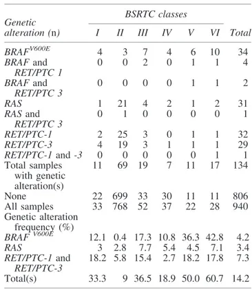
Cytological results and genetic alteration frequencies are displayed according to BSRTC classes in Table 1. Among 940 FNAB, 134 displayed at least one mutation (14.2%), specifically a BRAFV600E mutation in 4.2% of all nodules, RAS mutations in 3.4% (25 at N-RAS codon 61, one at H-RAS codon 13, one at H-RAS codon 61, two at K-RAS codon 12, one at K-RAS codon 13, two at K-RAS codon 61), and RET/PTC rearrangements in 7.3% (3.9% RET/PTC1 and 3.4% RET/PTC3). The highest incidence of RAS mutations was found within BSRTC class III and class VI samples, while the highest incidence of RET/PTC rearrangements was found among BSRTC class I samples (of which about 30% was operated on and had a benign histology) and among BSRTC class III and VI samples (Table 1).
The presence of a BRAFV600E mutation was significantly associated ( p< 0.01) with hypoechogenicity, microcalcifica-tions, and a diameter< 1 cm. RAS mutations were significantly ( p< 0.01) associated with isoechogenicity and a diameter > 1 cm. RET/PTC3 rearrangements were significantly ( p < 0.01) associated with isoechogenicity on US.
Overall, 72 patients underwent TT, and 45 patients un-derwent LT, which was completed in five patients (11.1% of LT), for a total of 117 operated patients. Among these, 62 patients (52.1%) had an indeterminate lesion on cytology: 23 AUS/FLUS (class III), 17 FN (class IV), and 22 SM (class V).
Table1. Genetic Alterations and Their Frequencies in Each Bethesda System for Reporting Thyroid Cytopathology
Class Nodules
BSRTC classes Genetic
alteration (n) I II III IV V VI Total
BRAFV600E 4 3 7 4 6 10 34 BRAF and RET/PTC 1 0 0 2 0 1 1 4 BRAF and RET/PTC 3 0 0 0 0 1 1 2 RAS 1 21 4 2 1 2 31 RAS and RET/PTC 3 0 1 0 0 0 0 1 RET/PTC-1 2 25 3 0 1 1 32 RET/PTC-3 4 19 3 1 1 1 29 RET/PTC-1 and -3 0 0 0 0 0 1 1 Total samples with genetic alteration(s) 11 69 19 7 11 17 134 None 22 699 33 30 11 11 806 All samples 33 768 52 37 22 28 940 Genetic alteration frequency (%) BRAF V600E 12.1 0.4 17.3 10.8 36.3 42.8 4.2 RAS 3 2.8 7.7 5.4 4.5 7.1 3.4 RET/PTC-1 and RET/PTC-3 18.2 5.8 15.4 2.7 18.2 17.8 7.3 Total(s) 33.3 9 36.5 18.9 50.0 60.7 14.2
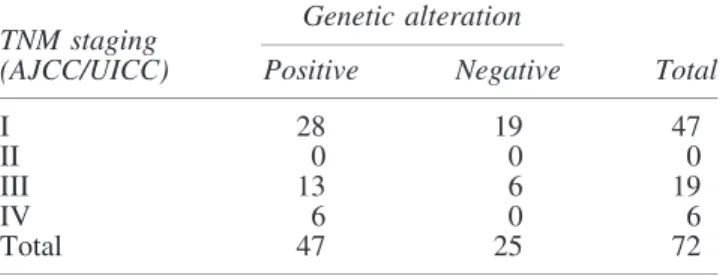
The presence of a cancer was histologically confirmed in 72 patients (61.5% of operated patients), including 70 papillary thyroid cancers (PTC; 96.05%), one follicular thyroid can-cer (FTC), and one anaplastic thyroid cancan-cer (ATC). Among the patients with a final malignant histology, more than half carried one or more somatic genetic alteration and displayed stage I disease (Table 2).
In particular, 40 patients who displayed a somatic BRAFV600E mutation (including six who also displayed a RET/PTC rearrangement) underwent TT and had a PTC on final histology.
Among the 31 patients who displayed an isolated somatic RAS mutation, 10 were submitted to LT and one to TT. His-tology revealed the presence of a cancer in two cases, in-cluding one ATC and one FTC (the latter initially submitted to LT and then to completion thyroidectomy). The remaining nine patients who were operated on showed a follicular ade-noma (FA) in six cases and hyperplastic nodules (HN) in three cases. Moreover, one patient with a malignant cytology, dis-playing a somatic RAS mutation, was not operated on due to several comorbidities. The remaining 19 patients refused surgery, mostly because of the finding of a benign cytology.
The presence of a RET/PTC rearrangement was found in 69 FNAB, six of which also harbored a BRAFV600Emutation and were therefore submitted to TT. One patient also carried a RAS mutation and was submitted to LT with final histology of a FA. One patient was to have both RET/PTC rearrange-ments and was submitted to TT with a final histology of PTC. Among the 62 patients displaying an isolated RET/PTC re-arrangement, five underwent TT (in the presence of a BSRTC class V in two patients and class VI in three patients) and 19 underwent LT. Histology revealed the presence of a cancer in five cases (all PTC), while 11 lesions were FA and eight HN. The remaining 38 patients refused surgery, mostly because of the finding of a benign cytology. No correlation was found between the presence of a malignant lesion and the amount of RET/PTC rearranged mRNA, preventing the identification of a threshold value that discriminates benign from malignant lesions.
Indeterminate lesions
We then evaluated cytology, molecular testing, and pa-thology findings in the group of indeterminate nodules, which were included in the whole group described above.
We found that 37 (26.4%) of the 140 cytologically inde-terminate lesions (corresponding to 14.8% of all FNAB), including 19 class III, seven class IV, and 11 class V lesions, displayed at least one genetic alteration. Among these pa-tients, two refused LT (class III cytology) and 35 underwent
TT. Final histology showed 24 thyroid cancers (23 PTC and 1 FTC), eight FA, and three HN. Among the 23 identified PTCs, 21 carried a somatic BRAFV600Emutation.
Among the 103 patients with a cytologically indeterminate lesion not displaying a genetic alteration, all the 11 patients with a class V lesion underwent TT, with a final histology of 10 PTC and one HN. Ten out of 30 patients with class IV lesions agreed to undergo LT, with a final histology of three PTC (then submitted to completion thyroidectomy) and se-ven FA. All 62 patients with a class III lesion underwent a second FNAB that confirmed an indeterminate lesion in 33 cases; six of these patients agreed to undergo LT, and the final histology showed one FTC, four FA, and one HN. Cytology showed a benign lesion in the other 29 patients who were then reclassified as BSRTC class II and subsequently followed with US. The management of these patients was chosen ac-cording to the ATA guidelines (8), in order to avoid unnec-essary surgery in keeping with the low cancer risk of BSRTC class III nodules (in contrast with the higher cancer risk of BSRTC class IV and V nodules).
Taken together, in our series, malignancy rates in each BSRTC class overlap those described by Cibas et al. (14). The cancer risk in thyroid nodules with indeterminate cy-tology according to BSRTC classification and genetic alter-ations is shown in Table 3.
Diagnostic value of cytology and molecular analyses
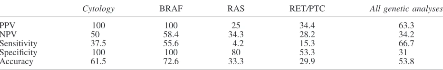
The diagnostic value of cytology and of the studied mutational analyses is reported in Table 4a, which also shows the results obtained by performing the three avail-able genetic analyses in combination. Our data show that cytology displays optimal PPV and specificity, while sen-sitivity for thyroid cancer is low. When performed alone, BRAFV600E analysis shows, as compared to cytology, a significantly higher diagnostic sensitivity ( p< 0.05), which increases by 20.8% ( p< 0.01) when the two evaluations are performed together (Table 4b). On the other hand, the presence of RAS mutations and RET/PTC rearrangements shows a very low sensitivity for thyroid cancer when evaluated alone (Table 4a) and does not significantly in-crease the diagnostic sensitivity of cytology (Table 4b). In addition, the increased sensitivity recorded when all three genetic analyses are performed in combination is not sig-nificantly higher compared to the sensitivity obtained by
Table2. Distribution According to TNM Stages and the Presence/Absence of a Genetic Alteration
Genetic alteration TNM staging
(AJCC/UICC) Positive Negative Total
I 28 19 47
II 0 0 0
III 13 6 19
IV 6 0 6
Total 47 25 72
Table3. Cancer Risk in Thyroid Nodules with Indeterminate Cytology According to BSTRC
Classification and Genetic Alteration
% Class III Class IV Class V
Indeterminate cytology Cytology alone 19.2 21.6 90.9 27.1 Any mutation 47.3 71.4 90.9 63.1 BRAF 100 100 100 100 RAS 0 50 0 14.2 RET/PTC-1 40 — 100* 57.1 RET/PTC-3 0 0 100* 33.3 No mutations 3 10 90.9 13.5
*The patients with a PTC displaying RET/PTC rearrangements also had a BRAFV600Emutation, or a class V or a class VI BSTRC cytology.
performing BRAFV600E analysis alone, even when com-bined with cytology. These data indicate that, in our set-ting, BRAFV600Eanalysis suffices to increase the diagnostic sensitivity of cytology for thyroid cancer.
We then evaluated the diagnostic sensitivity of the genetic analysis panel in the subset of the indeterminate lesions in order to understand whether and which mutation testing might be helpful in this group. We found that the diagnostic sensitivity for thyroid cancer of the three genetic analyses in the indeterminate group, performed alone or in combination, overlaps that identified in the whole group. We then analyzed each BSRTC class included in the indeterminate group (Ta-ble 4c) and found that the diagnostic sensitivity for thyroid cancer reaches 90% in class III when BRAFV600Eanalysis is performed. This value does not change when RAS mutations and RET/PTC rearrangements are simultaneously included. In class IV and V samples, when all three genetic abnor-malities are analyzed in combination, the diagnostic sensi-tivity for cancer is greater compared to BRAFV600Ealone, but the difference is not statistically significant. In addition, the analysis of RAS mutations and RET/PTC rearrangements does not seem to be important to increase further the high NPV of BRAFV600Eanalysis in class III and IV samples.
Discussion
This prospective study confirms the diagnostic utility of assessing the presence of a BRAFV600Emutation (16). On the
other hand, the investigation of two additional genetic ab-normalities (RAS mutations and RET/PTC rearrangements) did not significantly increase the diagnostic sensitivity of cytology toward thyroid cancer in this cohort, even in the category with indeterminate lesions. Despite the fact that the techniques employed in our study are very similar to those employed by others (5,15,18), the results do not overlap. It should be noted that the method employed here to assess RET/PTC rearrangements displayed a 10-fold higher sensi-tivity compared to that employed by Nikiforov et al. (15,18), but provided low sensitivity and specificity in detecting malignant lesions. Therefore, the identification of RET/PTC rearrangements by a very sensitive method may not be useful to increase FNAB diagnostic sensitivity for thyroid cancer. These data suggest that the contribution of this genetic marker to presurgical diagnosis of thyroid nodules may not be so relevant, since we also found a very high incidence of RET/PTC rearrangements in benign lesions.
US characteristics provide the basis of performing FNAB (8), and often accurately predict the presence of a BRAFV600E mutation (20). In our hands, the presence of a BRAFV600E mutation was significantly associated with hypoechogenicity, microcalcifications, and a diameter < 1 cm, strengthening the evidence that nodules displaying these US characteristics very likely reflect the presence of a cancer. Our study high-lights, for the first time, that RAS mutations and RET/PTC rearrangements correlate with specific US findings (i.e., iso-echogenicity and diameter > 1 cm). However, these genetic Table4. (a) Diagnostic Value of Cytology and of Genetic Analyses in All 940 Samples
Cytology BRAF RAS RET/PTC All genetic analyses
PPV 100 100 25 34.4 63.3
NPV 50 58.4 34.3 28.2 34.2
Sensitivity 37.5 55.6 4.2 15.3 66.7
Specificity 100 100 80 53.3 31
Accuracy 61.5 72.6 33.3 29.9 53.8
Table4. (b) Diagnostic Value of Cytology Combined with Genetic Analyses in All 940 Samples Cytology combined with
BRAF RAS RET/PTC All genetic analyses
PPV 100 76.3 61.1 66.7
NPV 72.6 45.6 38.1 51.9
Sensitivity 76.4 40.3 45.8 82.2
Specificity 100 80 53.3 31.8
Accuracy 85.5 55.6 48.7 63.2
Table4. (c) Diagnostic Value of Genetic Analyses in the 140 Indeterminate Lesions According to BSRTC Classification
Class III Class IV Class V
BRAF All genetic analyses BRAF All genetic analyses BRAF All genetic analyses
PPV 100 52.9 100 71.4 100 90.9
NPV 92.9 83.3 69.2 70 14.3 9.1
Sensitivity 90 90 50 62.5 40 50
Specificity 100 38.5 100 77.8 100 50
Accuracy 95.7 60.9 76.5 70.6 45.5 50
abnormalities do not indicate the presence of a cancer with high accuracy in our population, and therefore the re-lated US characteristics cannot be taken into account as predictive of cancer.
The distribution of our samples among BSRTC classes is in line with literature data, indicating that the investigated nodules had been selected according to the indications of the ATA guidelines (8). In particular, > 80% of FNAB cytolo-gies turned out to be a benign lesion, and *12% of the samples displayed an indeterminate cytology. The latter re-sult is very similar to the percentage of indeterminate lesions that were retrieved in our previous study (17), which included an unselected nodule population, indicating that the appli-cation of strict selection criteria for FNAB does not influence the number of indeterminate lesions. While the percentage of malignant lesions identified by cytology in our series (2.9%) is comparable to the literature data, the incidence of ND reports is quite high (3.5%). This may be because the re-trieved FNAB material was used for several diagnostic pro-cedures, which may have reduced the sample quantity dedicated to cytology.
The present series shows that 14.2% of the investigated nodules harbored at least one mutation, a higher incidence than the previously reported (*9%) (18), probably due to the different inclusion criteria. In addition, 6% of mutated FNAB samples displayed more than one genetic alteration, con-firming that BRAF and RAS mutations, as well as RET/PTC rearrangements, are not mutually exclusive, as previously indicated (21). Our data also show that the applied FNAB criteria allowed diagnosing thyroid cancers at an early stage of disease, since 65.3% of the diagnosed cancers were Stage I. In addition, nearly 50% of Stage I cancers had a negative cytology but displayed at least one genetic alteration, most commonly a BRAFV600E mutation, which allowed a cor-rect diagnosis to be established. These data indicate that BRAFV600E mutation analysis helps PTC to be identified at an earlier stage, possibly resulting in a more conservative treatment with potential consequences on patient health and healthcare resources. Moreover, 76% of Stage III and IV cancers displayed a genetic alteration, in line with the hy-pothesis that the latter may characterize a more aggressive behavior (22,23), as previously indicated (24). Last, the ap-plied protocol allowed 31 out of 46 false negative lesions to be diagnosed correctly as cancers, corresponding to 43% of the diagnosed malignant lesions. Among these 31 patients, 21 harbored a BRAFV600E mutation and an indeterminate cy-tology, and were therefore submitted to TT rather than to a diagnostic LT. Moreover, seven patients were submitted to TT only based on positivity for a BRAFV600E mutation and turned out to have a PTC (six Stage I and one Stage III). The latter finding strengthens the evidence that BRAFV600E mu-tation analysis facilitates early diagnosis. On the other hand, in our setting, RAS mutations have a poor diagnostic value, in keeping with their rarity, and are predominantly associated with follicular lesions, mainly represented by FA that may, in part, be considered as precursors of malignant lesions (25). In keeping with the latter hypothesis, RAS mutated cancers were characterized by an aggressive histology and a high disease stage. In our patients, each RET/PTC rearrangement was nearly as frequent as BRAFV600E mutations, but had a poor diagnostic value, since the rearranged lesions were mostly found in benign nodules (64.5% of the cases), contrary to
what was observed by Cantara et al. (5) and Nikiforov et al. (18), but in line with Marotta et al. (26), even if a prognostic significance cannot be ruled out (27). These differences may be due to different genetic backgrounds and to geographic factors, but may also be due to the applied selection crite-ria. Among the samples harboring RET/PTC rearrangements, the 11 PTC cases had a BRAFV600E mutation and/or a sus-picious or malignant cytology, and were therefore submit-ted to TT independently of the presence of a RET/PTC rearrangement.
A previous report (18) showed an increased diagnostic sensitivity for thyroid cancer in a large group of indetermi-nate nodules submitted to multiple genetic analyses (in-cluding BRAFV600Eand RAS mutations as well as RET/PTC1, RET/PTC3, and PAX8/PPARc rearrangements). The study showed a high NPV for this panel of molecular markers, indicating that the absence of a genetic mutation very likely excludes the presence of a malignant lesion. On the contrary, we did not obtain high NPV values in the indeterminate group when performing the three analyses together (BRAFV600Eand RAS mutations, as well as RET/PTC1 and RET/PTC3 re-arrangements), but we found a high NPV for BRAFV600E mutation analysis alone, which is even higher in class III nodules. The latter finding, together with the low cancer risk, suggests that in the absence of a BRAFV600Emutation, diag-nostic LT may not be necessary in class III nodules. In class IV nodules without mutations, we found a slightly higher cancer risk, which importantly increased when a RAS muta-tion was present. These data, together with a suboptimal NPV of BRAFV600Eanalysis in class IV lesions, do not support a conservative management in these settings (i.e., avoiding a LT). On the other hand, cancer risk is high in class V nodules, indicating that an aggressive surgical management (i.e., TT) is justified in these patients, independently of the presence of a mutation, such as in class VI lesions. Taken together, these data demonstrate that, among the investigated molecular markers, only BRAFV600E mutation may modify patient management and has an impact on the surgical approach. Therefore, our data concerning indeterminate lesions are only partially in keeping with previous findings (18), probably due to the different inclusion criteria, which may play an im-portant role in molecular studies.
In conclusion, our results confirm that BRAFV600Eanalysis performed in all BSRTC classes increases the diagnostic sensitivity of cytology for thyroid cancer, which is not further enhanced by investigating the presence of RAS mutations or RET/PTC rearrangements, even among indeterminate nod-ules. In addition, our data demonstrate that BRAFV600E analysis, when negative, may be useful for identifying class III nodules at very low risk of being cancerous, suggesting that these cases may be treated more conservatively and do not need to be submitted to a LT. Moreover, we conclude that BRAFV600E analysis is useful for the diagnosis of thyroid cancer at an early stage, possibly reducing the clinical impact of a delayed diagnosis, which may result in higher costs for the patient and the healthcare system.
Acknowledgments
This work was supported by grants from the Italian Min-istry of Education, Research and University (FIRB RBAP11884M, FIRB RBAP1153LS, 2010TYCL9B_002),
Fondazione Cassa di Risparmio di Ferrara, in collaboration with Laboratorio in rete del Tecnopolo ‘‘Tecnologie delle terapie avanzate’’ (LTTA) of the University of Ferrara.
Author Disclosure Statement
E.d.U. received consulting fees from Novartis and Pfizer. M.C.Z. received consulting fees from Novartis and Gen-zyme. The other authors have nothing to disclose and have no conflict of interest.
References
1. Ferraz C, Eszlinger M, Paschke R 2011 Current state and future perspective of molecular diagnosis of fine-needle aspiration biopsy of thyroid nodules. J Clin Endocrinol Metab 96:2016–2026.
2. Keutgen XM, Filicori F, Fahey TJ 3rd 2013 Molecular diagnosis for indeterminate thyroid nodules on fine needle aspiration: advances and limitations. Expert Rev Mol Diag 13:613–623.
3. Freitas BC, Cerutti JMc 2010 Genetic markers differenti-ating follicular thyroid carcinoma from benign lesions. Mol Cell Endocrinol 321:77–85.
4. Xing M, Haugen BR, Schlumberger M 2013 Progress in molecular-based management of differentiated thyroid cancer. Lancet 381:1058–1069.
5. Cantara S, Capezzone M, Marchisotta S, Capuano S, Bu-sonero G, Toti P, Di Santo A, Caruso G, Carli AF, Brilli L, Montanaro A, Pacini F 2010 Impact of proto-oncogene mutation detection in cytological specimens from thyroid nodules improves the diagnostic accuracy of cytology. J Clin Endocrinol Metab 95:1365–1369.
6. Eszlinger M, Paschke R 2010 Molecular fine-needle aspi-ration biopsy diagnosis of thyroid nodules by tumor specific mutations and gene expression patterns. Mol Cell En-docrinol 322:29–37.
7. Mehta V, Nikiforov YE, Ferris RL 2013 Use of molecular biomarkers in FNA specimens to personalize treatment for thyroid surgery. Head Neck 35:1499–1506.
8. Cooper DS, Doherty GM, Haugen BR, Kloos RT, Lee SL, Mandel SJ, Mazzaferri EL, McIver B, Pacini F, Schlumberger M, Sherman SI, Steward DL, Tuttle RM 2009 The American Thyroid Association (ATA) Guide-lines Taskforce on Thyroid Nodules and Differentiated Thyroid Cancer. Revised American Thyroid Association management guidelines for patients with thyroid nod-ules and differentiated thyroid cancer. Thyroid 19:1167– 1214.
9. Durante C, Costante G, Filetti S 2013 Differentiated thy-roid carcinoma: defining new paradigms for postoperative management. Endocr Relat Cancer 20:R141–R154. 10. Tuttle RM, Sabra MM 2013 Selective use of RAI for
ab-lation and adjuvant therapy after total thyroidectomy for differentiated thyroid cancer: a practical approach to clin-ical decision making. Oral Oncol 49:676–683.
11. Durante C, Attard M, Torlontano M, Ronga G, Monzani F, Costante G, Ferdeghini M, Tumino S, Meringolo D, Bruno R, De Toma G, Crocetti U, Montesano T, Dardano A, Lamartina L, Maniglia A, Giacomelli L, Filetti S; Papillary Thyroid Cancer Study Group 2010 Identification and op-timal postsurgical follow-up of patients with very low-risk papillary thyroid microcarcinomas. J Clin Endocrinol Me-tab 95:4882–4888.
12. Roti E, degli Uberti EC, Bondanelli M, Braverman LE 2008 Thyroid papillary microcarcinoma: a descriptive and meta-analysis study. Eur J Endocrinol 159:659–673. 13. Batawil N, Alkordy T 2014 Ultrasonographic features
as-sociated with malignancy in cytologically indeterminate thyroid nodules. Eur J Surg Oncol 40:182–186.
14. Cibas ES, Ali SZ 2009 The Bethesda System for Reporting Thyroid Cytopathology. Thyroid 19:1159–1165.
15. Nikiforov YE, Steward DL, Robinson-Smith TM, Haugen BR, Klopper JP, Zhu Z, Fagin JA, Falciglia M, Weber K, Nikiforova MN 2009 Molecular testing for mutations in improving the fine-needle aspiration diagnosis of thyroid nodules. J Clin Endocrinol Metab 94:2092–2098.
16. Zatelli MC, Trasforini G, Leoni S, Frigato G, Buratto M, Tagliati F, Rossi R, Cavazzini L, Roti E, degli Uberti EC 2009 BRAF V600E mutation analysis increases diagnostic accuracy for papillary thyroid carcinoma in fine-needle aspiration biopsies. Eur J Endocrinol 161:467–473. 17. Rossi M, Buratto M, Bruni S, Filieri C, Tagliati F,
Tras-forini G, Rossi R, Beccati MD, degli Uberti EC, Zatelli MC 2012 Role of ultrasonographic/clinical profile, cytology, and BRAF V600E mutation evaluation in thyroid nodule screening for malignancy: a prospective study. J Clin En-docrinol Metab 97:2354–2361.
18. Nikiforov YE, Ohori NP, Hodak SP, Carty SE, LeBeau SO, Ferris RL, Yip L, Seethala RR, Tublin ME, Stang MT, Coyne C, Johnson JT, Stewart AF, Nikiforova MN 2011 Impact of mutational testing on the diagnosis and man-agement of patients with cytologically indeterminate thy-roid nodules: a prospective analysis of 1056 FNA samples. J Clin Endocrinol Metab 96:3390–3397.
19. Guerra A, Carrano M, Angrisani E, Puzziello A, Izzo G, Di Crescenzo V, Vatrella A, Vitale M 2014 Detection of RAS mutation by pyrosequencing in thyroid cytology samples. Int J Surg 12:S91–94.
20. Kabaker AS, Tublin ME, Nikiforov YE, Armstrong MJ, Hodak SP, Stang MT, McCoy KL, Carty SE, Yip L 2012 Suspicious ultrasound characteristics predict BRAFV600E-positive papillary thyroid carcinoma. Thyroid 22:585–589. 21. Guerra A, Zeppa P, Bifulco M, Vitale M 2014 Concomitant BRAF(V600E) mutation and RET/PTC rearrangement is a frequent occurrence in papillary thyroid carcinoma. Thy-roid 24:254–259.
22. Fugazzola L, Mannavola D, Cirello V, Vannucchi G, Muzza M, Vicentini L, Beck-Peccoz P 2004 BRAF muta-tions in an Italian cohort of thyroid cancers. Clin En-docrinol (Oxf) 61:239–243.
23. Xing M, Alzahrani AS, Carson KA, Viola D, Elisei R, Bendlova B, Yip L, Mian C, Vianello F, Tuttle M, Ro-benshtok E, Fagin JA, Puxeddu E, Fugazzola L, Czarniecka A, Jarzab B, O’Neill CJ, Sywak MS, Lam K, Riesco-Ei-zaguirre G, Santisteban P, Nakayama H, Tufano RP, Pai SI, Zeiger MA, Westra WH, Clark DP, Clifton-Bligh R, Si-dransky D, Ladenson PW, Sykorova V 2013 Association between BRAF V600E mutation and mortality in patients with papillary thyroid cancer. JAMA 309:1493–1501. 24. Xing M, Clark D, Guan H, Ji M, Dackiw A, Carson KA,
Kim M, Tufaro A, Ladenson P, Zeiger M, Tufano R 2009 BRAF mutation testing of thyroid fine-needle aspiration biopsy specimens for preoperative risk stratification in papillary thyroid cancer. J Clin Oncol 27:2977–2982. 25. Schulten HJ, Salama S, Al-Ahmadi A, Al-Mansouri Z,
Mirza Z, Al-Ghamdi K, Al-Hamour OA, Huwait E, Gari M, Al-Qahtani MH, Al-Maghrabi J 2013 Comprehensive
sur-vey of HRAS, KRAS, and NRAS mutations in proliferative thyroid lesions from an ethnically diverse population. An-ticancer Res 33:4779–4784.
26. Marotta V, Guerra A, Sapio MR, Vitale M 2011 RET/PTC rearrangement in benign and malignant thyroid diseases: a clinical standpoint. Eur J Endocrinol 165:499–507. 27. Sapio MR, Guerra A, Marotta V, Campanile E, Formisano
R, Deandrea M, Motta M, Limone PP, Fenzi G, Rossi G, Vitale M 2011 High growth rate of benign thyroid nodules bearing RET/PTC rearrangements. J Clin Endocrinol Me-tab 96:E916–919.
Address correspondence to: Maria Chiara Zatelli, MD, PhD Section of Endocrinology and Internal Medicine Department of Medical Sciences University of Ferrara Via Savonarola 9 44121 Ferrara Italy E-mail: [email protected]