Case report
Lateralized periodic discharges in insular status epilepticus: A case
report of a periodic EEG pattern associated with ictal manifestation
Fedele Dono
a,⇑, Mirella Russo
a, Claudia Carrarini
a, Vincenzo Di Stefano
a, Stefania Nanni
b,
Camilla Ferrante
a, Marco Onofrj
a, Francesca Anzellotti
ba
Department of Neuroscience, Imaging and Clinical Sciences, University G. d’Annunzio of Chieti-Pescara, Italy
b
Department of Neurology, SS Annunziata Hospital, Chieti, Italy
a r t i c l e i n f o
Article history: Received 5 October 2018
Received in revised form 5 January 2019 Accepted 14 January 2019
Available online 24 January 2019 Keywords:
Focal status epilepticus Insular epilepsy LPDs + F Brain tumuor
a b s t r a c t
Objective: Insular lobe seizures generally represent a misconceived ictal phenomenon characterized by specific neurological signs. Aphasia can be a rare presenting sign associated with insular lobe epilepsy which could be easily mistaken for a manifestation of other acute brain diseases.
Method: We describe an insular status epilepticus (SE) characterized by sudden onset of language distur-bance associated with hypersalivation and paraesthesia. A concomitant EEG recording showed the pres-ence of Lateralized Periodic Discharges plus superimposed fast activity (LPDs + F). After an adequate acute endovenous anti-seizure treatment, a normalization of the EEG abnormalities with a complete res-olution of all the neurological symptoms was achieved.
Discussion: Language disturbances can be usually found in various pathological acute pictures involving the dominant frontal and temporal lobes. The presence of certain EEG pattern, could rise the suspect of aphasia as a critical manifestation. LPDs pattern is usually correlated with structural lesions. The associ-ation between LPDs and seizure is controversial but it seems to be more consistent when they are asso-ciated with ‘‘Plus modifiers” and with an high periodic frequency.
Conclusion: Our case underlines the importance of considering focal SE in the differential diagnosis of patients presenting aphasia, even in the absence of previous history of epilepsy. We describe how LPDs can be associated with SE in a patient affected by a brain tumour, supporting the idea that some characteristic periodic patterns could be associated with seizure occurrence.
Ó 2019 International Federation of Clinical Neurophysiology. Published by Elsevier B.V. This is an open access article under the CC BY-NC-ND license (http://creativecommons.org/licenses/by-nc-nd/4.0/).
1. Introduction
Insular lobe seizures (ILSs) generally represent a misconceived ictal phenomenon. Ictal sequence occurs in full consciousness, beginning with sensation of laryngeal constriction and paraesthe-sia affecting large cutaneous territories eventually followed by dysarthria and focal motor convulsive symptoms (Ryvlin, 2006). Aphasia is rarely described as accompanying sign of ILSs.Ryvlin (2006)divided insular epilepsy into three subtypes: (1) Perisylvian form, with somatosensitive symptoms (2) Frontal form, with hypermotor manifestations (3) Temporal form, with epigastric ascending sensation associated with anxiety, simple auditory hal-lucinations, laryngeal constriction, apnoea, tonic postures, eyes and head versive movements. This is usually a drug-resistant epi-lepsy that is challenging to treat with surgical procedures, due to
the localization of the epileptic focus and proximity to highly elo-quent brain structure.
2. Case study
A 66 year-old right-handed man with no familiar history of epi-lepsy was admitted to our Emergency Unit because of sudden onset of language disturbance associated with hypersalivation and paraesthesia at the right forehand about 20 min before the arrival. The anamnestic record showed the presence of similar brief episodes in the previous days, sometimes associated with a sense of laryngeal constriction. During the neurological examination, the patient was restless and showed a fluent aphasia with an incongruous and meaningless speech and the inability to compre-hend simple instructions. To best define the severity of aphasia, Aphasia Rapid Test (ART) (Azuar et al., 2013) was performed result-ing in total score of 23/26 points. With exception of paraesthesia at the right forehand, there were no other sensory-motor deficits, and examination of cranial nerves was normal. Since the patient’s med-https://doi.org/10.1016/j.cnp.2019.01.002
2467-981X/Ó 2019 International Federation of Clinical Neurophysiology. Published by Elsevier B.V.
This is an open access article under the CC BY-NC-ND license (http://creativecommons.org/licenses/by-nc-nd/4.0/).
⇑ Corresponding author at: Via dei Vestini 5, 66013 Chieti, Italy.
E-mail addresses: [email protected] (F. Dono), [email protected] (C. Carrarini),[email protected](M. Onofrj),[email protected](F. Anzellotti).
Clinical Neurophysiology Practice 4 (2019) 27–29
Contents lists available atScienceDirect
Clinical Neurophysiology Practice
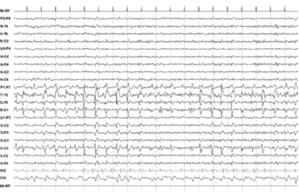
ical history included diabetes and hypercholesterolemia, in order to rule out any cerebrovascular accident, a brain Computed Tomog-raphy (CT) scan was performed that showed an hypodense lesion within the insular-temporal region of the left hemisphere. A 21 derivations EEG recording was then performed which showed mid-temporal continuous spike-wave complexes at 1.5–2 Hz and 100–150 uV of amplitude with phase opposition on T3 with imposed fast activity (Lateralized Periodic Discharges plus super-imposed fast activity, LPDs + F pattern), (Fig. 1). A concomitant ECG performed during the EEG recording showed several asystoles with a maximum pause of 2 s. The patient was then treated with a bolus of Lorazepam 10 mg and underwent to continuous EEG mon-itoring. Over the next 30 min there was a complete normalization of the EEG abnormalities with a resolution of all the neurological symptoms including aphasia (ART = 0/26). A Magnetic Resonance Imaging (MRI) of the brain, performed after the recovery, showed a large neoplastic lesion interesting the left temporo-insular area (Fig. 2). After the resolution of SE, an anti-seizure therapy was introduced. A first attempt to control the symptoms through the combination of Levetiracetam 1500 mg twice per day (serum level 12
l
g/ml) and Valproic Acid 800 mg twice per day (serum level 65.9l
g/ml) was unsuccessful because aphasic seizures were fre-quent in the first month after status. The final therapeutic setting, with an evident reduction of ictal events documented in a four weeks of follow-up visit, consisted of the association of Eslicar-bazepine 800 mg per day with Levetiracetam 1000 mg twice per day; patient referred only two aphasic seizures. Eslicarbazepine serum level measuring test was not available. The patient was then transferred to Neurosurgery Unit and after few days a stereotactic biopsy was performed revealing a WHO II grade glial neoplasia. Hence, the patient underwent neurosurgery and started chemotherapy with Temozolomide but unfortunately died three months later due to pulmonary embolism.3. Discussion
Language disturbances can be usually found in various patho-logical acute pictures involving the dominant frontal and temporal
lobes. In the present report, aphasia was the main manifestation of the focal status epilepticus (SE) in association with other symp-toms related to insular involvement. Routine EEG was helpful in demonstrating the epileptic origin of the symptoms though the hidden distant location of insular lobe cortex compared with the localization of the scalp electrodes. According to recent evidences (Isnard et al., 2000, 2004) the direct stimulation of the insular lobe induces some characteristic symptoms that can appear in individ-uals with insular epilepsy. In particular, sense of laryngeal con-striction, paraesthesia, dysarthria, auditory hallucinations, somatosensory auras, autonomic nervous system symptoms, hypermotor crisis and retrosternal pain could represent the leading signs of insular lobe epilepsy. Even though a stereotactic EEG was not performed, the presence of mid-temporal discharge in the EEG, MRI findings and the characteristic clinical picture highlighted the insular lobe origin of the epileptic discharge. The present case report stresses the electro-clinical correlation between insular sta-tus epilepticus and Lateralized Periodic Discharges. LPDs pattern is
Fig. 1. EEG findings during the ictal phase. Shows periodic spike-wave complexes at 1.5–2 Hz and 100–150 uV of amplitude with phase opposition on T3 mixed with poly-spikes, described as Lateralized Periodic Discharges plus superimposed fast activity (LPDs + F) on the left fronto-temporal derivations.
Fig. 2. Magnetic Resonance Imaging (MRI) of the brain performed after SE recovery. In (A) Axial Fluid Attenuated Inversion Recovery (FLAIR) sequence and in (B) T2-weightened sequences showing a large neoplastic lesion involving the left temporo-insular area.
usually correlated with structural lesions of cortical or subcortical areas due to some pathological conditions such as acute stroke, brain tumours, infections, traumas and metabolic diseases (Chong and Hirsch, 2005). The origin of LPDs is a controversial issue and only a few existing neurophysiological hypotheses address causes and circumstances of LPDs onset and if they repre-sent an ictal or inter-ictal pattern. It has been recently reported that the association between LPDs and seizure is more consistent in the presence of particular LPDs features with an increased sei-zure risk with higher periodic discharges frequency and ‘‘Plus mod-ifier” such as superimposed fast activity (Rodriguez Ruiz et al., 2017). In previous literature, different cases of focal status epilep-ticus characterized by the acute onset of aphasia were described. In those contexts, routine EEG were not always conclusive for SE (Flügel et al., 2015) even though itctal EEG patterns have been reported (Patil and Oware, 2012). LPDs have been sometimes described but they have never been marked as ictal pattern even though, in some cases, a clear electro-clinical correlation was described with patient’s good clinical response to the anti-seizure therapy (Quintas et al., 2018). In the largest clinical records (Ericson et al., 2011) nine patients affected by aphasic status epilepticus have been described of whom LPDs were reported in five. In each cases, LPDs were interrupted by evolving electro-graphic seizure patterns and persisted after the seizure patters had ceased and as the patients began to improve clinically. For those reasons, authors considered LPDs, including those with plus modifying features, as interictal abnormalities. However, authors did not focus on LPDs features and it is not clear if an evolution in frequency or morphology was presented at the time of anti-seizure therapy administration. In our case, the evident electro-clinical correlation and the unequivocal response to anti-seizure drug, according to Salzburg Criteria for non-convulsive status epilepticus (NCSE) (Leitinger et al., 2015), led us to consider LPDs as ictal pattern. The choice of introducing Eslicarbazepine in a patient with a recent diagnosis of cerebral tumour has been made in consideration of the specific anti-neoplastic treatment with Temozolomide. As reported in previous literature, Eslicarbazepine has shown a reduced drug-to-drug interaction through a minimal cytochrome induction power (Bialer and Soares-da-Silva, 2012) 4. Conclusion
Our case underlines the importance of considering focal SE in the differential diagnosis of patients presenting aphasia, even in the absence of previous history of epilepsy. Aphasia should not necessarily be linked to acute cerebrovascular events, even if in association with other focal neurological signs. In addition, we
describe how LPDs can be associated with SE in a patient affected by a brain tumour, supporting the idea that some characteristic periodic patterns could be considered ictal ones. In conclusion, the fluctuating feature and its correlation with a suggestive EEG pattern, should raise suspicion that aphasia might be an ictal man-ifestation rather than merely a focal sign due to a cerebral lesion. A correct interpretation either the clinical picture either the EEG pat-tern is important in order to warrant an adequate anti-seizure therapy.
Funding
This research did not receive any specific grant from funding agencies in the public, commercial, or not-for-profit sector. Conflict of interest
The authors affirm that they do not have conflicts of interest.
References
Azuar, C., Leger, A., Arbizu, C., Henry-Amar, F., Chomel-Guillaume, S., Samson, Y., 2013. The aphasia rapid test: an NIHSS-like aphasia test. J. Neurol. 260, 2110– 2117.
Bialer, M., Soares-da-Silva, P., 2012. Pharmacokinetics and drug interactions of eslicarbazepine acetate. Epilepsia 53, 935–946.
Chong, D.J., Hirsch, L.J., 2005. Which EEG patterns warrant treatment in the critically ill? Reviewing the evidence for treatment of periodic epileptiform discharges and related patterns. J. Clin. Neurophysiol. 22, 79–91.
Ericson, E.J., Gerard, E.E., Macken, M.P., Schuele, S.U., 2011. Aphasic status epilepticus: electroclinical correlation. Epilepsia 52, 1452–1458.
Flügel, D., Kimb, O.C., Felbecker, A., Tettenborn, B., 2015. De novo status epilepticus with isolated aphasia. Epilepsy Behav. 49, 198–202.
Isnard, J., Guénot, M., Ostrowsky, K., Sindou, M., Mauguière, F., 2000. The role of the insular cortex in temporal lobe epilepsy. Ann Neurol. 48, 614–623.
Isnard, J., Guénot, M., Sindou, M., Mauguière, F., 2004. Clinical manifestations of insular lobe seizures: a stereo-electroencephalographic study. Epilepsia 45, 1079–1090.
Leitinger, M., Beniczky, S., Rohracher, A., Gardella, E., Kalss, G., Qerama, E., et al., 2015. Salzburg consensus criteria for non-convulsive status epilepticus – approach to clinical application. Epilepsy Behav. 49, 158–163.
Patil, B., Oware, A., 2012. De-novo simple partial status epilepticus presenting as Wernicke’s afasia. Seizure 21, 219–222.
Quintas, S., Ródriguez-Carrillo, J.C., Toledano, R., de Toledo, M., Navacerrada Barrero, F.J., Berbís, M.Á., et al., 2018. When aphasia is due to aphasic status epilepticus: a diagnostic challenge. Neurol. Sci. 39, 757–760.
Rodriguez Ruiz, A., Vlachy, J., Lee, J.W., Gilmore, E.J., Ayer, T., Haider, H.A., Gaspard, N., Ehrenberg, J.A., Tolchin, B., Fantaneanu, T.A., Fernandez, A., Hirsch, L.J., LaRoche, S., 2017. Critical care EEG monitoring research consortium. Association of periodic and rhythmic electroencephalographic patterns with seizures in critically ill patients. JAMA Neurol. 74, 181–188.
Ryvlin, P., 2006. Avoid falling into the depths of the insular trap. Epileptic Disord. 8 (Suppl 2), 37–56.
