Cellular factors that interact with acetylated
integrase: new insights in HIV-1 integration
Awatef Allouch
PhD Thesis in Molecular Biology 2011
Supervisor: Prof. Anna Cereseto
Abstract
AIMS OF THE THESIS 1
INTRODUCTION 3
HIV-1 replication 3
1- HIV-1 genome and structure 3
2- HIV-1 replication cycle 5
2.1- Cellular entry 5
2.2- Reverse transcription 6
2.3- Nuclear import 7
2.4- Integration 8
2.5- Gene expression 8
2.6- Assembly and release 9
2.7- Roles of accessory proteins in HIV-1 replication 10
3- HIV-1 integration 12
3.1- Integration steps 12
3.2- Post-integration repair 14
3.3- The un-integrated viral cDNA forms 15
3.4- Integration site selection 16
4- HIV-1 integrase structure and functions 17
4.1- Integrase domains 17
4.2- Integrase structure 20
5- HIV-1 integrase interacting host factors 22
5.1- Lens Epithelium-derived Growth Factor/p75: LEDGF/p75 22
5.2- Histone acetyl transferases: p300 and GCN5 28
5.3- Integrase interactor 1: INI1 32
5.4- Cellular factors binding integrase and involved in HIV-1 nuclear import 33 a-karyophilic properties of integrase and its association with importin factors 33
b- Transportin-SR2 35
c- JNK and Pin1 35
d- NUP153 36
5.5- Gemin2 36
5.6- Human polycomb group EED protein 37
5.7- Heat shock protein 60: HSP60 37
5.8- Uracil DNA glycosylase isoform 2: UNG2 38
5.9- Von Hippel-Lindau binding protein 1: VBP1 38
HIV-1 restriction factors 39
1- TRIM family proteins 39
1.1- TRIM proteins: characteristics and implications in the innate immunity 39
1.2- TRIM5and TRIMCyp 43
1.3- TRIM22 45
2- 2- APOBECs (A3G and A3F)
3- 46
3- Tetherin (CD317/BST-2) 47
4- Factors inhibiting HIV-1 integration and nuclear pre-integration events 48
4.1- p21CIP1/Waf1 48
4.2- RAD52 49
4.3- Rad18 50
4.4- XPB and XPD 50
KAP1 (TRIM28) 51
1- KAP1 structure and functions 51
2- Transcriptional co-repressor activity of KAP1 54
3- Involvement of KAP1 in the deacetylation of non-histone proteins 58
4- Involvement of KAP1 in the DNA damage response 57
5- Involvement of KAP1 in the retroviral inhibition 59
Acetylation and deacetylation of histone and non-histone proteins 60
1- Acetylation mediated by HATs 61
2- Deacetylation mediated by HDACs 64
MATERIALS AND METHODS 67
1- Vectors and constructs 67
2- Yeast assays 68
2.1- Immunoprecipitations from yeast cells 68
2.2- Yeast two hybrid screen 68
4- In vitro binding assays 70 5- Cell lines and purification of primary blood lymphocytes (PBLs), Naïve (CD45RA+)
and Memory (CD45RO+) CD4+ T cells 71
6- Anti-Flag beads pull down assays 71
7- Co-immunoprecipitation experiments 72
8- Western blot analysis and antibodies 73
9- In vitro HDAC assay 74
10- Virus and vector productions 74
11- Transient and stable knockdowns and back-complementation experiments 74 12- Cell infections and measurement of HIV-1 infectivity 76 13- Quantifications of retroviral cDNA species by real time quantitative PCR (Q-PCR)
and their statistical analysis 77
14- HIV-1 LTR transcription assays in KAP1 knockdown HeLa and J-lat A1 cells 78
RESULTS 79
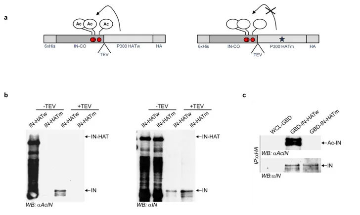
1- Production of constitutive acetylated integrase fused to the HAT domain of p300
79
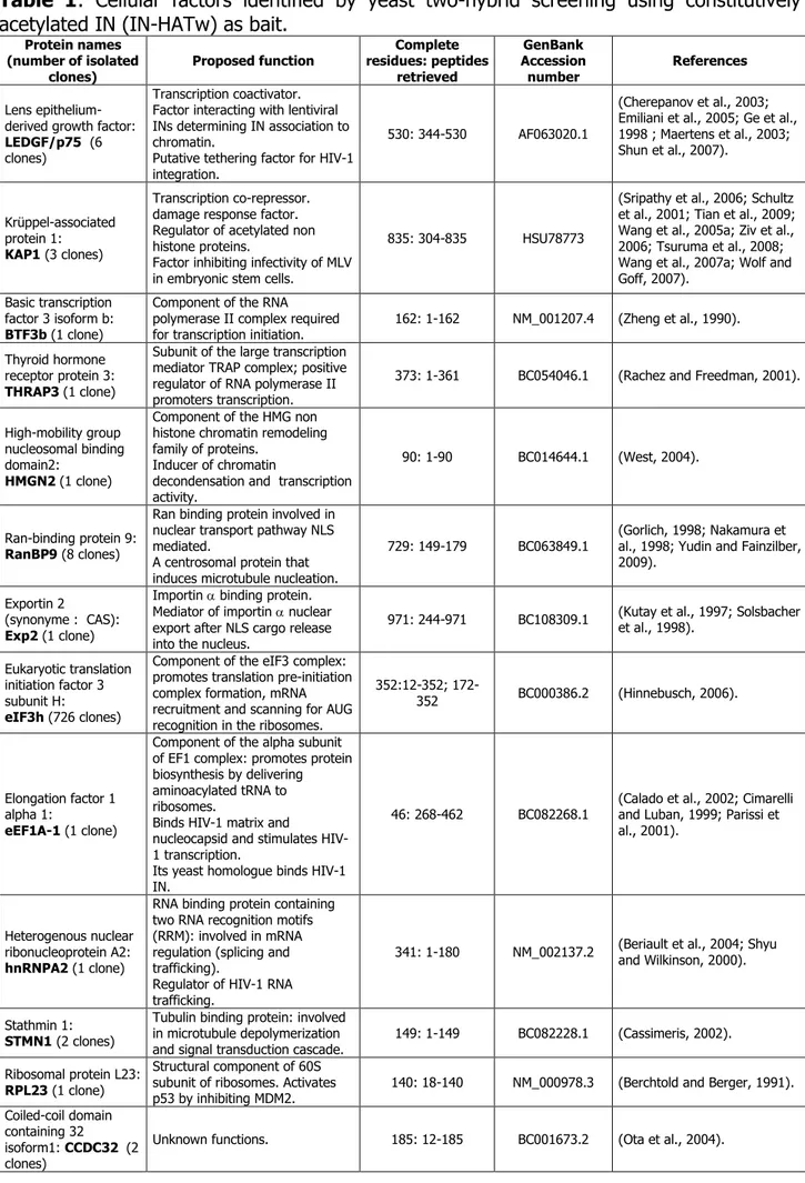
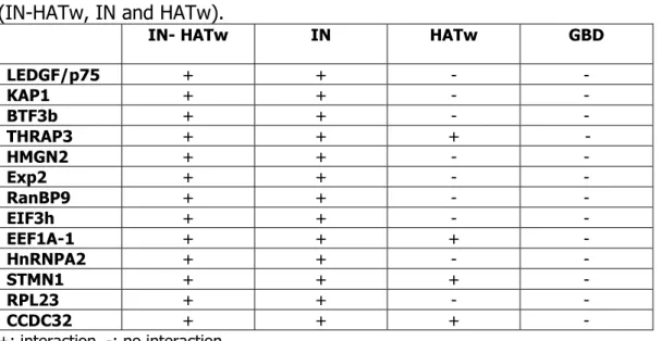
2- Two-hybrid screening analysis using the constitutively acetylated integrase 83 3- Analysis of binding interactions between two-hybrid factors and acetylated or un-
modified integrase 86
4- Interaction between KAP1 and acetylated integrase 88
5-Transient knockdown of KAP1 increases HIV-1 infectivity and integration 93 6- KAP1 stable knockdown increases HIV-1 infectivity and integration 96 7- KAP1 over-expression decreases HIV-1 infectivity and integration 99
8- KAP1 does not affect HIV-1 transcription 100
9- Human KAP1 does not inhibit integration of murine leukemia virus (MLV) 103
11- KAP1 mediates HDAC1 binding to integrase 105 12- KAP1 inhibits HIV-1 integration by targeting acetylated lysines of integrase
through HDAC1 108
13- Validation of KAP1 inhibition of HIV-1 integration in natural HIV-1 target T cells:
CEMss and primary blood lymphocytes (PBLs) 111
14- Determination of the region of KAP1 required for the inhibition of HIV-1
integration 113
15- Expressions of KAP1 and HDAC1 in cells relevant in HIV-1 pathogenesis 116 16- Could KAP1 phosphorylation during HIV-1 infection be a way for the virus to
escape KAP1 inhibition? 118
DISCUSSION 123
1- Potential roles in HIV-1 replication of the identified host factors binding
acetylated integrase 123
2- KAP1 targets acetylated integrase and inhibits HIV-1 integration through HDAC1
126
3- KAP1 does not affect HIV-1 gene expression and likely is not involved in viral
latency 130
4- KAP1 as a restriction factor for HIV-1 infectivity 130
5- Models for KAP1 inhibition of HIV-1 integration 131
6- Could HIV-1 escape from KAP1 inhibition through the induction of KAP1
phosphorylation? 132
CONCLUSIONS AND FUTURE PERSPECTIVES 135
References 137
Publications 169
Abstract
The viral protein integrase (IN) catalyzes the integration of the HIV-1 cDNA into the host cellular genome. We have recently demonstrated that IN is acetylated by a cellular histone acetyltransferase, p300, which modifies three lysines located in the C-terminus of the viral factor (Cereseto et al., 2005). This modification enhances the catalytic activity and the DNA affinity of IN, as demonstrated by in vitro assays (Cereseto et al., 2005). Consistently, mutations introduced in the targeted lysines greatly decrease the efficiency of HIV-1 integration (Cereseto et al., 2005; Terreni et al., 2010; Apolonia et al., 2007). Acetylation was proven to regulate protein functions by modulating protein-protein interactions. HIV-1 to efficiently complete its replication steps, including the integration reaction, requires the interaction with numerous cellular factors. Therefore, we sought to investigate whether acetylation might modulate the interaction between IN and cellular factors. To this aim we performed a yeast two-hybrid screening that differs from the screenings so far performed for using as bait IN constitutively acetylated. From this analysis we have identified thirteen cellular factors (seven are nuclear and four are cytoplasmic) involved in transcription, chromatin remodeling, nuclear transport, RNA binding, protein synthesis regulation and microtubule organization. Binding assays showed that acetylation increases the association of IN with four identified factors (KAP1 (TRIM28), Exp2, eIF3h and RanBP9), while for three two-hybrid hits (BTF3b, THRAP3 and HMGN2), the acetylation does not modulate their binding with IN. KAP1, which belongs to the TRIM family of antiviral proteins, was investigated for its role in HIV-1 infection and for the determination of the molecular mechanisms that underlie its interaction with IN. We found that KAP1 binds preferentially acetylated IN and induces its deacetylation through the formation of a protein complex including the deacetylase HDAC1. Modulation of intracellular KAP1 levels in different cell types including T-cells, the primary HIV-1 target, revealed that KAP1 curtails viral infectivity by selectively affecting HIV-1 integration. KAP1-dependent viral inhibition was found to require HDAC1 activity since cells deficient for this deacetylase were insensitive to KAP1-induced resistance. This study reveals that KAP1 is a novel cellular factor restricting HIV-1 infection and underscores the relevance of IN acetylation as a crucial step in the viral infectious cycle.
1
AIMS OF THE THESIS
Human immunodeficiency virus type 1 (HIV-1) is a retrovirus that belongs to the lentivirus genus (Coffin et al., 1997). HIV-1 infects mainly CD4+ human T lymphocytes and causes the acquired immunodeficiency syndrome (AIDS) (Barre-Sinoussi et al., 1983; Popovic et al., 1984). AIDS is a human infectious disease that causes a progressive profound state of immune suppression characterized by a drop of the circulating CD4+ T lymphocyte number and leaves infected individuals susceptible to opportunistic infections and prone to develop tumors (Barre-Sinoussi et al., 1983; Popovic et al., 1984).
An essential step in the retroviral life cycle is the integration of the viral DNA into the host cellular genome; a reaction catalyzed by the viral protein integrase (IN) (Coffin et al., 1997). Purified HIV-1 integrase displays 3‘ end processing and DNA strand transfer activities that are sufficient to catalyze the cDNA integration reaction in vitro (Lewinski and Bushman, 2005; Vandegraaff and Engelman, 2007). However, in vivo numerous cellular proteins are required for efficient integration. The host factors regulate integrase enzymatic functions by modulating its stability and by mediating nuclear import and access to specific regions of the chromatin (Busschots et al., 2009). We have recently demonstrated that p300, a histone acetyl transferase, binds integrase and acetylates three lysines (K264, K266, K273) located in its C-terminus leading to enhanced integrase activity and DNA binding affinity (Cereseto et al., 2005). Acetylable lysines are necessary for virus integration and thus for optimal replication, as demonstrated by the inefficient infectivity observed following their mutations into arginine residues (K264, 266, 273R) (Apolonia et al., 2007; Cereseto et al., 2005; Terreni et al., 2010). Since it has been demonstrated that acetylation modulates the activities of cellular and viral proteins by affecting protein-protein interactions (Bannister et al., 2000; Berro et al., 2006; Bres et al., 2002; Das and Kundu, 2005; Dorr et al., 2002; Glozak et al., 2005; Kouzarides, 2000; Mujtaba et al., 2002; Mujtaba et al., 2004; Polesskaya and Harel-Bellan, 2001; Spange et al., 2009; Sterner and Berger, 2000), the aim of this study is to investigate whether integrase acetylation could affect its interaction with cellular factors and to reveal the possible functional role of these interactions in the HIV-1 replication cycle.
2
To reach this aim, four research steps were involved:
1- The identification of cellular proteins binding acetylated integrase by employing the tethered catalysis two-hybrid system in yeast, a method previously reported to efficiently identify factors binding specifically to acetylated proteins (p53, histones H3 and H4) (Acharya et al., 2005; Guo et al., 2004).
2- The validation of the interactions between acetylated integrase and the candidate cellular factors, identified from the yeast two-hybrid screening, by in vitro pull down assays and in vivo co-immunoprecipitation experiments.
3- The study of one identified candidate (KAP1 or TRIM28) through the investigation of its role in HIV-1 replication and specifically at the level of integration.
4- The search of the molecular mechanisms that underlie the role of KAP1 in HIV-1 infection.
Identification and study of novel cellular proteins interacting with HIV-1 acetylated integrase would provide more knowledge on host factors that promote or inhibit HIV-1 infection. Such information might be useful for alternative HIV-1 therapies based on the inhibition of viral and cellular protein-protein interactions when the interactions enhance HIV-1 replication or on the stimulation of the viral and cellular protein associations when the interactions impair HIV-1 infectivity (Busschots et al., 2009). In fact, the current highly active antiretroviral therapy (HAART) for HIV-1 that is based on the combination of effective drugs that inhibit specifically the viral proteins (reverse transcriptase, protease and recently integrase) resulted in the emergence of HIV-1 resistant strains. This resistance is due to the virus short replication cycle and to the high error rate of its reverse transcriptase that allow the virus to mutate very quickly (Busschots et al., 2009). Therefore, targeting interactions between cellular and viral proteins is a novel therapy strategy that might decrease the risk of HIV-1 drug resistance induced by targeting directly viral proteins (Busschots et al., 2009; Christ et al., 2010).
3
INTRODUCTION HIV-1 replication
HIV-1 was discovered on 1983 by Montagnier‘s group, two years after the first description of AIDS. In this study, HIV-1 was called LAV (lymphadenopathy associated virus) and was described to be involved in several infectious syndrome including AIDS (Barre-Sinoussi et al., 1983). In 1984, Gallo‘s group gave also evidence that HIV-1 is the causal agent of AIDS; HIV-1 was termed as HTLVIII (Popovic et al., 1984). In 1985 came the cloning and sequencing of HIV-1 genome with identification of new open reading frames specific for lentiviruses (Ratner et al., 1985; Sanchez-Pescador et al., 1985; Wain-Hobson et al., 1985). In 1986, a second retrovirus strain, known as HIV-2 and having 40-60% homology with HIV-1, was isolated from West African patients with AIDS (Clavel et al., 1986). The majority of the studies reported that HIV-2 is less pathogenic than HIV-1 and causes AIDS only in the minority of infected individuals; HIV-2 infection is mainly asymptomatic (de Silva et al., 2008).
1- HIV-1 genome and structure
HIV-1 is a human retrovirus that is related most closely to other animal lentiviruses (Coffin et al., 1997). Lentiviruses are ―complex‖ retroviruses that encode a number of regulatory and accessory proteins not encoded by the genome of prototypical ―simple‖ retrovirus (Coffin et al., 1997).
The HIV-1 genome is approximately 9 Kb in length and encodes 15 distinct proteins (Figure 1) (Frankel and Young, 1998). From the 5‘- to 3‘- ends of the genome are found the gag (for group-specific antigen), pol (for polymerase) and env (for envelope glycoprotein) genes. The gag gene encodes a polyprotein precursor, pr55Gag that is
cleaved by the viral protease (PR) to produce Gag proteins matrix (also known as MA or p17), capsid (CA or p24), nucleocapsid (NC or p7) and p6. Two spacer peptides p2 and p1 are also generated upon pr55Gag processing. The pol gene encodes HIV-1 enzymes,
protease (PR), reverse transcriptase (RT), integrase (IN) which are cleaved from pr160GagPol precursor by the viral protease (Frankel and Young, 1998). The envelope (Env) glycoprotein is also synthesized as a polyprotein precursor (Figure 1). Unlike the Gag and Pol precursors which are cleaved by the viral protease the Env precursor, known as gp160, is processed by a cellular protease (furin or furin like enzyme) during Env trafficking to the
4
cell surface. gp160 processing results in the generation of the surface (SU) Env glycoprotein gp120 and the trans-membrane (TM) glycoprotein gp41 (Freed, 2001). gp120 contains the determinants that interact with target cell receptor and co-receptors, while, gp41 anchors the gp120/gp41 complex in the membrane and also contains domains that are critical for catalyzing the membrane fusion reaction between viral and host lipid bilayers during virus entry (Freed, 2001). Comparison of env sequences from large number of virus isolates revealed that gp120 is organized into five conserved regions (C1-C5) and five highly variable domains (V1-V5). gp41 is composed of three major domains: the ectodomain (which contains determinants essential for membrane fusion), the trans-membrane anchor sequence and the cytoplasmic tail (Freed, 2001).
Figure 1: . Organization of the HIV-1 genome. The relative locations of the HIV-1 open reading frames
gag, pol, env, vif, vpr, vpu, nef, tat, and rev are indicated. The 5' and 3' long terminal repeats (LTRs) are shown, with U3, R, and U5 regions noted. The indicates the position of the RNA packaging signal. The major Gag domains (MA, CA, NC, p6) and the Gag spacer peptides (p2 and p1) are shown under the gag
gene. The site of Gag N-terminal myristylation is denoted as ―myr‖. Under the pol gene are indicated the PR, RT (p66 and p51 sub-domains), and IN coding regions. The SU and TM Env glycoproteins (gp120 and gp41, respectively) are enlarged to show the position in gp120 of the major conserved (C1-C5) and variable (V1-V5) regions and in gp41 the location of the fusion peptide, the N- and C-helices, membrane-spanning domain, and the cytoplasmic tail (Freed, 2001).
In addition to gag, pol and env genes HIV-1 encodes two regulatory proteins: Tat and Rev and four accessory proteins: Vpu (viral protein u), Vpr (viral protein r), Vif (viral infectivity factor) and Nef (negative effector) (Frankel and Young, 1998) (Figure 1).
5
2- HIV-1 replication cycle
HIV-1 replication proceeds in a series of events that can be divided in infection phase and expression phase or early and late steps (Figure 2) (Freed, 2001; Peterlin and Trono, 2003).
Figure 2: The replication cycle of HIV-1. The viral envelope protein (Env) of HIV-1 binds CD4 first, undergoes a conformational change, then binds one of two chemokine receptors — CCR5 (R5 strains) or CXCR4 (X4 strains) — and enters cells by fusion of the viral and cellular membranes. Reverse transcription begins, yielding double-stranded viral cDNA. The HIV-1 PIC enters the nucleus without cell division with the help of the viral and cellular proteins and the viral cDNA flap. Integrase (IN) catalyzes the insertion of the viral cDNA into the host geneome. The un-integrated viral cDNA is circularized in the nucleus into 1-LTR or 2-LTR circles. The provirus transcription, which is stimulated by Tat, yields the viral mRNAs. Rev transports partially spliced and unspliced genomic transcripts from the nucleus to the cytoplasm. Viral structural and enzymatic proteins are synthesized and transported to the plasma membrane, where they localize in lipid rafts. Late domains in group-specific antigen (Gag) then recruit components of multivesicular bodies to the site of budding, so that progeny virions are released from the infected cell (Peterlin and Trono, 2003). 2.1- Cellular entry
HIV-1 enters the body through the exchange of bodily fluids, and it infects mainly T helper cells, macrophages and to some extent microglial cells and dentritic cells (Peterlin and Trono, 2003). This tropism is determined at the level of viral entry by the use of CD4 as a
6
primary receptor and the use of co-receptors that are strain and target specific. R5 strains of HIV-1 use CC-chemokine receptor 5 (CCR5) as their co-receptor and can therefore enter macrophages, dentritic cells and T cells, whereas X4 strains of HIV-1 use CXCR4 co-receptors and infect only T cells (Doms and Trono, 2000). R5/X4 HIV-1 isolates have dual tropism and uses both CCR5 and CXCR4 co-receptors. The V3 loop of gp120 is demonstrated to play major role in determining 1 tropism (O'Brien et al., 1990). HIV-1 entry in target cells was showed to occur mainly by viral and cellular membranes fusion that involve the following events: gp120 first binds CD4 receptors, a ternary complex composed of gp120, CD4 and co-receptor then forms, and finally conformational changes in gp41 ultimately trigger membrane fusion (Berger et al., 1999). Recent work entailing live cell imaging demonstrates that HIV-1 entry also occurs after virion endocytosis and shows that the cellular protein dynamin plays a pivotal role in this process (Miyauchi et al., 2009). Following entry, the viral core is delivered into the cytoplasm. The viral core is composed of a capsid (CA) protein shell that encapsidates the single stranded, dimeric viral RNA genome in complex with the viral nucleocapsid (NC) protein and the viral enzymes reverse transcriptase (RT) and integrase (IN) (Adamson and Freed, 2007; Ganser-Pornillos et al., 2008).
2.2- Reverse transcription
Following entry, the core is partially disassembled in a poorly understood process known as uncoating to form reverse transcription complex (RTC) and then pre-integration complex (PIC) (Adamson and Freed, 2007; Ganser-Pornillos et al., 2008). It should be noted that distinction between RTCs and PICs is somewhat arbitrary, since uncoating is believed to occur progressively, but PICs are usually defined as the integration competent complexes, whereas reverse transcription is incomplete in RTCs (Iordanskiy et al., 2006; Nisole and Saib, 2004). Reverse transcription, a hallmark step of retroviruses, which converts HIV-1 RNA genome into double stranded DNA, early post-infection is catalyzed by the reverse transcriptase enzyme (Coffin et al., 1997). The HIV-1 reverse transcriptase is a heterodimer of two subunits p66 and p51 domains (Freed, 2001). These two subunits are both derived from Pr160GagPol precursor protein; p51 is formed when the C-terminal, 15 KDa RNaseH domain of p66 is removed by viral protease. The HIV-1 reverse transcription is primed by the annealing of the cellular tRNALys3 to the primer binding site (PBS) at the 5‘ end of the viral RNA downstream the LTR and involves ―jumps‖ from one template to
7 another along the viral RNA (Kleiman, 2002). Reverse transcription generates a formation of a trimeric structure in the middle of HIV-1 cDNA called ―central DNA flap‖ which has been demonstrated to play an important role in the PIC nuclear import and in HIV-1 replication (De Rijck and Debyser, 2006; Zennou et al., 2000). HIV-1 reverse transcriptase enzyme has a high mutation rate (3 x 10⁻⁵ per cycle of replication) resulting in highly heterogeneous HIV-1 sequence populations (Mansky and Temin, 1995). As a consequence, HIV-1 is able to rapidly evade host immune response and develop resistance to antiviral drugs.
2.3- Nuclear import
Unlike other retroviruses, HIV-1 does not require disintegration of the nuclear membrane during cell division to enter nucleus, thus it replicates efficiently in non-dividing cells such as differentiated macrophages and dentritic cells (Fassati, 2006). Moreover, HIV-1 was found to be able to infect cells arrested in cell cycle by treatments with aphidicolin or -irradiation and HIV-1 derived vectors infect hematopoietic stem cells and neurons (Fassati, 2006). The viral DNA is eventually transported to the nucleus as part of pre-integration complex (PIC). Nevertheless, the composition of the PIC and the mechanism by which the PIC translocates into the nucleus are still subject of debate (Fassati, 2006; Yamashita and Emerman, 2006). Matrix, Vpr and integrase were demonstrated to be components of the PIC and reported to contain several putative nuclear localization signals (NLSs) that showed to be important for the PIC nuclear import in non-dividing cells (Bukrinsky et al., 1993; Heinzinger et al., 1994; Popov et al., 1998; von Schwedler et al., 1994). However, there is no agreement in the existence of NLSs in HIV-1 matrix argued by the similar effect of HIV-1 mutant viruses in matrix putative NLS on nuclear import of the PICs in cycling and non-dividing cells (Depienne et al., 2000; Fouchier et al., 1997; Freed et al., 1995). The transferable NLSs on Vpr protein were extensively confirmed but their role in HIV-1 PIC nuclear import in non-dividing cells is under discussion since Vpr mutations in NLSs reduce mildly the ability of HIV-1 to replicate in macrophages (Fouchier et al., 1998; Le Rouzic and Benichou, 2005; Rey et al., 1998). The role of integrase in HIV-1 PIC nuclear import was also widely investigated and many reports attributed its role to its cellular interacting factors (see chapter HIV-1 replication section n°5).
8
2.4- Integration
Following nuclear import of the viral pre-integration complex, the 32 KDa integrase catalyzes the insertion of the linear double stranded viral DNA into the host cell chromosome preferentially (see chapter HIV-1 replication section n°3) (Lewinski and Bushman, 2005; Vandegraaff and Engelman, 2007).
In resting lymphocytes there are several barriers that preclude the completion of the early HIV-1 replication steps, described above, as an incomplete reverse transcription (Zack et al., 1990; Zack et al., 1992) or an inefficient nuclear import of the PICs (Bukrinsky et al., 1992). It has been also shown that in resting cells double stranded viral cDNA accumulate extra-chromosomally unable to integrate (Stevenson et al., 1990). Nevertheless, once they have been activated, even partially, T cells become fully permissive for HIV-1 infection. A recent study described that in activated T cells, HIV-1 integrase is phophorylated by c-jun kinase (JNK) and then stabilized by Pin1 isomerization which allow the virus to achieve an efficient nuclear import and integration (Manganaro et al., 2010). The lack of these integrase modifications in resting T cells, due to the dowregulated levels of JNK in these cells, contributes to the non-permessivness to HIV-1 infection.
2.5- Gene expression
Once integrated into the host genome, the provirus behaves like any human gene, with transcription being initiated at the 5‘ end and terminating at the 3‘ end. The 5‘ LTR contains enhancer and promoter sequences, with binding sites for several transcription factors and the 3‘ LTR contains a polyadenylation signal (Peterlin and Trono, 2003). Moving upstream from the transcription start site, the initiator, the TATA BOX and three SP1-binding sites are found (Peterlin and Trono, 2003). These elements position RNA polymerase II at the correct site for initiating transcription. In the absence of Tat, HIV-1 transcription begins but elongation is inefficient (Jones and Peterlin, 1994). Tat acts upon an RNA structure known as transactivation response region (TAR) which is found at the 5‘ end of all the viral transcripts (Berkhout et al., 1989). Tat binds TAR and recruits the positive elongation transcription factor b (P-TEFb) complex that contains cyclin T1 (CYCT1) and cyclin dependent kinase 9 (CDK9) heterodimer (Wei et al., 1998). The recruitment of P-TEFb to TAR results in the phosphorylation of the Carboxy-terminal domain of RNA polymerase II by CDK9 and a dramatic stimulation of transcription elongation (Wei et al., 1998). Memory CD4 lymphocytes have been demonstrated to be
9 involved in the establishment of a latent reservoir of infected cells harbouring a silent integrated provirus (Chun et al., 1995; Han et al., 2007). The mechanisms of HIV-1 post-integration latency are poorly understood, however, many studies suggested a main role of a transcription inhibition involving Tat and P-TEFb (Ghose et al., 2001; Kao et al., 1987; Marcello, 2006).
Transcription from HIV-1 LTR leads to the generation of more than 30 viral mRNAs (Freed, 2001). These fall into three major classes: 1) unspliced RNAs which function as the mRNAs for Gag and GagPol and are packaged into progeny as genomic RNA; 2) partially spliced mRNAs encoding to Env, Vif, Vpu and Vpr proteins and 3) multiply spliced mRNAs which are translated into Rev, Tat and Nef (Freed, 2001; Peterlin and Trono, 2003). To export the unspliced and partially spliced viral mRNAs from nucleus to cytoplasm, HIV-1 uses the Rev protein which acts on the cis-acting RNA element, the Rev responsive element (RRE) (Freed, 2001; Peterlin and Trono, 2003). RRE is a highly stem-looped RNA structure located in the env gene and is present in all unspliced and partially spliced HIV-1 mRNAs (Pollard and Malim, 1998). Rev binds RRE and forms a complex capable of the interaction with the cellular nuclear export machinery resulting in the export of HIV-1 mRNAs to the cytoplasm and then Rev shuttles back to the nucleus using its nuclear localization signal (Pollard and Malim, 1998).
2.6- Assembly and release
Following the synthesis of the full component of viral proteins using the cellular translation machinery, the assembly process begins within the plasma membrane (Adamson and Freed, 2007). Assembly is directed by Gag, which coordinates the incorporation of each of the viral components, together with the number of host cell factors, into the assembling particle (Adamson and Freed, 2007). The N-terminus domain of matrix is co-translationally modified by myristic acid; this fatty acid modification is essential for Gag-membrane binding. Capsid, the central domain of Gag, homo-oligomerizes in an ordered manner during assembly and is a critical determinant of particle morphology (Bieniasz, 2009). The GagPol precursor (Pr160GagPol), which is synthesized as the result of a frame shifting event,
is packaged into virions via its Gag domain, largely using the same Gag-Gag interactions that drive Gag assembly (Freed, 2001). The zing finger motifs and basic residues domain of nucleocapsid bind specifically the RNA packaging signal located in 5‘ of the gag initiation codon and encapsidates two positive single-stranded copies of the genomic RNA
10
into each viral particle (Freed, 2001). The Env glycoprotein precursor, gp160, is synthesized in the rough endoplasmic reticulum where gp120 domain is heavily glycosylated and oligomerized (Freed, 2001). gp160 is transported to the cell surface via the secretory pathway; during its trafficking through the Golgi, gp160 is cleaved by a host protease (furin or furin like enzyme) to generate the mature envelope glycoproteins, gp120 and gp41 (Freed, 2001). Although, the process by which the Env glycoproteins are incorporated into virions remains incompletely understood, a number of lines of evidence suggest that a direct or mediated interaction between the gp41 cytoplasmic tail and matrix domain of Gag recruits Env into virions (Lopez-Verges et al., 2006; Murakami and Freed, 2000). HIV-1 Gag particles are also able to incorporate heterologous glycoprotein envelopes such as amphotropic Env of murine leukemia virus (A-MLV), VSV-G of vesicular stomatitis virus (VSV) (Reiser et al., 1996), and many other viral envelopes (e.g. Ebola virus).
Virus particle production is completed upon budding of the nascent virion from the plasma membrane (Adamson and Freed, 2007). To facilitate virus release, the p6 domain of Gag hijacks components of the cellular endosomal sorting machinery complexes (ESCRTs), which normally function to promote the budding of vescicles into late endosomes to form multi-vescicular bodies (Bieniasz, 2009). The p6 domain of Gag contains two small peptides, called ―late domains‖ PTAP and YPLTSL which bind Tsg101 (a component of ESCRTI) and ALIX (an ESCRT-I and ESCRT-III binding protein), respectively and trigger viral budding (Bieniasz, 2009). Concomitant with virus release, viral protease (PR) cleaves Gag and GagPol precursors into their respective protein domains leading to virion maturation. Following cleavage, capsid forms a conical shell around the RNA/protein complex within the core, a hallmark of mature HIV-1 virions (Freed, 2001).
2.7- Roles of accessory proteins in HIV-1 replication
In addition to the roles of HIV-1 structural (MA, CA, NC and Env), enzymatic (PR, RT and IN) and regulatory (Tat and Rev) proteins in the HIV-1 replication, HIV-1 accessory proteins (Vpu, Vif, Vpr and Nef) were shown to increase markedly HIV-1 infectivity and production. These proteins are known as ―accessory‖ proteins because they are dispensable for virus growth in some cell culture. Nevertheless, they have essential roles in viral replication and progression to AIDS in vivo (Peterlin and Trono, 2003).
11
Vpu (viral protein u) is unique to HIV-1 except the highly related lentivirus chimpanzee
simian immunodeficiency virus (SIVcpz). Vpu stimulates the viral release of budded particles from the plasma membrane and induces CD4 degradation in order to liberate gp160 from Env/CD4 complexes in the endoplasmic reticulum thereby increasing the amount of Env glycoprotein available for transport to cell surface (Freed, 2001). The virus particle release of HIV-1 viruses defective in Vpu is inhibited in certain human cells (such as HeLa) and is not affected in other human (for example HOS, HEK293T and HT1080) or in African green monkeys (COS-7) cell lines (Neil et al., 2006; Neil et al., 2007; Varthakavi et al., 2003).
Vif (viral infectivity factor) is conserved among lentiviruses except equine infectious
anaemia virus (EIAV) (Freed, 2001). HIV-1 deleted in Vif is defective in viral cDNA synthesis (von Schwedler et al., 1993). The defective phenotype is cell-type dependent and is determined not by the target cells but by the virus producing cells (von Schwedler et al., 1993). For instance, HeLa, HEK293T, SupT1, CEMss and Jurkart cell lines are ―permissive‖ for Vif deleted viruses; virus produced from these cell lines is fully infectious regardless of the target cell used (Gabuzda et al., 1992; Madani and Kabat, 1998; Simon et al., 1998). In contrast, CEM T cells, primary lymphocytes and macrophages are ―non permissive‖ cells (Gabuzda et al., 1992; Madani and Kabat, 1998; Simon et al., 1998). The cell type dependency of the permissiveness to HIV-1 defective in Vpu or in Vif has suggested for long time the existence of specific cellular restriction factors that are counteracted by these accessory proteins. Recent studies have revealed new HIV-1 restriction factors tetherin (CD317/BST2) and APOBEC3G (CEM15) that are antagonized respectively by Vpu and Vif in the ―non permissive‖ cells types (see chapter HIV-1 restriction factors) (Neil et al., 2008; Sheehy et al., 2002).
Vpr (viral protein r) is incorporated into the viral particles through its specific interaction
with a Leucine rich motif located near the C-terminus of p6 (Freed, 2001). Vpr was found in the PIC and shown to promote HIV-1 nuclear import (Heinzinger et al., 1994). Vpr was reported to induce cell arrest in G2 phase probably to delay or to prevent apoptosis of infected cells (He et al., 1995).
Nef (negative effecter) is a membrane-associated protein that is expressed at high levels
in infected cells and was shown to stimulate HIV-1 pathogenesis progression by stimulating the viral load in the infected individuals (Deacon et al., 1995). Nef was reported to down-regulate the cell-surface expression of the major histocompatibility
12
complex I (MHC-I) and of the CD4 chemokine receptors (Aiken et al., 1994; Collins et al., 1998). The correlation between Nef functions and the induction of the disease in vivo is not yet established, although CD4 down-regulation may prevent super-infection and impair the functions of T helper cells and MHC-I down-modulation may impair the cytotoxic T lymphocytes (CTLs) to detect and eliminate virus-expressing cells (Adamson and Freed, 2010).
In every step of its life cycle HIV-1 takes advantage of host cell factors and pathways to promote successful replication (Adamson and Freed, 2010; Al-Mawsawi and Neamati, 2007; Goff, 2007; Suzuki and Craigie, 2007; Van Maele et al., 2006; Vandegraaff and Engelman, 2007). However, it has become clear in recent years that the host cell has set up antiretroviral barriers in the form of restriction factors that impair specific steps in the replication cycle (chapter HIV-1 restriction factors) (Bieniasz, 2007; Goila-Gaur and Strebel, 2008; Huthoff and Towers, 2008; Malim, 2009; Nakayama and Shioda, 2010; Nisole et al., 2005; Strebel et al., 2009). In some cases, HIV-1 have responded by evolving counter-defense mechanisms to overcome these restriction factors (Neil et al., 2008; Sheehy et al., 2002).
3- HIV-1 integration
For an efficient production of progeny virions, the retroviral DNA must become covalently integrated into the host cell chromosome (Coffin et al., 1997). Some expression from un-integrated viral DNA can be detected but is not sufficient to sustain spreading infection (Engelman et al., 1995). The study of integrase mutants revealed that viral integrase (IN) encoded by the C-terminal pol gene is essential to catalyze retroviral cDNA insertion into the host genome (Panganiban and Temin, 1984).
3.1- Integration steps
Integration of HIV-1 cDNA into the host cell chromosome involves three main steps (Figure 3): 1) 3‘ processing of the viral DNA ends, 2) DNA strand transfer by joining the processed viral ends to the cellular target DNA and 3) the repair of the gaps of the recombination intermediate. The first two reactions are catalyzed by integrase, whereas the last is mediated by still undefined factors likely cellular DNA repair proteins (Lewinski and Bushman, 2005). Reverse transcription yields a copy of the long terminal repeat (LTR) at each end of the nascent reverse transcript. Integrase binds to short DNA sequences
13 called attachment sites at the U3 and U5 viral DNA ends of respectively 5‘ and 3‘ LTRs and cleaves two nucleotides from each 3‘ termini end (Katzman and Katz, 1999; Li et al., 2006). Following terminal cleavage, a recessed hydroxyl group (3‘-OH) is exposed that immediately follows a CA dinucleotide (Figure 3a). This CA motif is the more conserved among retroviruses and many related transposons and it showed fundamental for initial integrase-viral DNA binding (Esposito and Craigie, 1998; Lewinski and Bushman, 2005). The 3‘ processing reaction is a defining moment in the formation of the pre-integration complex (PIC) (Vandegraaff et al., 2006). PICs are nucleoproteins complex isolated from acutely infected cells and able to catalyze endogenous retroviral cDNA integration inside a heterologous target DNA in vitro (Farnet and Haseltine, 1990). Only purified PICs containing viral cDNA correctly cleaved by integrase at the 3‘ ends were demonstrated competent to integrate in vitro (Miller et al., 1997).
Figure 3: Mechanism of HIV-1 DNA integration. (a) A tetramer of integrase (grey circles) engages two ends of human immunodeficiency virus type 1 (HIV-1)DNA(blue lines) within a synaptic nucleoprotein complex. During 3‘ processing, water is used by integrase to effect hydrolytic clips (vertical arrows), which trim off dinucleotides from both HIV-1 ends. Opened triangle, U3 DNA sequences in the upstream LTR recognised by integrase; closed triangle, downstream U5 sequences recognised by integrase. (b) After nuclear entry and locating a suitable target acceptor site within chromatin (orange lines), integrase uses the 3‘-hydroxyl groups of the cleaved viral DNA to cut the target in a staggered fashion (vertical arrows), which concomitantly joins the viral 3‘ ends to the 5‘-phosphates of the cut. (c) The recombination intermediate formed by integrase‘s DNA-strand-transfer activity comprises joined viral 3‘ends but free 5‘ends. (d) Gap repair of the DNA recombination intermediate yields the integrated provirus flanked by a 5 bp duplication of target DNA (bracketed green lines). The sequence of the target-site duplication is defined by the sequence of the double-stranded staggered cut in panel (b) (Vandegraaff and Engelman, 2007).
14
In cells, PICs must access the nucleus to target chromosomes for integration. Unlike the ―simple‖ gammaretroviruses, such as the Moloney murine leukemia virus (M-MLV), which infection is restricted to cycling cells (Roe et al., 1993), lentiviruses including HIV-1 infect cycling and non-dividing cells (Lewis et al., 1992). Although, several mechanisms were suggested about the ability of HIV-1 to infect non-dividing cells explaining the nuclear import of the PICs, the consensus mechanism is far from complete (Fassati, 2006; Yamashita and Emerman, 2006). The role of integrase and its cellular interacting factors in HIV-1 PIC nuclear import are discussed below in the section N° (5.4-) of this chapter. Once inside the nucleus, the PIC must engage a DNA sequence within chromatin for integrase to catalyse DNA-strand transfer. In this trans-esterification reaction integrase first uses the 3‘-OH groups at the viral DNA ends to attack phosphodiester bonds on opposite strands of the target DNA at positions staggered by five nucleotide bases in the 5‘ direction and then uses the energy of the broken phosphodiester bonds to join covalently the recessed 3‘ ends of the viral cDNA to the 5‘ phosphates of the cleaved chromosomal DNA (Figure 3b) (Vink et al., 1990).
3.2- Post-integration repair
The product of DNA-strand transfer reaction is a recombination intermediate harboring single-stranded gaps flanking either side of the virus. Gap repair of the DNA recombination intermediate yields the integrated provirus flanked by a 5 bp duplication of target DNA (Figures 3c and d) (Vink et al., 1990). The gap repair consists on filling in the missing nucleotides, removing the short flap on the 5‘ ends of the viral DNA, ligation to newly synthesized 3‘ end of the host DNA and, likely, reconstitution of appropriate chromatin structure and composition. The mechanisms of this post-integration repair are not yet fully understood. Studies based on depletion of DNA repair proteins and monitoring of cell death following retroviral infection, as surrogate marker of defective DNA repair, suggested the involvement of components of non homologous end joining repair pathway (NHEJ) such as DNA-PK, Ku, XRCC4 and ligase IV (Daniel et al., 2004; Daniel et al., 1999). In addition, it has been also suggested that the gaps of the integration intermediate are sensed by ATM and ATR kinases, usually activated by DNA double-strand breaks (DSB), which in turn trigger the DNA repair response by activating the NHEJ pathway (Daniel et al., 2003; Daniel et al., 2001; Daniel et al., 2005; Lau et al., 2005). Nevertheless, the described roles of DNA-PK, Ku, ATM and ATR in HIV-1 replication were criticized in several
15 reports. On one hand, because the NHEJ associated proteins are known to repair DSBs and not single-stranded gaps and on the other hand because no effect on HIV-1 infectivity was shown following depletion of these proteins in different cell types (Ariumi et al., 2005; Baekelandt et al., 2000; Dehart et al., 2005). Another studies proposed that the activation of the NHEJ pathway is due to the un-integrated viral cDNA ends sensed by cells as DSB instead of the un-repaired proviruses (Jeanson et al., 2002; Kilzer et al., 2003; Li et al., 2001). In these reports it has been shown that NHEJ proteins, specifically Ku, XRCC4 and ligase IV, catalyze the ligation of the 5‘ and 3‘ LTRs leading to the formation of the 2-LTR circles (Jeanson et al., 2002; Kilzer et al., 2003; Li et al., 2001). The circularization of the viral cDNA was proposed as a way to prevent cell apoptosis induced by the un-integrated DNA (Kilzer et al., 2003; Li et al., 2001).
3.3- The un-integrated viral cDNA forms
Not all reverse transcribed retroviral cDNAs are integrated into host genome (Coffin et al., 1997). It has been estimated that about 5% and 15% of total HIV-1 reverse transcripts are integrated in HEK293T and in SupT1 cells, respectively (Butler et al., 2001). The un-integrated viral cDNA is usually circularized by non homologous end joining repair (NHEJ) to form 2-LTR circles or by homologous recombination between the 5‘ and 3‘ LTRs to form the 1-LTR circles (Kilzer et al., 2003; Li et al., 2001). A third circularized un-integrated viral cDNA, that does not require the cellular DNA repair machinery, is formed by the integration of the viral cDNA inside itself, yielding internally rearranged circular forms (Coffin et al., 1997). The formation of circularized viral cDNA is believed to be the result of failure in integration process since retroviruses carrying integrase mutants catalytically inactive are characterized by the nuclear accumulation of the 1-LTR and 2-LTRs circles (Coffin et al., 1997; Engelman, 1999; Engelman et al., 1995; Jurriaans et al., 1992). These circle un-integrated forms can be transiently expressed but they are unable to sustain a spreading infection (Engelman, 1999; Engelman et al., 1995). The exclusively nuclear location of the 1-LTR and 2-LTRs circles has been confirmed among diverse combinations of retroviruses and host cells, therefore, they are used as a surrogate marker for retroviruses nuclear import (Coffin et al., 1997).
16
3.4- Integration site selection
Retroviral DNA integration is not tightly sequence specific, however, integration site selection is not random. Genome-wide surveys of retroviral DNA-integration site selection revealed that retroviruses target the genome in different ways (Bushman et al., 2005). Lentiviruses like HIV-1 prefer to integrate into genes, displaying a greater propensity for active genes, whereas, gammaretroviruses such as M-MLV display marginal preference for genes and integrate preferentially within 5 Kb of either side of transcriptional start site (Mitchell et al., 2004; Schroder et al., 2002; Wu et al., 2003). Consistently with the preference of HIV-1 to integrate in transcription units, these studies showed that human endogenous retroviruses (HERVs), long interspersed nuclear elements (LINEs) and -satellite DNA, which are all genome DNA sequences repeats depleted from gene-rich regions and enriched in heterochromatin regions, strongly disfavor HIV-1 integration. Moreover, recent studies performed by sequence analysis using the ENCODE annotation (Wang et al., 2007b) and by a visualization analysis (Albanese et al., 2008) demonstrated that HIV-1 targets decondensed regions of the chromatin. All these lines of evidence suggest that one the mechanisms that define the HIV-1 integration site selection is the accessibility of the host DNA defined by the chromatin structure. HIV-1 integrase would also seem to mediate global access to specific regions of chromatin. An HIV-1-M-MLV chimera virus carrying gammaretroviral integrase targeted integration to regions nearby gene start sites similar to wild type M-MLV (Lewinski et al., 2006). LEDGF/p75, a cellular protein that binds tightly HIV-1 integrase, has been reported to be involved in HIV-1 targeting. LEDGF/p75 partial depletion from human cells diminished HIV-1 integration in transcription units without changing its preference to active genes (Ciuffi et al., 2005). More recent studies using LEDGF knockout mouse cells or more intensified LEDGF/p75 knockdown human cells showed a greater reduction of HIV-1 integration in transcription units and an increase of integration near transcription start sites, as M-MLV, and in CpG islands, normally disfavored for HIV-1 integration (Marshall et al., 2007; Shun et al., 2007). Although in both studies integration remained still favored in transcription units, the significant reduction of the integration events in genes in different cells types attributed a predominant role of LEDGF/p75 in lentiviral target site selection (Shun et al., 2007). Importantly, LEDGF/p75 demonstrated to bind exclusively lentiviral integrases and not integrases from other retrovirus genera (Busschots et al., 2005; Cherepanov, 2007; Llano et al., 2004b). Moreover, another integrase binding factor the histone acetyl
17 transferase p300 which is a broad transcriptional co-activator has been proposed to be a good candidate to target HIV-1 integration in active gene (Cereseto et al., 2005; Van Maele et al., 2006; Vandegraaff and Engelman, 2007). Integrase interactor 1 (INI1) is a component of SWI/SNF chromatin remodeling complex that interacts with integrase has been also hypothesized as integration targeting factor (Kalpana et al., 1994). However, INI1 binds only HIV-1 integrase and not integrases from related lentiviruses that integrate preferentially in active genes, such as SIV, which reduced its potentiality as integration site selection factor (Vandegraaff and Engelman, 2007; Yung et al., 2004).
4- HIV-1 integrase structure and functions
HIV-1 integrase catalyzes two essential reactions for the virus integration into host genome: the 3‘ end processing and DNA strand transfer (Figure 3). Purified HIV-1 integrase is able to catalyze in vitro the 3‘ processing and integration of recombinant DNA substrates that mimic the ends of the viral reverse transcripts (Bushman and Craigie, 1991). HIV-1 integrase belongs to a protein super-family of nucleotidyl transferases that include RNase H, Holliday junction resolvase (RuvC), bacterial Mu and Tn5 transposases and RAG1/2 recombinase (Dyda et al., 1994; Rice and Baker, 2001; van Gent et al., 1996). These enzymes break and/or join nucleic acids via their phosphodiester backbones. Moreover, they have active sites that harbor conserved amino acid residues aspartic acid (D) and glutamic acid (E) that coordinate Mg2+ or Mn2+ metal ions to catalyze bimolecular in-line nucleophilic substitution reactions (Sn2) at the scissile phosphodiester bond (Dyda et al., 1994).
4.1- Integrase domains
HIV-1 integrase comprises three protein domains (Figure 4): the N-terminal domain (NTD), the catalytic core domain (CCD) and the C-terminal domain (CTD), which function together to catalyze 3‘ processing and DNA strand transfer (Engelman et al., 1993; van Gent et al., 1993). The NTD (1-49 amino acid residues) adopts a helix-turn-helix protein fold and contains a pair of conserved histidine and cysteine residues (HHCC zinc binding motif) that coordinate a single zinc atom (Zn2+) (Cai et al., 1997). Zinc binding contributes to proper integrase multimerization and catalytic function (Lee et al., 1997; Zheng et al., 1996).
18
Figure 4: Retroviral integrase protein domains and structures. Domain organisation and amino acid residues conserved among retroviral integrase proteins. The enzyme comprises the N-terminal domain (NTD, HIV-1 residues 1–49), catalytic core domain (CCD, residues 50–212) and C-terminal domain (CTD, residues 213– 288). The histidine (H) and cysteine (C) residues within the NTD are additionally conserved among retrotransposon integrase proteins. The aspartic acid (D) and glutamic acid (E) residues in the CCD form the DDE motif (red font) that likewise forms the catalytic centers of retrotransposon integrases and some bacterial transposases (Vandegraaff et al., 2006). (b) Structure of the prototype foamy virus integrase in complex with recombinant DNA mimicking processed LTR (PFV intasome). Views along (left) and perpendicular to (right) the crystallographic two-fold axis. The inner subunits of the IN tetramer, engaged with viral DNA, are blue and green; outer IN chains are yellow. The reactive and non-transferred DNA strands are magenta and orange, respectively. Side chains of D128, D185 and E221 active-site residues are red sticks; Zn2+ atoms are grey spheres. Locations of the canonical IN domains (NTD, CCD and CTD) are
19 The CCD (50-212 amino acids residues) is composed of mixed alpha helix and beta sheets and harbors three acidic residues D64, D116 and E152 known as DDE active site responsible for coordination of a pair of Mg2+ ions for Sn2 chemistry (Dyda et al., 1994; Engelman and Craigie, 1992; Engelman et al., 1995; van Gent et al., 1992). Site-directed mutagenesis of conserved amino acids (DDE) in the catalytic core resulted in integrase catalytically inactive in 3‘ end processing and DNA strand transfer (Engelman and Craigie, 1992; van Gent et al., 1992). HIV-1 viruses carrying mutations in the DDE active site, called class I IN mutants, are integration defective and characterized by the accumulation of the dead end products (1-LTR and 2-LTRs circles) (Engelman, 1999). In addition to catalysis of the integration reactions, CCD contains conserved amino acid residues tyrosine 143 (Y143) and glutamine (Q148 ) that showed responsible for the binding to the viral DNA ends in vitro (Esposito and Craigie, 1998). Moreover, lysines 156 and 159 (K156 and K159), were suggested to be essential for the interaction between integrase and viral LTRs (Jenkins et al., 1997). HIV-1 virus containing mutations in K156 and K159 residues is integration defective (Jenkins et al., 1997). Furthermore, the serine 119 (S119) of CCD was shown to critical for the integrase-cellular DNA interaction and target site selection (Harper et al., 2001).
The CTD (213-288 amino acid residues) which is the least conserved among the three integrase domains, adopts a Src homology region 3 (SH3) protein fold (Vandegraaff and Engelman, 2007). SH3 domains are small (approximately 60 amino acids) structures that are involved in protein-protein interactions and in signal transduction pathways (Mayer, 2001). The CTD binds the viral cDNA ends and might also contribute to binding of chromosomal DNA during integration (Engelman et al., 1994). The CTD has strong but nonspecific DNA binding activity and thus has been called DNA binding domain (Engelman et al., 1994). The minimal region of CTD required for DNA binding comprises residues from 220 to 270 and mutations in K264 showed a strong reduction of integrase DNA binding and catalytic activities in vitro (Lutzke et al., 1994). In addition to K264, the CTD domain contains other amino acid residues, that have been identified important for DNA binding by protein-DNA cross-linking assays: E246, K258, P261, R262 and with some weaker involvement : S230 and R231 (Gao et al., 2001). Moreover, mutations in leucines 241 and 242 (L241 and L242) along CTD dimer interface have been shown to disrupt integrase tetramerization and to reduce its catalytic activity in vitro (Lutzke and Plasterk, 1998). Interestingly, lysines K264, K266 and K273 in the CTD are acetylated by the
20
histone acetyl transferase p300 (Cereseto et al., 2005). Moreover, GCN5 HAT acetylates these lysines and also K258 (Terreni et al., 2010). Acetylation of the CTD enhances DNA binding affinity and catalytic activity of integrase (Cereseto et al., 2005; Terreni et al., 2010).
4.2- Integrase structure
The three-dimensional structure of full length HIV-1 integrase either separately or in complex with viral DNA is still not determined due to the insolubility of the protein which hampered crystallization assays. The introduction of mutation points inside the integrase protein, that increase protein solubility, led to the resolution of single domains or fused domains crystals. The isolated NTD, CCD, CTD, NTD-CCD and CCD-CTD showed self association proprieties usually in dimers (Jaskolski et al., 2009). Integrase has been shown to function in multimeric form since mixing deleted mutants, each individually inactive, was sufficient to catalyze 3‘ end processing and DNA strand transfer in vitro (Engelman et al., 1993). Furthermore, a catalytically inactive integrase, mutant in DDE motif, could be complemented by an inactive integrase truncated at its C-terminal domain (van Gent et al., 1993). Such functional complementation was also reported in virions (Fletcher et al., 1997). Based on biochemical, mutagenesis and atomic force microscopy assays, the most suggested functional form of HIV-1 integrase is a tetramer (Faure et al., 2005; Li et al., 2006; Ren et al., 2007). Moreover, a recent study that resolved the crystal structure of NTD-CCD in complex with the integrase binding domain (IBD) of LEDGF/p75 showed a tetrameric structure of integrase composed of two dimers that were flexibly stabilized inside the integrase tetramer via salt bridges and hydrophobic interactions involving residues of NTD and CCD from each dimer (Hare et al., 2009). NTD-CCD tetramer structure showed an important role of salt bridge interaction between residues E11 of NTD and K186 of CCD in maintaining dimer-dimer interface stability (Hare et al., 2009). Interestingly, single point mutations of these residues E11K and K186E separately or in the same protein abolished concerted integration in vitro and viral infectivity by disturbing the integrase tetramer structure (Hare et al., 2009). However, complementation assays by mixing equal quantities of each integrase mutant or by introducing E11K and K186E mutations in the same protein restored the salt bridge interaction of the dimer-dimer interface and thus restored the concerted integration and the viral infectivity (Hare et al., 2009). In the same study, the addition of the IBD LEDGF/p75 stabilized the integrase
21 tetramer and rescued some activities of E11K and K186E IN mutants in vitro. A more recent study succeed to report for the first time a crystal structure of a full length retroviral integrase of prototype foamy virus (PFV) in complex with its cognate pre-processed viral DNA; all the structure was called PFV intasome (Figure 4b) (Hare et al., 2010). The integrase-DNA complex structure revealed a tetramer of integrase associated with a pair of viral DNA ends (Hare et al., 2010). As previously reported for HIV-1 NTD-CCD structures (Hare et al., 2009), the integrase tetramer was formed by a pair of dimers stabilized by intermolecular NTD-CCD interactions, however, the dimer-dimer interface was constrained and not flexible like in HIV-1 NTD-CCD structure. The inner subunits of the tetramer were responsible for all contacts involved in tetramerization and viral DNA binding. The CCDs of the outer subunits seemed to provide supporting function. In this intasome, the integrase-DNA interactions involved amino acid residues from each domain of the inner subunits, their interdomain linkers and 17 nucleotides from each viral cDNA end. The integrase-DNA intasome showed an intimate interaction between the pre-processed viral DNA end and the active site loop containing the DDE motif. Moreover, each DDE active site loop of the inner subunits coordinated two metal ions (Mn2+); one ion
was bound by the DD amino acid residues and the second ion was bound by the DE carboxylates of the same active site. This observation confirmed the expected two-metal binding mode of retroviral integrases suggested by their similarities to Tn5 transposase and RNAse H. In this model, the metal ions would serve as Lewis acids during Sn2 chemistry: the metal ion bound by DE would activate the 3‘-OH group of the pre-processed viral end, whereas the other metal ion coordinated by DD would destabilize the scissile phosphodiester group in the target DNA; during the 3‘ end processing the DE bound metal ion would activate the nucleophile water.
In addition to its role in the integration reactions, integrase may play several roles in HIV-1 replication cycle. Indeed, several replication defective HIV-HIV-1 viruses carrying integrase mutations, different from the DDE active site residues, called class II mutants, have shown pleiotropic effects on reverse transcription, viral release and morphogenesis (Engelman, 1999).
22
5- HIV-1 integrase interacting host factors
Although purified HIV-1 integrase is sufficient to catalyze 3‘ end processing and DNA strand transfer in vitro, mounting evidence highlights important roles for host cell factors in enabling HIV-1 to accomplish integration in infected cells. Such factors might influence non catalytic aspects of pre-integration complex (PIC) biology such as stability, nuclear import and access to specific regions of chromatin and/or more directly influence integrase enzymatic function. Several host proteins have been shown to interact with HIV-1 integrase in vivo; however, so far the functional role of only few factors has been validated in HIV-1 integration process.
5.1- Lens Epithelium-derived Growth Factor: LEDGF/p75
Lens epithelium-derived growth factor/p75 (LEDGF/p75), a member of the hepatoma derived growth factor (HDGF) related protein (HRP) family, was initially implicated in lentiviral biology through its association with ectopically expressed Flag-tagged HIV-1 integrase in HEK 293T cells by immunoprecipitation assays (Cherepanov et al., 2003; Maertens et al., 2003). The association of HIV-1 integrase and LEDGF/p75 was independently confirmed by analyzing cellular proteins associated with integrase in HeLa cells (Turlure et al., 2004) and by yeast two hybrid screen for integrase interactors (Emiliani et al., 2005).
LEDGF/p75 was proven to bind specifically lentiviral integrases but not alpharetroviral, betaretroviral, gammaretroviral, deltaretroviral and spumaretroviral integrases (Busschots et al., 2005; Cherepanov, 2007; Llano et al., 2004b). LEDGF/p75 was found as component of purified HIV-1 pre-integration complexes (PICs) that were functional in the in vitro integration assays (Llano et al., 2004b). LEDGF/p75 is a ubiquitous nuclear protein tightly associated with chromatin throughout the cell cycle (Engelman and Cherepanov, 2008; Van Maele et al., 2006). LEDGF/p75 contains 530 amino acids and several functional domains (Figure 5a). In the N-terminal region of LEDGF/p75, a PWWP (proline-tryptophan-tryptophan-proline) domain is present and that functions as protein-protein interaction domain and/or chromatin binding domain. In the same region, a functional nuclear localization signal (NLS) and dual copy of the AT-hook DNA binding motif are present (Van Maele et al., 2006). The binding of LEDGF/p75 to DNA in vitro is mediated by the NLS and AT-hook motif, whereas the PWWP domain supplies a critical chromatin recognition function (Engelman and Cherepanov, 2008; Turlure et al., 2006).
23
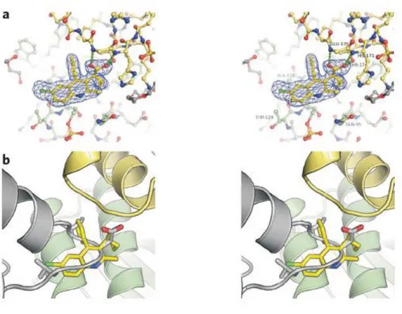
Figure 5: (a) Domain Organization of LEDGF/p75. The binding of LEDGF/p75 to DNA in vitro is mediated by the NLS and a nearby dual copy of the AT-hook DNA binding motif, whereas the N-terminal PWWP domain supplies a critical chromatin recognition function. Charged regions (CRs) 1–3 work in concert with the PWWP domain and AT-hooks to affect the wild type chromatin binding phenotype. The LEDGF/p75 IBD is critical for stimulation of HIV-1 IN function in vitro and for HIV-1 infection. The N-terminal 325 residues within LEDGF/p75 and LEDGF/p52 are identical, whereas the p52 isoform harbors a unique 8–amino acid residue tail. (b)Crystal structure of the LEDGF/p75-IN interaction. Left panel: cartoon representation of the CCD-IBD complex. IN CCD molecules are colored green and blue, whereas the LEDGF/p75 IBDs are magenta and yellow. The side chains of IN active site residues D64, D116, and E152 (Figure 5) are shown as red sticks. Right panel: details of the CCD-IBD interface. LEDGF/p75 hotspot residues I365 and D366, situated at the base of the loop between IBD helices 1 and 2, project into a pocket at the CCD dimer interface. I365 is buried into a hydrophobic pocket predominantly formed by IN residues (A128), W132, Leucine 102 (L102) and Methionine 178 (M178) if HIV-1 IN. Hydrogen bonds and salt bridges are shown as dotted lines (Engelman and Cherepanov, 2008).
24
Consistent with its ability to interact with integrase, an evolutionary conserved integrase-binding domain (IBD) (347-429 residues) was mapped on the LEDGF/p75 C-terminus (Cherepanov et al., 2004). LEDGF gene (PSIP1) encode two splice variants the p75 and p52 isoforms (Figure 5a). The p52 isoform shares the N-terminal 325 amino acid residues and lacks the IBD and consequently failed to bind integrase (Maertens et al., 2003).
In live cells, the N-terminal and the core catalytic domains (NTD and CCD) of integrase were shown involved in the binding to LEDGF/p75 (Maertens et al., 2003). CCD is the minimal domain required for this interaction, whereas NTD enhances the affinity of binding between integrase and LEDGF/p75 (Maertens et al., 2003). By site directed mutagenesis, isoleucine 365 (I365), aspartic acid 366 (D366) and phenylalanine 406 (F406) in the IBD region of LEDGF/p75 were revealed important for the interaction with integrase in vitro (Cherepanov et al., 2005b). Their principal involvement in the protein-protein interaction was confirmed through determination of the crystal structure of IBD in complex with the CCD (Figure 5b) (Cherepanov et al., 2005a). The complex of IBD with CCD consists of a dimer of CCD bound to two IBDs in a fully symmetric fashion. Each IBD burrows into a cleft created by the integrase dimer interface. The side chains carbonyl of LEDGF/p75 D366 forms a bidentate hydrogen bond with the backbone amides of residues glutamic acid 170 (E170) and histidine 171 (H171) from one integrase monomer, while I365 and F406 participate in multiple hydrophobic interactions with residues primarily donated from the other integase monomer. In particular, the side chain of I365 becomes buried within a hydrophobic pocket (Cherepanov et al., 2005a). Two regions within the CCD were identified to be indispensable for the interaction with LEDGF/p75 (Busschots et al., 2007). The first region centers around residues W131 and W132, while the second extends from I161 up to E170 amino acids of integrase (Busschots et al., 2007). A number of single amino acid substitutions to alanine (A) within these CCD two regions were shown to impair integrase-LEDGF/p75 interaction: W131A, I161A, arginine 166A (R166A), E170A (Busschots et al., 2007), glutamine 168 A (Q168A) (Busschots et al., 2007; Emiliani et al., 2005) and valine 165A (V165A) (Turlure et al., 2004). Interestingly, based on the graphical analysis of IBD-CCD crystal structure (Figure 5b), W131 forms hydrogen bond with IBD R405 and Q168 has electrostatic interaction with IBD lysine 402 (K402) (Busschots et al., 2007). Moreover, analysis of modeled mutant structures of W131A and Q168A CCD with IBD showed a disruption of the hydrogen and electrostatic interactions
25 that involve these residues and additionally an alteration of significant number of hydrophobic interactions between CCD and IBD (Busschots et al., 2007).
The purified recombinant LEDGF/p75 stimulated the DNA strand transfer activity of HIV-1 integrase in vitro (Cherepanov et al., 2003) and increased its affinity to DNA (Busschots et al., 2005). The LEDGF/p75 knockdown was shown to have three consequences on the feature of the ectopically expressed HIV-1 integrase: 1) re-distribution of integrase from the nucleus to the cell cytoplasm (Emiliani et al., 2005; Llano et al., 2004b; Maertens et al., 2003); 2) loosen of integrase chromosomal association (Emiliani et al., 2005; Llano et al., 2004b; Maertens et al., 2003) and 3) significant reductions in the steady-state levels of integrase (Emiliani et al., 2005; Llano et al., 2004a). Thus LEDGF/p75 tethers integrase to the chromatin and participates in its karyophilic proprieties, although no evident role of LEDGF/p75 in integrase or pre-integration complex (PIC) nuclear import was demonstrated. In addition, LEDGF/p75 protects integrase from proteasomal degradation (Llano et al., 2004a). The accumulation of integrase in the nucleus might be a consequence of the chromosomal tethering of integrase by LEDGF/p75 and the protection of integrase from proteasomal degradation. In fact, a mutant integrase (Q168A) that fails to bind LEDGF/p75 was unable to bind chromosomes (Emiliani et al., 2005) and proteasome inhibitor added to cells defective for LEDGF/p75 restored the nuclear accumulation of integrase (Llano et al., 2004b).
The role of LEDGF/p75 in HIV-1 infection was studied by three approaches: 1) using HIV-1 viruses carrying mutant integrases impaired for LEDGF/p75 interaction but catalytically active in vitro; 2) down-regulation of the endogenous LEDGF/p75 by knockdown assays or using LEDGF mouse knockout cells and 3) trans-dominant over-expression of the IBD. A Q168A integrase mutant HIV-1 virus is replication defective and showed a specific block at the integration step, whereas, reverse transcription and nuclear import were mildly hampered (Busschots et al., 2007; Emiliani et al., 2005). Furthermore, the HIV-1 virus harboring a mutant integrase (W131A) exhibited mildly impaired replication and integration (Busschots et al., 2007). The first studies performed partial LEDGF/p75 knockdowns and consequently failed to reveal an important role of the cellular factor in HIV-1 infection (Llano et al., 2004b; Vandegraaff et al., 2006; Zielske and Stevenson, 2006). Subsequently, efficient transient and stable LEDGF/p75 knockdowns resulted in a residual HIV-1 integration of 20-50% comparing to not silenced cells, without affecting