Diversity of non-typhoidal
Salmonella in Algeria
by
Bilal Djeghout
This thesis is submitted to the University of Sassari for the degree of
Doctor of Philosophy
Tutor
Prof. Salvatore Rubino
Co-tutors
Dr. Ammar Ayachi
List of publications……….. Declaration……….. List of Figures………. List of Tables……… Abbreviations………..
Abstract
………..………Background and aim of the study ………...……….………..
INTRODUCTION………. Taxonomy of Salmonellae………..………...
Species definition………..…………..… Sub-species typing………..…………... Serotyping………..….
Infection and clinical disease………...
Host restriction and adaptability in Salmonella………...………..….
Typhoidal Salmonella………..….. Human-restricted typhoid………... Host-adapted typhoid………..……... Non-typhoidal Salmonella………... Pathogenesis………... Gastroenteritis..……….…. Bacteraemia………..….. Extra-intestinal infections………..…….
Treatment for NTS infection………..…….
Mechanisms of pathogenesis in non-typhoidal Salmonella infection……... Antimicrobial resistance in non-typhoidal Salmonella………...………...
7 8 9 10 10 15 15 17 17 18 19 20 20 20 21 22 II III IV V VI 23 25 1 3
Salmonella genomic island 1………..………...…… Variants in SGI1………..………...………….……… Salmonella pathogenicity islands………..……….………. Plasmids………..………... Bacteriophages………..……….……….
Epidemiology of Non-typhoidal Salmonella infection………..……….…….
STUDY DESIGN AND METHODS
………...…….Study design and methods……….…………...…...……
Bacterial isolation and identification……….………...…..…... Antimicrobial resistance phenotyping……….……….……..…...…… Whole Genome Sequencing (WGS) and in silico analysis….………...…….
RESULTS
………....……..Genomic characterization of non-typhoidal Salmonella isolated from human and poultry in four cities in Algeria………..…
Genomic characterization of non-typhoidal Salmonella………...
Salmonella Gallinarum isolated from poultry………..… Salmonella Typhimurium isolated from humans………..…... Salmonella Kentucky isolated from humans……….….….. Salmonella Enteritidis isolated from humans………..….…
35 36 36 36 37 40 28 29 31 31 32 33 41 41 42 43 44 39
Molecular mechanisms of resistance to beta-lactamases……….…… Molecular mechanisms of other antibiotic resistance……….………….
Salmonella Genomic Island 1……….…………...……...…
Genomic analysis of multi-drug resistant S. Kentucky ST198 isolated from humans in Algeria………..………..
Antimicrobial resistance phenotype of S. Kentucky isolated from human in Algeria ………...………..
In silico analysis of S. Kentucky genome………...…….
Genetic characterization of the MDR region in SGI1 variant K7 (SGI1-K7) identified in
S. Kentucky strains HSK31, HSK61 and HSK71………
OVERALL DISCUSSION
………..……...Prevalence of non-typhoidal Salmonella in poultry and humans in four cities in Algeria ……….
Salmonella Gallinarum in the poultry industry in Algeria………...……..……….. Salmonella Kentucky in travellers………...………..…..
Antimicrobial resistance among non-typhoidal Salmonella isolated in four cities in Algeria………..…
Burden of NTS infection………...…………...
CONCLUSION
.………..………LIMITATIONS OF THE STUDY
……….……….……51 52 55 56 58 59 60 63 64 76 77
REFERENCES
……...…………..……….……….…78 67 69 70 73researchers that have been inspiring and motivating me during all years of my studies. I would like to thank my supervisors: Pr. Salvatore Rubino who gave me the opportunity
to conduct my Ph.D. project in his laboratory at the University of Sassari in Italy, Pr. Ammar Ayachi for his immense support during my research period at the University
of Batna in Algeria and Dr. Bianca Paglietti for the continuous support of my Ph. D.
study and related research, for her patience and motivation. Beside my supervisors, I would like to thank Dr. Manuela Murgia, a researcher fellow at the University of
Sassari, for her insightful encouragement, but also for advising me during my work in the laboratory.
I would like to express my special appreciation and thanks to Pr. John Wain, who provided me an opportunity to join his team as post-graduate intern, and who gave me access to the laboratory and research facilities in Norwich Medical School at University of East Anglia in United Kingdom. My sincere thanks to Dr. Gemma C. Langridge, Senior Fellow Researcher in JW’s team, for her precious support and immense knowledge.
My sincere thanks to all members at the University of Batna, University of Sassari and University of East Anglia.
Djeghout B., Ayachi A., Paglietti B., Langridge G.C., Rubino S. (2017) An Algerian perspective on non-typhoidal Salmonella infection. J Infect Dev
Ctries 11: 583. https://doi.org/10.3855/jidc.9456.
Food-borne Salmonella in Algeria. The 3rd Euro-Global Conference on
Infectious Diseases. September 5-6, 2016 Frankfurt, Germany. http://dx.doi.org/10.4172/2332-0877.C1.012.
whole or in part, in any previous application for a degree. Except where states otherwise by reference or acknowledgment, the work presented is entirely my own.
October 2017 Bilal Djeghout
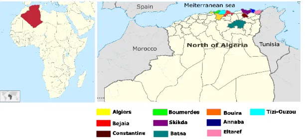
Figure 1. Geographical location and distribution of reported NTS infection in North
Algeria………....
Figure 2. General overview of Salmonella enterica classification………. Figure 3. Transmission of Salmonella in the human-animal-environment web………... Figure 4. Schematic illustration of invasive non-typhoidal salmonella infecting lower
intestine……….
Figure 5. Schematic view of the genetic organization of the MDR gene clusters of
SGI1……….….
Figure 6. Schematic view of the genetic organization of the MDR gene clusters of
SGI1-K7………
Figure 7. Geographical spread of epidemic population of MDR S. Kentucky ST198,
with a focus on the Mediterranean region and Middle East……….. 5 24 30 62 70 13 16 IV
Table 2. Common typing methods in Salmonella………... Table 3. In silico typing of Salmonella enterica isolated from humans and poultry in
four cities in Algeria……….………...
Table 4. Antimicrobial resistance patterns of non-typhoidal Salmonella (NTS) isolated
from humans and poultry in four cities in Algeria………...
Table 5. Prevalence of antibiotic resistant genes detected in S. enterica serovars……...
14
50 54 46
DNA Deoxyribonucleic acid
ET Electromorph type
FDA Food and Drug Administration
KCN Potassium Cyanide
LPS Lipopolysaccharide
MDR Multi-drug resistant
MLEE Multi-locus enzyme electrophoresis
MLST Multi-locus sequence typing
NTS Non-typhoidal Salmonella
ONPG Ortho-Nitrophenyl-B-galactoside
PCR Polymerase chain reaction
PFGE Pulsed field gel electrophoresis
RNA Ribonucleic acid
rRNA Ribosomal ribonucleic acid
SCV Salmonella containing-vacuole
SGI Salmonella genomic island
SPI Salmonella pathogenicity island
ST Sequence type
T3SS Type three secretion system
WGS Whole genome sequencing
WHO World Health Organization
Salmonella nomenclature
For simplicity, in this dissertation Salmonella serovars are referred to by their serovar names, the preceding Salmonella enterica subspecies enterica is abbreviated by the letter “S.”.
ABSTRACT
Non-typhoidal Salmonella (NTS) are globally recognized as important pathogens associated with gastroenteritis. In most cases, humans are infected through consumption of contaminated food products, especially food of animal origin. Poultry is reported to be the major source or reservoir of these pathogens. Most data on incidence and prevalence of NTS infection in both human and poultry are available from industrialized countries. In developing countries including Algeria, there is a lack of documentation, surveillance projects and initiatives. Thus, this has led to an underreporting of Salmonella serovars. This situation is making it harder for health authorities to implement and design preventive approaches for NTS infections. This concern has heightened after the emergence of multidrug-resistant Salmonella strains, as these pathogens are more virulent and responsible for adverse outcomes in infected patients. In Algeria, unregulated use of antibiotics is thought to have caused an increase in resistance by these organisms. Therefore, it is required more than before to initiate more projects documenting the background of NTS serovars in circulation, in order to build a strong and reliable data, useful in making policies for the establishment of routine surveillance systems. The overall aim of this thesis was to identify the different Salmonella serovars isolated from human and poultry in four cities in Algeria, including Guelma, Setif, Batna and Algiers, and to determine the prevalence of antimicrobial resistance in these isolates. Antimicrobial testing was conducted on the isolates using genotypic and phenotypic approaches.
Full genome sequences of the isolates were obtained using Whole Genome Sequencing (WGS) technology, and were analysed in silico for molecular characterization. Different serovars have been identified among the human isolates, naming S. Typhimurium as the most dominant, followed by S. Kentucky, S. Enteritidis, S. Heidelberg, S. Ohio, S. Lindenburg, S. Indiana, S. Virchow, and S. Bonn. Instead, S. Gallinarum was the only serovar found among the poultry isolates. The isolates displayed resistance to multiple antimicrobials. Genotyping showed that the resistance was mediated by various genes encoding for resistance to β-lactam antibiotics, carbapenems, quinolones, aminoglycosides and to co-trimoxazole (trimethoprim-sulfamethoxazole). Classical
Salmonella genomic island 1 was identified in serovar Typhimurium, while new variant
of SGI1 was identified in serovar Kentucky isolated from human, and it was given a name SGI1-K7. Fifty-four isolates (79%) carried various Salmonella pathogenicity islands (SPIs), including SPI-5, SPI-9, SPI-13, SPI-14 and C63PI. Fifty-four isolates (76%) carried at least one plasmid each. Plasmids belonging to incompatibility group FIB and FII were the most commonly identified among the isolates.
These findings are vital to public care system, and helpful for epidemiological control programmes. Furthermore, results presented above contribute in building strong and reliable databases, than can be effective to describe NTS infection and trace its public health consequences among the Algerian population.
BACKGROUND
At the global level, diseases caused by non-typhoidal Salmonella (NTS) still have a massive impact on public health with a relevant impact in developing countries (Schlundt et al., 2004; Djeghout et al., 2017). It is estimated that NTS gastroenteritis are around 94 million cases, resulting 115,000 deaths every year (Majowicz et al., 2010). On the other hand, NTS infections represent a socio-economic burden in both high- and low-income countries as it requires epidemiological surveillance systems and monitoring programs in order to effectively detect and control outbreaks (Crump et al., 2011). Different vehicles have been shown to be implicated in the transmission of these pathogens, and it is mainly associated with the consumption of contaminated raw meat, eggs and chicken, milk and other dairy products, fish and other sea foods, fruits and vegetables (FDA/NSTA, 2008).
Multi-drug-resistance (MDR) among Salmonella strains is another increasing concern for public health, as infections with MDR strains are causing more morbidity and mortality than those caused by susceptible strains (Fluit, 2005). Indeed, MDR
Salmonella is giving rise to clinical worries for the antimicrobial therapy in both
systemic gastroenteritis and bacteraemia caused by NTS serovars (Cooke et al., 2007). There is a paucity of data on Salmonella serovars in humans and from food sources in many developing countries. In Algeria, NTS represent one of the primary causes of
salmonellosis in both humans and food animal production, especially poultry (Ayachi et
al., 2010). The lack of data is probably linked to the inadequate setting of resources and
shortage of epidemiological investigation.
To date, few studies have been reported information on NTS infection from various cities in Algeria, including Algiers, Boumerdes, Tizi-Ouzou, Bouira, Bejaia, Constantine, Batna, Annaba and El Taref (Fig. 1) (Aboun et al., 2000; Elgroud et al., 2009; Ayachi et al., 2010; Bouzidi et al., 2012; Mezali and Hamdi, 2012; Elgroud et al., 2015; Djeffal et al., 2017). Further research investigations are needed to effectively describe NTS infection and trace its public health consequences among the Algerian population.
Fig. 1. Geographical location and distribution of reported NTS infection with a focus on North Algeria. Colors represent cities where NTS infection has been
AIM OF THE STUDY
The purpose of the Ph. D. project was to identify the different Salmonella serovars isolated from human and poultry in four cities in Algeria, including Guelma, Setif, Batna and Algiers, and to determine the prevalence of antimicrobial resistance in these isolates using phenotypic and genotypic approaches.
The specific studies focused on the following objectives:
To identify the different non-typhoidal Salmonella serovars isolated from human and poultry in four cities in Algeria
To determine the prevalence of antimicrobial resistance in the isolates, using genotypic and phenotypic approaches.
To characterise the multi-drug resistant invasive S. Kentucky ST198 from Algeria.
INTRODUCTION
Serological and biochemical characteristics are the main methods used for Salmonella differentiation (Achtman et al., 2012). Serotyping has been the core of public health monitoring of Salmonella infections for years. The Kauffmann White scheme is simplified and is commonly used in routine clinical laboratories, because it contains all serotypes and variants of existing serotypes confirmed and accepted by the WHO Collaborating Centre for Reference and Research on Salmonella (Bale, 2007). However, currently scientists commonly use DNA- based method such multi-locus sequence typing (MLST) to further divide every serotype into more subtypes, to detect outbreaks and effectively identify the pathogen in cause. Furthermore, Whole genome sequencing (WGS) technology have replaced traditional typing, and it is used now as routine typing tool in different reference laboratories including Public Health England (PHE) and Centers for Disease and Control Prevention (Ashton et al., 2016).
Taxonomy of Salmonellae
Salmonella represents a leading cause of food-borne disease worldwide (Patrick A.D.
Grimont, 1, 2001). Salmonella, a bacterial strain named after the American pathologist Daniel Elmer Salmon. It was first isolated in 1855 by Theobald Smith from the intestines of in infected pigs with swine fever (Eng et al., 2015).
Genetic relatedness among Salmonella serovars put in evidence their clonal origin, and the degree of the sequence divergence permits to estimate that a common ancestor of the genus existed more than 25 years ago (Bäumler et al., 1998). To present, nomenclature used for Salmonella is the one recommended by World Health Organization (WHO) and used by the Centers for Disease Control and Prevention (CDC) (Popoff et al., 2003).
Species definition
In the 1880s, species were named according to the disease they cause and the host they infect (Ford Doolittle and Zhaxybayeva, 2009). Currently, the most adapted concept is the phylogenetic species concept (PSC). According to this concept, assignments to species is mostly made on the basis of overall genotypic similarity (Ford Doolittle and Zhaxybayeva, 2009). The assignment of two isolates to one species is made when the degree of the identity has a value of ≥70-80% in a standardized DNA–DNA hybridization experiment (Crosa et al., 1973; Staley, 2006). This concept considers as well the similarity of 16S rRNA sequences, strains with <95-97% identity are placed in the same species (Staley, 2006). Multi-locus enzyme electrophoresis (MLEE) is also used to characterize the species of organisms, as every strain produces a different and unique profile known as eletromorph type (ET). This method detects mutations among amino acids responsible of the differences in genes loci that codes the enzymes (Stanley and Wilson, 2003). Based on the measurement of sequence divergence or what is known
as the nucleotide sequence variation, members in the genus Salmonella are assigned to two species enterica and bongori (Crosa et al., 1973; Reeves et al., 1989; Patrick A.D. Grimont, 1, 2001).
Subspecies typing
Subspecies typing is defined using biotyping, DNA hybridization (Crosa et al., 1973), 16S rRNA analysis and MLEF (Reeves et al., 1989). Biotyping or biochemical tests was widely used by taxonomists (Patrick A.D. Grimont, 2001). This method uses typical biochemical tests to distinguish species and sub-species such as the ability to ferment sugar, the presence of organic acids, and many other components (Bale, 2007). Currently, seven different sub-species have been classified based on the agreement between DNA hybridization and biotyping tests (Patrick A.D. Grimont, 1, 2001). The committee referee formed the sub-species S. enterica subsp. enterica (equivalent to subspecies I), including also other six subspecies naming; S. enterica subsp. salamae (subspecies II); S. enterica subsp. arizonae (subspecies IIIa); S. enterica subsp.
diarizonae (subspecies IIIb); S. enterica subsp. houtenae (subspecies IV); S. enterica
Serotyping
Serological typing or serotyping is based on two antigenic determinants: the somatic (O) antigen, and the flagellar (H: phases 1 and 2) antigen (Brenner et al., 2000; Porwollik, 2011). Each serotype of Salmonella has a unique antigenic formula that is a certain combination of the antigens O and H (Patrick A.D. Grimont, 1, 2001) and few of them produces the capsular antigen Vi (Kauffmann, 1961). The rfb locus encodes for the enzymes that synthesizes the antigen O, whilst the fliC and fliB genes encode the phase 1 and phase 2 flagellins, respectively (Li et al., 1994). Sixty seven somatic O-antigens and 117 flagellar H-antigens have been identified to date (Grimont and Weill, 2008) in more than 2600 serotypes of Salmonella enterica (Gal-Mor et al., 2014). The genetic variation for the Vi antigen locus is used to describe the serovar Typhi (Ferris et al., 1990). The serotyping concept; each serovar carries a unique antigenic formula is untenable, as in some cases one serovar was found to be heterogeneous and in other cases, serovars shared several traits. This situation seems to pull-out the serotyping from the species rank (Patrick A.D. Grimont, 1, 2001).
Current identification of Salmonella strains is mostly based on serology that classifies more than 2600 serotypes (Patrick A.D. Grimont, 1, 2001; Cooke et al., 2007). Thus, different typing methods have been designed to describe the ancestry of Salmonella
They are assorted as phenotypic, genotypic and sequence-based methods (Table 2) (Cooke et al., 2007).
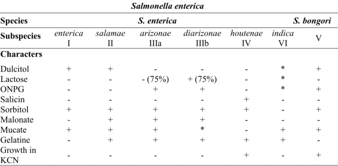
Table 1. Biochemical differentiation characters for Salmonella species and subspecies.
Salmonella enterica
Species S. enterica S. bongori
Subspecies enterica
I salamae II arizonae IIIa diarizonae IIIb houtenae IV indica VI V
Characters Dulcitol + + - - - * + Lactose - - - (75%) + (75%) - * - ONPG - - + + - * + Salicin - - - - + - - Sorbitol + + + + + - + Malonate - + + + - - - Mucate + + + * - + + Gelatine - + + + + + - Growth in KCN - - - - + - +
Adapted from (Bale, 2007).
+: positive reaction with 90% average. -: negative reaction with 90% average
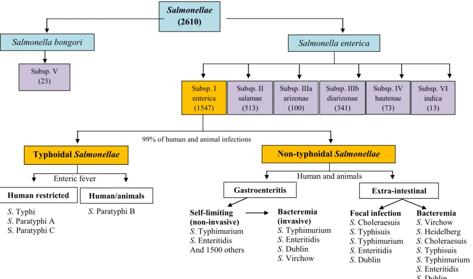
Fig. 2. General overview of Salmonella enterica classification. Two species have been defined using DNA-DNA hybridization and MLEE (Crosa et al.,
1973; Reeves et al., 1989). Seven sub-species in Salmonella are defined by biotyping, DNA hybridization (Crosa et al., 1973), 16sRNA analysis and MLEE (Reeves et al., 1989). Serotypes within each subspecies are defined by serotyping (Guibourdenche et al., 2010). Between brackets are numbers of serovars/serotypes identified in each sub-species.
Salmonella enterica Salmonella bongori Subsp. V (23) Non-typhoidal Salmonellae Typhoidal Salmonellae Gastroenteritis Extra-intestinal Self-limiting (non-invasive) S. Typhimurium S. Enteritidis And 1500 others And 1500 others Focal infection S. Choleraesuis S. Typhisuis S. Typhimurium S. Enteritidis S. Dublin S. Paratyphi B S. Typhi S. Paratyphi A S. Paratyphi C Bacteremia S. Virchow S. Heidelberg S. Choleraesuis S. Typhisuis S. Typhimurium S. Enteritidis S. Dublin Bacteremia (invasive) S. Typhimurium S. Enteritidis S. Dublin S. Virchow
Human restricted Human/animals
Subsp. I enterica (1547) Subsp. II salamae (513) Subsp. IIIa arizonae (100) Subsp. IIIb diarizonae (341) Subsp. IV hautenae (73) Subsp. VI indica (13)
99% of human and animal infections
Human and animals Enteric fever
somatic “O” and Flagellar “H” with a
specific antibody. Does not reflect true classification as it depends on markers of choice. 1950; Popoff et al., 2003)
Phage typing Susceptibility of bacterial isolate to a panel of bacteriophage.
Useful for epidemiology. Widely used for S. Typhi, S. Typhimurium and S. Enteritidis
Limited use and is not applicable on all serotypes. Does not reflect true classification.
(Demczuk et al., 2003)
Resistance typing Susceptibility of bacterial isolate to a
panel of antibiotics. Inexpensive and easily performed. Useful data at population level. It is not a classification, because phenotypic resistance pattern is not stable in the same serovar.
(Cooke et al., 2007)
Genotypic Pulsed field gel electrophoresis gel (PFGE)
Genomic DNA restriction into fragments and migration in an electrical field of alternating polarity.
Highly discriminative molecular typing technique. Useful in epidemiological studies.
Expensive and limited to research
laboratories. (Cooke et al., 2007)
Amplified fragment length polymorphism (AFLP)
PCR based modification of PFGE. Fluorescent markers used to better discriminate the fragment.
Robust and supplies higher level of discrimination.
Expensive equipment required. (Nair et al., 2000)
Multilocus enzyme electrophoresis (MLEE)
Analyses the relative mobilities under electrophoresis of a large number of intracellular enzymes.
Helpful in surveillance and
epidemiological investigations. Difficult technique. Manual use only. (Ferris et al., 1990)
PCR for specific
genes or islands PCR based method for resistance genes and virulence factors. Useful for identification within serotypes. Does not identify serotypes. (Wain et al., 2003)
Plasmid profiling Identification and analysis of plasmids harbored by a bacterial isolate.
Useful for outbreaks investigation. Not considered a classification. Bacterial isolates may lose or gain plasmids easily. (Connerton et al., 2000) Sequence-based Multilocus sequence typing (MLST)
Analysis of 7 conserved genes to separate strains into sequence types.
Reproducible. Based on phylogenetic concept to define salmonella subtypes.
Expensive. Restricted to research/reference laboratories.
(Kidgell et al., 2002)
Microarrays DNA-DNA hybridization of the whole
genome against a reference sequence. Measure gene content. Excellent tool to describe genetic variation. Cannot detect novel insertions. (Thomson et al., 2004)
Whole genome sequencing (WGS)
Entire genome sequencing and analysis. Provides raw nucleotide sequence of an individual’s DNA.
Excellent research tool “in silico typing”, is being introduced to clinical laboratories.
Requires a capacious computing power and skills to use in silico for genome analysis.
(Gilissen et al., 2014)
Infection and clinical disease
Salmonella can be transmitted by the oral–fecal route via contaminated food and water
(Figure 3) and are mostly associated with inadequate sanitation and hygiene (McElhaney, 1992). Clinically, Salmonella enterica is divided into two groups based upon the disease caused: typhoidal that is usually human-host restricted, causing systemic infection known as enteric fever. The other group includes all non-typhoidal
salmonella (NTS) that causes self-limiting gastroenteritis (Figure 1) (Cooke et al.,
2007).
Host restriction and adaptability in Salmonella
Host adaptation in Salmonella is mainly based on epidemiological evidence and surveillance investigations that can contribute to disclose characteristics of reservoirs of
Salmonella serotypes. In salmonella, host adaptation can be defined as the ability of
bacteria to circulate and cause disease in different populations of vertebrate hosts (Kingsley and Bäumler, 2000). For example, S. Typhimurium and S. Enteritidis are considered broad-host-range serovars, because they are frequently associated with
salmonella infection in different animal species and also human (Hormaeche et al.,
1991; Cogan and Humphrey, 2003). Indeed, S. Dublin, which is mainly hosted in cattle, is predominantly responsible for the systemic form of salmonellosis in humans (Chen et
al., 2013). On the other hand, host restricted Salmonella is defined as the ability to cause
with only to disease incidents reported from human causing typhoid fever, and it is unable to infect other vertebrate species (Kingsley and Bäumler, 2000; McClelland et
al., 2004). S. Gallinarum and S. Abortusovis are highly adapted to animal hosts in
particular in poultry and sheep respectively, and may only produce very mild symptoms in humans (Rubino et al., 1993; Chen et al., 2013).
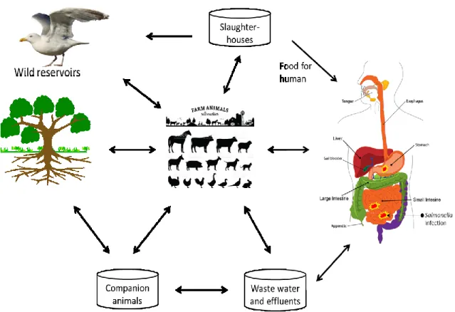
Fig. 3. Transmission of Salmonella in the human-animal-environment web. Salmonella is
habitually spread after the consumption of contaminated raw meat, eggs and chicken, milk and other dairy products, fish and other sea foods, fruits and vegetables (FDA/NSTA, 2008). These pathogens can be transmitted between people and companion animals such as pets. Animals may become infected with Salmonella after roaming in contaminated environment. Wild animals such as birds may play a role in transmitting the bacteria to human (Patrick A.D. Grimont, 1, 2001).
Typhoidal Salmonella
Typhoid and paratyphoid fever are a systemic bacterial infection caused by Salmonella
enterica subspecies enterica serovar Typhi and Paratyphi (hereafter S. Typhi)
(McClelland et al., 2004).
Clinically, paratyphoid fever is indistinguishable from typhoid fever. Therefore, S. Typhi, Paratyphi A, B and C are collectively referred to as typhoidal Salmonella serotypes (Sudeepa Kumar et al., 2013). This disease is contagious, the bacteria can be transmitted from infected individuals via the fecal-oral route (Pang, 1998). At the global level and annually, typhoid infection comprises around 75% to 80% with more than 21 650 000 cases (with 216 500 deaths), and around 5 410 000 cases of paratyphoid fever (Ayele et al., 2011).
Human-restricted typhoid
Human constitutes the only natural host and reservoir of infection caused by S. Typhi and S. Paratyphi A and C. These serovars are a highly host-adapted pathogens (McClelland et al., 2004; Ayele et al., 2011). S. Typhi was first isolated in 1880 by Karl J. Erberth and Robert Koch. It is known by the antigenic formula [9]Vi:d:-, and certain biochemical characters (Langridge, 2010). Substantially, during the late nineteenth and early twentieth centuries, typhoid fever caused by serovar S. Typhi was endemic in the majority of European countries and in both North and South America (Wolman and Gorman, 1931). Currently, the disease is uncommon in developed countries (Ackers et
al., 2000). Similarly to S. Typhi, S. Paratyphi identified with antigenic formula
1,2,12:a[1,5] is thought to cause milder disease, with mainly gastrointestinal symptoms (Grimont and Weill, 2008; Farrar et al., 2013). Indeed, in some countries, particularly in Asia, enteric fever caused by S. Paratyphi A is increasing comparing with other enteric fevers (Woods et al., 2006).
Although they are from different serogroup, genome analysis of S. Paratyphi A indicates that is genetically similar to S. Typhi, putting in suggestion that it contains more recent evolutionary origin because it has fewer pseudogenes. Both genomes carry around 4,400 protein coding sequences; over 173 pseudogenes in S. Paratyphi A and ~ 210 pseudogenes in S. Typhi (McClelland et al., 2004). S. Paratyphi C, human restricted serovar (Patrick A.D. Grimont, 1, 2001), identified by the antigenic formula 6,7:c:1,5 and distinguished recently from other variants sharing the same antigenic formula by MLST analysis with the sequence types (STs) 146, 90 and 114 (Uzzau et al., 2000; Grimont and Weill, 2008; Langridge, 2010). This serovar causes enteric fever with similar symptoms caused by S. Typhi and S. Paratyphi, but it is not commonly isolated.
Host-adapted typhoid
S. Paratyphi B, host-adapted and identified by the antigenic formula 2;25, appears to not
have an important role in causing enteric fever but primarily causes gastroenteritis (Chart, 2003). This serotype is frequently isolated from terrapins and shares the same antigenic formula with S. Java. The subdivision of S. Paratyphi B and S. java is based on
the ability to produce a slime wall and the inability to utilize D-tartrate (Patrick A.D. Grimont, 1, 2001). Based on the fermentation of dextrorotatory (d-tartrate), S. Paratyphi B can be characterized into d-tartrate fermenting (dT+) and non-fermenting (dT-) (Barker, 1985). dT- variant S. Paratyphi B mainly causes paratyphoid fever while dT+ variant S. Paratyphi B (previously called S. Java)only provokes gastroenteritis in human (Chart, 2003; Ahmad et al., 2012).
Non-typhoidal Salmonella
Salmonella strains other than S. Typhi and S. Paratyphi are referred to as non-typhoidal Salmonella (NTS) (Figure 2). Animals are the primary reservoir of NTS strains (Steve
Yan et al., 2003; Wales and Davies, 2013). Infection due to NTS is mainly associated with the consumption of contaminated raw meat, eggs and chicken, milk and other dairy products, fish and other sea foods, fruits and vegetables (FDA/NSTA, 2008). Despite close genetic relatedness, typhoidal and non-typhoidal Salmonella (NTS) provoke different illnesses and a divergent response from the human immune system (Gal-Mor et
al., 2014). Incubation period in NTS infection is shorter (6–12 hours) compared to
typhoid infection, and symptoms are usually self-limiting (Crump et al., 2008; Eng et
al., 2015).
Pathogenesis
Infection with NTS strains in human is clinically manifested with gastroenteritis, bacteraemia and other extra-intestinal complications (Darby and Sheorey, 2008). The
severity of infection varies depending on two factors; the serotype implicated and the health of the patient. Infants, the elderly, and immuno-compromised patients are more susceptible to NTS infections than healthy adults (Eng et al., 2015).
Gastroenteritis
NTS commonly cause an acute gastroenteritis, an inflammatory condition of the gastrointestinal tract, which is accompanied by typical symptoms of salmonellosis such as diarrhea, nausea, vomiting, abdominal cramps and headache (Steve Yan et al., 2003; Gharieb et al., 2015). The symptoms appear 12 hours post-incubation, they last approximately for 10 days and they are usually self-limiting (Crump et al., 2008; Eng et
al., 2015). Salmonella is excreted in faeces after infection, and remain detected for
median of five weeks (Chen et al., 2013). Patients with immunodeficiency may have prolonged or chronic Salmonella infection (Yuan et al., 2011).
Bacteraemia
NTS actively invade intestinal epithelial cells Invasive NTS infection (bacteraemia) occurs when the bacteria enter the bloodstream, the meninges, bone, and joint spaces after invading the intestinal barrier (Acheson and Hohmann, 2001; Woods et al., 2008; Eng et al., 2015). Mostly, all NTS serotypes may cause bacteraemia. However, serovars
S. Dublin and S. Choleraesuis are highly linked with invasive infections (Woods et al.,
2008). Bacteraemia is clinically manifested with high fever, but in the contrary of patients with enteric fever, no formation of rose spots is observed. These symptoms are
seen more in infections caused by NTS serovars than typhoidal serovars, and the infection can be developed to acute condition causing mortality when the immune system is trigged, resulting what is known as septic shock (Eng et al., 2015).
Extra-intestinal infections
Five per cent of infected patients with NTS develop a bacteraemia, and in some of them extra-intestinal manifestations occur (Eng et al., 2015). These manifestations are clinically important, in some cases the pathogen may even affect brain function (Arii et
al., 2002). For example, S. Typhimurium has the ability to adhere, invade, and penetrate
human brain microvascular endothelial cells, the single-cell layer constituting the blood– brain barrier (BBB). It is believed to be a primary stage in the pathogenesis of meningitis (van Sorge et al., 2011). NTS strains also are a leading cause of meningitis in Africa and Latin America, causing high level of mortality (GRAHAM et al., 2000; Owusu-Ofori and Scheld, 2003). Other complications may be occurred in NTS infection, including cellulitis,pneumonia, urinary tract infections and endocarditis (Shimoni et al., 1999; Arii
et al., 2002).
Treatment for non-typhoidal Salmonella infection
NTS infection is a self-limiting disease, mostly manifested with gastroenteritis and rarely requires antimicrobial therapy. Nevertheless, infections may become severe involving hospitalization and some patients may even become septic with bacteraemia (GRADEL et al., 2007; Chen et al., 2013). Antimicrobials are not recommended,
because these do not reduce the duration or acuteness of gastroenteritis, and instead may result in extended carriage and appearance of resistant strains (Steve Yan et al., 2003; Su and Chiu, 2007). Indeed, fluid replacement is highly recommended in NTS infection (Chen et al., 2013). However, antimicrobial therapy is considered for patients with severe diarrhea, and is commonly used for invasive salmonellosis or when individuals with immunodeficiency are affected (Steve Yan et al., 2003).
In cases of extra-intestinal or focal infection, and similar to bacteraemia, antibiotics are chosen depending on the susceptibility pattern of the strains in cause, considering as well the clinical condition of the patients. Mostly, the therapy includes ampicillin, trimethoprim-sulfamethoxazole, fluoroquinolones or third generation cephalosporins, such as ceftriaxone (Wong et al., 2000; Chen et al., 2013). With potential risk of causing arthroplasty, fluoroquinolones remain last option in NTS infection for children, when there is no other alternative treatment available (Wong et al., 2000; Chen et al., 2013; Choi et al., 2013).
Mechanisms of pathogenesis in non-typhoidal Salmonella infection
A range of virulence genes contribute to Salmonella pathogenicity (Marcus et al., 2000). Virulence determinants can be harboured on the chromosome, usually encoded on pathogenicity islands, or on plasmids and bacteriophages (Fluit, 2005). Moreover,
salmonella are surrounded by an outer membrane containing lipopolysaccharide (LPS)
(Messina, 1993). Another component of virulence is the ability to synthesize enterobactin (Pollack and Neilands, 1970). Enterobactin is a siderophore secreted by
Salmonella allowing it to sequester iron from the host and use it for growth (Nagy et al.,
2013).
Following the consumption of contaminated food with NTS strains, these pathogens colonize the intestines and may invade epithelium barrier (Hurley et al., 2014). Serovars that succeed in passing this barrier have to face macrophages and may be subjected to phagocytosis, in their best state they actively invade these macrophages using T3SS-1 and fimbriae (Figure 4) (Rescigno et al., 2001).
While Salmonella actively invade intestinal epithelial cells using a type III secretion system (TTSS) encoded in Salmonella pathogenicity island 1 (SPI-1) to inject effector proteins into host cells (Galán and Wolf-Watz, 2006), SPI-2 encodes genes involved in intracellular survival and replication in phagocytic and non-phagocytic cells, and has a crucial role in systemic infection (Abrahams and Hensel, 2006).
Fig. 4. Schematic illustration of invasive non-typhoidal salmonella infecting lower intestine.
adapted from (Hurley et al., 2014). (A) After reaching the lower intestine of the host, the pathogen adheres to the mucosal membranes and actively invades epithelial cells (Rescigno et al., 2001). (B) Then, Salmonella follow two different paths; translocation across M cells of Peyer’s patches or using SPI-1 encoded T3SS-1 to inject effector proteins into host cells (Galán and Wolf-Watz, 2006). (C) Salmonella is then surrounded by macrophages. SPI-2 encoded T3SS-2 releases effector proteins into the cytosol of the host cell (SigD/SopB, SipA, SipC, SodC-1, SopE2, and SptP) preventing fusion of the phagosome with the lysosome. Salmonella propagate within the
Salmonella-containing vacuole (SCV) inducing secretion of cytokines by macrophage. As result,
macrophage undergoes in apoptosis, Salmonella escape and will invade basolateral side of epithelial wall and even other phagocytic cells of innate immune system of the host (Hurley et al., 2014).
Antimicrobial resistance and virulence in non-typhoidal Salmonella
Antimicrobial resistance in NTS is mostly promoted by the use of those antimicrobials in food animals (Angulo et al., 2000; Gupta et al., 2003; Dutil et al., 2010; Crump et al., 2011). It can be due to genetic mutations or through the acquisition of resistance encoding genes on mobile elements (Fluit, 2005). Plasmids are typical carriers of determinants that confer resistance against conventional antibiotics such as ampicillin, chloramphenicol and tetracycline (Guerra et al., 2001). However, the chromosome can also harbour these determinants, e.g. on the multidrug resistance region of Salmonella Genomic Island 1 (SGI1) (Fluit, 2005).
In Salmonella, virulence determinants have been identified in different coding genes, mostly linked with a combination of chromosomal and plasmid factors (Oliveira et al., 2002). For instance, The Salmonella-encoded fimbria lpf operon provides bacteria affinity for Peyer’s patches and adhesion to intestinal M cells. While agf operon promotes interaction between the bacteria and intestine by stimulating bacterial self-aggregation, improving its survival rates within the intestine. On the other hand, sef
operon enhances interactions between the bacteria and macrophages (Collinson et al.,
1996; Bäumler et al., 1998; Edwards and Puente, 1998; Borges et al., 2013).
Transmission of virulence and antibiotic resistance in non-typhoidal Salmonella
Likewise, other gram-negative organisms, genes in NTS are transmitted via genetic mobile elements. For instance, many plasmids and integrons are associated in the
dissemination of resistance to antibiotics via horizontal gene transfer. These genes play an important role in infection, host adaptation and disease development (Porwollik and McClelland, 2003).
IS elements
Insertion-sequence (IS) elements, firstly identified in E. coli in the gal operon, are genetic mobile elements originally from bacterial DNA. They are able to move from a position to another on the same or different genome. When IS elements are inserted in the middle of a gene sequence, they may interrupt the coding sequence, inhibiting the expression of that gene (Harper, 2012). Generally, genome of salmonella carries several copies of IS element naming IS200, IS3 and IS1617. Presence of an IS element may be an indication of variability in genetic content of a specific genetic region (Haack and Roth, 1995).
Transposons
Transposons known also as jumping genes, areDNA sequences that are able to change their location within a genome. They were first identified by Barbara McClintock more than fifty years ago. They are found in both eukaryotes and prokaryotes (Pray, 2008). Originally, they were distinguished from IS elements because transposons contain detectable genes, often conferring resistance to antibiotics (Calos and Miller, 1980). Generally, transposons are flanked between two copies of the same IS element.
Transposons and IS elements are now grouped together under the single term transposable elements (Griffiths, 2005).
Integrons
Integrons are mobile genetic units containing determinants of the components of site-specific recombination system that has the ability to capture and translocate genes contained in mobile elements known as gene cassettes that can be inserted into or deleted from their receptor elements (Hall and Collis, 1995a; Hall, 1997). Gene cassettes are known to encode for antibiotic resistance in gram negative pathogens, for example genes conferring resistance to β-lactams, aminoglycosides, trimethoprim, chloramphenicol, streptothricin (Hall, 1997). Three classes of integrons involved in antibiotic resistance have been characterized in detail (Ploy et al., 2003). However, Class 1 integron is the most common class found in Salmonella enterica serovars (Hall and Stokes, 1993). It has been detected as well in many Salmonella Genomic Islands 1 SGI1 (Boyd et al., 2002; Meunier et al., 2002; Ebner et al., 2004).
Salmonella genomic island 1 (SGI1)
Salmonella genomic island 1 (SGI1), a 43 kb integrative chromosomal island clustering
antibiotic resistance genes in a 13 kb region known as Multi-drug resistant (MDR) region. It confers the bacteria resistance to antimicrobials. It was firstly identified in the epidemic S. Typhimurium DT104 that emerged during the 1980s, and had caused a
global health concern in both human and animals. Later this genomic island was identified in several Salmonella enterica serovars (Boyd et al., 2002; Doublet et al., 2005).
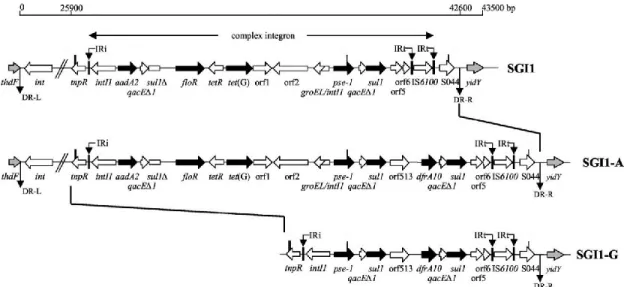
Antibiotic resistance genes cluster is located near the 3’ end of SGI1, forming a complex class 1 integron belonging to the In4 group (Boyd et al., 2002). The gene cluster of SGI1 is bounded by inverted repeats of 25 bp IRi and IRt and have a 3’-CS (conserved sequence) that includes a copy of IS6100 (Brown et al., 1996). Further, it is surrounded by 5 bp direct repeats, which suggests that it was integrated in SGI1 by a transposition event. SGI1 contains also a duplication of a part of the 5’-CS, that leads to a second attI1 site followed by a gene cassette (Partridge, Brown, et al., 2001; Boyd et al., 2002; Doublet et al., 2005). SGI1 contains also a duplication of a part of the 5-CS, that leads to a second attI1 site followed by a gene cassette (Doublet et al., 2005).
Variants in SGI1
Variants of SGI1 are identified based on the variation of the genetic organization in their MDR region. Different variants of SGI1 containing different sets of resistance genes have been identified (Boyd et al., 2002; Carattoli et al., 2002; Benoît Doublet et al., 2004; Levings et al., 2005; Cloeckaert et al., 2006). The original SGI1 was isolated from
S. Typhimurium phage type DT104. It contains five genes aadA2, sul1, floR, tetA(G),
and blaP1 (variant form blaPSE-1 or blaCARB-2). These genes respectively confer resistance to streptomycin and spectinomycin, sulfonamides, chloramphenicol and
florfenicol, tetracyclines, and β-lactam antibiotics (Briggs and Fratamico, 1999; Boyd et
al., 2001), and all of them are located within the boundaries of a complex class 1
integron named In104 (Levings et al., 2005) (Fig. 5). Sequence of the MDR region in SGI1 has revealed that the integron contains a backbone similar to that of In4 (Briggs and Fratamico, 1999; Partridge, Brown, et al., 2001; Partridge, Recchia, et al., 2001; Boyd et al., 2001) , but it has in addition duplications of parts of the integron conserved segments (CS) naming 5′-CS and part of the 3′-CS (qacEΔ1 and partial sul1 genes), where integrated gene cassettes are usually flanked (Hall and Collis, 1995b; Hall and Collis, 1998). A second integrated element named retron phage, is found inserted between end of SGI1 and the yidY gene, but it is detected only in the serovar Typhimurium (Boyd et al., 2000; Boyd et al., 2001).
Variants from SGI1-A to SGI1-J, seem to have gained, lost, or exchanged resistance genes by gaining and/or losing various segments of DNA, mostly occurred by homologous recombination. Some of them have lost parts of In104, and others have different gene cassettes from those found in SGI1, naming the dfrA1-orfC or the
aacCA5-aadA7 pairs (Doublet et al., 2003; Benoit Doublet et al., 2004; Levings et al.,
2005). Further variants with different gain/loss have been identified (Boyd et al., 2002; Levings et al., 2005).
The genetic variation in the MDR region seems to be a useful tool to track the evolution of the structure of SGI1, especially that it is occurring in a defined context and location, the backbone of the SGI1, which is integrated within the end of the thdF gene in the
Salmonella enterica chromosome (Levings et al., 2007).
Fig. 5. Schematic view of the genetic organization of the MDR gene clusters of SGI1.
Adapted from (Benoît Doublet et al., 2004). The variants SGI1-A and SGI1-G are represented below to illustrate the variation. Left and right junctions (thdf and yidY), bracketing SGI1 are represented in grey arrows. Black arrows correspond to antibiotic resistance genes. Vertical thick black bars indicate homology regions between SGI1 variants.
Salmonella pathogenicity islands (SPIs)
SPIs are genomic island that contain important genes conferring virulence to Salmonella
enterica. They are located on the chromosome and can be transferred from an organism
to another through horizontal gene transfer. Usually, pathogenicity islands accommodate vast clusters of genes that contribute to a specific virulence phenotype. In general, this
phenotype is manifested at a specific moment during the infection, and may convert normally benign microorganism into a pathogen (Marcus et al., 2000). Some SPIs are conserved in the genus Salmonella and others are serovar-restricted (Siriken, 2013). Different SPIs have been identified, but the most important ones namely SPI1 and SPI2 that code for genes involved in the intestinal phase of the infection. The remaining SPIs are required for fimbrial expression, magnesium and iron uptake, antibiotic resistance and other functions required for survival inside the host (SIRIKEN, 2013).
Plasmids
Many Salmonella serovars harbour virulence plasmids that are important for systemic infection (Gulig, 1990; Wallis et al., 1995). Plasmids in Salmonella are heterogeneous in size, depending on the serovar, ranging from 50 to 110 kb (Chu et al., 1999). Moreover, all of them share a 7.8 kb region named spv, required for bacterial multiplication in the reticulo-endothelial system. Plasmids carrying virulence determinants can be transferred from a bacterium to another by horizontal gene transfer, this mechanism is known by transformation (Johnston et al., 2014). However, current evidence suggests that the contribution of virulence plasmids to pathogenesis in Salmonella is less important (Casadesüs, 1999), as these last do not affect the ability of Salmonella of causing a gastroenteritis (Gulig et al., 1993).Plasmids found in Salmonella enterica can contain a variety of genes encoding antimicrobial and heavy metal resistance, toxins or virulence genes that are beneficial to adapt to different environments. These plasmids have been
found in different Salmonella serovars namely S. Enteritidis, S. Typhimurium, S. Dublin,
S. Choleraesuis, S. Gallinarum, S. Pullorum and S. Abortusovis. These plasmids ranging
in size from few to several hundred kbp. They contribute in the spread of genes in bacterial populations. (Rychlik et al., 2006). They are mostly classified into incompatibility groups (Inc) based on their mode of replication and maintenance inside the bacterial cell. Consequently, plasmids exploiting the same replication machinery are mutually incompatible and unable to persist in the same cell for extended period (Ou, 1993). Knowledge on plasmids is limited. However, their presence or absence is frequently used for strain differentiation in epidemiological studies.
Bacteriophages
Bacteriophages (phages) are the most abundant organism in the biosphere (Clokie et al., 2011). They can transfer antibiotic resistance from a bacterium to another using a mechanism known by transduction. This process is a significant contributing factor to dissemination of antibiotic resistance genes in food-borne pathogens of the
Enterobacteriaceae family, naming non-typhoidal Salmonella (Colavecchio et al.,
2017).
Epidemiology of non-typhoidal Salmonella infection
Food-borne diseases have become a global economic burden on health care systems. More than 550 million individuals fall sick every year, including 220 million children under the age of 5 years. Salmonella is one of the most reported causes of this disease
globally (WHO, 2016). In fact, the prevalence of Salmonella infection is higher than is reported, because most infected people do not undergo copro-culture diagnosis (kay M.Tomashek, Tyler M.Sharp, 2015).
Epidemiology related to NTS varies depending upon serovar, and its ability to cause gastrointestinal or invasive infection. With certain subtypes, S. Typhimurium is recognized as capable of causing bacteraemia, whereas S. Heidelberg, S. Dublin, and S. Choleraesuis represent a significant potential to cause hospitalization, and even death. In comparison, serovar S. Newport was observed to cause fewer fatalities than S. Typhimurium (Crump et al., 2011).
Several outbreaks of food-borne infections of antibiotic resistant NTS have been reported globally while individual epidemiology studies in more-developed African countries provide some basic insight until more data are available (Enwere et al., 2006). Such information is crucial to understand the spread of multidrug-resistant Salmonella strains (Harrois et al., 2014). In Algeria, limited data is available on epidemiology of NTS infection. S. Typhimurium, S. Heidelberg and S. Enteritidis are commonly reported in both human and animal food sources (Ayachi et al., 2009; Elgroud et al., 2009; Bounar-Kechih et al., 2012; Bouzidi et al., 2012; Mezali and Hamdi, 2012). In other parts of Africa, particular serovars are prevalent in specific regions, namely S. Concord in Ethiopia (Beyene et al., 2011), S. Bovismorbificans in Malawi (Bronowski et al., 2013), S. Stanleyville and S. Dublin in Mali (Tennant et al., 2010), and S. Isangi in South Africa (Wadula et al., 2006). In Kenya, an average of 166 per 100,000 children
under five acquire an NTS infection every year (Oundo et al., 2002; Berkley et al., 2005). In sub-Saharan Africa, a serious invasive form of NTS linked to S. Typhimurium sequence type ST313 has emerged, and become a leading public health issue in this region. This invasive S. Typhimurium had an estimated mortality rate of 20-25% in children and up to 50% in adults (Kingsley et al., 2009).
In the US, food-borne Salmonella is the largest health burden of all bacterial pathogens (Scallan et al., 2011). During 2014, more than 19,000 laboratory-confirmed cases of food-borne infections were identified. Salmonella was linked to 7439 of these cases, in which 2144 persons were hospitalized, and among 32 patients died (CDC, 2014). The most commonly detected serovars of Salmonella in humans include S. Enteritidis and S. Typhimurium, followed by S. Newport, S. Javiana, and Salmonella with the antigenic formula 4, [5],12:i:- (CDC, 2014).
Although developed countries have a better awareness among food handlers and optimized surveillance programs, NTS infection has been present and responsible for patient morbidity and mortality for decades. For developing countries, the situation is worsened with the limited sources currently available. In addition, massive human migration from African countries and the increase in food trade between developed and developing countries may play a significant role in spreading such pathogens all over the world.
STUDY DESIGN AND METHODS
Bacterial isolation and identification
Sixty-nine Salmonella were isolated according to a standard ISO method (ISO, 2007). Human clinical isolations was obtained from stools of diarrheagenic patients admitted at different hospitals in three cities in Algeria: Guelma, Setif and Algiers during 2014-2015. Animal origin strains were isolated from poultry houses in Batna city. The isolates were serotyped by the slide agglutination method using Omni-O Salmonella antisera (Bio-Rad Laboratories, USA).
Antimicrobial resistance phenotyping
The isolates were tested for susceptibility to antimicrobials on Muller-Hinton agar following the Kirby-Bauer disk diffusion method, using a panel of 12 antibiotics, and interpreted according to the recommendation of the Clinical and Laboratory Standards Institute 2016. Antimicrobial disks (Bio-Rad Laboratories, USA) with the following drug contents were used: ampicillin (10 μg), ceftriaxone (30 µg), chloramphenicol (30 μg), nalidixic acid (30 µg), ciprofloxacin (5 μg), trimethoprim/sulfamethoxazole (1.25/23.75 μg), streptomycin (10 μg), tetracycline (30 μg), meropenem (10 µg), imipenem (10 µg), cefotaxime (30 µg) and gentamicin (10 µg). Isolates resistant to three or more classes of antimicrobials were specified as MDR.
Whole Genome Sequencing (WGS) and in silico analysis
Genomic DNA of the isolates was extracted using Wizard Genomic DNA Purification Kit. DNA extracts were converted into a Nextera XT library for sequencing on an Illumina NextSeq 500 platform according to the manufacturer’s instructions. The
Salmonella library was diluted to 4nM (as determined by analysis on an Agilent
Technologies 2200 Tapestation and using the Qubit HS dsDNA assay) and pooled in equimolar amounts with other barcoded libraries. The entire library pool was then diluted to 1.8 pM and sequenced using the NextSeq 500 v2 2x150 bp paired-end protocol.
Genomes were assembled using Velvet de novo genomic assembler (Afgan et al., 2016). Web-based tool SeqSero 1.0 was used to determine the serotype and the antigenic profile of the isolates (Zhang et al., 2015). The genome assemblies were then subjected to sequence type (ST) analysis using Salmonella in Silico Typing Resource platform (SISTR) [https://lfz.corefacility.ca/sistr-app/] (Yoshida et al., 2016). Further investigation was conducted on the genome assemblies for acquired resistance genes,
Salmonella Pathogenicity Islands (SPI), plasmids and incompatibility group using
ResFinder (Zankari et al., 2012), SPIFinder-1.0 (CGE online platform:
http://www.genomicepidemiology.org/) and PlasmidFinder 1.3 platforms respectively (Carattoli et al., 2014). Presence of the Salmonella Genomic Island 1 (SGI1) was
investigated by PCR primers DR-S004 targeting SGI1 left junction, int, xis and rep, and with S044-DR targeting SGI1 right junction with retronphage (Carattoli et al., 2002). SGI1 mapping was conducted in silico using Geneious R10 software.
Genomic characterization of non-typhoidal
Salmonella isolated from human and poultry in
Genomic characterization of non-typhoidal Salmonella
Information and surveillance on NTS serotypes are limited in Africa, and also in Algeria. Reliable source of data is not always easy to be found, whilst few individual studies supply some basic information. One of the aims of this Ph. D. study is to identify and characterize the different non-typhoidal Salmonella serovars isolated from humans and poultry in four cities in Algeria. Serotypes of the isolates were determined utilizing whole genome sequencing data.
Human isolates were collected from hospitalized patients suffering from diarrhea in three cities, including Guelma, Setif and Algiers. Poultry isolates were collected from slaughterhouses in Batna city. Altogether, 10 different serotypes were recovered from poultry and humans, being S. Typhimurium the dominant serotype among human isolates, followed by S. Kentucky, S. Enteritidis, S. Heidelberg, S. Ohio, S. Lindenburg,
S. Indiana, S. Virchow, and S. Bonn, while S. Gallinarum was the only serovar found
among the poultry isolates (Table 5).
Salmonella Gallinarum isolated from poultry
Twenty-nine (42%) Salmonella enterica isolated from poultry were of serovar Gallinarum. They belonged to serogroup D1, holding antigenic formula 9:g,m:-, and sequence type ST78. Five strains harbored two plasmid each, belonging to incompatibility group N and FII. Eighteen strains harbored one plasmid each of IncFII
group. Six strains did not contain any plasmid. In silico analysis of genome sequences identified different Salmonella pathogenicity islands (SPIs), including SPI-5 carrying
sopB, sigD, pipB genes encoding for effector proteins required in function of SPI-1 and
SPI-2. This SPI was acquired from S. Typhimurium strains LT2 and has inserted in the
serT allele on the chromosome. SPI-13 have been found inserted in the allele pheV on
the chromosome, carrying gtrB and gtrA genes encoding for LysR transcriptional regulator, those genes are implicated in the bacterial stringent response and in the virulence of Salmonella (Maddocks and Oyston, 2008). This SPI was originally found in
S. Gallinarum strain SGA-10. SPI-14 was detected in the genome carrying two genes, naming gpiA encoding for electron transfer favoprotein beta subunit, and gpiB implicated in the regulation of transcription. The origin of this SPI is S. Gallinarum strain SGA-8. Furthermore, another SPI was detected, named C63PI, inserted in fhlA allele. This SPI is acquired from S. Typhimurium strain SL1344. It is implicated in transcription of the sit operon under iron-lack growth conditions, forming Putative Iron Transport System (PITS) within the Centisome of this pathogenicity island (Zhou et al., 1999).
Salmonella Typhimurium isolated from humans
Nineteen strains (27%) were identified as S. Typhimurium among the human clinical isolates. They all belonged to serogroup B, with antigenic formula 4:i:1,2, and sequence
type (ST19). 84% (16 out of 19) Typhimurium carried plasmids belonging to incompatibility group FII, and FIB. Different SPIs were found in the chromosome of these strains. Sixteen strains had similar SPIs previously found in S. Gallinarum in this study, including SPI-5 inserted in the chromosomic allele serT and carrying SopB, SigD,
PipB genes. Three different functional SPI-13 were found inserted in the allele pheV on
the chromosome, one carrying gacD originally acquired from S. Gallinarum strain SGD-3, encoding for Acetyl-coA dehydrolase, and other two harbouring gtrB and gtrA genes, originally from S. Gallinarum SGG-1 and S. Gallinarum SGA-10 respectively. SPI-14 was detected in the genome carrying two genes, naming gpiA and gpiB. The origin of this SPI is S. Gallinarum strain SGA-8. The pathogenicity island C63PI were found inserted in fhlA allele of the chromosome, with the similar functions as in strains of S. Gallinarum isolated from poultry. Two strains of S. Typhimurium found to be carried only two SPIs on their chromosomes, including SPI-13 and SPI-14.
Salmonella Kentucky isolated from humans
Seven strains (10%) were identified as S. Kentucky. They belonged to serogroup C2-C3 with antigenic formula 8:i:z6, and sequence type ST198. Three strains carried one plasmid each belonging to incompatibility group I2. A plasmid of IncHI2 was found in one strain, while three strains did not harbor any plasmid. Only pathogenicity island C63PI was detected on the chromosome of two isolates. It was inserted in the allele fhlA.
Salmonella Enteritidis isolated from humans
Five S. Enteritidis have been identified from humans. They belonged to serogroup D1, with antigenic formula 9:g,m:-, and sequence type ST11. Two plasmids IncFII and IncFIB were found in each of forth S. Enteritidis isolates, the fifth strains did not harbor any plasmid. Two strains were found to harbor SPI-5, SPI-13, SPI-14 and the pathogenicity island C63PI inserted in the chromosomal allele fhlA, encoding for iron uptake system.
On the other hand, three strains were identified as S. Heidelberg. They belong to serogroup B, with antigenic formula 4:r:1,2, with sequence type ST15. Two of the three strains carried two plasmids each, belonging to incompatibility group X1 and I1. One of the three strains carried one only plasmid belonging to incompatibility group X1. They carried three different SPIs, including SPI-9, SPI-13 and SPI-14. Two strains were identified as S. Indiana. They belong to serogroup B, with antigenic formula 4:z:1,7, and do not harbor any plasmids or SPIs.
Two other isolates were identified as S. Ohio with sequence type ST329 and antigenic formula 7:b:l,w, and as S. Bonn sequence type ST2522 with antigenic formula 7:l,v:e,n,x, Both isolates carried one plasmid each belonging to IncA/C2 group. Four different SPIs were found inserted on the chromosome of S. Bonn including 5,
SPI-13, SPI-14 and C63PI, while S. Ohio harboured only C63PI inserted in fhlA allele on the chromosome.
Finally, one S. Lindenburg with antigenic formula 8:i:1,2 and one S. Virchow with antigenic formula 7:r:1,2 carrying one plasmid of IncA/C2 group were identified among the Salmonella isolates. S. Lindenburg carried four different SPIs inserted on the chromosome, including SPI-5, SPI-13, SPI-14 and C63PI. In S. Virchow only SPI-13 was found inserted in pheV allele on the chromosome. Genotypic characters are summarised in Table 3.