A
n important issue for language rehabilitation after stroke is the relationship between the effects of therapeutic interventions and the functional changes observed in brain language-related areas. Most studies focused on the spontane-ous recovery of language,1–7 but only a few, often single casedescriptions, focused on patients submitted to language reha-bilitation.8–13 All of these were conducted on chronic patients.
The rate of complete spontaneous (ie, without language therapy) aphasia recovery poststroke has been estimated at ≈33% in the first month, 43% after 4 months, and 50% 12 months later.14 However, the concept of spontaneous
recov-ery of language may not be appropriate (as human beings are extensively exposed to language). The term language therapy commonly relates to formal interventions provided by speech therapists aimed at improving different language abilities with specific exercises.
Evidence supports the use of language rehabilitation in stroke, particularly if intensively administered for short courses in chronic aphasics.15,16 In a pilot study, Godecke et al17
found that daily aphasia therapy in early stroke, that is, mean 3 days poststroke (dps), resulted in improved communication outcomes in moderate to severe aphasia.
Brain plasticity induced by language training has not been extensively investigated to date. Several studies, conducted in chronic patients, suggested that functional correlates of aphasia recovery could be either the activation of predomi-nantly left hemisphere (LH) language-related areas18–22 or of
the homologous right hemisphere (RH) areas,2,23–25 but often
the authors do not clarify whether patients were formally treated and to what extent, or not. Saur et al4 suggest that in
the postacute stages of recovery (2 weeks poststroke), the LH areas would be reduced in activation, whereas after months
Background and Purpose—Early poststroke aphasia rehabilitation effects and their functional MRI (fMRI) correlates were investigated in a pilot, controlled longitudinal study.
Methods—Twelve patients with mild/moderate aphasia (8 Broca, 3 anomic, and 1 Wernicke) were randomly assigned to daily language rehabilitation for 2 weeks (starting 2.2 [mean] days poststroke) or no rehabilitation. The Aachen Aphasia Test and fMRI recorded during an auditory comprehension task were performed at 3 time intervals: mean 2.2 (T1), 16.2 (T2), and 190 (T3) days poststroke.
Results—Groups did not differ in terms of age, education, aphasia severity, lesions volume, baseline fMRI activations, and in task performance during fMRI across examinations. Rehabilitated patients significantly improved in naming and written language tasks (P<0.05) compared with no rehabilitation group both at T2 and T3. Functional activity at T1 was reduced in language-related cortical areas (right and left inferior frontal gyrus and middle temporal gyrus, right inferior parietal lobule and superior temporal gyrus) in patients compared with controls. T2 and T3 follow-ups revealed a cortical activation increase, with significantly greater activation in the left hemisphere areas in rehabilitated patients at T2 and T3, and a time×treatment effect at T2 in the left inferior Broca area after rehabilitation. Left inferior frontal gyrus activation at T2 significantly correlated with naming improvement.
Conclusions—Early poststroke aphasia treatment is useful, has durable effects, and may lead to early enhanced recruitment of brain areas, particularly the left inferior frontal gyrus, which persists in the chronic phase. (Stroke. 2014;45:545-552.)
Key Words: aphasia ◼ language therapy ◼ magnetic resonance imaging, functional ◼ neuroimaging
◼ rehabilitation outcome
Reactivation of the Left Inferior Frontal Gyrus
A Pilot Study
Flavia Mattioli, MD; Claudia Ambrosi, MD; Lorella Mascaro, PhD; Cristina Scarpazza, PhD;
Patrizia Pasquali, PG; Marina Frugoni, PG; Mauro Magoni, MD; Laura Biagi, PhD;
Roberto Gasparotti, MD
Received August 17, 2013; accepted October 30, 2013.
From Neuropsychology Unit (F.M., C.S., P.P., M.F.), Department of Diagnostic Imaging, Medical Physics Unit (L.M.), and Stroke Unit (M.M.), Spedali Civili di Brescia, Brescia, Italy; Department of Diagnostic Imaging, Neuroradiology Unit, University of Brescia, Brescia, Italy (C.A., R.G.); and IRCCS Stella Maris Foundation, Pisa, Italy (L.B.).
The online-only Data Supplement is available with this article at http://stroke.ahajournals.org/lookup/suppl/doi:10.1161/STROKEAHA. 113.003192/-/DC1.
Correspondence to Mattioli Flavia, MD, Neuropsychology Unit, Spedali Civili di Brescia, Via Nikolajewka 13, 25123 Brescia, Italy. E-mail flaviacaterina. [email protected]
© 2013 American Heart Association, Inc.
they would increase their activation, reflecting the language improvement. Patients examined in the study were reportedly submitted to language therapy, but details on its duration and type were not provided.
A maladaptive role of the RH language-related areas in apha-sia recovery has also been hypothesized; Breier et al11 and Heiss
and Thiel26 suggested that the RH could be implicated when
large LH lesions are present or when aphasia recovery is incom-plete. Finally, brain modulation with transcranial direct current stimulation or transcranial magnetic stimulation also supports this hypothesis, because anodal inhibition over the RH has been reported to improve naming in chronic aphasics.6,27 The
recruit-ment of RH may reflect maladaptive functional reorganization, brought about by the presence of the lesion itself, or a transcal-losal disinhibition, unrelated to the level of recovery.
Furthermore, some studies clearly addressing the functional correlates of language rehabilitation, particularly anomia, per-formed on chronic patients found LH,10 bilateral,9,28 or RH29
activation. It is worth noting that functional MRI (fMRI) stud-ies using different tasks8,19,20,24,30,31 also found variable activation
patterns within the language-related areas (see Thompson and den Ouden31 for a review), suggesting that the whole network
could be consistently activated by different language modalities. In conclusion, language improvement was consistently found to be associated with modifications at a neural level, but the effect of rehabilitation on brain plasticity, particularly in the acute phase, is unclear and the way in which training modulates functional recovery remains to be determined.
Therefore, the aim of the present pilot study was double-fold. First, we aimed to replicate previous findings on the efficacy of early poststroke aphasia rehabilitation in people with moderate to severe aphasia17 on a group of patients with mild/moderate
aphasia. We predicted that daily language rehabilitation leads to the improvement of communicative outcomes. Second, we aimed to investigate the functional correlates of language reha-bilitation. We predicted that language therapy should be able to address brain plasticity,32 promoting an early shift from RH
to LH language-related areas in rehabilitated patients. Hence, early recruitment of LH perilesional areas would be the corre-late of language improvement in early treated patients.
Material and Methods
PatientsInclusion criteria were: (1) first ever acute stroke in the territory of the middle cerebral artery; (2) aphasia with mildly impaired oral com-prehension, that is, comprehension sufficient to perform a screening task of the fMRI paradigm; (3) native Italian speakers; (4) right-handedness33; (5) absence of previous history of other neurological
or psychiatric diseases; (6) absence of general contraindications to MRI; (7) age <80 years; (8) absence of hearing deficits. Twelve first-ever-stroke patients participated, with 6 randomly assigned to Rehab group and 6 to NRehab group. A random number generator was used to randomize participants. The study was conducted in accordance with the Helsinki Declaration and was approved by the Spedali Civili of Brescia institutional board. Informed oral consent was obtained from each patient and written informed consent by the closest rela-tive. See the online-only Data Supplement for additional information.
Methods
All patients had a CT scan and a NIHSS (National Institute of Health Stroke Scale) evaluation performed in the first 24 hours poststroke.
Details of lesion volume analysis are available in the online-only Data Supplement.
Patients who entered the study were assessed 3 times, both with neuropsychological evaluation and with fMRI. Examinations were conducted ≈2 dps (T1: mean [SD], 2.2 [1.3] dps), 2 weeks post-stroke (T2: 16.2±1.3 dps), and 6 months later (T3: 190±25.5 dps). Neuropsychological assessment and fMRI were separated by ≤24 hours. Ten sex- and age-matched healthy subjects were also exam-ined with the same fMRI paradigm only once.
Neuropsychological Evaluation
The Aachen Aphasia Test (AAT)34 was used in all 3 examinations,
given the moderate severity language impairment of our patients and the high test–retest reliability of the AAT. The test includes subtests of repetition, naming, reading, writing, oral, and written comprehen-sion; a 50-item version of the Token test; and a semiquantitative scor-ing of several aspects of spontaneous speech (communicative ability, articulation and prosody, automatic speech, semantic, phonemic, and syntactic structure). Neuropsychological assessment was performed by a blinded speech therapist. We separately considered oral and written comprehension in the AAT comprehension subtest to better understand the behavioral data. The number of correct responses was used for all the subtests, except for spontaneous speech, which was scored with a mean of each spontaneous speech aspect’s score.
Severity (based on the Italian AAT version) and subtyping of pa-tients’ aphasia was also obtained, according to the AAT procedure.
Aphasia Treatment
For each treated patient, language therapy started 2 dps after the first evaluation and lasted until the subsequent examination, ≈2 weeks post-stroke. Treatment consisted of a 1-hour session per day, for 5 days per week. After T2, no patient was further rehabilitated. Given the low severity of their aphasia and the low impact of the disorder on their everyday activities, this was not considered an ethical violation. For details on rehabilitation, see the online-only Data Supplement. NRehab patients did not undergo any language rehabilitation, as was the clini-cal practice of our hospital in the acute phase of stroke. However, they were all exposed to the natural speech environment of people they were visited, and this could be considered an unstructured language therapy.
fMRI Examination
fMRI Paradigm
An oral comprehension task was used in an event-related fMRI experi-ment at T1, T2, and T3, similar to the task used in Saur et al.4 This
task was chosen because comprehension is thought to be the earliest linguistic-improving ability poststroke and because of the well-stud-ied functional correlates of the comprehension task in the poststroke acute phase. Details of fMRI paradigm are available in the online-only Data Supplement.
Image Analysis
Details of fMRI acquisition and preprocessing are available in the online-only Data Supplement. For each subject, analysis was per-formed by pooling correct and incorrect predictors (intelligible sen-tences) versus reverse speech and noise [1 1 −1 −1], and relative contrast maps were calculated with a confidence level ≤0.001.
First of all, within-group analysis was performed, using a gen-eral linear model approach with statistical threshold of 0.001 (un-corrected), considering contrasts in each examination (T1, T2, T3) for both controls and patients. To understand the difference in ac-tivation between Rehab and NRehab, a between-group compari-son was performed for each examination (T1Rehab>T1NRehab, T2Rehab>T2NRehab, T3Rehab>T3NRehab), considering significant effects at P<0.001. A cluster size threshold of 160 mm3 was used.
Moreover, to study the longitudinal effect of rehabilitation on brain activations, a within-group analysis of contrasts between examina-tions (T2>T1 and T3>T2) was performed as well for both Rehab and NRehab groups (error level <0.005; uncorrected).
Finally, as postrehabilitation differences were expected to be found, a region of interest (ROI) selection was carried out to perform a single ROI statistical analysis. To perform ROI selection, controls’ and patients’ activation at all examinations (Rehab and NRehab at T1, T2, and T3) were pooled. ROIs were defined besed on the result-ing whole group activation: bilateral inferior frontal gyrus (IFG) and bilateral superior temporal gyrus (STG). A 2-way ANOVA on ROI activations using Group (2 levels: Rehab and NRehab) as a between-factor and Time (3 levels: T1, T2, and T3) as a within-between-factor was performed for each ROI, with a confidence level of 0.05.
Results
Sociodemographical and Clinical Features
Rehab patients included 2 women and 4 men, whereas NRehab patients included 3 women and 3 men. Age, education (years of schooling), and NIHSS were not statistically different between the groups. At baseline, the patients showed Broca aphasia in 8 cases, anomic aphasia in 3, and mild Wernicke aphasia in 1 case. Lesions mainly involved areas in the left frontal lobe: insula, thalamus, internal and external capsula, and the parietotemporal junction. Neither thrombolytic nor clonidine, antipsychotic, or antiepileptic drugs were adminis-tered to the included patients. No differences in lesion volume were detected between the 2 groups. The Broca area was dam-aged by the lesion in 3 patients both in the Rehab and in the NRehab groups. All the included patients completed the study, except 1 participant in the NRehab group who died after T2 because of a cardiac arrest (for details about sociodemographic features, see Table I in the online-only Data Supplement; for details of lesion volume analysis, see Figure I and the Results section in the online-only Data Supplement).
Aphasia Evolution Results
Because the neuropsychological data were not all normally dis-tributed at Shapiro–Wilk test, the data were analyzed by means of nonparametric Mann–Whitney U-tests, with Bonferroni cor-rection. AAT subtest scores for Rehab and NRehab groups at T1, T2, and T3 are reported in Table II in the online-only Data Supplement. At T1 evaluation, groups were not statistically different in all AAT subtests, whereas at T2, Rehab patients obtained a statistically higher score in both naming and writ-ten language subtests compared with NRehab patients (mean
naming: 110/120, 80/120; P=0.01; mean written language: 84.5/90, 48.7/90; P=0.02). Although all the patients examined singularly or as separate groups improved in all AAT scores at T2, the remaining AAT subtests were not statistically different between groups. The same was observed at T3 (mean naming: 113/120, 98/120; P=0.004; mean written language: 85.5/90, 71/90; P=0.03), confirming a significantly better performance of Rehab patients compared with NRehab patients in written language and naming tests. During the clinical interview at T2, Rehab patients and their relatives appeared satisfied about the improvement in language functions.
fMRI Results
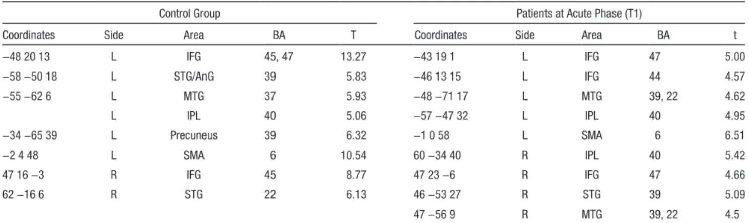
Task performance analysis did not reveal differences between groups across examinations (see the online-only Data Supplement). In control subjects, the analysis revealed strong bilateral activation of several brain language-related areas: left and right IFG, anterior part of left and right STG, left infe-rior parietal lobule (IPL) and precuneus, left supplementary motor area in the medial frontal gyrus, and left middle tem-poral gyrus. Two days after the stroke, all Rehab and NRehab aphasic patients, as a group, showed a markedly reduced cor-tical activation both in the RH and in the LH compared with controls. In particular, clusters of activation were found in left and right IFG, left and right middle temporal gyrus, right IPL, and right STG (Table 1 and Figure 1). Based on activations of controls and patients, we selected the following ROIs: bilat-eral IFG, left middle temporal gyrus, right STG, and left IPL. Group analysis at T2 follow-up showed different patterns of activation in Rehab and NRehab groups. Rehab patients acti-vated left and right IFG, left and right STG, left IPL/supra-marginal gyrus, and left supplementary motor area, whereas in NRehab patients, the main activated area at T2 was only the right IFG. The remaining language-related areas, except left IPL/supramarginal gyrus, showed less cortical activation. At 6-month follow-up (T3), an increase in both hemispheres’ corti-cal activations was similarly seen in both groups, mainly locorti-cal- local-ized in LH, particularly the left and right IFG, left STG, left and right IPL/supramarginal gyrus, and left supplementary motor area (Figure 2 and Table 2). This pattern was confirmed by the between-group comparison. At T1, no difference between Rehab and NRehab groups was found, as was expected,
Table 1. Baseline Brain Activation During Functional MRI Comprehension Task in Control Group and Patients
Control Group Patients at Acute Phase (T1)
Coordinates Side Area BA T Coordinates Side Area BA t
−48 20 13 L IFG 45, 47 13.27 −43 19 1 L IFG 47 5.00 −58 −50 18 L STG/AnG 39 5.83 −46 13 15 L IFG 44 4.57 −55 −62 6 L MTG 37 5.93 −48 −71 17 L MTG 39, 22 4.62 L IPL 40 5.06 −57 −47 32 L IPL 40 4.95 −34 −65 39 L Precuneus 39 6.32 −1 0 58 L SMA 6 6.51 −2 4 48 L SMA 6 10.54 60 −34 40 R IPL 40 5.42 47 16 −3 R IFG 45 8.77 47 23 −6 R IFG 47 4.66 62 −16 6 R STG 22 6.13 46 −53 27 R STG 39 5.09 47 −56 9 R MTG 39, 22 4.5
P<0.001; uncorrected. AnG indicates angular gyrus; BA, broadman area; IFG, inferior frontal gyrus; IPL, inferior parietal lobule; L, left, MTG, middle temporal gyrus; R, right; SMA, supplementary motor area; and STG, superior temporal gyrus.
whereas at T2, left IFG revealed to be predominantly activated by Rehab patients followed, at 6 months poststroke, by a simi-lar recruitment of areas predominantly located in the LH, again with a higher recruitment observed in Rehab patients compared with NRehab patients (Figure 2 and Table 2). The opposite NRehab>Rehab contrast showed no areas with increased acti-vation in the NRehab versus the Rehab group.
Within-group changes in brain activation over time (T2>T1 and T3>T2) are visualized in Figure 3 and reported in Table 3. After a 2-week course of language rehabilitation, an increased activation was found in left IFG (Broca area) and in STG in Rehab patients, whereas NRehab group showed change in activation of right IFG and left IPL. The 6-month follow-up (no language rehabilita-tion condirehabilita-tion for both groups) change in activarehabilita-tion was simi-larly present in LH Broca area, STG, and IPL in both groups. To summarize, left IFG was predominantly recruited 2 weeks after the stroke only in patients submitted to language rehabilitation, whereas after 6 months, all the patients had a prevalent recruit-ment of brain language-related areas located in the LH.
These results were subsequently confirmed using ANOVA: a treatment×time interaction was found in the left IFG at T2 only (F=10.2; P=0.009), with significantly higher activation in the Rehab group as revealed by post hoc test (P<0.05). No significant difference was found between groups at T1 and T3, neither in other ROIs, indicating left IFG as the crucial site of different brain activation between groups, at T2 only.
Analysis of Correlations
To further corroborate the previous results, a correlation between functional changes after rehabilitation in ROIs and naming and written language changes (T2>T1) was performed. A significant correlation was found between functional imag-ing changes in the left IFG (T2>T1) only and improvement in naming (r=0.957; P<0.003) in Rehab, and changes in the right
IFG only and improvement in naming (r=0.821; P<0.015) in NRehab. See Figure 3 for details.
Discussion
Two main results arise from this pilot study: first, the clinical efficacy of aphasia rehabilitation has been well demonstrated in acute poststroke patients; second, the possible functional correlate of this treatment-related language improvement is supposed to be the left IFG early activation during the process of aphasia recovery.
Rehabilitation was found to improve naming abilities as well as writing and reading aloud in aphasic patients. The lack of significant difference in comprehension and repetition may, in our opinion, be because of the milder deficits pre-sented by some of the patients, possibly inducing a ceiling effect. The language improvement of Rehab patients, which was evident after 15 days, persists 6 months after stroke. The present results are in line with Godecke et al’s17 findings and
may have relevant clinical implications. Although the results need to be confirmed on larger samples of patients harboring aphasic syndromes of diverse severity, they seem to support early daily rehabilitation program of aphasic stroke patients as a valuable intervention. The results also provide at least partial answers to clinicians in deciding whether to wait for spontaneous improvement to occur or provide intensive early aphasia treatment. On the contrary, the present findings are not in line with the negative results of the ACT Now Study,35 in
which communication therapy and unstructured social contact were compared in the first 4 months after stroke. This latter study seems to rule out any added benefit of communication therapy. However, important methodological difference could explain the different results: contrary to the present study, the ACT Now Study was not focused on the first-ever-LH-stroke patients, but also included patients with dysarthria, and critical
Figure 2. T2 and T3 brain activation during functional MRI (fMRI) tasks. T2 (upper line) and T3 (lower line) activation in Rehab and
NRe-hab patients, and ReNRe-hab>NReNRe-hab in left hemisphere (left) and right hemisphereRH (right) at T2 and T3. Activation shown in the inferior
frontal gyrus (white arrow), superior temporal gyrus (dotted arrow), inferior parietal lobule, and angular gyrus (open arrow). Statistical significance level, P<0.001; uncorrected.
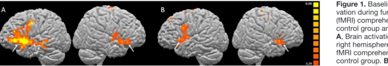
Figure 1. Baseline brain
acti-vation during functional MRI (fMRI) comprehension task in control group and patients.
A, Brain activation in left and
right hemisphere during the fMRI comprehension task in control group. B, Brain
activa-tion in left and right hemisphere during the fMRI comprehension task in patients (both Rehab and NRehab) at acute phase (T1). Activations in language-related areas of inferior frontal gyrus, mainly in the operculum (white arrow), in superior and middle temporal gyrus (dotted line). Statistical significance level, P<0.001; uncorrected.
time window for rehabilitation was not considered in the first 2 weeks poststroke.
The second main finding of the present study was that patients submitted to early intensive (2 weeks) poststroke reha-bilitation showed a different recruitment of language-related cortical areas compared with the no treatment group, particu-larly involving the LH. Although all patients showed a dramatic decrease in cortical activations compared with controls 2 dps, Rehab patients showed larger predominantly left-sided areas at the second examination, particularly the left Broca area, com-pared with NRehab patients, correlating with language behav-ioral improvement. A further increase in activation in LH at 6-month (T3 to T2) follow-up (when both groups were not sub-mitted to language rehabilitation), with a persistently higher activation in Rehab patients, was also found. In our opinion, the intensive aphasia training protocol seems to promote the early shifting of activation to the LH in the first 2 weeks post-stroke, as observed in our patients submitted to early aphasia therapy. On the other hand, NRehab group showed a pattern of activation which is similar to the one found in the Saur et al (2006) patient group, ie, a reduced activation in the L lan-guage area in the acute phase, followed by a recruitment of homologue language zone in subacute phase and by a normal-ization of activation in chronic phase. Importantly, we could reasonably rule out the practice effect of task on fMRI results.
In fact, we did not find differences over time in fMRI task per-formance, especially between T1 and T2, and if practice had influenced the findings, we would have expected decreased activation between T1 and T2,36 which is the opposite of what
we actually found.
These findings suggest that a possible correlate of early language poststroke rehabilitation could be the earlier recruit-ment of the LH, namely the left Broca area, whereas the cor-relate of spontaneous, and possibly less efficient, language recovery in NRehab patients could be the activation of right IFG, which we found to be related with language improve-ment in NRehab only.
Our results support Saur et al4’s hypothesis of a different
temporal dynamics in LH and RH activation after stroke, but most importantly, they point to a specific correlation between early language training and early recruitment of the left Broca area. The frequency of left IFG involvement in the ischemic lesion was identical in the 2 groups, so the possibility that the results may be driven by a sparing of the area seems unlikely. The fall in activation of many language-related areas not involved in the lesion at T1, and their subsequent activation over time, may be otherwise ascribed to their early functional suppression because of diaschysis and the eventual reduction of the ischemic penumbra, associated with a specific exercise-induced brain plasticity. It may be speculated that the more the Table 2. Clusters of Activation in Rehab and NRehab Patients at T1, T2, and T3 Examinations
Rehab NRehab Rehab>NRehab
Coordinates Side Area BA t Coordinates Side Area BA t Coordinates Side Area BA t T1 −45 20 1 L IFG 45, 37 3.91 −51 29 1 L IFG 47 3.77 Nothing
−60 −46 28 L IPL 40 3.65 −57 −25 28 L IPL 40 2.94 −56 −59 0 L MTG 22 3.42 −60 −40 4 L MTG 22 3.22 50 23 −3 R IFG 47 4.22 −18 11 67 L SMA 6 3.89 48 −38 55 R IPL 40 3.92 48 17 4 R IFG 45 3.95 3 7 64 R SMA 6 4.15 51 −34 53 R IFG 40 2.83 18 5 70 R SMA 6 4.30
T2 −48 20 10 L IFG 45, 47 6.68 −48 21 0 L IFG 47 4.80 −52 16 3 L IFG 44, 45 7.35 −54 −58 21 L STG/AnG 39 3.52 −37 −56 32 L STG/AnG 39 4.05 −49 28 9 L IPL 40 7.18 −48 29 24 L MFG 46 4.10 −55 −62 28 L STG 39 6.37 −57 −42 29 L IPL/SmG 40 4.22 −2 6 52 L SMA 6 5.26 −1 20 56 L SMA 6 5.53 −56 −32 6 L STG 22 5.20
−3 −13 52 L SMA 6 6.50
46 20 5 R IFG 45 4.21 48 20 10 R IFG 45 5.67 61 −29 30 R IPL 40 6.80
59 −38 13 R STG 22 5.73 54 4 13 R IFG 44, 46 4.57
40 −43 38 R SmG 40 4.40
T3 −46 19 9 L IFG 45, 46 7.24 −49 19 9 L IFG 45, 44 8.72 −49 −35 36 L IPL 40 5.42 −54 −56 14 L STG 39 5.28 −58 −41 6 L STG 22 7.72 −52 25 9 L IFG 45 6.10 −50 −30 33 L IPL 40 6.70 −57 −39 22 L IPL 40 6.13 −34 25 0 L IFG 47 6.10
−2 0 54 L SMA 6 6.36 −1 0 58 L SMA 6 7.20
47 25 5 R IFG 45, 44 6.62 42 19 6 R IFG 45 6.76 50 24 4* R IFG 45 4.64 63 −29 22 R IPL/SmG 40 5.65 46 −54 38 R IPL/SmG 40 4.80 63 −37 25* R IPL 40 3.73 Between-group analysis in acute (T1), subacute (T2), and chronic (T3) stage after stroke. P<0.001; P<0.005. AnG indicates angular gyrus; BA, broadman area; IFG, inferior frontal gyrus; IPL, inferior parietal lobule; L, left; MFG, middle frontal gyrus; MTG, middle temporal gyrus; R, right; SMA, supplementar motor area; SmG: supramarginal gyrus; STG, superior temporal gyrus; and t, Student t.
naming abilities after language training, the higher and earlier would be the recruitment of the left Broca area, possibly caus-ing persistent effects after 6 months.
The hypothesis of an incomplete aphasia amelioration sus-tained mainly by the right-sided language-related areas, partic-ularly by the right IFG (right Broca jomologue) and, conversely, a better outcome related to activation in left perilesional areas,
has been sustained by many authors,7,11,30,37,38 if not all,39 when
examining chronic aphasics. Only in some patients, right IFG activation would be essential for residual language function; however, its compensatory potential would be less effective than in patients who recover left IFG function.
Our results support the role of left IFG activation in acute poststroke patients submitted to language rehabilitation and
Table 3. Within-Group Analysis at Baseline and Follow-Ups (Change in Activation T2>T1 and T3>T2)
Rehab NRehab
Coordinates Side Area BA t Coordinates Side Area BA t T2>T1 −48 14 7 L IFG 44 4.63 −45 −49 28 L IPL 40 3.60 54 −34 10 R STG 22 4.58 39 23 4 R IFG 45 3.45 T3>T2 −37 24 3 L IFG 45 5.02 −50 35 10 L IFG 44 6.67 −34 26 1 47| −44 29 0 46 −46 11 9 47 −55 14 −2 L STG 22 5.21 −58 −53 9 L STG 22 5.06 −39 −31 36 L IPL 40 5.18 −64 −49 −5 L MTG 21 4.66 −47 −72 20 L AnG 39 5.03 −62 −22 15 L SmG 40 4.35 33 29 1 R IFG 45–47 4.65 47 17 11 R IFG 45 4.91 47 −55 37 R IPL 40 3.88 P<0.005; uncorrected. AnG indicates angular gyrus; BA, broadman area; IFG, inferior frontal gyrus; IPL, inferior parietal lobule; L, left; MTG, middle temporal gyrus; R, right; SmG, supramarginal gyrus; STG, superior temporal gyrus; and t, Student t.
Figure 3. Brain activation changes over time and correlations with clinical data. The functional MRI contrast show changes in activation at
different follow-ups in Rehab (upper line) and NRehab (lower line) patients. In the scatter plots (left column) are shown the results of cor-relation analysis between T2>T1 activation of the inferior frontal gyrus (IFG; left [L] IFG for Rehab and of right [R] IFG for NRehab; y axis) and naming ability improvement (T2>T1; x axis). T3>T2 activation of the L IFG and T2>T1 activation of the R and L superior temporal gyrus for both groups do not correlate with clinical performance. Statistical significance level, P<0.005; uncorrected.
point to the potential role of right IFG as the neural substrate for aphasia spontaneous recovery.
The activation of left IFG has also been described as a reflection of a therapy-induced improvement of naming8,12 in
chronic aphasics, whereas right IFG has been described to be activated both by healthy subjects who were taught to name objects in a foreign language40 and in aphasics with left IFG
lesions.41 Our study suggests a possible dichotomy between
LH and RH IFG activation, respectively, in early rehabilitated and spontaneously recovered aphasics. The differential activa-tion of left IFG reported in patients who received rehabilita-tion seems to specifically reflect an effect of language therapy in the first 2 weeks poststroke.
Neural correlates of the training-induced neuropsychologi-cal improvement have been reported to be different in rela-tion to the linguistic task used,42,43 as well as in relation to the
training used both in aphasia12 and in other nonstroke-induced
neuropsychological symptoms, that is, the executive functions and information processing training in multiple sclerosis.44
This pilot study, however, has several limitations. First, the study used a small number of patients with relatively low severity of aphasia, probably because of the small lesion size. This small sample size reflects on the difficulty in including homogeneous acute stroke aphasics in an fMRI study. Second, we did not use a control therapy, because the daily social inter-action of patients in the stroke unit could, in our opinion, be considered a unstructured language treatment. Third, the inclu-sion of patients with moderate/mild baseline impairment may have induced a ceiling effect that prevented a higher improve-ment in their performance over time. Fourth, data were not corrected for multiple comparisons because of the small sam-ple size, causing the statistical power to be low. Finally, we found improved output language tests in our patients, which we tended to associate with brain activation induced by a comprehension task. On this point, we argue that, in conjunc-tion with several studies,31 the same language-related areas
have been activated by different tasks (either comprehension or naming). Hence, it is not unlikely that different language tasks activate the same language-related areas. Further studies with different fMRI language tasks, larger samples including patients with more severe aphasia, and using ecological scales on communicative abilities will be useful.
Acknowledgments
We acknowledge A. Duina and M. Tosetti for technical support.
Disclosures
None.
References
1. Müller RA, Rothermel RD, Behen ME, Muzik O, Chakraborty PK, Chugani HT. Language organization in patients with early and late left-hemisphere lesion: a PET study. Neuropsychologia. 1999;37:545–557. 2. Abo M, Senoo A, Watanabe S, Miyano S, Doseki K, Sasaki N, et al.
Language-related brain function during word repetition in post-stroke aphasics. Neuroreport. 2004;15:1891–1894.
3. Crinion J, Price CJ. Right anterior superior temporal activation predicts auditory sentence comprehension following aphasic stroke. Brain. 2004; 128:1858–1871.
4. Saur D, Lange R, Baumgaertner A, Schraknepper V, Willmes K, Rijntjes M, et al. Dynamics of language reorganization after stroke. Brain. 2006;129(pt 6):1371–1384.
5. Hillis AE. Aphasia: progress in the last quarter of a century. Neurology. 2007;69:200–213.
6. Winhuisen L, Thiel A, Schumacher B, Kessler J, Rudolf J, Haupt WF, et al. The right inferior frontal gyrus and poststroke aphasia: a follow-up investigation. Stroke. 2007;38:1286–1292.
7. van Oers CA, Vink M, van Zandvoort MJ, van der Worp HB, de Haan EH, Kappelle LJ, et al. Contribution of the left and right inferior fron-tal gyrus in recovery from aphasia. A functional MRI study in stroke patients with preserved hemodynamic responsiveness. Neuroimage. 2010;49:885–893.
8. Lerger A, Demonet JG, Roff S, Aithamon B, Touyeras B, Puel M, et al. Neural substrates of spoken language rehabilitation in an aphasic patients: an fMRI study. NeuroImage. 2002;17:174–183.
9. Fridriksson J, Morrow-Odom L, Moser D, Fridriksson A, Baylis G. Neural recruitment associated with anomia treatment in aphasia.
Neuroimage. 2006;32:1403–1412.
10. Fridriksson J, Moser D, Bonilha L, Morrow-Odom KL, Shaw H, Fridriksson A, et al. Neural correlates of phonological and semantic-based anomia treatment in aphasia. Neuropsychologia. 2007;45:1812–1822. 11. Breier JI, Juranek J, Maher LM, Schmadeke S, Men D, Papanicolaou
AC. Behavioral and neurophysiologic response to therapy for chronic aphasia. Arch Phys Med Rehabil. 2009;90:2026–2033.
12. Rochon E, Leonard C, Burianova H, Laird L, Soros P, Graham S, et al. Neural changes after phonological treatment for anomia: an fMRI study.
Brain Lang. 2010;114:164–179.
13. Szaflarski JP, Eaton K, Ball AL, Banks C, Vannest J, Allendorfer JB, et al. Poststroke aphasia recovery assessed with functional magnetic res-onance imaging and a picture identification task. J Stroke Cerebrovasc
Dis. 2011;20:336–345.
14. Huber W, Poeck K, Kerschensteiner M. Acquired organic pathologies of linguistic behavior: neurolinguistic disorders. In: Blanken G, ed.
Linguistic Disorders and Pathologies: An International Handbook. Berlin: Walter de Gruyter & Co; 1977;114.
15. Bhogal SK, Teasell R, Speechley M. Intensity of aphasia therapy, impact on recovery. Stroke. 2003;34:987–993.
16. Robey RR. A meta-analysis of clinical outcomes in the treatment of aphasia. J Speech Lang Hear Res. 1998;41:172–187.
17. Godecke E, Hird K, Lalor EE, Rai T, Phillips MR. Very early poststroke aphasia therapy: a pilot randomized controlled efficacy trial. Int J Stroke. 2012;7:635–644.
18. Fridriksson J, Richardson JD, Fillmore P, Cai B. Left hemisphere plastic-ity and aphasia recovery. Neuroimage. 2012;60:854–863.
19. Fridriksson J, Bonilha L, Baker JM, Moser D, Rorden C. Activity in preserved left hemisphere regions predicts anomia severity in aphasia.
Cereb Cortex. 2010;20:1013–1019.
20. Fridriksson J. Preservation and modulation of specific left hemisphere regions is vital for treated recovery from anomia in stroke. J Neurosci. 2010;30:11558–11564.
21. Fernandez B, Cardebat D, Demonet JF, Joseph PA, Mazaux JM, Barat M, et al. Functional MRI follow-up study of language processes in healthy sub-jects and during recovery in a case of aphasia. Stroke. 2004;35:2171–2176. 22. Meinzer M, Mohammadi S, Kugel H, Schiffbauer H, Flöel A, Albers
J, et al. Integrity of the hippocampus and surrounding white matter is correlated with language training success in aphasia. Neuroimage. 2010;53:283–290.
23. Perani D, Cappa SF, Tettamanti M, Rosa M, Scifo P, Miozzo A, et al. A fMRI study of word retrieval in aphasia. Brain Lang. 2003;85:357–368. 24. Richter M, Miltner WH, Straube T. Association between therapy out-come and right-hemispheric activation in chronic aphasia. Brain. 2008;131(pt 5):1391–1401.
25. Fridriksson J, Baker JM, Moser D. Cortical mapping of naming errors in aphasia. Hum Brain Mapp. 2009;30:2487–2498.
26. Heiss WD, Thiel A. A proposed regional hierarchy in recovery of post-stroke aphasia. Brain Lang. 2006;98:118–123.
27. Baker JM, Rorden C, Fridriksson J. Using transcranial direct-current stim-ulation to treat stroke patients with aphasia. Stroke. 2010;41:1229–1236. 28. Cherney LR, Small SL. Task-dependent changes in brain activation fol-lowing therapy for nonfluent aphasia: discussion of two individual cases.
J Int Neuropsychol Soc. 2006;12:828–842.
29. Breier JI, Maher LM, Novak B, Papanicolaou AC. Functional imaging before and after constraint-induced language therapy for aphasia using magnetoencephalography. Neurocase. 2006;12:322–331.
30. Vitali P, Abutalebi J, Tettamanti M, Danna M, Ansaldo AI, Perani D, et al. Training-induced brain remapping in chronic aphasia: a pilot study.
Neurorehabil Neural Repair. 2007;21:152–160.
31. Thompson CK, den Ouden DB. Neuroimaging and recovery of language in aphasia. Curr Neurol Neurosci Rep. 2008;8:475–483.
32. Luft AR. Rehabilitation and plasticity. Front Neurol Neurosci. 2013;32:88–94.
33. Oldfield RC. The assessment and analysis of handedness: the Edinburgh inventory. Neuropsychologia. 1971;9:97–113.
34. Huber W, Poeck K, Weniger D, Willmess K. Aachener Apahsie Test.
Verlag Fur Psychologie. 2nd ed. Florence, Italy: Organizzazioni Speciali Firenze;1996.
35. Bowen A, Hesketh A, Patchick E, Young A, Davies L, Vail A, et al. Effectiveness of enhanced communication therapy in the first four months after stroke for aphasia and dysarthria: a randomised controlled trial. BMJ. 2012;345:e4407.
36. Henson RN. Neuroimaging studies of priming. Prog Neurobiol. 2003;70: 53–81.
37. Winhuisen L, Thiel A, Schumacher B, Kessler J, Rudolf J, Haupt WF, et al. Role of the contralateral inferior frontal gyrus in recovery of
language function in poststroke aphasia: a combined repetitive transcra-nial magnetic stimulation and positron emission tomography study. Stroke. 2005;36:1759–1763.
38. Heiss WD, Thiel A, Kessler J, Herholz K. Disturbance and recovery of language function: correlates in PET activation studies. Neuroimage. 2003;20(suppl 1):S42–S49.
39. Musso M, Weiller C, Kiebel S, Müller SP, Bülau P, Rijntjes M. Training-induced brain plasticity in aphasia. Brain. 1999;122(pt 9):1781–1790. 40. Raboyeau G, De Boissezon X, Marie N, Balduyck S, Puel M, Bézy C,
et al. Right hemisphere activation in recovery from aphasia: lesion effect or function recruitment? Neurology. 2008;70:290–298.
41. Turkeltaub PE, Messing S, Norise C, Hamilton RH. Are networks for residual language function and recovery consistent across aphasic patients? Neurology. 2011;76:1726–1734.
42. Nudo RJ. Plasticity. NeuroRx. 2006;3:420–427.
43. Price CJ, Crinion J. The latest on functional imaging studies of aphasic stroke. Curr Opin Neurol. 2005;18:429–434.
44. Mattioli F, Stampatori C, Bellomi F, Capra R, Rocca M, Filippi M. Neuropsychological rehabilitation in adult multiple sclerosis. Neurol Sci. 2010;31(suppl 2):S271–S274.
1
Early aphasia rehabilitation is associated with functional reactivation of the left
inferior frontal gyrus: a pilot study
Mattioli F, Ambrosi C, Mascaro L, Scarpazza C, Pasquali P, Frugoni M, Magoni M, Biagi L , Gasparotti R.
Supplemental Methods:
Patients: Patients were recruited from the Stroke Unit of Spedali Civili of Brescia (Italy)
during a 24 month period from January 2008 to December 2009. 85 patients with aphasia were
screened for the study. The reasons for exclusion were the following: no native Italian speakers
(n=6); age > 80 (n=19); previous diagnosis of dementia or psychiatric disorders (n=8); stroke in the
territory other of the MCA (n=21); severe aphasia with severe comprehension impairment (n=7);
pacemaker carriers (n=6); claustrophobia (n=2); severe obesity, i.e. impossibility to put the patient
in the MRI scanner (n=1) and deafness (n=4). A total of 12 stroke patients were included.
Lesions analysis: For each patients, lesion extent and location were documented by using the
T1 Diffusion Weighted (DW) scan. Lesions were manually traced by a neurologist expert in image
analysis on the MRI template scan from the Montreal Neurological Institute provided with the
MRIcron software (Rorden and Brett, 2000;
free available at http://www.mricro.com/mricron). This
scan is normalized to Talairach space and it is a widely used template for normalization in
functional brain imaging (e.g. Moretti et al. 2009). The original DW MRI of each patients was
always available during the tracement, and anatomical landmarks were used to better define the
boundaries of each lesions. After superimposing individual brain lesions onto the standard Ch2
brain template, MRIcron automatically calculated the total brain lesion volume (in cc). The
individual lesion volume are reported in table S1.
Language Rehabilitation: No ethics were violated using the treatment/no treatment
randomization, since rehabilitation of aphasia in the acute post stroke phase was not widely
accepted as useful at the time of this study. Language rehabilitation was conducted on an in patient
basis, mainly focusing on verbal comprehension and lexical retrieval. In each session, after a short
and simple dialogue with the patient, covering his mood and status, as well as any relevant episodes
occurred during the day, a naming task was usually conducted, where patients had to spontaneously
2
facilitations were given. Single word as well as sentence comprehension was also treated with the
help of available common objects and objects pictures. A semi structured rehabilitation setting was
used, instead of a rigidly predetermined set of tasks identical for all the subjects, due to the clinical
condition of the acute phase and the location (the stroke unit) where the rehabilitation was
conducted. Generally, a stimulation of the impaired linguistic functions was conducted by the
therapist, according to the deficits shown by the AAT.
fMRI paradigm:
Patients listened to a recorded voice pronouncing semantically correct
sentences, randomly intermixed with semantically violated ones and reversed ones, for the control
condition and had to press a button with the right hand only if they heard a semantically correct
sentence. The sentences were simple (subject-verb-object, i.e. the boy eats the apple, for a
semantically correct phrase, or the boy reads the apple for a semantically violated; balanced for
gender, number) and short (<2500 ms). 28 sentences were used for each run (7 semantically correct,
7 semantically violated and 14 reverse), which was repeated 4 times in each examination. The
correct response number was measured for all the patients each time. Presentation was randomized
across runs. fMRI examinations were performed on a 1.5 Siemens Avanto (Siemens, Erlangen
Germany) equipped with an 8 channel head coil (Invivo-Gainesville), using an integrated
audio-video system (eloquence Invivo-Gainesville) connected to an external computer.
An event-related paradigm was generated with EPRIME software (Psycology Software Tools
Pittsburgh). Recording and reverse processing was performed with Adobe Audition software
(Adobe system incorporated, San Jose’, California).
fMRI acquisition and preprocessing: We used cushions to minimize patients head movement
inside the scanner. The acquisition protocol consisted of the repetition of 126 BOLD volumes
(SE-EPI/ TR=2500ms, TE=50ms, Matrix 64*64, FOV= 224*224 mm, 3.5mm slice thickness) with the
following scheme: a volume with a ‘bip’ (noise) sound to alert the patient of the incoming
sentence, a volume with the recorded sentence and 2 or 3 volumes of rest. fMRI acquisition time
was about 21’20’’. The first 2 volumes were discarded to eliminate T1 effects. After functional
scanning, an anatomical image was collected using a magnetized prepared rapid acquisition
gradient-echo echo planar imaging (MPRAGE) sequence, (TR/TE 2050/2.56 ms, 256 mm FOV,
256*256 matrix, 144 sagittal slices for an effective resolution of 1.0 mm.
fMRI images were processed with Brain Voyager QX (vs. 1.9- Brain Innovation – Maastricht
The Nederland). A pre-processing with high pass filtering for drifts removal and slice scan time
correction (cubic spline interpolation) was performed, as well as a 3D motion correction with
images spatially realigned to the first volume. Functional data were co registered with the 3D
anatomical images, which were normalized to the Talairach space. A mask of the lesion was created
on the base of co-registered MRI in order to take into account the possible errors in normalization
due to lesions. A correction for the delay of the hemodynamic response was also applied to the
boxcar design matrix. A general linear model approach was used for contrast evaluations.
3
Supplemental Results:
Lesions analysis: Lesion volume in Rehab (669,04±438,21 mm
3) and NRehab
(716,92±581,30 mm
3) patients was not statistically different (two independent sample t-test:
p=0.87). We performed a correlation analysis between lesions volume and the improvement in
naming and written language between T1 and T2 to investigate the possibility that bigger
improvement in clinical tests were due to smaller lesion size. Both the correlations were not
significant (lesions volume and written language improvement: Spearman R = 0.23; p=0.46; lesions
volume and naming improvement: Spearman R = 0.40; p=0.19), supporting the assumption that
improvement in clinical performance is not driven by lesion size.
fMRI Behavioral results: the percentages of correct answer were analyzed. Mean correct
answers at T1 were 0.82 and 0.93 in Rehab and NRehab groups respectively, at T2 were 0.83 and
0.89 and at T3 were 0.81 and 0.83. Since the data of both groups at each time were normally
distributed (T1: W=0.9, p=0.19 at Shapiro Wilk test; T2: W=0.92, p=0.29; T3: W=0.91, p=0.26), a
repeated measure ANOVA was conducted on percentage of correct answer, using Group (two level)
as between subjects variable, and Time (three level) as within subjects variable. The results revealed
no significant results (main effect of Group: F[1,10]=1,51, p=0,24; main effect of Time:
F[2,20]=1,97, p=0,16; Group x Time Interaction: F[2,20]=1,35, p=0,28).
4
M= male, F= female; NonFlu = non fluent aphasia; MCA = middle cerebral artery , SFG = Superior frontal gyrus, MFG = middle frontal gyrus, IFG
= inferior frontal gyrus, CR = corona radiate, STG: superior temporal gyrus, MTG: middle temporal gyrus; NIHSS = National Institute of Health
Stroke Scale: no deficit = 0, maximal deficit = 34. Significance = p value at non parametric Mann Whitney U test.
5
Values denotes row scores obtained from patients. *p denotes significance at Mann-Whitney U test.
6
In the upper line Non Rehabilitated patients’ lesions are shown; in the lower line Rehabilitaed patients’
lesions are shown.
Supplementary References:
Rorden, C., Brett, M. Stereotaxic display of brain lesions. Behav Neurol. 2000;
12(4):191-200.
Moretti, L., Dragone, D., di Pellegrino, G. Reward and social valuation deficits following
ventromedial prefrontal damage. J Cogn Neurosci. 2009; 21 (1): 128-140.
Snodgrass, JG, Vanderwart M. A standardized set of 260 pictures: Norms for name
agreement, image agreement, familiarity, and visual complexity. J Exp Psychol: Hum Learn Mem.
1980; 6(2):174-215.
Marina Frugoni, Mauro Magoni, Laura Biagi and Roberto Gasparotti
Flavia Mattioli, Claudia Ambrosi, Lorella Mascaro, Cristina Scarpazza, Patrizia Pasquali,
Print ISSN: 0039-2499. Online ISSN: 1524-4628
Copyright © 2013 American Heart Association, Inc. All rights reserved.
is published by the American Heart Association, 7272 Greenville Avenue, Dallas, TX 75231
Stroke
doi: 10.1161/STROKEAHA.113.003192
2014;45:545-552; originally published online December 5, 2013;
Stroke.
http://stroke.ahajournals.org/content/45/2/545
World Wide Web at:
The online version of this article, along with updated information and services, is located on the
http://stroke.ahajournals.org/content/suppl/2013/12/05/STROKEAHA.113.003192.DC1.html
Data Supplement (unedited) at:
http://stroke.ahajournals.org//subscriptions/
is online at:
Stroke
Information about subscribing to
Subscriptions:
http://www.lww.com/reprints
Information about reprints can be found online at:
Reprints:
document.
Permissions and Rights Question and Answer
process is available in the
Request Permissions in the middle column of the Web page under Services. Further information about this Once the online version of the published article for which permission is being requested is located, click
can be obtained via RightsLink, a service of the Copyright Clearance Center, not the Editorial Office.
Stroke
in
Requests for permissions to reproduce figures, tables, or portions of articles originally published