www.ajhg.org The American Journal of Human Genetics Volume 80 March 2007 539
REPORT
Localization of a Gene for Nonsyndromic Renal Hypodysplasia
to Chromosome 1p32-33
Simone Sanna-Cherchi, Gianluca Caridi, Patricia L. Weng, Monica Dagnino, Marco Seri, Anita Konka, Danio Somenzi, Alba Carrea, Claudia Izzi, Domenica Casu, Landino Allegri, Kai M. Schmidt-Ott, Jonathan Barasch, Francesco Scolari, Roberto Ravazzolo,
Gian Marco Ghiggeri, and Ali G. Gharavi
Nonsyndromic defects in the urinary tract are the most common cause of end-stage renal failure in children and account for a significant proportion of adult nephropathy. The genetic basis of these disorders is not fully understood. We studied seven multiplex kindreds ascertained via an index case with a nonsyndromic solitary kidney or renal hypodysplasia. Systematic ultrasonographic screening revealed that many family members harbor malformations, such as solitary kid-neys, hypodysplasia, or ureteric abnormalities (in a total of 29 affected individuals). A genomewide scan identified significant linkage to a 6.9-Mb segment on chromosome 1p32-33 under an autosomal dominant model with reduced penetrance (peak LOD score 3.5 at D1S2652 in the largest kindred). Altogether, three of the seven families showed positive LOD scores at this interval, demonstrating heterogeneity of the trait (peak HLOD 3.9, with 45% of families linked). The chromosome 1p32-33 interval contains 52 transcription units, and at least 23 of these are expressed at stage E12.5 in the murine ureteric bud and/or metanephric mesenchyme. These data show that autosomal dominant nonsyndromic renal hypodysplasia and associated urinary tract malformations are genetically heterogeneous and identify a locus for this common cause of human kidney failure.
From the Department of Medicine, Division of Nephrology, Columbia University College of Physician and Surgeons (S.S.-C.; P.L.W.; A.K.; K.M.S.-O.; J.B.; A.G.G.), and Department of Pediatrics, Division of Nephrology, Mount Sinai School of Medicine (P.L.W.), New York; Department of Clinical Medicine, Nephrology and Health Science, University of Parma, Parma, Italy (S.S.-C.; D.S.; L.A.); Laboratory on Pathophysiology of Uremia (G.C.; M.D.; M.S.; A.C.; G.M.G.) and Laboratory of Molecular Genetics (M.S.; R.R.), G. Gaslini Institute, Genoa, Italy; Division and Chair of Nephrology, Spedali Civili, University of Brescia, Brescia, Italy (C.I.; F.S.); and Division of Nephrology and Dialysis, Hospital of Alghero, Alghero, Italy (D.C.)
Received July 12, 2006; accepted for publication January 2, 2007; electronically published January 26, 2007.
Address for correspondence and reprints: Dr. Ali G. Gharavi, Department of Medicine, Columbia University College of Physicians and Surgeons, 630 W. 168th Street, P&S 10-432, New York, NY 10032. E-mail: [email protected]
Am. J. Hum. Genet. 2007;80:539–549. 䉷 2007 by The American Society of Human Genetics. All rights reserved. 0002-9297/2007/8003-0017$15.00 DOI: 10.1086/512248
The human kidney is composed of 500,000 to 1.8 million independent functional units, called “nephrons.”1,2
Kid-ney failure occurs when acquired or hereditary disorders cause progressive nephron loss and reduce their numbers to below the threshold required for maintenance of fluid-electrolyte homeostasis and elimination of nitrogenous wastes. Congenital abnormalities that impair nephron de-velopment comprise 30%–50% of congenital anomalies detected on prenatal screening.3,4 There is wide
interin-dividual variability in the anatomy and clinical course of these malformations, but, altogether, they account for up to 50% of pediatric and 10% of adult end-stage renal
disease worldwide.5,6 Renal agenesis/adysplasia (MIM
%191830) is one of the most severe forms of malforma-tions; bilateral renal agenesis occurs in∼1 in 2,000 births and is nearly always fatal because of oligohydramnios and consequent pulmonary hypoplasia.3,4,7,8 Other related
phenotypic variants, such as unilateral agenesis and uni-lateral or biuni-lateral renal hypodysplasia, occur more fre-quently (1 in 1,000 births).3,4Affected kidneys frequently
exhibit dysplastic parenchyma, reduced nephron mass, and impaired function, predisposing the individual to re-nal failure.8Kidney malformations are also commonly
ac-companied by anatomic abnormalities in the lower uri-nary tract, such as ureteropelvic junction obstruction
(UPJO) or vesicoureteral reflux (VUR).8–12Impairment of
urinary drainage due to these lower-tract defects further contributes to nephron injury and augments the risk of chronic renal failure.
The etiology of the majority of nonsyndromic forms of urinary tract malformations remains unknown. The over-lap in phenotypic expression, together with the limita-tions of morphological classification, has complicated ef-forts to understand the primary pathogenesis of these disorders and to formulate clear diagnostic categories. For example, it is not known whether the observation of a congenital solitary kidney in an adult represents a primary failure of kidney development (agenesis) or the end-result of a dysplastic kidney that underwent involution.3,8
Sev-eral risk factors for human urinary tract malformations are recognized, including maternal diabetes13 or
intra-uterine exposure to drugs such as ACE inhibitors.14 In
addition, the genetic bases for several rare, syndromic disorders featuring renal developmental defects have been elucidated (e.g., renal-coloboma syndrome [MIM #120330], Fraser syndrome [MIM #219000], and bran-chiootorenal syndrome [MIM #113650]). Gene-targeting studies have shown that alteration in gene dosage or dis-ruption in the temporospatial sequence of gene expression in intermediate mesodermal tissues (the metanephric
mesenchyme [MM] or the ureteric bud [UB]) can result in developmental defects that affect both the upper and lower urinary system.8,12,15–22Moreover, because of
inter-dependence of developmental pathways, similar renal and urologic phenotypes arise by inactivation of different genes.8,12,15–22These findings are consistent with the “bud
theory” of Mackie and Stephens, which proposes that ab-normalities in the kidney and ureter stem from the same common mechanism—namely, abnormal ureteral bud-ding from the Wolffian duct into the bladder results in malposition of the ureteral orifice.12These data together
with the phenotypic overlap observed in human renal and urologic disorders emphasize the potential for genetic het-erogeneity of disease. Candidate-gene screening has con-firmed heterogeneity, revealing mutations in the Uroplakin
3A gene in a subset of patients with renal dysplasia.23,24
Other recent surveys of patients with renal hypodysplasia, many of whom had extra-renal defects suggestive of syn-dromic disease, identified mutations in PAX2, TCF2, EYA1,
SIX1, or SALL1 in up to 17%–25% of patients.25,26Thus,
mutations identified to date account for a fraction of non-syndromic renal developmental defects, leaving the ma-jority of cases unexplained.
Several studies have reported familial aggregation of nonsyndromic renal malformations. For example, ultra-sonographic screening of first-degree relatives of 41 index patients with bilateral renal agenesis/dysgenesis identified asymptomatic renal malformations (mostly unilateral re-nal agenesis) in 9% of relatives.9Additional malformations
were also documented, including UPJO and VUR. This increased recurrence risk among relatives has been con-firmed in several other studies and is estimated at 4%– 20%.9–11,27 Multigenerational occurrence of disease has
suggested multifactorial or dominant inheritance in most kindreds,9–11but families with probable recessive
inheri-tance have also been reported.27 Altogether, these data
strongly argue in favor of genetic causation and suggest that familial aggregation is under-recognized because an-atomical defects in many family members are often silent. No linkage studies in such families have been reported to date.
We set out to investigate the genetic basis of nonsyn-dromic urinary tract malformations. Our previous studies of primary VUR had suggested that gene mapping for such heterogeneous disorders would necessitate large numbers of medium-sized pedigrees or uniquely large kindreds.28
In the situation of uncertainty about clinical classification and the potential for genetic heterogeneity, we strived to assemble a cohort with a strong genetic contribution by concentrating on patients with the most severe clinical phenotype—namely, solitary kidneys and/or renal hypo-dysplasia. We identified seven Italian pedigrees ascer-tained through an index patient with solitary kidney/hy-podysplasia documented by a renal sonogram and/or isotopic scintigraphy (fig. 1). Solitary kidney was diag-nosed when one kidney only was seen in the imaging study. Hypodysplasia was defined as kidney length below
the 95% tolerance limit based on height- and
weight-adjusted sonographic nomograms.29,30 A pediatric
ne-phrologist and clinical geneticist with expertise in diag-nosis of developmental disorders of the urinary tract evaluated all the patients. Histories and physical exami-nations were conducted to search for evidence of mul-tiorgan malformations, which would suggest syndromic forms of renal hypodysplasia. In particular, we focused on the possibility of disease associated with PAX2 or TCF2 mutations, which may masquerade as nonsyndromic malformations.25,26We performed ophthalmologic exams
to search for retinal colobomas and investigated the pres-ence of deafness or genital malformations, which would be indicative of renal-coloboma syndrome. In addition, we searched for abnormalities suggestive of TCF2 mu-tations, such as presence of renal cysts, diabetes, and el-evated liver enzymes or uric acid levels. The exclusion of
PAX2 and TCF2 by clinical criteria was further confirmed
by linkage analysis in all pedigrees and by direct sequenc-ing of TCF2 exons in one kindred (K101) that had a min-imally positive LOD score at the TCF2 locus. There was also no history of intrauterine exposure to teratogenic agents, such as ACE inhibitors and angiotensin receptor antagonists, and no history of maternal diabetes.
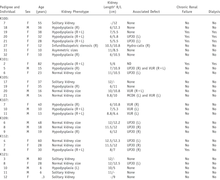
To detect additional affected relatives, we performed ul-trasonographic screening and chart review of family mem-bers. We identified 22 family members with anatomic ab-normalities (table 1). Most family members had congenital solitary kidneys/hypodysplasia, but other abnormalities were also identified, such as UPJO (11 individuals), VUR (6 individuals), infundibulopelvic stenosis (1 individual), and multicystic dysplastic kidney (1 individual). These ad-ditional defects mostly presented in association with the hypodysplasia phenotype but also occurred in isolation in a few family members (table 1). One individual (31 in K100, age 10 years) had asymmetric kidney sizes that fell within the normal range, but she was classified as affected because she had an MZ twin with unilateral renal hypo-dysplasia (this individual and her twin were considered a single affected individual in the genome scan).
Altogether, 29 individuals were classified as affected on the basis of abnormal urinary anatomy seen on imaging studies (12 males and 17 females, listed in table 1). Of these affected individuals, 10 were asymptomatic and re-ceived a diagnosis at the time of screening, and 5 had renal dysfunction (as defined by serum creatinine 11.5 mg/dl) or had developed end-stage renal failure requiring dialysis or transplantation. Twenty-nine family members had normal imaging studies and normal renal function and were classified as unaffected (fig. 1). In addition, chart review revealed that five other family members had died in childhood from end-stage renal failure, but renal im-aging studies for these individuals were not available to make a definitive diagnosis (fig. 1). These individuals and all other family members who did not undergo imaging studies were considered to have an unknown phenotype. All phenotypes were assigned prospectively. In these
ped-www.ajhg.org The American Journal of Human Genetics Volume 80 March 2007 541
Figure 1. Pedigree structure of the seven families studied. Patients with childhood end-stage renal disease (ESRD) but no sonographic
data available are indicated by a blackened rectangle inside the symbol. Arrows identify the index cases. Asterisks (*) mark the individuals from whom DNA was available for the study. Individual identification numbers correspond to table 1.
igrees, the mode of inheritance was most consistent with multifactorial inheritance or autosomal dominant trans-mission with incomplete penetrance.
All individuals gave informed consent, and the study protocol was approved by the Western Institutional Re-view Board for Columbia University and the ethics com-mittee at the Gaslini Institute. Total genomic DNA was isolated from peripheral white blood cells by use of stan-dard procedures. Genomewide screens were performed with two independent methodologies. In the largest
kin-dred (K100), we performed an ∼10-cM microsatellite
screen of autosomes (382 markers from Linkage Mapping Set v2.5 MD10 [Applied Biosystems]), using seven affected and seven unaffected individuals. The seven unaffected individuals (8, 10, 11, 12, 14, 15, and 23) were included, to increase inheritance information. During that time,
ge-nomewide SNP genotyping technology became readily available. We therefore genotyped all affected individuals from our seven kindreds for 10,204 SNPs, using the GeneChips Mapping 10K 2.0 Arrays (Affymetrix). DNA processing and gene-chip hybridization were performed as suggested by Affymetrix. This provided a uniform data set across all pedigrees.
We performed pairwise analysis of linkage, using FAST-LINK 4.1,31and multipoint analysis, using ALLEGRO 1.2c32
and Simwalk 2.89.33 Parametric analysis was performed
under a model of autosomal dominant transmission with reduced penetrance (85%, estimated from the pedigrees), disease-gene frequency of 0.001, and phenocopy rate of 0.001. We computed parametric LOD scores under genetic homogeneity and heterogeneity (calculation of HLODs). Nonparametric statistics were concurrently computed
Table 1. Clinical Data of the Affected Individuals from the Seven Pedigrees
Pedigree and Individual Sex
Age
(years) Kidney Phenotype
Kidney LengthaR/L (cm) Associated Defect Chronic Renal Failure Dialysis K100:
7 F 55 Solitary kidney -/12 None No No
18 M 36 Hypodysplasia (R) 6/12.3 None No No
19 F 38 Hypodysplasia (R⫹L) 7/5.5 None Yes Yes
20 F 32 Hypodysplasia (R⫹L) 6/5.8 UPJO (L) Yes Yes
21 F 28 Hypodysplasia (R⫹L) 5/5.5 UPJO (L) Yes Yes
27 F 12 Infundibulopelvic stenosis (R) 10.5/10.8 Hydro-calix (R) No No
31 F 10 Asymmetric sizes 11/8.5 None No No
32 F 10 Hypodysplasia (R) 6/10.5 None No No
K101:
2 F 82 Hypodysplasia (R⫹L) 5/6 ND Yes Yes
5 M 15 Hypodysplasia (R) 7/10.9 UPJO (R) and VUR (R⫹L) No No
6 F 23 Normal kidney size 11/10.5 UPJO (L) No No
K105:
17 F 37 Solitary kidney 12/- None No No
19 F 35 Hypodysplasia (R) 6/11 None No No
20 M 16 Normal kidney size 10/10.8 VUR (R⫹L) No No
21 M 14 Normal kidney size 9.8/10 MCDK (L) and VUR (L) No No
K107:
7 F 40 Hypodysplasia (R) 6/10.8 VUR (R) No No
10 M 10 Hypodysplasia (R⫹L) 7/5.3 VUR (L) No No
11 M 13 Hypodysplasia (R⫹L) 8.8/6.4 VUR (L) No No
K109:
6 M 48 Normal kidney size 12/12.2 UPJO (L) No No
8 M 18 Normal kidney size 11.5/12 UPJO (R) No No
9 M 19 Hypodysplasia (R) 6/12 UPJO (R) No No
K112:
4 F 60 Normal kidney size 12.5/12.3 UPJO (L) No No
7 F 28 Normal kidney size 11.5/12 UPJO (R) No No
8 F 30 Hypodysplasia (R⫹L) 8/7 UPJO (R) Yes No
K121:
3 M 80 Solitary kidney 12/- None No No
8 F 28 Normal kidney size 12/12.5 UPJO (L) No No
10 M 6 Hypodysplasia (L) 10/5 None No No
11 M 6 Solitary kidney 11/- None No No
12 F .3 Solitary kidney -/9 None No No
NOTE.—R p right; L p left; MCDK p multicystic dysplastic kidney; ND p not determined.
a A hyphen (-) indicates that the kidney was not visualized. For reference, the normal range for kidney size in an adult is 10–15 cm; the 95th
percentile interval for a 10-year-old is 7–11 cm.
with Allegro (the NPL score and associated exact P value). Allele frequencies were calculated on the basis of the fre-quencies observed in the data set for the microsatellites; for the SNP data, frequencies were based on public, eth-nically matched, frequencies provided by Affymetrix. Pub-lished thresholds for suggestive and significant linkage were used.34,35To detect our study power, 200 simulations
under the assumption of linkage with 85% penetrance were performed using the SLINK program.36The simulated
pedigrees were next tested for linkage by use of the au-tosomal dominant model delineated above. This analysis demonstrated a maximal expected LOD score of 4.1 (av-erage3.1Ⳳ 1.3) in K100.
We therefore initially focused on K100, because this ped-igree was sufficiently large to localize a trait locus inde-pendently. With analysis of affected individuals only (hereafter, “affected-only analysis”), several loci showed LOD scores 11 in K100 for both microsatellite and SNP
analyses: 1p32-33 (LOD p 1.4), 1q31-33 (LOD p 1.5), and 6p24-25 (LOD p 1.5) (fig. 2). When the phenotype of un-affected individuals was introduced into the analysis (with the microsatellite scan), the LOD increased to 2.7 at the chromosome 1p32-33 interval (D1S2770) but decreased at the other two intervals. Together, these data suggested that the linkage peak on chromosome 1p32-33 was unlikely to be an artifactual finding produced by linkage disequi-librium between SNP markers. We genotyped 42 addi-tional microsatellite markers across these three promising intervals and genotyped all 16 available family members to extract full inheritance information. After the incor-poration of microsatellite markers, we achieved inheri-tance information10.9 at these loci. Evidence of linkage
to chromosomes 1q31-33 and 6p24-25 became less sig-nificant (LOD⭐ 1.2). However, the LOD score on chro-mosome 1p32-33 improved significantly, resulting in a peak parametric LOD score of 3.5 between D1S2652 and
Figure 2. LOD score distribution across the genome. The continuous lines (A) represent the HLOD scores obtained from the SNP scan with affected-only analysis of all seven pedigrees (minimumHLOD p 0). The dashed lines (B) represent the parametric LOD scores obtained from microsatellite scan in pedigree K100 alone. Values!⫺6 are not shown. Chr p chromosome.
Figure 3. LOD plot of chromosome 1p32-33 locus. The multipoint LOD score in K100 and the HLOD in all seven pedigrees combined are shown. The X-axis shows genetic distance based on the deCODE map. The location of microsatellite markers genotyped is shown above the graph. The LOD-1 support interval is indicated by the thick horizontal bar above the LOD curve.
rs1341347 (fig. 3). The likelihood of linkage to 1p32-33
was 200-fold greater than the next most likely interval at 1q31-33 (LOD p 1.2). As shown in table 2, these results were robust to changes in estimates of disease-allele frequencies (0.001–0.01), penetrances (65%–95%), and phenocopy rates (0.001–0.01). This analysis therefore achieved genomewide significance on the basis of pub-lished criteria34,35and established localization of a gene for
nonsyndromic renal hypodysplasia on chromosome 1p32-33 (fig. 3).
We next determined whether our six other pedigrees with renal hypodysplasia demonstrate linkage to this in-terval. After genotyping 24 microsatellites across 1p32-33 in these remaining pedigrees, affected-only analysis pro-vided an HLOD of 1.1 (with 45% of families linked) and an NPL of 2.2 (P p .02) at 1p32-33, demonstrating that some but not all families linked to this interval. Two kin-dreds (K105 and K109) showed near-maximal expected LOD scores between D1S200 and D1S220, encompassing the same linkage interval as K100. In these two kindreds, the conditional probability of linkage to 1p32-33 was
10.95 (table 3). In K112, a LOD score of 0.3 was achieved by affected-only analysis, but it decreased to⫺0.6 when the phenotype of unaffected individuals was introduced in the calculations. Results for the other three kindreds (K101, K107, and K121) were negative in the affected-only analysis and after incorporation of unaffected phenotypes. To further verify that the data for the kindreds with pos-itive LOD scores point to the same interval as K100, we combined all seven families and analyzed linkage to 1p32-33 under a model of locus heterogeneity. This analysis resulted in a peak HLOD of 3.9 with 45% of families linked; this HLOD peak coincides perfectly with the LOD curve obtained with K100 alone (fig. 3). As before, varying linkage parameters had little effect on the HLOD results (table 2), exceeding the threshold for genomewide sig-nificance in all cases.34,35Finally, evidence of linkage to
1p32-33 was also supported by nonparametric analysis (NPL p 5.3 P p 1.5 # 10; ⫺4). Altogether, these data pro-vide genomewide epro-vidence for localization of a gene for renal hypodysplasia on 1p32-33 and confirm that this trait is genetically heterogeneous.
We next analyzed the 1p32-33 haplotypes, which are defined by 51 SNP and microsatellite markers across our linked interval. This demonstrated that all affected mem-bers and obligate carriers of K100, K105, and K109 inher-ited the linked haplotypes at 1p32-33 (fig. 4). Consistent with incomplete penetrance of the trait, one unaffected member in pedigree K100 (individual 15) had inherited the linked haplotype, and his unaffected daughter (indi-vidual 28) had inherited the distal portion of the haplo-type. The recombination interval, inferred from affected-only analysis, localizes the disease gene within the 6.9-Mb region between D1S2661 and D1S203, corresponding to the region with the maximal LOD scores from SNP and microsatellite analysis. Comparison of haplotypes showed no evidence of shared segments between the three families
with positive LOD scores on 1p32-33, suggesting inde-pendent mutations. This is not surprising, because K100 originates from Sardinia, whereas the other two kindreds belong to other regions of Italy.
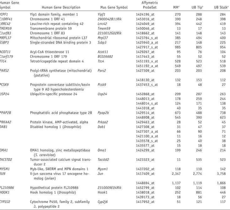
Examination of genes located within the LOD-1 interval on 1p32-33 (by use of the NCBI Map Viewer, build 36.1; Ensembl, v39; and University of California–Santa Cruz [UCSC] Genome Browser, March 2006 assembly) reveals 52 transcription units, of which 23 encode hypothetical or predicted proteins. None of the positional candidates have been previously implicated in structural abnormal-ities of the kidney or urologic tract. Consultation of On-line Mendelian Inheritance of Man (OMIM) revealed that mutations in six positional candidates have been impli-cated in human traits (PCSK9, BSDN, C8A, C8B, TACSTD2, and DHCR24). Mice carrying null alleles for seven other genes (HOOK1, DAB1, JUN, PRKAA2, PPAP2B, DHCR24, and SSBP3) have also been reported, but none feature uro-genital defects. Two other genes on 1p32-33 had been associated with kidney developmental defects (FOXD2 and CPT2) but were located outside our recombinant interval.37,38
A powerful method to annotate positional candidates is to determine their expression pattern within relevant tis-sues and developmental time points. Nephron develop-ment is a recursive process that starts in the 2nd month of gestation, when the UB invades the MM, initiating branching morphogenesis.21,22 Genetic manipulations in
www.ajhg.org The American Journal of Human Genetics Volume 80 March 2007 545
Table 2. Multipoint LOD Scores on Chromosome 1p32-33 with Varying Linkage Parameters
Penetrances (%)
LOD for K100 at HLOD at
Disease-Allele Frequency .001 Disease-Allele Frequency .01 and Phenocopy Rate .001 Disease-Allele Frequency .001 Disease-Allele Frequency .01 and Phenocopy Rate .001 Phenocopy Rate .001 Phenocopy Rate .01 Phenocopy Rate .001 Phenocopy Rate .01 65 3.2 3.2 3.2 3.6 3.5 3.3 75 3.4 3.4 3.4 3.7 3.7 3.5 85 3.5 3.5 3.5 3.9 3.8 3.6 95 3.3 3.3 3.3 3.7 3.7 3.4
NOTE.—Peak parametric HLODs are shown for all seven pedigrees combined (with 45% of families linked).
either compartment can produce the renal hypodysplasia phenotype.12–20Hence, one can predict that the phenotype
observed in our families originates from dysregulation of a gene that is normally expressed early in nephrogenesis but could be localized to the MM, the UB, or both. Ac-cordingly, we performed annotation of positional candi-dates with the results of a recently published study that characterized gene expression in the murine MM, UB tips, and UB stalk.39This expression profiling was performed at
the E12.5 time point, corresponding to the 2nd and 3rd round of cleavage in branching morphogenesis, among the earliest time points at which the MM can be anatom-ically differentiated from the UB in the mouse. We par-ticularly focused on genes that show differential expres-sion in UB tips, which coordinate UB branching and secrete factors necessary for nephron induction in the MM. Examination of the data revealed that 30 positional candidates had murine homologs and corresponding probe sets on the arrays; of these, 23 had detectable ex-pression in the developing MM, UB tip, or UB stalk (table 3). Among positional candidates, three genes initially stood out because they had high expression levels across metanephric compartments (JUN and HOOK1) or dis-played UB-specific expression in mouse (TACSTD2). JUN, a component of the AP-1 transcription factor complex, is essential for cell proliferation and organogenesis after mid-gestation.40It acts downstream of RET signaling and may
modify UB branching and nephrogenesis.41 HOOK1 is a
microtubule-binding protein involved in endocytosis in
Drosophila and is responsible for the abnormal
spermat-ozoon head shape phenotype in mice.42TACSTD2 encodes
a cell-surface receptor recognized as a carcinoma-associ-ated antigen, with mutations associcarcinoma-associ-ated with gelatinous
drop-like corneal dystrophy.43 TACSTD2 and TACSTD1
were identified as UB-specific secreted proteins in multiple studies; alteration in TACSTD2 may therefore modify UB branching and nephrogenesis.39,44We performed direct
se-quencing of the coding exons of the JUN, HOOK1,
TACSTD2, FOXD2, and CPT2 genes, using the index
pa-tients from our linked families, but found no evidence of pathogenic mutations.
We also tested whether the 1p32-33 interval may be relevant to inherited disorders that affect the ureters but
not the kidneys. This search was motivated by the pos-sibility that primary ureteric defects can produce chronic renal parenchymal injury and subsequent involution of the kidneys, resulting in the finding of small kidney size on sonogram. We tested linkage to 1p32-33 in 10 families segregating isolated ureteric abnormalities (7 kindreds with primary VUR,28 2 multiplex kindreds with isolated
UPJO, and 1 family with duplicated collecting system). All individuals in this second cohort had normal kidney size. Parametric linkage analysis was performed with 20 micro-satellite loci spanning to 1p32-33, under the same model used to map the hypodysplasia pedigrees (affected-only analysis). We found no evidence for linkage to 1p32-33 in these kindreds. Moreover, our 1p32-33 interval is190
Mb proximal to, and therefore distinct from, the 1p13 locus reported elsewhere for linkage to primary VUR and reflux nephropathy.45These data suggest the 1p32-33
in-terval harbors a gene that may be specific to familial dis-orders that feature the renal hypodysplasia phenotype.
Because several families displayed negative LOD scores on 1p32-33, we searched for additional loci across the ge-nome that account for disease. As K100 clearly linked to 1p32-33, we performed genomewide analysis of linkage after removing this kindred from our cohort (affected-only analysis with SNPs). There was no evidence of linkage across the genome under a model of genetic homogeneity. Under a model of locus heterogeneity, we identified sev-eral linkage peaks with HLOD11 on chromosomes
18p11-q11, 20q11-13, 1p32-33, and 3p21-22. We next added 40 additional markers at these four loci and typed all available family members, to yield inheritance information10.9.
After this analysis, only the HLOD signal on chromosome 3p21-22 improved (HLOD 1.5;a p 0.35 NPL p 2.3 P p; ; ; maximum between D3S1768 and D3S2409). These .02
data indicate that, other than 1p32-33, there are no sig-nificant intervals across the genome that account for the trait in remaining families (table 4).
In this study, we performed detailed phenotypic char-acterization of families with nonsyndromic renal hypo-dysplasia, identified a locus for this trait, and completed the initial steps for functional annotation and prioritiza-tion of posiprioritiza-tional candidates. Because of the complex sig-naling network involved in urogenital development, renal
Table 3. List of Positional Candidates with Detectable Expression in the Developing Murine Urinary Tract
Human Gene
Symbol Human Gene Description Mus Gene Symbol
Affymetrix
ProbeSet MMa UB Tipa UB Stalka
YIPF1 Yip1 domain family, member 1 Yipf1 1424196_at 270 288 284
C1ORF41 Chromosome 1 ORF 41 2900042B11Rik 1453016_at 390 248 398
LRRC42 Leucine-rich repeat containing 42 Lrrc42 1424049_at 394 442 419
TMEM59 Transmembrane protein 59 Tmem59 1450046_at 13 17 13
C1orf83 Chromosome 1 ORF 83 2210012G02Rik 1418662_at 141 148 143
MRPL37 Mitochondrial ribosomal protein L37 Mrpl37 1423764_s_at 385 494 400 SSBP3 Single-stranded DNA binding protein 3 Ssbp3 1425940_a_at 217 148 225
1427917_s_at 985 805 954
ACOT11 Acyl-CoA thioesterase 11 Acot11 1429267_at 95 76 104
C1orf179 Chromosome 1 ORF 179 BC026682 1417443_at 55 52 52
TTC4 Tetratricopeptide repeat domain 4 Ttc4 1451193_x_at 528 523 518
1451192_a_at 549 497 539
PARS2 Prolyl-tRNA synthetase (mitochondrial) (putative)
Pars2 1427309_at 203 203 208
1418130_at 132 153 112
PCSK9 Proprotein convertase subtilisin/kexin type 9 AD hypercholesterolemia
Pcsk9 1437453_s_at 18 48 27
USP24 Ubiquitin-specific protease 24 Usp24 1452868_at 209 287 263
1448013_at 178 259 244
1448014_s_at 124 171 138
1441018_at 40 35 35
PPAP2B Phosphatidic acid phosphatase type 2B Ppap2b 1429514_at 673 488 738
1448908_at 545 390 623
PRKAA2 Protein kinase, AMP-activated, alpha Prkaa2 1429463_at 28 52 45
DAB1 Disabled homolog 1 (Drosophila) Dab1 1427308_at 31 47 37
1427307_a_at 66 90 71
1421100_a_at 11 16 12
1435578_s_at 25 40 38
1435577_at 15 18 18
OMA1 OMA1 homolog, zinc metallopeptidase (S. cerevisiae)
Oma1 1424299_at 199 246 214
TACSTD2 Tumor-associated calcium signal trans-ducer 2
Tacstd2 1423323_at 11 535 523
MYSM1 Myb-like, SWIRM and MPN domains 1 Mysm1 1437202_at 118 110 142
JUN V-jun sarcoma virus 17 oncogene ho-molog (avian)
Jun 1417409_at 2,347 2,774 3,758
1448694_at 1,137 1,119 1,866
FLJ10986 Hypothetical protein FLJ10986 2310009E04Rik 1452799_at 102 114 108
HOOK1 Hook homolog 1 (Drosophila) Hook1 1438018_at 252 881 446
1439173_at 18 56 27
CYP2J2 Cytochrome P450, family 2, subfamily J, polypeptide 2
Cyp2j6 1417952_at 51 121 117
NOTE.—Genes reported to be present in at least one compartment are shown.
a The last three columns show raw expression levels across metanephric compartments (from Schmidt-Ott et al.39).
developmental phenotypes are likely to display a high de-gree of genetic heterogeneity, necessitating large numbers of pedigrees or uniquely large kindreds for gene map-ping.28 We therefore performed systematic sonographic
screening of families with renal hypodysplasia, to extend kindreds and maximize study power. We confirmed that this phenotype segregates as an autosomal dominant trait with reduced penetrance. Next, genomewide analysis of linkage demonstrated that this trait is genetically hetero-geneous, with about half of families showing linkage to a 6.9-Mb interval on 1p32-33. This linkage assignment is firmly established by a large family (K100), which pro-vided evidence for genomewide significance on its own.
Our present findings can be pursued by several ap-proaches. We can perform systematic sequencing of all
positional candidates to identify the pathogenic muta-tions. However, further prioritization of candidates may help accelerate gene identification. This can be achieved by determining renal gene expression at multiple devel-opmental time points and by examining tissue and cell-specific expression through in situ hybridization. In ad-dition, our largest kindred (K100) is of Sardinian origin, providing the opportunity to refine the interval by dis-equilibrium mapping. Sardinia is composed of subpopu-lations that descend from limited sets of ancestors and exhibit strong linkage disequilibrium.46This characteristic
has facilitated gene identification for several traits, such as uric acid nephrolithiasis.47 Hence, comparison of the
haplotypes between K100 and Sardinian patients from the same locality may identify chromosomal segments
inher-Figure 4. Haplotype structure of the three pedigrees that show linkage to the 1p32-33 locus. Genotypes for the most informative markers spanning the linkage interval are shown (19 of 51 SNPs and microsatellite loci genotyped are shown). The vertical bars highlight the linked (black) and the wild-type (white) haplotypes. The physical locations of the markers are indicated in a list at the top right. Patients with childhood end-stage renal disease (ESRD) but no sonographic data available are indicated by a blackened rectangle inside the symbol.
Table 4. Multipoint Parametric LOD Scores at the Most Significant Intervals in the Seven Pedigrees
Kindred
LOD
1p32-33a 3p21-22b,e 18p11-q11c,e 20q11-13d,e
100 3.5 ⫺4 ⫺2.6 ⫺5 101 ⫺2.5 ⫺1.3 ⫺2.8 .8 105 1.2 ⫺1.3 1 ⫺2 107 ⫺1.4 1.2 1.2 1.2 109 1.1 ⫺2.4 ⫺.5 ⫺1.9 112 ⫺.6 ⫺.4 .4 .6 121 ⫺2.3 1.8 ⫺.5 ⫺3
a HLOD p 3.9(45% of families linked). b HLOD p 1.5(35% of families linked). c HLOD p 1.2(50% of families linked). d HLOD p 1(35% of families linked).
e HLOD was calculated without K100 because this kindred shows
sig-nificant linkage to chromosome 1p32-33.
ited by descent and help refine the interval to a limited set of candidates.
Identification of the gene underlying the 1p32-33 link-age will provide insight into the molecular basis of renal developmental disorders and will help improve diagnostic schemes. Low nephron number at birth and consequent low renal reserve have been proposed as a major suscep-tibility factor for the development of nephropathy or hypertension.48,49 Some genes regulating kidney
devel-opment may also participate in tissue repair after renal injury.50 This suggests that identification of the 1p32-33
hypodysplasia gene may also inform us about interindi-vidual variability in nephron number and have additional diagnostic or therapeutic implications.
Acknowledgments
We thank the patients and family members for participating in this study. A.G.G. is supported by the Emerald Foundation, the Irving Clinical Scholar Program, and the National Kidney Foun-dation (NFK) Clinical Scientist Program. P.L.W. is supported by an NKF research fellowship. G.M.G. was supported by Telethon grant E.1122. We thank Richard Lifton, Cathy Mendelsohn, Juan Oliver, and Qais Al-Awqati for their insightful comments.
Web Resources
The URLs for data presented herein are as follows:
Ensembl, http://www.ensembl.org/Homo_sapiens/index.html (for v39)
NCBI Map Viewer, http://www.ncbi.nlm.nih.gov/mapview/ (for build 36.1)
Online Mendelian Inheritance in Man (OMIM), http://www.ncbi .nlm.nih.gov/Omim/ (for renal agenesis/adysplasia, renal-coloboma syndrome, Fraser syndrome, and branchiootorenal syndrome)
UCSC Genome Browser, http://genome.ucsc.edu/cgi-bin/ hgGateway (for March 2006 assembly)
References
1. Hughson M, Farris AB 3rd, Douglas-Denton R, Hoy WE, Ber-tram JF (2003) Glomerular number and size in autopsy kid-neys: the relationship to birth weight. Kidney Int 63:2113– 2122
2. Hughson MD, Douglas-Denton R, Bertram JF, Hoy WE (2006) Hypertension, glomerular number, and birth weight in Af-rican AmeAf-ricans and white subjects in the southeastern United States. Kidney Int 69:671–678
3. Hiraoka M, Tsukahara H, Ohshima Y, Kasuga K, Ishihara Y, Mayumi M (2002) Renal aplasia is the predominant cause of congenital solitary kidneys. Kidney Int 61:1840–1844 4. Helin I, Persson PH (1986) Prenatal diagnosis of urinary tract
abnormalities by ultrasound. Pediatrics 78:879–883 5. US Renal Data System (2005) USRDS 2005 annual data report:
atlas of end-stage renal disease in the United States. National Institutes of Health, National Institute of Diabetes and Di-gestive and Kidney Diseases, Bethesda, MD
6. Ardissino G, Dacco V, Testa S, Bonaudo R, Claris-Appiani A, Taioli E, Marra G, Edefonti A, Sereni F (2003) Epidemiology of chronic renal failure in children: data from the ItalKid project. Pediatrics 111:e382–e387
7. Stroup NE, Edmonds L, O‘Brien TR (1990) Renal agenesis and dysgenesis: are they increasing? Teratology 42:383–395 8. Woolf AS, Price KL, Scambler PJ, Winyard PJ (2004) Evolving
concepts in human renal dysplasia. J Am Soc Nephrol 15: 998–1007
9. Roodhooft AM, Birnholz JC, Holmes LB (1984) Familial na-ture of congenital absence and severe dysgenesis of both kid-neys. N Engl J Med 310:1341–1345
10. McPherson E, Carey J, Kramer A, Hall JG, Pauli RM, Schimke RN, Tasin MH (1987) Dominantly inherited renal adysplasia. Am J Med Genet 26:863–872
11. Carter CO, Evans K, Pescia G (1979) A family study of renal agenesis. J Med Genet 16:176–188
12. Mackie GG, Stephens FD (1975) Duplex kidneys: a correlation of renal dysplasia with position of the ureteral orifice. J Urol 114:274–280
13. Parikh CR, McCall D, Engelman C, Schrier RW (2002) Con-genital renal agenesis: case-control analysis of birth charac-teristics. Am J Kidney Dis 39:689–694
14. Cooper WO, Hernandez-Diaz S, Arbogast PG, Dudley JA, Dyer S, Gideon PS, Hall K, Ray WA (2006) Major congenital mal-formations after first-trimester exposure to ACE inhibitors. N Engl J Med 354:2443–2451
15. Vize PD, Woolf AS, Bard JBL (2003) The kidney: from normal development to congenital disease. Elsevier Science, San Di-ego, CA
16. Grieshammer U, Le M, Plump AS, Wang F, Tessier-Lavigne M, Martin GR (2004) SLIT2-mediated ROBO2 signaling restricts kidney induction to a single site. Dev Cell 6:709–717 17. Yu J, McMahon AP, Valerius MT (2004) Recent genetic studies
of mouse kidney development. Curr Opin Genet Dev 14:550– 557
18. Miyazaki Y, Oshima K, Fogo A, Ichikawa I (2003) Evidence that bone morphogenetic protein 4 has multiple biological functions during kidney and urinary tract development. Kid-ney Int 63:835–844
19. Durbec P, Marcos-Gutierrez CV, Kilkenny C, Grigoriou M, Wartiowaara K, Suvanto P, Smith D, Ponder B, Costantini F,
www.ajhg.org The American Journal of Human Genetics Volume 80 March 2007 549 Saarma M, et al (1996) GDNF signalling through the Ret
re-ceptor tyrosine kinase. Nature 381:789–793
20. Torres M, Gomez-Pardo E, Dressler GR, Gruss P (1995) Pax-2 controls multiple steps of urogenital development. Devel-opment 121:4057–4065
21. Schmidt-Ott KM, Lan D, Hirsh BJ, Barasch J (2006) Dissecting stages of mesenchymal-to-epithelial conversion during kid-ney development. Nephron Physiol 104:p56–p60
22. Vainio S, Lin Y (2002) Coordinating early kidney develop-ment: lessons from gene targeting. Nat Rev Genet 3:533–543 23. Jenkins D, Bitner-Glindzicz M, Malcolm S, Hu CC, Allison J, Winyard PJ, Gullett AM, Thomas DF, Belk RA, Feather SA, et al (2005) De novo Uroplakin IIIa heterozygous mutations cause human renal adysplasia leading to severe kidney failure. J Am Soc Nephrol 16:2141–2149
24. Schonfelder EM, Knuppel T, Tasic V, Miljkovic P, Konrad M, Wuhl E, Antignac C, Bakkaloglu A, Schaefer F, Weber S (2006) Mutations in Uroplakin IIIA are a rare cause of renal hypo-dysplasia in humans. Am J Kidney Dis 47:1004–1012 25. Weber S, Moriniere V, Knuppel T, Charbit M, Dusek J,
Ghig-geri GM, Jankauskiene A, Mir S, Montini G, Peco-Antic A, et al (2006) Prevalence of mutations in renal developmental genes in children with renal hypodysplasia: results of the ESCAPE study. J Am Soc Nephrol 17:2864–2870
26. Ulinski T, Lescure S, Beaufils S, Guigonis V, Decramer S, Morin D, Clauin S, Deschenes G, Bouissou F, Bensman A, et al (2006) Renal phenotypes related to hepatocyte nuclear factor-1beta (TCF2) mutations in a pediatric cohort. J Am Soc Nephrol 17: 497–503
27. Pasch A, Hoefele J, Grimminger H, Hacker HW, Hildebrandt F (2004) Multiple urinary tract malformations with likely re-cessive inheritance in a large Somalian kindred. Nephrol Dial Transplant 19:3172–3175
28. Sanna-Cherchi S, Reese A, Hensle T, Caridi G, Izzi C, Kim YY, Murer L, Scolari F, Ravazzolo R, Ghiggeri GM, Gharavi AG (2005) Familial vesicoureteral reflux: testing replication of linkage in seven new multigenerational kindreds. J Am Soc Nephrol 16:1781–1787
29. Dinkel E, Ertel M, Dittrich M, Peters H, Berres M, Schulte-Wissermann H (1985) Kidney size in childhood: sonograph-ical growth charts for kidney length and volume. Pediatr Ra-diol 15:38–43
30. Han BK, Babcock DS (1985) Sonographic measurements and appearance of normal kidneys in children. AJR Am J Roent-genol 145:611–616
31. Cottingham RW Jr, Idury RM, Schaffer AA (1993) Faster se-quential genetic linkage computations. Am J Hum Genet 53: 252–263
32. Gudbjartsson DF, Jonasson K, Frigge ML, Kong A (2000) Al-legro, a new computer program for multipoint linkage anal-ysis. Nat Genet 25:12–13
33. Sobel E, Lange K (1996) Descent graphs in pedigree analysis: applications to haplotyping, location scores, and marker-sharing statistics. Am J Hum Genet 58:1323–1337
34. Faraway JJ (1993) Distribution of the admixture test for the detection of linkage under heterogeneity. Genet Epidemiol 10:75–83
35. Lander E, Kruglyak L (1995) Genetic dissection of complex traits: guidelines for interpreting and reporting linkage re-sults. Nat Genet 11:241–247
36. Ott J (1989) Computer-simulation methods in human linkage analysis. Proc Natl Acad Sci USA 86:4175–4178
37. Bonnefont JP, Djouadi F, Prip-Buus C, Gobin S, Munnich A, Bastin J (2004) Carnitine palmitoyltransferases 1 and 2: bi-ochemical, molecular and medical aspects. Mol Aspects Med 25:495–520
38. Kume T, Deng K, Hogan BL (2000) Minimal phenotype of mice homozygous for a null mutation in the forkhead/ winged helix gene, Mf2. Mol Cell Biol 20:1419–1425 39. Schmidt-Ott KM, Yang J, Chen X, Wang H, Paragas N, Mori
K, Li JY, Lu B, Costantini F, Schiffer M, et al (2005) Novel regulators of kidney development from the tips of the ureteric bud. J Am Soc Nephrol 16:1993–2002
40. Johnson RS, van Lingen B, Papaioannou VE, Spiegelman BM (1993) A null mutation at the c-jun locus causes embryonic lethality and retarded cell growth in culture. Genes Dev 7: 1309–1317
41. Jijiwa M, Fukuda T, Kawai K, Nakamura A, Kurokawa K, Mu-rakumo Y, Ichihara M, Takahashi M (2004) A targeting mu-tation of tyrosine 1062 in Ret causes a marked decrease of enteric neurons and renal hypoplasia. Mol Cell Biol 24:8026– 8036
42. Mendoza-Lujambio I, Burfeind P, Dixkens C, Meinhardt A, Hoyer-Fender S, Engel W, Neesen J (2002) The Hook1 gene is non-functional in the abnormal spermatozoon head shape (azh) mutant mouse. Hum Mol Genet 11:1647–1658 43. Tsujikawa M, Kurahashi H, Tanaka T, Nishida K, Shimomura
Y, Tano Y, Nakamura Y (1999) Identification of the gene re-sponsible for gelatinous drop-like corneal dystrophy. Nat Ge-net 21:420–423
44. Caruana G, Cullen-McEwen L, Nelson AL, Kostoulias X, Woods K, Gardiner B, Davis MJ, Taylor DF, Teasdale RD, Grim-mond SM, et al (2006) Spatial gene expression in the T-stage mouse metanephros. Gene Expr Patterns 6:807–825 45. Feather SA, Malcolm S, Woolf AS, Wright V, Blaydon D, Reid
CJ, Flinter FA, Proesmans W, Devriendt K, Carter J, et al (2000) Primary, nonsyndromic vesicoureteric reflux and its nephro-pathy is genetically heterogeneous, with a locus on chro-mosome 1. Am J Hum Genet 66:1420–1425
46. Angius A, Bebbere D, Petretto E, Falchi M, Forabosco P, Maes-trale B, Casu G, Persico I, Melis PM, Pirastu M (2002) Not all isolates are equal: linkage disequilibrium analysis on Xq13.3 reveals different patterns in Sardinian sub-populations. Hum Genet 111:9–15
47. Gianfrancesco F, Esposito T, Ombra MN, Forabosco P, Man-inchedda G, Fattorini M, Casula S, Vaccargiu S, Casu G, Car-dia F, et al (2003) Identification of a novel gene and a com-mon variant associated with uric acid nephrolithiasis in a Sardinian genetic isolate. Am J Hum Genet 72:1479–1491 48. Brenner BM, Chertow GM (1994) Congenital
oligonephro-pathy and the etiology of adult hypertension and progressive renal injury. Am J Kidney Dis 23:171–175
49. Keller G, Zimmer G, Mall G, Ritz E, Amann K (2003) Nephron number in patients with primary hypertension. N Engl J Med 348:101–108
50. Mori K, Lee HT, Rapoport D, Drexler IR, Foster K, Yang J, Schmidt-Ott KM, Chen X, Li JY, Weiss S, et al (2005) Endo-cytic delivery of lipocalin-siderophore-iron complex rescues the kidney from ischemia-reperfusion injury. J Clin Invest 115:610–621