Use of the infra hyoid musculo-cutaneous flap in soft
palate reconstruction
P. Gangloff
a,*
, A. Deganello
b, M.L. Lacave
a, J.L. Verhaeghe
c,
M. Lapeyre
d, F. Maire
e, B. Phulpin
a,e, F. Guillemin
c, G. Dolivet
a aHead and Neck Surgery Unit, Oncologic Surgery Department, Centre Alexis Vautrin, Avenue de Bourgogne, Brabois, 54511 Vandoeuvre les Nancy Cedex, France
b
Department of Otolaryngology/Head and Neck Surgery, National Cancer Institute Regina Elena, Rome, Italy
c
Oncologic Surgery Department, Centre Alexis Vautrin, Avenue de Bourgogne, Brabois, 54511 Vandoeuvre les Nancy Cedex, France
d
Radiotherapy Unit, Centre Alexis Vautrin, Avenue de Bourgogne, Brabois, 54511 Vandoeuvre les Nancy Cedex, France
e
Dental Surgery Unit, Oncologic Surgery Department, Centre Alexis Vautrin, Avenue de Bourgogne, Brabois, 54511 Vandoeuvre les Nancy Cedex, France
Accepted 17 July 2006 Available online 1 September 2006
Abstract
Aims: To review a series of 23 consecutive patients with squamous cell carcinomas arising from oropharynx who underwent infra hyoid musculo-cutaneous flap reconstruction including soft palate in alternative to free radial forearm flap or maxillofacial prosthesis. Post operative radiotherapy was performed for all patients.
Results: Every reconstruction healed quickly without major wound complications. The functional results evaluated by speech and swallow-ing capacities, were good for 17 patients, fair for 4 patients and bad for 2.
Conclusions: The infra hyoid musculo-cutaneous flap is a versatile, reliable and convenient flap suitable for repairing small and medium sized defects; it can be used in combination with other flaps, and in selected cases obviates the need for a microvascular free radial forearm flap or maxillofacial prosthesis.
Ó 2006 Elsevier Ltd. All rights reserved.
Keywords: Infra hyoid musculo-cutaneous flap; Head and neck; Cancer; Soft palate; Rehabilitation
Introduction
Velopharyngeal function is often compromised by the resection and reconstruction of oropharyngeal and palatal tumours. While free tissue transfer has improved the outcomes of head and neck reconstruction, in general, palatal reconstruction remains a challenge.1
The use of microvascular free flaps is the most widespread method currently employed for the reconstruction of exten-sive defects after resection of head and neck cancer, so that they represent today the golden standard in many cases because of their versatility and reliability. The flap most com-monly used for head and neck reconstruction is the free radial forearm flap (FRFF).2 This FRFF can be used alone or
combinated with other local flaps. The study of Brown et al.3shows that the addition of the superiorly based pharyn-geal flap to the FRFF in soft palate reconstruction results in improved speech and swallowing. Brown et al.3recommend the use of the additional flap in resections in which more than one quarter of the soft palate is included.
The evidence that not all the patients are suitable for a free flap reconstruction, and also that not every defect strictly requires a free flap transfer to achieve a good func-tional result, rises the necessity to find good alternatives.
Pectoralis major flap and temporalis flap are the most used pedicled flaps in head and neck reconstruction, but the infra hyoid musculo-cutaneous flap (IHMCF) is one of the alternatives to be considered for the reconstruction of moderate defects following resection of the oral cavity, oropharynx or hypopharynx cancers in selected cases. It obviates the need for a microvascular free flap or other local flaps in many cases.4
* Corresponding author. Tel.:þ33 383 598 446. E-mail address:[email protected](P. Gangloff).
0748-7983/$ - see front matterÓ 2006 Elsevier Ltd. All rights reserved. doi:10.1016/j.ejso.2006.07.011
Here we report our experience of a series of 23 recon-structions for selected tumours of the soft palate by using the IHMCF, as a valid alternative to FRFF reconstruction or maxillofacial prosthesis.
Patients and methods
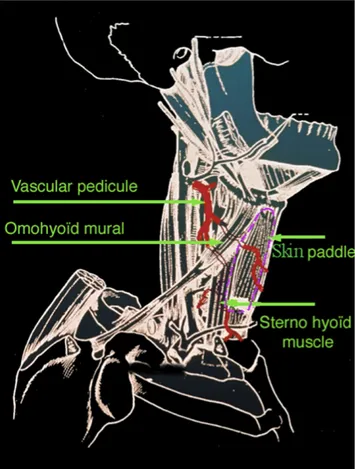
The infrahyoid muscles including sternohyoid (SH), thy-rohyoid (TH), sternothyroid (ST) and omohyoid (OH) con-stitute the anatomical substratum of the flap, completed by the platysma and the overlying skin.
Design of flap
The IHMCF is designed as an oval vertically shape in a paramedian situation and can measure up to 10 cm in its greatest length and up to 5 cm widthways (Figs. 1 and 2).5e7The extent of the resection goes from 1⁄4 to ½
of the soft palate. Patients for whom the resection reaches more than the half of soft palate were excluded from this series. The flap is dissected in order to separate it from the median cervical fascia. The inferior muscular part of the flap is defined by sectioning the muscles downwards (SH and ST) and outwards (intermediate tendon of OH). The venous drainage has two systems through the anterior jugular vein and the superior thyroid vein. Then the strap
muscles are separated from the thyroid plane in order to identify the superior thyroid artery and vein pedicle (Fig. 3).8,9 Collateral veins and superior laryngeal artery (carefully separated from the superior laryngeal nerve) can be ligated, allowing securing the flap to the external ca-rotid artery and the facial vein, or perhaps the internal jug-ular vein. The SH is usually upwardly sectioned at the insertion to the hyoid bone. The flap is then placed to repair
Figure 1. Infra hyoid musculo-cutaneous flap and its vascular network.
Figure 2. Pre operative skin design.
the defect site (Fig. 4). Cutaneous closure of the donor site is performed without important difficulties (Fig. 5).
Patients and treatments
From 1996 to 2005, 23 consecutive patients, 19 men and 4 women, underwent IHMCF reconstruction after orophar-ynx cancer ablation e including a part of soft palate e and neck dissection, in one stage procedure. The extension limits of the tumour had not to go beyond the midline of the soft palate. The ages of the patients ranged from 39 to 71 years, with median age of 58 years. The series ac-counts 23 squamous cell carcinomas (100%) arising from the mucosa of the oral cavity and oropharynx. The localiza-tion was velotonsil area for 20 patients and retro molar tri-gon for 3 patients. Twenty one patients (91%) admitted to tobacco consumption and alcohol abuse. The disease was staged according to the VIthedition of the TNM classifica-tion established by the UICC/AJCc.10 Four tumours were noted T1, 9 T2, 7 T3 and 3 T4. Nodes were staged as 5 N0, 5 N1, 3 N2a, 6 N2b, 3 N2c and 1 N3. Post operative radiotherapy was performed for 23 patients. All patients
underwent speech and swallowing evaluation and reeduca-tion after surgery and radiotherapy.
In this series IHMCF reconstruction has been chosen in-stead of FRFF reconstruction or maxillofacial prosthesis. Results
Nineteen patients had cicatrisation without complica-tions for the flap or the donor site. Local complicacomplica-tions oc-curred in 4 patients. In 2 cases we observed a partial skin paddle necrosis. In the other 2 cases the patients demon-strated a minor cervical dehiscence of the skin requiring only local care.
17/23 patients were able to eat normally (good degluti-tion) with good speech evaluation (good intelligibility). The remaining 6 patients had to adapt their eating habits by mincing (2/6) (fair deglutition) or by mixing (2/6) (bad deglutition) their food. The last 2 patients had fair speech evaluation (fair intelligibility) (Table 1). These six patients, for whom function was classified fair or bad, had T4 (2/6) or T3 (4/6) tumours. The two bad results were noted for patients who had presented in the past laryn-geal or pharynlaryn-geal tumours. The 1stwas a second localiza-tion and the 2ndwas a third localization. For fair results, the delay of surveillance after surgery was too short for three patients (less than 12 months), one presented a second lo-calization and the last obtained only fair results after reed-ucation. The extent of soft palate resection was varied: from the quarter to the half with no clear relation between the ex-tent of the resection and the function quality (Table 2).
The delay of surveillance after surgery ranged from 6 months to 9 years, with median delay of 2 years and 9 months.
Discussion
Since 1979, Wang et al.11 performed a long series of IHMCF. Earliest studies were published from 1986 to 1994.4,11e13 Wang et al.11 reported 112 flaps which were successful in 90% of the cases (101 of 112 cases). The same success rate of IHMCF is noted by Zhao et al.14 who have concluded that cervical pedicle flaps have clinical value in selected patients needing reconstruction of small e and medium e sized defects after intraoral cancer surgery. IHMCF is a versatile, reliable, and convenient flap suitable for repairing the defects in and around the oral cavity, par-ticularly in the oropharynx, even in aged and weak pa-tients.11 Since 1994, we performed routinely IHMCF to reconstruct mucous defects in the head and neck region with this technique, which we subsequently modified for head and neck surgery and immediate reconstruction.15
At best, the flap extremity can reach a distance of 15 cm (theoretical) around its rotation axis. The effective region includes the cervical trachea up to the velotonsil, including the inferior facial cutaneous covering (under the labial e tragus commissura). For soft palate, the maximum size of Figure 4. Post operative view. Right soft palate reconstruction with infra
hyoid musculo-cutaneous flap.
defect that could be safely reconstructed with the IHMCF is the half. Functionally, flap resection does not induce phona-tory, respiratory or swallowing complications. The size of the cutaneous flap sampled was always compatible with a direct suture of the donor site without cicatrisation complication.
In our experience, the results were comparable with those published in the literature.4,11e13The lack of ability to reconstruct the dynamic function of the soft palate con-tinues to be disappointing. Limited studies have shown promise in soft palate reconstruction without the complica-tions of velopharyngeal insufficiency. The lack of a uniform classification for palate defects has limited prospective comparison of reconstructive methods.16The usual respect of contraindications helped avoiding the complications en-countered by other authors.11Contra indications of IHMCF such as previous thyroid surgery or radical neck dissection must be respected; relative contra indication is represented by previous cervical radiotherapy. It is acceptable to use material from a metastatic neck for defect cover in the cases where the vascular pedicle of the flap and the IHMCF itself are not in the tumour and are at least at 30 mm of can-cerous tissue.
In case of soft palate reconstruction, it is useful to pre-serve the motor innervation of the infrahyoid muscles pro-vided by the descending branch of the hypoglossal nerve (the ansa cervicalis), that is kept with the flap during its new positioning. The main advantage of this voluntary in-nervated flap is the prevention of atrophies and the
improvement of scarring qualities of the reconstructed soft palate.17 The function qualities are also improved by this innervation conservation which allows synchronous contraction of the two sides of soft palate during swallow-ing. As Wang et al. published,11 a minor motricity reap-pears within 12 months after intervention.
The IHMCF is a versatile, reliable and convenient flap, with interesting plastic qualities, suitable for repairing small and medium sized defects;15 this is an additional tool in the therapeutic possibilities for cervicofacial recon-struction. It can be used in combination with other flaps, and in selected cases, as soft palate reconstruction, obviates the need for a microvascular FRFF or maxillofacial pros-thesis. This flap is thin, pliable, so that is particularly useful in oral cavity reconstructions and, in our experience, the functional results are comparable to those of the FRFF re-construction for small and medium sized defects. The IHMCF has the particularity to remain the anatomy after re-construction, which is less possible with FRFF or maxillo-facial prosthesis.
The realisation of a maxillofacial prosthesis is another solution for these patients. Prosthetic treatment of soft pal-ate defects varies based on the extent and site of the defect. The goal of treatment is to attain velopharyngeal closure during function, which allows normal speaking and swal-lowing and keeps the patient relatively comfortable. While maxillofacial prosthetic treatment is not a substitute for plastic and reconstructive surgery, in certain circumstances it may be an alternative. Certain patients may simply not be Table 1
Series description
Classification Localization Post operative treatment Local complications Function evaluation
4 T1 5 N0 20 velotonsil 23 radiotherapy 2 partial skin
paddle necrosis 17 good 9 T2 5 N1 3 retro molar trigon 23 speech and swallowing reeducation 2 minor cervical dehiscence 4 fair 7 T3 3 N2a 2 bad 3 T4 6 N2b 3 N2c 1 N3 Table 2
Fair or bad function description Classification TNM Extent of soft palate resection Function impairement
Main reason of function impairement
Delay of surveillance after surgery Fair result #1 T3 N1 M0 Quarter of soft palate Fair intelligibility Delay too short for
complete reeducation
8 months Fair result #2 T3 N1 M0 Quarter of soft palate Fair deglutition Fair reeducation results 16 months Fair result #3 T3 N2a M0 Third of soft palate Fair deglutition Delay too short for
complete reeducation
6 months Fair result #4 T3 N2c M0 Quarter of soft palate Fair intelligibility Second localization and
delay too short for complete reeducation
10 months
Bad result #1 T4 N1 M0 Half of soft palate Bad deglutition Third localization 23 months Bad result #2 T4 N2a M0 Third of soft palate Bad deglutition Second localization 47 months
good candidates for plastic surgery because of their ad-vanced age, poor health, very large deformity, or poor blood supply to irradiated tissue. Moreover, maxillofacial prosthetic treatment is indicated when anatomical parts of the head and neck are not replaceable by living tissue or when recurrence of malignancy is likely.
Nevertheless, in patients with soft palate defects, it is difficult to obtain sufficient retention, support, and stability of the obturator prosthesis. In addition, its mobility during various functions is considered to be large.
Although the system of speech evaluation was subjective in our series, but standardized by the same speech therapist, the results obtained seemed equivalent to those obtained by Wang et al.11and Zuydam et al.18Four fair results and two bad results were observed. On the one hand, these results seemed to be related to the tumour stage (T3 or T4) and not to the extent of soft palate resection (for some cases, good results have been obtained after resection of the half of soft palate) and on the other hand, three out of four fair results had a delay of surveillance after surgery less than 12 months, which can also explain these functional re-sults. The two bad results were noted for T4 tumours.
The indications of this flap remain numerous for the up-per aerodigestive tract allowing the repair of large mucous or cutaneous defects with acceptable functional or aesthetic sequelae.
Our surgical technical research has led us to the laryn-geal and pharyngolarynlaryn-geal reconstruction (i.e. after near total resection) with the IHMCF and in some specific case, partial reconstruction of cervical esophagus. Our pri-mary results seem to confirm the elective choice of this flap for these indications.
References
1. McCombe D, Lyons B, Winkler R, Morrison W. Speech and swallow-ing followswallow-ing radial forearm flap reconstruction of major soft palate defects.Br J Plast Surg 2005;58:306–11.
2. De Bree R, Hartley C, Smeele LE, Kuik DJ, Quak JJ, Leemans CR. Evaluation of donor site function and morbidity of the fasciocutaneous radial forearm flap.Laryngoscope 2004;114:1973–6.
3. Brown JS, Zuydam AC, Jones DC, Rogers SN, Vaughan ED. Func-tional outcome in soft palate reconstruction using a radial forearm free flap in conjunction with a superiorly based pharyngeal flap. Head Neck 1997;19:524–34.
4. Rojananin S, Suphaphongs N, Ballantyne AJ. The infrahyoid muscu-locutaneous flap in head and neck reconstruction.Am J Surg 1991; 162:400–3.
5. Calloc’h F, Prades JM, Chelikh L, Dalmonego V, Martin Ch. Infra-hyoid paramedian musculocutaneous flap. Anatomical bases. Review of the literature.J Fr Otorhinolaryngol 1996;45:203–9.
6. Deganello A, De Bree R, Dolivet G, Leemans CR. Infrahyoid myocu-taneous flap reconstruction after wide local excision of a Merkel cell carcinoma.Acta Otorhinolaryngol Ital 2005;25:50–4.
7. Faucher A, Verhulst J, Majoufre C, de Bonfils C. Infrahyoid musculo-cutaneous flaps: anatomical bases and indications in cervicofacial on-cologic surgery.Rev Laryngol Otol Rhinol (Bord) 1997;118:43–6. 8. Eliachar I, Marcovich A, Har Shai Y, Lindenbaum E. Arterial blood
supply to the infrahyoid muscles: an anatomical study.Head Neck Surg 1984;7:8–14.
9. Lockhart R, Menard P, Chout P, Favre-Dauvergne E, Berard P, Bertrand JC. Infrahyoid myocutaneous flap in reconstructive maxillo-facial cancer and trauma surgery.Int J Oral Maxillofac Surg 1998;27: 40–4.
10. UICC Union Internationale Contre le Cancer. In: Hermanek P, Sobin LH, editors.TNM classification of malignant tumours. 4thed.
Paris: Springer-Verlag; 1988, p. 13–32.
11. Wang HS, Shen JW, Ma DB, Wang J, Tian A. The infrahyoid myocu-taneous flap for reconstruction after resection of head and neck cancer. Cancer 1986;57:663–8.
12. Dolivet G, Faucher A, Majoufre C, Micheik J, Renaud-Salis JL. The infra-hyoid musculo-cutaneous flap in head and neck recon-structive surgery: technique and first results. Rev Laryngol 1994; 115:225–9.
13. Magrin J, Kowalski LP, Santo GE, Walksmann G, DiPaula RA. Infra-hyoid myocutaneous flap in head and neck reconstruction.Head Neck 1993;15:522–5.
14. Zhao YF, Zhang WF, Zhao JH. Reconstruction of intraoral defects af-ter cancer surgery using cervical pedicle flaps.J Oral Maxillofac Surg 2001;59:1142–6.
15. Dolivet G, Gangloff P, Sarini J, et al. Modification of the infra hyoid musculo-cutaneous flap.Eur J Surg Oncol 2005;31:294–8.
16. Van der Sloot PG. Hard and soft palate reconstruction.Curr Opin Oto-laryngol Head Neck Surg 2003;11:225–9.
17. Remmert SM, Sommer KD, Majocco AM, Weerda HG. The Neuro-vascular Infrahyoid Flap: a new method for tongue reconstruction. Plast Reconstr Surg 1997;99:613–8.
18. Zuydam AC, Lowe D, Brown JS, Vaughan ED, Rogers SN. Predictors of speech and swallowing function following primary surgery for oral and oropharyngeal cancer.Clin Otolaryngol 2005;30:428–37.