©Science and Education Publishing DOI:10.12691/jfnr-6-7-1
Dietary Modifications of Nitrogen Intake Decreases
Inflammation and Promotes Rejuvenation of Spleen in
Aged Mice
Claudia Romano1,#, Giovanni Corsetti1,#,*, Evasio Pasini2, Vincenzo Flati3, Francesco S Dioguardi4 1Department of Clinical & Experimental Sciences, Division of Human Anatomy and Physiopathology,
University of Brescia, Brescia, Italy
2“S. Maugeri Foundation”, IRCCS, Cardiology Division, Medical Centre of Lumezzane, Brescia, Italy 3Department of Biotechnological and Applied Clinical Sciences, University of L’Aquila, L’Aquila, Italy
4Department of Clinical Sciences and Community Health, University of Milan, Milan, Italy #These authors contribute equally to this work.
*Corresponding author: [email protected]
Abstract
The spleen is a lymphoid organ with multiple functions including blood filtration and immune activity. Aging changes the spleen’s anatomy for both immune and stromal cells and can lead to immunesenescence, contributing to the increased rates of mortality and morbidity commonly observed in the elderly. Much evidence indicates that the combination of food quantity and quality can influence chronic inflammatory states in the spleen. Quantitative amino-acids (AA) adequacy is pivotal to maintain cell integrity in mammals. Aged mice feed with balanced essential-AA (EAA) formulation improved mitochondrial biogenesis and morphological and molecular changes in many organs, as well as increased lifespan. Here, we evaluated the inflammatory state of the spleen in aged male mice (fifteen months old) chronically fed for twelve months with a particular EAA-rich diet compared to a standard laboratory diet. This study found that chronic consumption of an EAA-rich diet decreased inflammation and modulated reticular and mitochondrial chaperones, mitochondrial function and cells survival, maintaining the correct architecture of the spleen. These changes could also be beneficial for immune system integrity, providing a possible theoretical-speculative basis for the role of EAA improving the quality of life of the elderly by probably slowing immune-senescence.Keywords
: Essential Amino-acids, spleen, inflammation, chaperones, Klotho, agingCite This Article:
Claudia Romano, Giovanni Corsetti, Evasio Pasini, Vincenzo Flati, and Francesco S Dioguardi, “Dietary Modifications of Nitrogen Intake Decreases Inflammation and Promotes Rejuvenation of Spleen in Aged Mice.” Journal of Food and Nutrition Research, vol. 6, no. 7 (2018): 419-432. doi: 10.12691/jfnr-6-7-1.1. Introduction
The spleen is a lymphoid organ composed of two morphologically and functionally distinct compartments, red pulp (RP) and white pulp (WP) [1]. The RP is rich in macrophages and is responsible for the removal of damaged or old erythrocytes, platelets and apoptotic cells [2]. It is separated from the WP by the marginal zone (MZ), which also contains macrophages and many resident cells that maintain its integrity, as well as marginal zone B cells and dendritic cells [3]. A network of fibroblastic reticular cells mark the MZ [4].
The WP consists of lymphoid tissue and is subdivided into a periarterial lymphatic sheath (PALS), where T cells encircle the central arteriole and B-cell follicles [1]. Within these structures, there are stromal cells that allow effective blood filtration as it passes through T and B cell areas, enabling pathogens to be captured, facilitating exchanges between antigen-presenting cells
and pathogen-specific lymphocytes and providing the necessary signals for survival and differentiation of leucocytes [5].
Recently, a comparative study between young and older mice has shown differences in the spleen anatomy and in both immune and stromal cells. In particular, it has been shown that age-related changes are irreversible and consist of increased WP size, primarily within the T-cell zone, reducing the distinction-between T cells and B cells. Furthermore, the loss of MZ macrophages and accumulation of fibroblasts has also been found [6]. Given the essential role that the microenvironment plays in the development and activation of many immune cell types [5,7], changes in tissue architecture may also contribute to immunesenescence [8].
Immunesenescence is the decline of activity and efficiency of the immune system associated with age [9,10,11]. The consequent increased susceptibility to and severity of infection is also associated with a decline in response to vaccinations and a higher prevalence of cancer and auto-immune diseases [9,12,13]. It therefore
contributes to the increased rates of mortality and morbidity commonly observed in the elderly [14].
Considerable evidence indicates that the combination of food quantity and quality can influence a chronic inflammatory state [15]. Unhealthy nutritional patterns are associated with the aberrant activation of innate immune signalling, which triggers chronic low-grade systemic inflammation [16]. Dietary intake is crucial for providing macro- and micro nutrients and energy to fuel the body’s metabolism. Muscular proteins is a ready-to-use amino acid (AA) reservoir during fasting or stress. Therefore, inadequate protein dietary intake can easily impair protein balance and lead to muscle atrophy, impaired organ growth and/or functional decline, particularly in aged patients and/or with disease [17]. Thus, quantitative AA adequacy is pivotal for maintaining cell integrity in mammals. However, what can we say about AA qualitative adequacy? From a nutritional point of view, AAs can be classified as either essential (EAA) or non-essential (NEAA) depending on their ability to be synthesized endogenously [18] or not.
We have shown that aged mice with a diet supplemented with a balanced EAA formulation increases lifespan, improves mitochondrial biogenesis and morphological and molecular changes in the heart, skeletal muscle and adipose tissues [19,20,21,22]. The protective role of an EAA rich diet was also observed in rat livers with chronic ethanol consumption [23,24], in the kidneys of rosuvastatin-treated mice [25] and during wound healing [26]. Furthermore, in vitro data showed that variations in the EAA/NEAA concentration ratio regulates cancer cell survival or death [27]. These data suggest that the dietary balance of EAA/NEAA ratio may considerably affect the whole-body metabolism [17]. However, the impact of diet on spleen architecture and integrity has been little studied, probably given the fact that the spleen needs to be removed using an appropriate and rapid procedure. However, we believe that, since the spleen is an important organ in balancing the immune system, and also aging is known to lead to immunesenescence, it is important to investigate whether a special balanced EAA enriched diet with specific stoichiometric ratios. This would influence the morphological and functional microenvironment of the spleen, thus modulating inflammatory and stress markers expression and so improving spleen health. On this premise, we aimed to evaluate the morphologic and immunohistochemical features of the spleen of aged male mice, fed with a specific EAA-rich diet compared to a standard rodent diet.
2. Materials and Methods
2.1. Diets
We developed a specific EAA-rich diet containing a mixture of EAA (approx. 84%) and NEAA (approx. 16%) [28]. This special diet includes iso-nutrients, iso-caloric and iso-nitrogenous and was expressly prepared for Nutriresearch s.r.l. (Milan, Italy) by Dottori Piccioni (Milan, Italy) in accordance with AIN76-A/NIH-7 rules. A standard rodent laboratory diet (StD) (Mucedola srl, Milan, Italy) was used as a reference diet, where the
nitrogen source came from full proteins of both vegetal and animal sources. It should be noted that the AA composition of these proteins was not available and is not constant when supplied in bulk by different producers. The composition of the diets is summarized in Table 1.
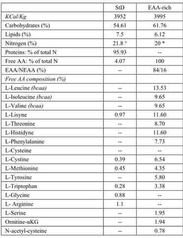
Table 1. Diet composition
StD EAA-rich KCal/Kg 3952 3995 Carbohydrates (%) 54.61 61.76 Lipids (%) 7.5 6.12 Nitrogen (%) 21.8 ° 20 * Proteins: % of total N 95.93 -- Free AA: % of total N 4.07 100
EAA/NEAA (%) -- 84/16 Free AA composition (%) L-Leucine (bcaa) -- 13.53 L-Isoleucine (bcaa) -- 9.65 L-Valine (bcaa) -- 9.65 L-Lisyne 0.97 11.60 L-Threonine -- 8.70 L-Histidyne -- 11.60 L-Phenylalanine -- 7.73 L-Cysteine -- -- L-Cystine 0.39 6.54 L-Methionine 0.45 4.35 L-Tyrosine -- 5.80 L-Triptophan 0.28 3.38 L-Glycine 0.88 -- L- Arginine 1.1 -- L-Serine -- 1.95 Ornitine-αKG -- 1.94 N-acetyl-cysteine -- 0.78 * Nitrogen (%) from free AA only. ° Nitrogen (N) (%) from vegetable and animal proteins and added AA. bold line represents the limit between EAAs (upside) and NEAAs (beneath) bcaa = branched chain amino-acids.
2.2. Animals
Sixteen male Balb/C mice (three months old, from Envigo, Holland) were divided into two experimental groups of eight animals each: 1) mice fed with a peculiar EAA-rich diet, and 2) mice fed with StD, used as control. The animals were individually housed in plastic cages with white wood chips for bedding, in a quiet room under controlled lighting (12h day/night cycle) and temperature (22±1°C) conditions. At all times, the animals had free access to food and water was supplied ad libitum. Every three days, body weight, diet and water consumption were measured and restored. Animals were regularly evaluated by veterinary physicians for their health and maintenance of normal daily and nocturnal behavioral activities and for criteria of increased disease burden following ethics standards for animal studies. The experimental protocol was approved and conducted in accordance with the Italian Ministry of Health and complied with the ‘The National Animal Protection Guidelines’. The Ethics Committee for animal experiments of the National Reference Centre for Animal Welfare (IZSLER of Brescia) and the Italian Ministry of Health all approved the procedures.
2.3. Experimental Protocol
Body weight (Bw), food (g) and water (mL) consumption for each animal were monitored every two days and the concentration of drinking solutions were adjusted accordingly. Water consumption was measured by weighing the bottle and Bw was determined using an electronic balance with an accuracy of 0.1 g.
Twelve months later, the animals (final age 15 months) were killed by cervical dislocation. Blood samples from right heart in both groups were used to analyze the hematocrit and hemoglobin content and serum proteins. The spleen from each animal was quickly removed, weighed, split and fixed with immunofix for 12 h at 4°C. The samples were then washed in phosphate buffer saline (PBS, 0.2M, pH 7.4) for 12 h and processed according to standard procedures for paraffin embedding. Thick sections (about 5 µm) obtained by a Leica Microtome, collected on poly-L-lysinated slides, were used for histochemical (HC) and immune-histochemical (IHC) procedures.
2.4. Histopathology
Deparaffinized sections of spleen samples were stained by haematoxylin and eosin (H&E), Sirius red, Prussian blue Pearls staining were used to check general features, the presence of fibrosis and free iron accumulation such as ferric [Fe(III)] ions [23].
2.5. Immunohistochemistry
Paraffin sections were stained according to the ABC-peroxidase method as previously described [23]. Paraffin sections were incubated over night at 4°C with primary polyclonal anti-macrophage (MAC387, from Abcam Inc., UK), anti-IL-10 (ABIN672051, from Antibodies-online Inc., Dunwoody, GA, U.S.A.), anti-IL1β (sc-7884), anti-IL-6 (sc-1265), anti-Klotho (sc-22220), anti-iNOS(sc-651), anti-eNOS (sc-654) anti-Grp78 (sc-1050), anti-Grp75 (sc-13967), anti-Cit-c-Ox (sc-7159), anti-SIRT3 (sc-99143), (1:100) all from Santa Cruz Biotechnology (CA, U.S.A.). In order to exclude any incorrect interpretation of immunostaining due to endogenous biotin, we also carried out experiments using the peroxidase-anti-peroxidase detection system. Results were overlapped using both methods. The IHC control (negative control) was performed by omitting the primary antibody in presence of isotype-matched IgGs. The sections were processed in accordance with the manufacturers’ protocol, visualized by a rabbit ABC-peroxidase staining system kit (ABC-peroxidase Elite kit from Santa Cruz Biotechnology) and mounted with DPEX synthetic mounting medium. The reaction product was visualized using 0.3% hydrogen peroxide and diaminobenzidine (DAB) at room temperature. Each set of experiment was carried out in triplicate, and carried out under the same experimental conditions. Splenic parenchyma was observed in three zones: white pulp (WP) comprising follicles, marginal zone (MZ), at the boundary between white and red area and red pulp (RP). Different researchers, blinded to the samples’ origin, independently analyzed all slides. Staining intensified in histochemical (HC) and IHC methods were
semi-quantitatively evaluated using an optical Olympus BX50 light microscope. According to Hall et al. [29] the scale of evaluation adopted for HC was: - undetectable; +/- barely detectable; + weak (10-30% positive cells); 2+ evident (30-50% positive cells); 3+ moderate (50-70% positive cells); 4+ intense/strong staining (>70% positive cells). The staining intensity in IHC slides was evaluated using light microscope equipped with image analysis program (Image Pro Plus 4.5.1). The integrated optical density (IOD) was calculated for arbitrary areas, by measuring at least five fields in each sample.
2.6. Blood Analysis
The analysis was performed by the “Division of Laboratory Animals” of IZSLER-Bs. Blood samples from each animal were split and collected into tubes containing K3-EDTA anti-coagulant (BD-Vacutainer 1.5 mL) for haemochromocytometric analysis and in a 1.5 ml vial (Eppendorf srl, Milan, Italy) without anticoagulant for serum analysis. Haemochromocytometric value were measured by Cell-Dyn 3700 laser-impedence cell counter (Abbott Diagnostics Division, Abbott Laboratories, IL, USA). Urea, albumin, creatinine and haptoglobin concentration were measured in blood serum by a biochemical automatic analyzer ILab Aries (Instrumentation Laboratory, Lexington, MA, USA) and its ready-to-use kits.
Hematologic indices were determined according to standard methods. Tests included red blood cell (RBCs), white blood cell (WBCs) and PLT counts, differential counts of neutrophils, lymphocytes, monocytes and eosinophils. The concentrations of hemoglobin (Hb), mean corpuscular volume (MCV), mean corpuscular hemoglobin (MCH), mean corpuscular hemoglobin concentration (MCHC), MPV, packed cell volume (PCV/hematocrit) and red cell distribution width (RDW) were also recorded. In addition to serum haptoglobin concentration, Neutrophils to Lymphocytes ratio (NLR) was also calculated and evaluated as a marker of inflammation [30,31].
2.7. Statistics
Organs weight was normalized as a ratio of final body weight. Statistical analysis was performed by a Student’s t-test for paired samples (t). A value of p<0.05 was considered statistically significant.
3. Results
3.1. Food and Water Intake, Body
Weight/Organs Weight Ratio
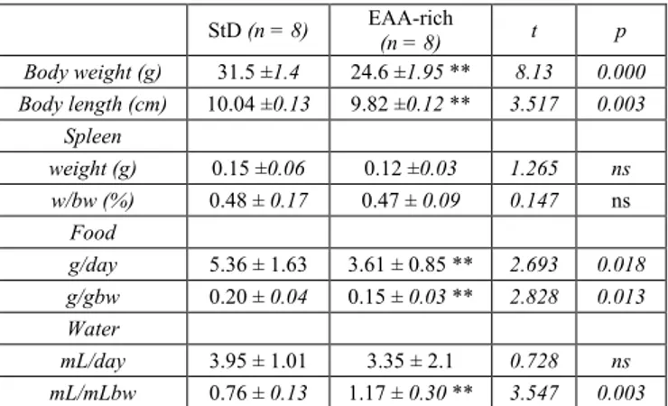
In EAA-fed mice, the daily food intake (and so caloric intake) was lower than those StD-fed. In spite of this, the EAA-fed animals were more active and had a better-looking fur than those fed with StD. An increase of mean daily water intake was observed in EAA-fed animals (Table 2). In mice fed with EAA, spleen weight decreased not significantly, as did the splenic index, defined as the [splenic weight (g)/body weight (g)]*100% (Table 2).
Table 2. Mean animal body weight (bw), animals length, spleen-weight (mean ± SD) and daily food and water consumption at the end of treatment (14 months) of StD-fed and EAA-fed animals. ns = not significant StD (n = 8) EAA-rich (n = 8) t p Body weight (g) 31.5 ±1.4 24.6 ±1.95 ** 8.13 0.000 Body length (cm) 10.04 ±0.13 9.82 ±0.12 ** 3.517 0.003 Spleen weight (g) 0.15 ±0.06 0.12 ±0.03 1.265 ns w/bw (%) 0.48 ± 0.17 0.47 ± 0.09 0.147 ns Food g/day 5.36 ± 1.63 3.61 ± 0.85 ** 2.693 0.018 g/gbw 0.20 ± 0.04 0.15 ± 0.03 ** 2.828 0.013 Water mL/day 3.95 ± 1.01 3.35 ± 2.1 0.728 ns mL/mLbw 0.76 ± 0.13 1.17 ± 0.30 ** 3.547 0.003
3.2. Blood
Blood values are summarized in Table 3. EAA-fed animals showed improved blood parameters compared to the StD-fed. In particular, we observed the increase of erythrocytes and haemoglobin and a large increase of leucocytes, in particular in lymphocytes and monocytes fraction and a decrease of neutrophils. In addition, the neutrophils (N) and neutrophils/lymphocytes ratio (N/L) decreased considerably when compared to StD-fed animals. In serum, the haptoglobin levels greatly decreased with the EAA diet, whereas albumin and creatinine concentration increased significantly.
3.3. Spleen Architecture and Iron Deposits
The WP of spleen of StD-fed animals appeared larger
and irregular in shape compared to the WP observed in EAA-fed mice. However, the germinative center of inner periarteriolar lymphoid sheath (or PALS) were easily identifiable and the MZ did not show any significant differences between the two diets (Figure 1A and D).
Sirius-red stain, under normal and polarized light, showed that in the spleen of StD-fed animals there was a moderate degree of fibrosis. On the other hand, the stroma did not show any signs of fibrosis in EAA-fed animals (Figure 1B-C and E-F).
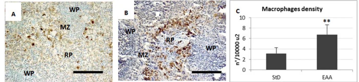
The RP of spleen from StD-fed mice showed only few macrophages with blue staining deposits of ferritin. Sometimes macrophages in the MZ and WP appeared moderately to poorly stained (Figure 2A). However, clearly evident hemosiderin deposits were present inside the macrophages of the RP and MZ (Figure 2B). With EAA feeding, the WP and more markedly the RP and MZ showed many macrophages with blue staining deposits of ferritin (Figure 2C), whereas the hemosiderin storage was scarce (Figure 2D). Furthermore, the mean density of macrophages, expressed as the number per unit area (n°/10000µ2), increased significantly, mainly in RP and MZ of the EAA-fed mice (Figure 3 A-C). Semi-quantitative evaluation of iron histochemistry staining is summarized in Table 4.
3.4. Inflammation
EAA-rich diet modulates ILs and NOS expression thus reducing inflammation.
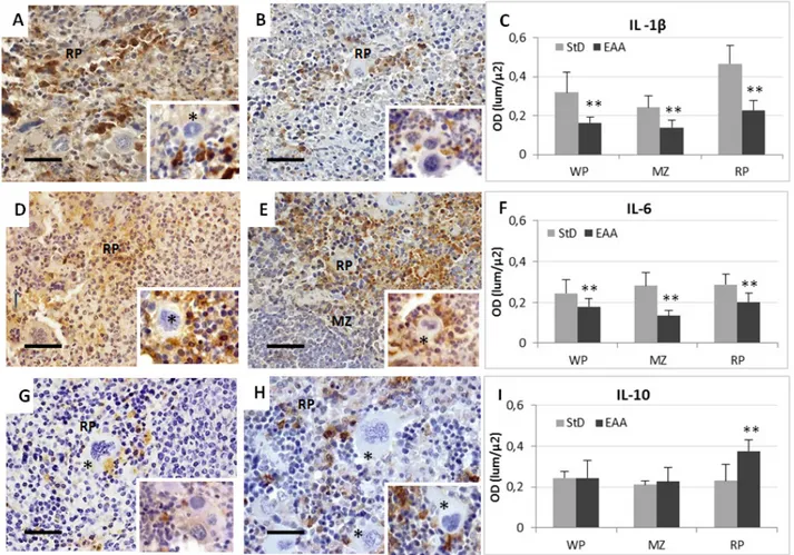
IL-1β. StD-fed animals showed intense IL-1β staining of the RP and MZ (Figure 4A) and almost all macrophages were strongly stained (Figure 4A insert). Sometimes WP macrophages were also stained.
Table 3. Blood and serum data according to diets (mean ± S.D.). ns = not significant StD
(n=8) EAA-rich (n=8) t P
Erythrocyte (RBC) (M/uL) 9.28 ±0.33 10.73 ±0.5 ** 6.846 0.000
Haemoglobin (g/dL) 14.46 ±1.54 15.9 ±0.62 ** 2.453 0.028
Haematocrit (HCT) % 47.43 ±4.1 50.15 ±2.33 1.631 ns
Mean globular volume (MCV) (fl) 49.37 ±0.51 48.26 ±0.46 ** 4.571 0.000
Mean globular haemoglobin (MCH) (pg) 14.13 ±0.29 15.22 ±0.17 ** 9.171 0.000
Mean globular haemoglobin concentration (MCHC) (g/dL) 30.40 ±0.8 31.54 ±0.09 ** 4.005 0.001
Erythrocyte distribution width (RDW) % 17.50 ±0.92 21.3 ±2.61 ** 3.884 0.002
Platelet (PLT) (K/uL) 1331.7 ±477.9 1227.3 ±202.9 0.560 ns
Leucocytes (WBC) (K/uL) 3.40 ±0.59 7.89 ±1.8 ** 6.704 0.000
Granulocytes neutrophils (K/uL) 1.77 ±0.9 1.65 ±0.7 0.298 ns
Lymphocytes (K/uL) 1.49 ±0.38 6.11 ±1.55 ** 8.188 0.000
Monocytes (K/uL) 0.07 ±0.01 0.13 ±0.04 ** 4.116 0.001
Granulocytes Eosinophil (K/uL) 0.01±0.002 0.12 ±0.007 ** 42.737 0.000
Granulocytes Basophils (K/uL) 0.07 ±0.01 0.04 ±0.08 1.052 ns
Granulocytes Neutrophils (%) 52.06 ±2.61 20.9 ±3.36 ** 20.715 0.000
Lymphocytes % 43.82 ±3.79 77.4 ±4.25 ** 16.679 0.000
Monocytes % 2.06 ±0.01 1.7 ±1.1 0.926 ns
Granulocytes Eosinophil (%) 0.3 ±0.1 1.5 ±0.46 ** 7.210 0.000
Granulocytes Basophils (%) 2.06 ±0.3 0.51 ±0.2 ** 12.159 0.000
Neutrophils / Lymphocytes ratio 1.19 ±0.12 0.27 ±0.08 ** 10.043 0.000 serum
Albumin (g/L) 26.8 ±0.7 28.7 ±0.75 ** 5.238 0.000
Urea (mM/L) 7.6 ±0.72 8.18 ±0.6 1.750 ns
Creatinine (µM/L) 37.6 ±1.56 46.98 ±2.26 ** 9.661 0.000
Figure 1. A and D. E&H staining. WP (asterisks) of spleen of StD-fed animals were larger and irregular in shape (A) compared to the WP observed in
EAA-fed mice (D). Scale bar 300 µm. B-C and E-F: Sirius-red staining of spleen for collagen detection under normal (B and E) and polarized light (C and F). StD-fed animals (B-C) showed a moderate degree of fibrosis. Differently, in EAA-fed animals (E-F) the stroma did not show fibrosis. Scale bar 150 µm.
Figure 2. Iron histochemistry. A-B. Pearls staining for ferritin. RP of spleen from StD-fed mice (A) had scarce macrophages with blue staining deposits
of ferritin (and insert). Macrophages in MZ and WP also appeared moderately to poorly stained. With EAA feeding (B) the RP but also MZ and WP showed numerous macrophages blue stained (insert). Scale bar 100 µm. C-D E&H staining of spleen from StD-fed mice (C) showed RP with evident orange-yellow hemosiderin deposits inside the macrophages (and insert). In spleen of EAA-fed mice (D) the hemosiderin storage is scarce. Scale bar 30 µm
Figure 3. Representative pictures of anti-macrophage IHC in spleen of StD-fed (A) and EAA-fed mice (B). Mean density of macrophages is expressed
as cells number for unit area (C). RP = red pulp. WP = white pulp. MZ = marginal zone. Scale bar 100 µm. ** t-test p<0.001.
Table 4. Semi-quantitative evaluation of iron histochemistry staining in spleen of StD- or EAA-fed mice
WP RP MZ
StD EAA-r StD EAA-r StD EAA-r
Ferritin + + + 4+ + 4+ Hemosiderin + +/- 2+ + 2+ +/- WP: white pulp; RP: red pulp; MZ: marginal zone; StD: standard diet. EAA-r: essential amino acids rich diet.
On the contrary, IL-1β staining in the RP of EAA-fed animals was faint and confined inside RP macrophages (Figure 4B and insert). Very occasionally we observed
some stained macrophage inside the WP. The OD values of IL-1β staining are shown in Figure 4C.
IL-6. Splenocytes of StD-fed mice had diffuse staining inside RP and MZ macrophages (Figure 4D and insert). Some intensely stained macrophages were observed inside the WP. The EAA administration decreased IL-6 staining in macrophages as well as in other cell types of RP and WP (Figure 4E and insert). OD values of IL-6 staining are shown in Figure 4F.
IL-10. In StD-fed animals there was a faint to moderate immune-staining confined to certain cells, principally macrophages of RP alone (Figure 4G and insert). The spleen of EAA-fed animals had staining inside many RP
cell types but it was most intense inside macrophages (Figure 4H and insert). The OD values of IL-10 staining are shown in Figure 4I.
eNOS. RP Splenocytes and less spleen MZ from StD-fed animals had diffuse faint staining (Figure 5A). Megakaryocytes did not have any immune-staining
(Figure 5A insert). Conversely in EAA-fed animals, RP
and MZ staining was diffuse and more intense, with some macrophages very intensely stained (Figure 5B). Sometimes megakaryocytes had pale staining (Figure 5B insert). The OD value of eNOS staining is resumed in Figure 5C.
Figure 4. Representative pictures of immunostaining for anti-IL-1β, -IL-6 and IL-10 in spleen from StD-fed (respectively A, D, G) and EAA-fed mice
(respectively B, E, H). See text for description. Scale bar 40 µm. Asterisks = megakaryocytes. C-I: optical density quantification according to antibodies. ** t-test p<0.01. RP = red pulp. MZ = marginal zone
Figure 5. Representative pictures of IHC for anti-eNOS (A-B) and –iNOS (D-E) in spleen from StD-fed (A and D) and EAA-fed (B and E) mice. C and
F, optical density quantification according to antibodies. ** t-test p<0.01. RP = red pulp. WP = white pulp. See text for description. Inserts: asterisks = megakaryocytes. Scale bar 100 µm.
iNOS. The spleen from StD-fed animals had intense immune-staining mainly located in RP and MZ macrophages. Furthermore, moderate positivity was observed inside WP cells. The megakaryocytes did not have any staining (Figure 5D and insert). In animals fed with the EAA-rich diet, the iNOS expression was very low and only occasionally did WP have any positive stained cells (Figure 5E and insert). The OD values of iNOS staining are shown in Figure 5F.
3.5. Chaperones
Chaperone expression increased with the EAA-rich diet. In particular, in spleens from StD-fed animals, Grp78 was moderately expressed in RP macrophages (Figure 6A) and in certain WP and MZ lymphocytes and macrophages. On the contrary, the immune-positivity for Grp78 was intense inside many macrophages and other RP cell types (perhaps activated lymphocytes and or plasma cells), while it was
less intense in the WP. Frequently, megakaryocytes were also faintly stained (Figure 6B). Similarly, Grp75 expression was faint to moderate in RP and StD-fed animals (Figure 6D and insert). Conversely, in EAA-rich fed animals, immune-staining greatly increased in many macrophages of all compartments, but less so in others cells, including the megakaryocytes (Figure 6E and insert). The OD values of Grp78 and Grp75 staining are shown in Figure 6C and 6F respectively.
3.6. Mitochondrial Markers
Since we observed that the mitochondrial chaperone Grp75 was enhanced with the special diet, we also evaluated other pivotal mitochondrial enzymes, such as SIRT3 and Cit-c-ox (COX). This is because they are essential to enhance mitochondrial antioxidant defense and the respiratory chain respectively.
Figure 6. Representative pictures of IHC for anti-Grp78 (A-B) and –Grp75 (D-E) in spleen from StD-fed (A and D) and EAA-fed (B and E) mice.
Frequently, numerous megakaryocytes of EAA-fed mice shows Grp immunostaining. C and F, optical density quantification according to antibodies. ** t-test p<0.01. RP = red pulp. WP = white pulp. MZ = marginal zone. See text for description. asterisks = megakaryocytes. Scale bar 40 µm
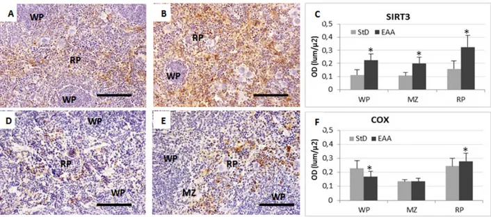
Figure 7. Representative pictures of IHC for anti-SIRT3 (A-B) and COX (D-E) in spleen from StD-fed (A and D) and EAA-fed (B and E) mice. C and
F, optical density quantification according to antibodies. * t-test p<0.05. RP = red pulp. WP = white pulp. MZ = marginal zone. See text for description. Scale bar 100 µm
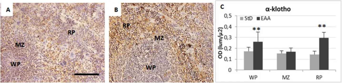
Figure 8. Representative pictures of anti-Klotho IHC in spleen of StD-fed (A) and EAA-fed mice (B). (C) optical density quantification of antibody
inside spleen’s compartments. ** t-test p<0.01; * p<0.05. RP = red pulp. WP = white pulp. MZ = marginal zone. See text for description. Scale bar 100 µm
SIRT3 expression was moderate but diffuse in many RP and MZ spleen cells types from StD-fed mice (Figure 7A). The EAA-rich diet considerably increased SIRT3 immune-staining in RP splenocytes of but less strongly inside MZ and WP ones. Many macrophages were intensely stained, whereas megakaryocytes were not (Figure 7B). The same tendency was seen with anti-COX staining. Indeed, in StD-fed spleens, RP cellular types of had moderate COX-immune-positivity (Figure 7D). On the contrary, EAA-rich diet induced diffuse and intense COX expression in many cells types but principally in RP macrophages (Figure 7E). The OD values of SIRT3 and COX staining are shown in Figure 7C and 7F respectively.
3.7. Klotho cell-survival Marker
In order to verify whether the special EAA-rich diet could also promote cell survival, we evaluated the Klotho expression level, a single-pass trans-membrane protein. When over-expressed, Klotho is known to extend life span [32] and could play a biological or immunological role in the spleen [33]. Our results showed that Klotho was weakly expressed in all compartments of StD-fed spleen (Figure 8A), while the EAA-rich diet improved Klotho expression mainly inside RP and MZ splenocytes (Figure 8B). The OD values of Klotho staining are shown in Figure 8C.
4. Discussion
The main message from our data is that the EAA-rich diet could improve spleen health of aged animals by reducing inflammation and by increasing chaperones and cell survival markers. Similar results have already been reported in muscles, liver, heart and kidneys [19,21,23,25] but to our knowledge, it is the first time that these results have been reported in the far less considered spleen.
In particular, we observed many changes in RP and MZ as components of the functional compartment entrusted with filtering blood, removing aged erythrocytes and recycling iron trough macrophage activity. Indeed, RP consists of a network of fibroblasts and reticular fibres that form cords containing many resident macrophages, and so constitutes of an open blood system. From the cords, blood is forced to pass into venous sinuses, characterized by discontinuous endothelium, where stress
fibres, composed of actin- and myosin-like filaments, run parallel to the endothelial axis. Contraction of stress fibres regulate space size between the endothelium, and so the blood flow becomes difficult for aged erythrocytes, due to stiffened membranes. As a result, old erythrocytes stick to cords and are phagocytised by resident macrophages [1]. In previous work, we demonstrated that EAA supplementation improves the integrity and function of muscular and cardiac fibres [19,21], so we can hypothesize that the EAA-rich diet also contributes to maintaining the integrity and function of stress fibres and of micro-organization of the splenic stroma. Indeed, in a different set of studies, we found marked alterations of collagen metabolism also in the skin of animals fed low levels of EAA [26].
Macrophages in the spleen play a major role in iron metabolism by recycling iron from erythrocytes. Indeed, haem derived from erythrophagocytosis is catabolized into biliverdin, carbon monoxide and ferrous iron (Fe2+). The iron is then either released from the cells or stored as ferritin, a cytosolic protein that can be observed in RP macrophages. For cell storage of larger amounts of iron, ferritin can aggregate into hemosiderin, which is an insoluble complex of partially degraded ferritin [34]. When necessary, iron can be released from macrophages linked to ferritin and rapidly bind to transferrin, a transporter protein present in the plasma.
Conversely, large hemosiderin deposits in the spleen may increase due to the stimulated sequestration and destruction of erythrocytes [35]. As a result hemosiderin may be found in many diseases associated with iron overload [36]. We found an increased number of macrophages with large amounts of ferritin and lower amounts of hemosiderin in RP of EAA-fed animals, suggesting an intense iron turnover. This is also supported by increased levels of hemoglobin in EAA-fed animals. Noticeably, few macrophages loaded with largest amount of hemosiderin and the lowest ferritin and hemoglobin concentrations, were observed in StD-fed animals. This would suggest functional sequestration of iron and scarce erythrocytes turnover. Collectively, our data support the idea that the EAA-rich diet could maximally improve haemoglobin synthesis, erythrocyte production and iron turnover.
In addition to the erythrophagocytosis inside the spleen, a considerable proportion of erythrocytes are also destroyed intra-vascularly throughout the body, as a result of continuing damage to their plasma membrane. This
leads to the release of haemoglobin, which is bound rapidly by haptoglobin (Hp).
Hp is a hemoglobin-binding protein with immunomodulatory properties that in humans is encoded by the Hp gene [37]. It is mainly produced by hepatocytes and to a lesser extent by cells in other tissues such as lung, skin and kidney, under inflammatory conditions. Its synthesis is induced by IL-1β, IL-6 and TNF. So Hp is considered a typical marker of inflammation in clinical practice. In the blood plasma, Hp binds free haemoglobins released from erythrocytes with high affinity so preventing oxidative activity. The Hp-hemoglobin complex will then be removed by the reticulo-endothelial system, mostly in the spleen [38].
Hp also has been demonstrated to modulate several aspects of the innate and adaptive immune response and to have anti-inflammatory activities [39,40]. Indeed, an interesting results from our present work is the strong decrease of Hp in EAA-fed animals. We can therefore suppose that the decrease of Hp serum level in EAA-fed animals may reflect the anti-inflammatory role of the diet, that could have positive effects on the entire organism, confirming benefits observed in previous works [21]. This hypothesis is also supported by our hematological data that also identified an increase in lymphocytes and monocytes, and contemporarily the decrease of neutrophils with a consequent reduction of N/L ratio, confirming the lowest grade of systemic inflammation [30,31].
On the other hand, aging is accompanied by a progressive decline in immunity in all mammal species so far studied [41]. These age-associated changes in immune competence could be responsible for the increased incidence of auto-immune diseases commonly found in the elderly, as well as their enhanced susceptibility to infectious agents, disease severity and risk of cancer [41]. The immune system is known to depend upon the proper functioning of a subtle and well-balanced network of cytokines that control the survival, proliferation and differentiation of lymphoid cells. Thus, the age-related alterations in immune competence could also be due to the complex remodeling of cytokine production that characterizes immunesenescence.
IL-1β is a potent pro-inflammatory cytokine that plays an important role in normal homeostasis and in the inflammatory response. It is deemed to be responsible for the development of major chronic diseases that are highly prevalent in the elderly [42,43], since overproduction could exacerbate damage, induce acute tissue injuries and chronic diseases [44]. Additionally, cells from aged mice produce increased levels of IL-6, a proinflammatory and immunoregulatory cytokine contributing to host defense against infections and tissue injuries, while persistent dysregulated IL-6 synthesis and/or its elevated concentrations contribute to development of various diseases and immunosuppression [45].
Another important immune-related citokyne is IL-10. This is a pleiotropic anti-inflammatory cytokine that plays an important role in immune regulation. During infection it inhibits the activity of Th1 cells, NK cells and macrophages, all of which are required for optimal pathogen clearance but also contribute to tissue damage [46,47,48,49,50]. Considered the wide range of IL-10 biologic activities, a dysregulation in the mechanisms that
control IL-10 production could be a major contributor to immunosenescence [51].
Our data showed that EAA-feeding induces marked reduction of pro-inflammatory cytokines such as IL-1β and IL-6, while the anti-inflammatory IL-10 was increased in spleen of aged animals, compared to StD-fed animals. Considered together, these data strongly support the protective role of the EAA-rich diet in spleen senescence, suggesting possible improvement of immunosenescence as a whole, due to the modulation of pro-inflammatory cytokine production. Modulation of the inflammatory state due to the EAA-rich diet is also supported by the strong reduction of iNOS staining, expressed in the presence of inflammatory stimuli [52].
Further finding reported here is the increased Grp78 (Glucose regulated protein 78 kDa, alias BiP or HSPA5) immunostaining in all spleen compartments of EAA-fed animals. Grp78 is a member of the HSP70 protein family [53]. Traditionally, it is regarded as a major endoplasmic reticulum (ER) chaperone facilitating protein folding and assembly, protein quality control, regulating calcium homeostasis, and regulating transmembrane ER inducers [54,55]. However, intriguingly, Grp78 has also been described in other intracellular sites apart from ER, such as nucleus [56], mitochondria [57], cytosol [58] and plasma membrane [59,60]. Surface Grp78 plays a critical role in cell signaling, proliferation and survival [61].
We speculate that the intense Grp78 level observed in EAA-fed mice may be due to increased ER efficiency coupled with increased protein synthesis for higher EAA availability. However, because RP and MZ macrophages were strongly immunoreactive, it is possible that Grp78 over-expression could also be due to an excess of iron/erythrocytes uptake. Indeed, it has been shown that Grp78 is directly involved in proper antigen binding to specific receptors in MZ macrophages associated with iron metabolism alteration and inflammation [62]. However, we did not detect any tissue inflammation, so we are inclined to exclude the alteration of iron metabolism.
Another chaperone, Grp75 (alias Mortalin/mthsp70/PBP74), was expressed more in the spleen with the EAA-rich diet. This is a mitochondrial matrix chaperone, with various sub-cellular localizations and implicated in multiple functions [63,64,65]. Its expression levels correlate with muscle activity, mitochondrial efficiency and biogenesis [66,67]. Constitutional low levels of Grp75 are present in adult rats’ spleens [68].
Grp75 also acts as a guardian against senescence and apoptosis promoted by various stress, or serves as a safeguard to promote cell proliferation and survival [69]. Indeed, Grp75 was shown to inhibit p53-Bax interactions and activate AKT, that are required for apoptotic signalling [70]. In addition, in neuro-degeneration of human cells in vitro and ex vivo it has been demonstrated that reduced Grp75 function leads to activation of the mitochondrial unfolded protein response, increased intramitochondrial proteolytic stress, increased autophagic degradation of fragmented mitochondria and reduced mitochondrial mass. These alterations drive cells to apoptotic death [65].
In line with these data, it was recently shown that Grp75 over-expression inhibits inflammation and potentially
inhibits neuronal apoptosis in a rat model of intracerebral haemorrhage [71]. In addition, Grp75 can inhibit reactive oxygen species accumulation, a mechanism that may be involved in the cytoprotective effect of Grp75 over-expression on glucose deprivation [72]. Our results show that in all spleen compartments of elderly animals fed with an EAA-rich diet, Grp75 levels increased when compared to the spleen of StD animals. This would suggest a protective role of our special diet against functional and morphological spleen damage induced by senescence, improving mitochondrial activity and biogenesis. This also agrees with our previous work showing that in middle aged mice fed with EAA, mitochondrial function was improved as well as survival of muscular skeletal and cardiac cells [21].
Because mitochondria play essential roles in the supply of energy, regulation of calcium levels and control of apoptotic cell death, it is plausible that improvement in their morphology and function could improves cellular longevity. We therefore also explored SIRT3 and COX expressions. SIRT3 is a mitochondrial deacetylase and essential player in enhancing the mitochondrial glutathione antioxidant defense system during caloric restriction (CR) diets [73], or when diets are enriched with EAA [21]. This would suggest that SIRT3-dependent mitochondrial adaptations may be a central mechanism for slowing aging in mammals. Indeed, mice without SIRT3 are not protected from oxidative stress or damage [74,75]. SIRT3 is expressed in various organs including spleen [76] where exhibits a broad pattern of cellular localization in the immune cells. Our data confirm the constitutional diffuse and moderate SIRT3 expression in many cellular types mostly in RP of StD aged mice, but its expression was increased by the EAA-rich diet. A similar expression pattern was observed for COX. Indeed, old spleen mitochondria exhibited a high level of COX defect, with a 40% decrease in the respiratory control index and a decline in specific activity of COX [77].
Our data also showed an increase in eNOS in EAA-rich diet mice spleen. eNOS is an enzyme that produces nitric oxide (NO) constitutively, which has different functions in biological processes. It is known that the NO produced by this enzyme, compared to the NO produced by iNOS, has beneficial effects [52]. Several studies demonstrated that EAA increase eNOS expression and how it is related to mitochondria. Indeed eNOS-/- mice have been show not to be able to affect mitochondrogenesis [78]. Taken together, these data support the improvement of mitochondrial function in splenocytes of elderly animals fed with an EAA-rich diet.
The beneficial effect of the EAA-rich diet in preventing age-related spleen damage was also demonstrated by the increased expression of the anti-aging protein Klotho. Indeed, Klotho is known to extend life span when over-expressed and when disrupted aging is accelerated. Klotho-deficient mice exhibit a syndrome resembling accelerated human aging in conjunction with extensive arteriosclerosis [32]. Furthermore, Klotho has been demonstrated to improve vascular endothelial dysfunction, increase nitric oxide production, reduce elevated blood pressure and prevent medial hypertrophy and perivascular fibrosis in animal model of metabolic syndrome [79].
Experimental and clinical studies have shown that Klotho is involved in the genesis and progression of various cardiovascular diseases [80,81,82] by preventing cell death [83,84]. Klotho proteins (trans-membrane and circulating) influence the endocrine activity of FGFs, regulate mineral transport and cell metabolism, thus preventing organ aging an death [32,85]. Our observations are in line with recent immunohistochemical assessment of the distribution of spleen cells positive for Klotho. The authors showed that cells expressing Klotho were present in both the MZ and RP of the spleen, and that FGFR1 was expressed in Klotho-expressing cells [33]. Although we did not measure FGF levels in this study (more detailed work in this specific topic is in progress), we believe that the increase in Klotho protein following an EAA-rich diet could correlate with the modulation of inflammation markers and chaperone expression that we evaluated. This could be a further useful element to support the beneficial role of a diet rich in EAA on aging spleen.
In summary, our study indicates that the chronic consumption of EAA-rich diet is correlated with the modulation of proteins related to inflammation, reticular and mitochondrial chaperones, mitochondrial function and cells survival, maintaining the correct architecture of the spleen. These protein variations could also be beneficial for the immune system, thus providing a possible theoretical-speculative basis for the role of EAA to improve the quality of life of aged human by slowing immune-senescence. It has been demonstrated that the intact microarchitecture of the spleen supports productive lymphocyte-antigen-presenting cell interactions and is critical for the ability of an organism to maintain adequate immune response ability [86]. Furthermore, because the splenic environment is uniquely suited as a site for extramedullary haematopoiesis, it is possible that the stromal micro-environment is similar enough to bone marrow to allow for near-normal development of hematopoietic cell lines [87]. Thus, it does not seem too risky to imagine that the benefit gained by EAAs consumption in the spleen could also affect the bone marrow.
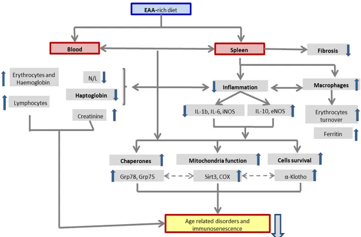
We need to underline that this study was conducted on a mouse experimental model, therefore extrapolating these data on humans requires caution. Interestingly however, it has been shown that in animals, EAA supplementation to a normal diet increases life-span [21] and in humans, diet supplementation with EAAs improves glycaemic metabolism, peripheral muscle and heart performances [88,89] and reduces infection risks in hospitalized elderly [90]. With this premise, we believe that the message that emerges from our data could be useful when considering the role of EAA in preventing immunesenescence also in human. Further studies are in progress to better understand effects of an EAA diet on the immune system, as well as the application of this animal model to humans. Potential events which link the EAA consumption and spleen changes are shown schematically in Figure 9.
4.1. Clinical Implications
The role of an EAA-rich diet on spleen architecture of aged animals may have effects on maintaining an adequate
immune response. Recently, we showed that specific AA mixture orally administered reduced infection by 30% and C-Reactive Protein compared to well-nourished controls in geriatric rehabilitative center [90]. It is known that specific AAs modulate immune-function favoring protein synthesis [91] which is a biological process fundamental for the proliferation and the function of immune cells [92]. Notably, our EAA-rich diet does not contain Glutamine, which is considered the most important molecule for the immune system [93]. However, Leucine is present in the EAA-rich diet used in our study. Leucine is both a
precursor of Glutamine and it is an important AA for protein cell synthesis. Nevertheless, here we showed that nutrition supplements with specific AA maintained spleen structure, reduced age-related enzymatic disarrangements and global inflammation, increasing the number of leucocytes (Table 3). Interestingly, the elderly may have selective nutritional protein intake deficit and/or digestive disarrangement with scarce intake and/or blood availability of EAA and consequently reduce protein synthesis and/or structure/cell functions involved in maintaining an appropriate immune response.
Figure 9. The graph shows the potential events which link EAA exposure with spleen and blood changes.
4.2. Study Limitations
A possible limitation of this study is the exclusive application of IHC to support our results. This choice was dictated by the fact that the spleen contains a very heterogeneous cell population (macrophages, lymphocytes, dendritic cells, megakaryocytes etc) and was divided into well-defined morphologically recognizable areas (WP, PALS, RP, MZ). Consequently, histopathological changes and especially the exact location of markers is possible with only IHC and HC.
In contrast, molecular analysis, although much more sensitive in highlighting proteins and very often used exclusively, does require immediate freezing and homogenization of the sample. As a consequence, it does not take into account the specificities of protein location, such as tissue morphology and organization [94]. This is a major limitation in the exclusive use of molecular analysis, which we believe is comparable if not superior to IHC. For these reasons, we believe that our data, even if obtained solely by IHC, should be considered useful since
they indicate a precise hierarchy of future targets for molecular research.
Acknowledgments
This study was supported by Nutriresearch s.r.l. (Milan, Italy) grant of CG. We thanks Dr.ssa I.L. Archetti (from National Reference Centre for Animal Welfare - IZSLER-Bs) for blood analysis. We would like to thank prof. Robert Coates (Centro Linguisitico, Bocconi University, Milan, Italy), medical writer, for his linguistic revision.
Author Contributions
G.C. and C.R. designed research; C.R. and G.C. conducted research; C.R., and G.C. analyzed and interpreted data; F.S.D., E.P., F.S.D. and V.F. contributed to data interpretation and the drafting of the manuscript; G.C. and C.R. wrote the paper. G.C. and F.S.D. had
primary responsibility for the final content. All authors read and approved the final manuscript.
Compliance with Ethical Standards
All animal procedures followed the European Communities Council Directive of November 24, 1986 (86/609/EEC), and the Italian Ministry of Health and complied with The National Animal Protection Guidelines. The Ethics Committee for animal experiments of the National Reference Centre for Animal Welfare (IZSLER of Brescia) and the Italian Ministry of Health all approved the procedures.
Conflict of Interest Statement
The authors declare that there are no conflicts of interest.
References
[1] Mebius, R.E., and Kraal., G., “Structure and function of the spleen”, Nature review. Immunology, 5 (8). 606-616. Aug 2005.
[2] Brendolan, A., Rosado, M.M., Carsetti, R., Selleri, L., and Dear, T.N., “Development and function of the mammalian spleen”,
BioEssays 29 (2). 166-177. February 2007.
[3] Martin, F., and Kearney, J.F., “Marginal-zone B cells”, Nature
Review. Immunology, 2 (5). 323-335. Review. May 2002. [4] Balogh, P., Horvath, G., and Szakal, A.K., “Immunoarchitecture
of distinct reticular fibroblastic domains in the white pulp of mouse spleen”, Journal of histochemistry & cytochemistry. 52 (10). 1287-1298. October 2004.
[5] Junt, T., Scandella, E., and Ludewig, B. Form follows function: lymphoid tissue microarchitecture in antimicrobial immune defence. Nature review. Immunology, 8 (10). 764-775. Oct 2008.
[6] Aw, D., Hilliard, L., Nishikawa, Y., Cadman, E.T., Lawrence, R.A. and Palmer, D.B., “Disorganization of the splenic microanatomy in ageing mice”, Immunology, 148 (1). 92-101. May 2016.
[7] Anderson, G., and Jenkinson, E.J., “Lymphostromal interactions in thymic development and function”, Nature Review.
Immunology, 1 (1). 31-40. 1:31-40 October 2001.
[8] Su, D.M., Aw, D., and Palmer, D.B., “Immunosenescence: a product of the environment?” Current Opinion in Immunology, 25 (4). 498-503. August 2013.
[9] Aw, D., Silva, A.B., and Palmer, D.B., “Immunosenescence: emerging challenges for an ageing population”, Immunology, 120 (4). 435-446. Apr 2007.
[10] Haynes, L., and Swain, S.L., “Why aging T cells fail: implications for vaccination”, Immunity, 24 (6). 663-666. June 2006.
[11] Pawelec, G., Akbar, A., Caruso., C., Solana, R., Grubeck-Loebenstein, B., and Wikby, A., “Human immunosenescence: is it infectious?”, Immunological Review 205. 257-268. Jun 2005.
[12] Chen, W.H., Kozlovsky, B.F., Effros, R.B., Grubeck-Loebenstein, B., Edelman, R., and Sztein, M.B., “Vaccination in the elderly: an immunological perspective”, Trends in Immunology, 30 (7). 351-319. Jul 2009
[13] Gardner, I.D., “The effect of aging on susceptibility to infection”,
Review of Infectious Diseases; 2 (5). 801-810. Sep-Oct 1980. [14] McElhaney, J.E., Zhou, X., Talbot, H.K., Soethout, E., Bleackley,
R.C., Granville, D.J., and Pawelec, G., “The unmet need in the elderly: how immunosenescence, CMV infection, co-morbidities and frailty are a challenge for the development of more effective influenza vaccines”, Vaccine, 30 (12). 2060-2067. March 2012.
[15] Neustadt, J., “Western diet and inflammation”, Integrative
Medicine, 5. 14-18. Aug-Sept 2006.
[16] Galland, L., “Diet and inflammation”, Nutrition in Clinical
Practice, 25 (6). 634-640. Dec 2010.
[17] Pasini, E., Corsetti, G., Aquilani, R., Romano, C., Picca, A., Calvani, R., and Dioguardi, F.S., “Protein-amino acid metabolism
disarrangements: the hidden enemy of chronic age-related conditions” Nutrients, 10 (4). Pii: E391. March 2018.
[18] Hou, Y., Yin, Y., and Wu, G., “Dietary essentiality of "nutritionally non-essential amino acids" for animals and humans”,
Experimental Biology and Medicine (Maywood), 240 (8).
997-1007. August 2015.
[19] Corsetti, G., Pasini, E., D’Antona, G., Nisoli, E., Flati, V., Assanelli, D., Dioguardi, F.S., and Bianchi R., “Morphometric changes induced by amino acid supplementation in skeletal and cardiac muscles of old mice”, The American journal of cardiology, 101 (11A). E26-E34. June 2008.
[20] Flati, V., Pasini, E., D’Antona, G., Speca, S., Toniato, E., and Martinotti, S., “Intracellular mechanisms of metabolism regulation: the role of signaling via the mammalian target of rapamycin pathway and other routes”, The American journal of cardiology, 101 (11A). 16E-21E. June 2008.
[21] D’Antona, G., Ragni, M., Cardile, A., Tedesco, L., Dossena, M., Bruttini, F., Caliaro, F., Corsetti, G., Bottinelli, R., Carruba, M.O., Valerio, A., and Nisoli E., “Branched-chain amino acid supplementation promotes survival and supports cardiac and skeletal muscle mitochondrial biogenesis in middle-aged mice”,
Cell metabolism, 12 (4). 362-372. October 2010.
[22] Stacchiotti A, Corsetti G, Lavazza A, Rezzani R. “Microscopic features of mitochondria rejuvenation by amino acids”, in: Antonio Méndez-Vilas editor, Current microscopy contributions
to advances in science and technology. Formatex Research Center,
Microscopy Series nº 5, vol. 2. 286-294. 2012.
[23] Corsetti, G., Stacchiotti, A., Tedesco, L., D’Antona, G., Pasini, E., Dioguardi, F.S., Nisoli, E., Rezzani R., “Essential amino acid supplementation decreases liver damage induced by chronic ethanol consumption in rats”, International journal of
immunopathology and pharmacology, 24 (3). 611-619. Jul-Sep
2011.
[24] Tedesco, L., Corsetti, G., Ruocco, C., Ragni, M., Rossi, F., Carruba, M.O., Valerio, A., and Nisoli, E., “A specific amino acid formula prevents alcoholic liver disease in rodents”, American
journal of physiology. Gastrointestinal and liver physiology, 314
(5). G566-G582. May 2018.
[25] Corsetti, G., D’Antona, G., Ruocco, C., Stacchiotti, A., Romano, C., Tedesco, L., Dioguardi, F., Rezzani, R., and Nisoli, E., “Dietary supplementation with essential amino acids boots the beneficial effects of rosuvastatin on mouse kidney”, Amino acids, 46 (9). 2189-2203. September 2014.
[26] Corsetti, G., Romano, C., Pasini, E., Marzetti, E., Calvani, R., Picca, A., Flati, V., and Dioguardi, F.S., “Diet enrichment with a specific essential free amino acid mixture improves healing of undressed wounds in aged rats”, Experimental gerontology, 96. 138-145. October 2017.
[27] Bonfili, L., Cecarini, V., Cuccioloni, M., Angeletti, M., Flati, V., Corsetti, G., Pasini, E., Dioguardi, F.S., and Eleuteri, A.M., “Essential amino acid mixtures drive cancer cells to apoptosis through proteasome inhibition and autophagy activation”, The
FEBS journal, 284 (11). 1726-1737. June 2017.
[28] Corsetti, G., Pasini, E., Romano, C., Calvani, R., Picca, A., Marzetti, E., Flati, V., and Dioguardi. F.S. “Body weight loss and tissue wasting in late middle-aged mice on slightly imbalanced essential/non-essential amino acids diet”, Frontiers in Medicine, 5. 1-11. May 2018.
[29] Hall, P., Davies, W., Stamp, K., Clamp, I., and Bigley, A. - “Comparison of computerized image analysis with traditional semiquantitative scoring of Perls’ Prussian Blue stained hepatic iron deposition”. Toxicology and Pathology, 41 (7). 992-1000. February 2013.
[30] Prats-Puig, A., Gispert-Saüch, M., Díaz-Roldán, F., Carreras-Badosa, G., Osiniri, I., Planella-Colomer, M., et al. “Neutrophil-to-lymphocyte ratio: an inflammation marker related to cardiovascular risk in children”, Thromb Haemost, 114(4). 727-34. July 2015.
[31] Ouellet, G., Malhotra, R., Penne, E.L., Usvya, L., Levin, N.W., Kotanko, P. “Neutrophil-lymphocyte ratio as a novel predictor of survival in chronic hemodialysis patients”, Clinical Nephrology, 85(4). 191-198. April 2016.
[32] Kuro-o, M., Matsumura, Y., Aizawa, H., Kawaguchi, H., Suga, T., Utsugi, T., Ohyama, Y., Kurabayashi, M., Kaname, T., Kume, E., Iwasaki, H., Iida, A., Shiraki-Iida, T., Nishikawa, S., Nagai, R., Nabeshima, Y.I.. “Mutation of the mouse klotho gene leads to a syndrome resembling ageing”, Nature, 390. 45-51. November 1997.
[33] Nakashima, Y., Mima, T., Yashiro, M., Sonou, T., Ohya, M., Masumoto, A., Yamanaka, S., Koreeda, D., Tatsuta, K., Hanba, Y., Moribata, M., Negi, S., Shigematsu, T. “Expression and localization of fibroblast growth factor (FGF) 23 and Klotho in the spleen: its physiological and functional implications”, Growth
Factors. 34 (5-6). 196-202. December 2016.
[34] Knutson, M., Wessling-Resnick, M. “Iron metabolism in the reticuloendothelial system”, Critical Reviews in Biochemistry and
Molecular Biology, 38. 61-88. September 2003.
[35] Barlas, N., Aydogan, M. “Histopathologic effects of maternal 4-tert-octylphenol exposure on liver, kidney and spleen of rats at adulthood”, Archives of Toxicology, 83 (4). 341-349. April 2009.
[36] Iancu, T.C. “Ferritin and hemosiderin in pathological tissues”,
Electron Microscopy Review. 5 (2). 209-229. July 1992.
[37] Dobryszycka, W. “Biological functions of haptoglobin-new pieces to an old puzzle”, Eur J Clin Chem Clin Biochem 35 (9). 647-54. September 1997.
[38] Vanuytsel, T., Vermeire, S., Cleynen, I. “The role of Haptoglobin and its related protein, Zonulin, in inflammatory bowel disease”,
Tissue Barriers. 1 (5). e27321. December 2013.
[39] Galicia G, Maes W, Verbinnen B, Kasran, A., Bullens, D., Arredouani, M., Ceuppens J.L. “Haptoglobin deficiency facilitates the development of autoimmune inflammation”, European
Journal of Immunology. 39. 3404-3412. December 2009. [40] Van Vlierberghe, H., Langlois, M., Delanghe, J. “Haptoglobin
polymorphisms and iron homeostasis in health and in disease”,
Clinica Chimica Acta. 345 (1-2). 35-42. July 2004.
[41] Miller R.A. “Aging and immune function”, International Review
of Cytology. 124. 187-214. 1991.
[42] Paulus W.J. “Cytokines and heart failure”, Heart Failure Monitor. 1 (2). 50-56. 2000.
[43] Dinarello C.A. “Biologic basis for interleukin-1 in disease”, Blood, 87 (6). 2095-2147. March 1996.
[44] Dinarello C.A. “Anti-inflammatory agents: present and future”,
Cell.140 (6). 935-950. March 2010.
[45] Zhou, D., Chrest, F.J., Adler, W., Munster, A., Winchurch, R.A. “Increased production of TGF-beta and Il-6 by aged spleen cells”,
Immunology Letters 36 (1). 7-11. April 1993.
[46] Couper N.K., Blount D.G., Riley E.M. “IL-10: The Master Regulator of Immunity to Infection”, Journal of Immunology. 180 (9). 5771-5777. May 2008.
[47] Redpath, S.A., Fonseca, N.M., Perona-Wright, G. “Protection and pathology during parasite infection: IL-10 strikes the balance”,
Parasite Immunology. 36. 233-252. June 2014.
[48] Moore, K.W., de Waal,M.R., Coffman, R.L., O’Garra, A. “Interleukin-10 and the interleukin-10 receptor”, Annual Review of
Immunology. 19. 683-765. 2001.
[49] Banchereau, J., Pascual, V., O’Garra, A. “From IL-2 to IL-37: the expanding spectrum ofanti-inflammatory cytokines”. Nature
Immunology. 13(10). 925-931. October 2012.
[50] Taga, K., Mostowski, H., Tosato, G. “Human interleukin-10 can directly inhibit T-cell growth”, Blood. 81 (11). 2964-71. June 1993.
[51] Spencer, N.F.L., Norton, S.D., Harrison, L.L., Li, G.Z., Daynes, R.A. “Dysregulation of IL-10 production with aging: Possible linkage to the age-associated decline in DHEA and its sulfated derivative”,
Experimental Gerontology. 31 (3). 393-408. May-June 1996. [52] Laroux F.S., Pavlick K.P., Hines I.N., Kawachi S, Harada H.,
Bharwani S., Hoffman J.M., Grisham M.B. “Role of nitric oxide in inflammation”, Acta Physiologica Scandinava. 173 (1). 113-118. September 2001.
[53] Ni, M., Lee, A.S. “ER chaperones in mammalian development and human diseases”, FEBS Letters. 581 (19). 3641-3651. July 2007.
[54] Wang, M., Wey, S., Zhang, Y., Ye, R., Lee, A.S. “Role of the unfolded protein response regulator GRP78/BiP in development, cancer, and neurological disorders”, Antioxidants & Redox Signaling. 11 (9). 2307-2316. September 2009.
[55] Hendershot, L.M, “The ER function BiP is a master regulator of ER function”, Mount Sinai Journal of Medicine, 71 (5): 289-297. October 2004.
[56] Matsumoto, A., Hanawalt, P.C, “Histone H3 and heat shock protein GRP78 are selectively cross-linked to DNA by photoactivated gilvocarcin V in human fibroblasts”, Cancer
Research, 60 (14). 3921-3926. July 2000.
[57] Sun, F.C., Wei, S., Li, C.W., Chang, Y.S., Chao, C.C., Lai, Y.K, “Localization of GRP78 to mitochondria under the unfolded protein response”, Biochemical Journal, 396 (1). 31-39. May 2006.
[58] Ni, M., Zhou, H., Wey, S., Baumeister, P., Lee, A.S, “Regulation of PERK signaling and leukemic cell survival by a novel cytosolic isoform of the UPR regulator GRP78/BiP”, PLoS One, 4 (8). e6868. August 2009.
[59] Quinones, Q., de Ridder, G., Pizzo, S, “GRP78: a chaperone with diverse roles beyond the endoplasmic reticulum”, Histology and
Histopathology, 23 (11). 1409-1416. November 2008.
[60] Ni, M., Zhang, Y., Lee, A, “Beyond the endoplasmic reticulum: atypical GRP78 in cell viability, signalling and therapeutic targeting”, Biochemical Journal, 434 (2).181-188. March 2011.
[61] Zhang, Y., Liu, R., Ni, M., Gill, P., Lee, A.S, “Cell Surface Relocalization of the Endoplasmic Reticulum Chaperone and Unfolded Protein Response Regulator GRP78/BiP”, Journal of
Biological Chemistry, 285 (20). 15065-15075. May 2010. [62] Zughayer, S., Stauffer, B., Mc Carty, N, “Inflammation and ER
stress downregulate BDH2 expression and dysregulate intracellular iron in macrophages”, Journal of Immunology
Research, 2014. ID140728. December 2014.
[63] Wadhwa, R., Taira, K., Kaul, S.C, “An Hsp70 family chaperone, mortalin / mthsp70 / PBP74 / Grp75: what, when, and where?”,
Cell Stress & Chaperones, 7 (3): 309-316. July 2002.
[64] Burbulla, L.F., Krüger, R, “Mortalin Biology: Life, Stress and Death”, In book: Mortalin Biology: Life, Stress and Death, March 2012.
[65] Burbulla, L.F., Fitzgerald, J.C., Stegen, K., Westermeier, J., Thost, A.K., Kato, H., Mokranjac, D., Sauerwald, J., Martins, L.M., Woitalla, D., Rapaport, D., Riess, O., Proikas-Cezanne, T., Rasse, T.M., Krüger, R, “Mitochondrial proteolytic stress induced by loss of mortalin function is rescued by Parkin and PINK1”, Cell Death
& Disease, 5 (4). e1180. April 2014.
[66] Ornatsky, O.I., Connor, M.K., Hood, D.A, “Expression of stress proteins and mitochondrial chaperonins in chronically stimulated skeletal muscle”, Biochemical Journal, 311 (Pt 1). 119-123. October 1995.
[67] Takahashi, M., Chesley, A., Freyssenet, D., Hood, D.A, “Contractile activity-induced adaptations in the mitochondrial protein import system”, American Journal of Physiology, 274 (5 Pt 1). C1380-1387. May 1998.
[68] Kaul, S.C., Matsui, M., Takano, S., Sugihara, T., Mitsui, Y., Wadhwa, R, “Expression Analysis of Mortalin, a Unique Member of the Hsp70 Family of Proteins, in Rat Tissues”, Experimental
Cell Research, 232(1). 56 -63. April 1997.
[69] Flachbartová, Z., Kovacech, B, “Mortalin, a multipotent chaperone regulating cellular processes ranging from viral infection to neurodegeneration”, Acta Virologica, 57 (1). 3-15. 2013.
[70] Yang, L., Liu, X., Hao, J., Yang, Y., Zhao, M., Zuo, J., Liu, W, “Glucose-regulated protein 75 suppresses apoptosis induced by glucose deprivation in PC12 cells through inhibition of Bax conformational change”, Acta Biochimica et Biophysica Sinica (Shanghai), 40 (4). 339-348. April 2008.
[71] Lv, L.J., Li, J., Qiao, H.B., Nie, B.J., Lu, P., Xue, F., Zhang, Z.M. Overexpression of GRP75 inhibits inflammation in a rat model of intracerebral haemorrhage. Molecular Medicine Reports, 15 (3). 1368-1372. March 2017.
[72] Liu, Y., Liu, W., Song, X.D., Zuo, J, “Effect of GRP75/mthsp70/PBP74/mortalin overexpression on intracellular ATP level, mitochondrial membrane potential and ROS accumulation following glucose deprivation in PC12 cells”,
Molecular and Cellular Biochemistry, 268 (1-2). 45-51. January
2005.
[73] Someya, S., Yu, W., Hallows, W.C., Xu, J., Vann, J.M., Leeuwenburgh, C., Tanokura, M., Denu, J.M., Prolla, T.A, “Sirt3 mediates reduction of oxidative damage and prevention of age-related hearing loss under caloric restriction”. Cell, 143 (5). 802-812. November 2010.
[74] Qiu, X., Brown, K., Hirschey, M.D., Verdin, E., Chen, D, “Calorie restriction reduces oxidative stress by SIRT3-mediated SOD2 activation”. Cell Metabolism, 12 (6). 662-667. December 2010.
[75] Tao, R., Coleman, M.C., Pennington, J.D., Ozden, O., Park, S.H., Jiang, H., Kim, H.S., Flynn, C.R., Hill, S., Hayes McDonald, W., Olivier, A.K., Spitz, D.R., Gius, D, “Sirt3-mediated deacetylation of evolutionarily conserved lysine 122 regulates Mn-SOD activity in response to stress”, Molecular Cell, 40 (6). 893-904. December 2010.
[76] Kwon, Y., Kim, J., Lee, C-Y., Kim, H, “Expression of SIRT1 and SIRT3 varies according to age in mice”, Anatomy & Cell Biology, 48 (1). 54-61. March 2015.