Peripheral accumulation of newly produced T and B
lymphocytes in natalizumab-treated multiple
sclerosis patients
Cinzia Zanotti
a, Marco Chiarini
a, Federico Serana
a, Alessandra Sottini
a,
Emirena Garrafa
a, Fabio Torri
b, Luigi Caimi
a, Sarah Rasia
c,
Ruggero Capra
c, Luisa Imberti
a,⁎
a
Biotechnology Laboratory, Diagnostics Department, Spedali Civili di Brescia, P.le Spedali Civili 1, 25123 Brescia, Italy
b
Pediatric Surgery Department, Spedali Civili di Brescia, P.le Spedali Civili 1, 25123 Brescia, Italy
cMultiple Sclerosis Center, Spedali Civili di Brescia, Presidio di Montichiari, Via Ciotti 154, 25012 Montichiari, Italy
Received 18 June 2012; accepted with revision 13 July 2012 Available online 21 July 2012
KEYWORDS Endothelial transmigration; Multiple sclerosis; Natalizumab; TRECs; KRECs
Abstract The anti-α4 monoclonal antibody natalizumab inhibits lymphocyte extravasation into the central nervous system and increases peripheral T and B lymphocytes in multiple sclerosis patients. To investigate whether the lymphocyte accumulation was due to a higher lymphocyte production, an altered homeostasis, or a differential transmigration of lymphocyte subsets through endothelia, T-cell receptor excision circles and kappa-deleting recombination excision circles were quantified before and after treatment, T-cell receptor repertoire was analyzed by spectratyping, and T- and B-lymphocyte subset migration was studied using transwell coated with vascular and lymphatic endothelial cells. We found that the number of newly produced T and B lymphocytes is increased because of a high release and of a low propensity of naïve subsets to migrate across endothelial cells. In some patients this resulted in an enlargement of T-cell heterogeneity. Because new lymphocyte production ensures the integrity of immune surveillance, its quantification could be used to monitor natalizumab therapy safety.
© 2012 Elsevier Inc. All rights reserved.
Abbreviations: MS, multiple sclerosis; VLA-4, very late antigen-4; VCAM-1, vascular cell adhesion molecule 1; BBB, blood–brain barrier; CNS, central nervous system; BM, bone marrow; TCR, T-cell receptor; TRECs, T-cell receptor excision circles; KRECs, kappa-deleting recombination excision circles; BECs, blood endothelial cells; LECs, lymphatic endothelial cells; HDs, healthy donors; PBMCs, peripheral blood mononuclear cells; TCRAC, TCR constant alpha chain; mAbs, monoclonal antibodies; TCRBV, TCR variable beta; TEMRA, CD45RA+T effector memory; PML, progressive multifocal leukoencephalopathy.
⁎ Corresponding author. Fax: +39 0303995095.
E-mail addresses:[email protected](C. Zanotti),[email protected](M. Chiarini),[email protected](F. Serana),
[email protected](A. Sottini),[email protected](E. Garrafa),[email protected](F. Torri),[email protected](L. Caimi),
[email protected](S. Rasia),[email protected](R. Capra),[email protected](L. Imberti). a v a i l a b l e a t w w w . s c i e n c e d i r e c t . c o m
C l i n i c a l I m m u n o l o g y
w w w . e l s e v i e r . c o m / l o c a t e / y c l i m
1521-6616/$ - see front matter © 2012 Elsevier Inc. All rights reserved. doi:10.1016/j.clim.2012.07.007
1. Introduction
Natalizumab is the first monoclonal antibody approved for the treatment of relapsing–remitting multiple sclerosis (MS) [1,2]. Its mechanism of action is thought to be mediated through the block of the binding between theα4 subunit of theα4β1 (very late antigen-4; VLA-4) and α4β7 integrins on the lymphocytes of systemic circulation with their ligand, the vascular cell adhesion molecule 1 (VCAM-1), on vascular endothelium. Natalizumab also induces a continuous de-crease of α4 expression [3] that results in a reduced transmigration of inflammatory cells across the blood– brain barrier (BBB) into the central nervous system (CNS) and in an enhancement of the number of B cells, in particular immature B lymphocytes, CD8+, CD4+ and NK
cells in peripheral blood [4–7]. In treated patients, the increase of CD34+ precursor cells [8], together with the
transient increase of nucleated immature red cells[9]and immature B lymphocytes[4]has suggested that natalizumab can preferentially mobilize immature cell subsets, probably because of its interference with the binding between VLA-4 and the VCAM-1 on the bone marrow (BM) stromal cells[8]. It is likely, therefore, that the drug can also affect T- and B-cell output from their production sites. Furthermore, the different expression of VLA-4 on lymphocyte subpopulations, with its basal levels higher on B than on T cells, on CD8+than
on CD4+T cells, and on memory than on naïve cells[10,11]
could also imply that natalizumab treatment can differen-tially influence the homeostasis of lymphocyte subsets. To verify these possibilities, we monitored the changes in the number of T and B lymphocytes recently released from thymus and BM in patients treated with natalizumab by quantifying the number of T-cell receptor (TCR) excision circles (TRECs) and kappa-deleting recombination excision circles (KRECs)[12,13]. KRECs and TRECs are DNA excision products generated during B- and T-cell receptor gene rearrangements. Because they do not replicate when cells proliferate in the periphery, and are lost when the cells die, their quantification is considered a reliable estimate of the amount of newly produced B and T lymphocytes[12,13]. The composition of peripheral lymphocyte pool was studied by phenotyping T- and B-cell subsets, and by analyzing the heterogeneity of the TCR repertoire[12]and the replication history of B lymphocytes [14]. Furthermore, because natalizumab alters the leukocyte migration, and peripheral homeostasis could be influenced by the extent of immune cell trafficking across endothelial barriers, we investigated whether there is a differential transmigration of lymphocyte subsets of natalizumab-treated patients through monolayers of primary cultures of endothelial cells originated from blood (BECs) or lymphatic (LECs) vessels.
2. Material and methods
2.1. Subjects
The characteristics of the 19 patients with relapsing– remitting MS included in the study are listed in Supplemen-tary Table 1. Peripheral blood was obtained before the first infusion of natalizumab (T0) and then at 6 (T6) and 12 (T12) months of therapy. Controls included age- and
gender-matched healthy donors (HDs). The study was reviewed by the Ethical Committee of the Spedali Civili of Brescia and informed consent was obtained from all participants. Peripheral blood was drawn into PAXgene tubes (PreAnalytiX, Hombrechtikon, Germany) for RNA preparation and into heparinized tubes for peripheral blood mononuclear cells (PBMCs) isolation by Ficoll–Hypaque density gradient centrifugation.
2.2. Quantification of TRECs, KRECs, average number of B-cell divisions, and TCR repertoire analysis
KRECs and TRECs were simultaneously quantified, together with the reference gene, a segment of TCR constant alpha chain (TCRAC), by a duplex quantitative Real-Time PCR performed on the 7500 Fast Real-Time PCR. The sequences and the quantity of primers and probes used for the assay, as well as the amplification schedule, were described else-where[12,13]. KREC, TREC, and TCRAC copy numbers have been calculated by extrapolating the respective sample quantities from the standard curve obtained by tenfold dilutions (from 106 to 10) of a linearized plasmid DNA,
containing three inserts corresponding to fragments of KRECs, TRECs and TCRAC. The average number of B-cell divisions was evaluated as previously reported [12]. The method used for TCR repertoire analysis was reported in the Supplementary Material and methods.
2.3. Cytofluorimetric characterization of T- and B-lymphocyte subpopulations
One million PBMCs of MS patients and HDs were phenotyped to obtain the percentage of ex vivo cell populations. Phycoerythrin anti-CD3, allophycocyanin-H7 anti-CD4, phyco-erythrin-Cy7 anti-CD8, fluorescein isothiocyanate anti-CD45RA (BD Pharmingen, Heidelberg, Germany), peridin-chlorophyll protein-Cy5.5 anti-CCR7 (BioLegend, San Diego, CA), and allophycocyanin anti-CD31 (Miltenyi Biotec GmbH, Bergisch Gladbach, Germany) monoclonal antibodies (mAbs) were used for T-cell subpopulation characterization. For B-cell analysis, PBMCs were stained with different amounts of peridin-chlorophyll protein-Cy5.5 anti-CD19, allophycocyanin anti-CD21, fluorescein isothiocyanate anti-IgD, and phycoery-thrin anti-CD27 (BD Pharmingen) mAbs. Data acquisition was performed using a six-color two-laser BD FACSCanto II cytometer and analyzed with the FACS Diva software. The gating strategy is shown in Supplementary Fig. 1. The same combinations of mAbs were used to stain cells transmigrated through the BEC and LEC monolayers, as specified in the Supplementary Material and methods.
2.4. Culture of BECs and LECs and transmigration assay
BECs and LECs were isolated from human foreskins of 22 children and adults undergoing surgical procedures. The methods used for BEC and LEC growth and characterization as well as the transmigration assay procedure were reported in the Supplementary Material and methods. Before the transmigration assay, purified BECs and LECs from different
donors were pooled in order to avoid individual donor-related differences.
2.5. Statistical analysis
TREC and KREC changes through therapy were analyzed after log-transformation as specified in the figure legend. The percentages of T- and B-cell subpopulations, obtained before and after therapy, and transmigrated through the BEC or LEC monolayer, were compared by a two-factor ANOVA model with repeated measures on both factors, and planned contrasts were performed to test the main effects and interactions, as detailed in the Supplementary Material and methods. The significance threshold was set at p = 0.05.
3. Results
3.1. New T- and B-cell production and lymphocyte subset quantification
The number of TRECs and KRECs of natalizumab-treated patients followed for 12 months of therapy was compared to that found in HDs (Fig. 1a). The mean level of TRECs in samples obtained before therapy was only slightly reduced
compared to HDs, but increased significantly after therapy initiation. The lowest levels observed at each time point belonged to the same patient. Similarly, KRECs increased during the follow-up, becoming significantly higher than in HDs after 6 and 12 months of therapy. The natalizumab-induced expansion of newly produced T and B cells was confirmed by flow cytometry because both CD4+CCR7+
CD45RA+CD31+ “recent thymic emigrants” and CD8+CCR7+
CD45RA+naïve lymphocytes, which are the T-cell subtypes
more likely containing the highest number of TRECs[15], as well as CD19+CD21− immature and CD19+CD21+IgD+CD27−
naïve B cells, which contain the highest number of KRECs [14], were significantly increased after 6 and 12 months of therapy (Fig. 1b).
The number of all memory T- and B-lymphocyte subsets was increased in treated patients in comparison to untreated patients and, in several cases, to HDs (Supplementary Fig. 2). After calculating the fold-change of the various cell populations in treated patients, we found that the median of the T0-to-T12 changes ranged from 1.2- to 1.7-fold within the various CD4+cell subsets, while they ranged from
1.8-and 3.1-fold within the CD8+populations, and from 1.8- to
4.0-fold within the B-cell subsets. In particular, the T0-to-T12 fold-increase in both the switched (median: 3.1) and unswitched (median: 4.0) memory B-cell subsets was
Figure 1 Number of newly produced T and B lymphocytes. a. TREC and KREC numbers in natalizumab-treated patients (black dots) were calculated by Real-Time PCR before therapy initiation (T0) and after 6 (T6) and 12 (T12) months of therapy, and compared to that of HDs (white dots). b. Number of“recent thymic emigrants”, naïve CD8+cells, immature and naïve B cells obtained by flow cytometric analysis in ex vivo samples at the indicated time points. P values were obtained either by Bonferroni-corrected multiple comparisons, run after a significant one-factor ANOVA for repeated measures, or by the Dunnett's test (to compare T0, T6, T12 with HDs, set as the reference group) following a significant one-way ANOVA. The mean values are indicated. Only significant p are shown as follows: *: pb0.05, **: pb0.01, and ***: pb0.001.
significantly higher than in the naïve B cells (median: 1.8; pb0.01, Dunn's multiple comparison test run after the Friedman test).
3.2. TCR repertoire and B-cell proliferation
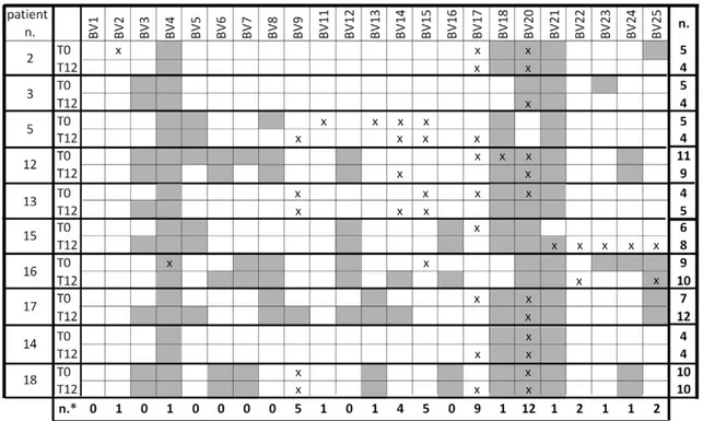
The TCR repertoire analysis, performed in samples obtained before therapy initiation and after 12 months from 10 randomly chosen patients, showed that TCR heterogeneity was slightly modified by the therapy with the number of TCR variable beta (TCRBV) chain perturbations decreasing in four patients (n. 2, 3, 5, and 12) and increasing in four (n. 13, 15, 16 and 17); in the remaining two patients (n. 14, 18), the same number of perturbed TCRBV chains were observed before and after the therapy (Fig. 2). In half of the patients clonal expansions were present in less than 6 TCRBV chains over the 23 segments analyzed at T0 and/or T12 while, in the other half, between 6 and 12 TCRBV chains were perturbed. It is also evident that clonal expansions prefer-entially involved specific TCRBV chains: the heterogeneity of TCRBV4+ cells was limited in all patients, both before and
after therapy, whereas that of cells expressing TCRBV18, TCRBV20 and TCRBV21 segments was limited in most of the patients. The preferentially expanded TCRBV segments were TCRBV20 and TCRBV17. These expansions were not associ-ated with clinical or laboratory signs of infection or with MS relapses.
The replicative history of the B-cell subpopulations of MS patients, calculated as the average number of B-cell
divisions, was not significantly different between genders (median at T12: 4.04 in males and 4.39 in females, Mann– Whitney test, p = NS), did not change significantly with age, nor was affected by natalizumab therapy, and was similar to that observed in HDs (Fig. 3a). As found in controls, the average number of divisions in the total B-cell population of MS patients correlated inversely with the number of logKRECs/ml in samples obtained before treatment and 12 months after therapy initiation (Fig. 3b).
3.3. Transmigration of lymphocyte subsets through BEC and LEC monolayers
Six and 12 months of therapy with natalizumab had only a little effect on lymphocyte subset transendothelial migra-tion because the percentage of recovery of the total population of CD8+ and CD19+ lymphocytes (solid lines of Fig. 4a), and of naïve and memory unswitched and switched CD19+counterparts (solid lines ofFig. 4b) at the bottom of
the BEC- or LEC-coated transwells reflected the effect of the drug seen in the ex vivo samples. Only the in vitro migration of CD45RA+T effector memory (TEMRA) cells through LECs
(bold solid line ofFig. 4b), but not through BECs, appeared to be selectively modified by 12 months of therapy. A very slight influence of the therapy was also observed on CD4+
cells because a reduced percentage of CD4+ lymphocytes
was observed after 6 months of therapy in the transmigrated samples (solid lines ofFig. 4a), while in ex vivo samples the decrease became apparent only after 12 months of treat-ment (bold solid line ofFig. 4a). However the drug, that did
Figure 2 Analysis of TCR repertoire. The percent usage of TCRBV (from BV1 to BV25) segments and the perturbations of TCRBV CDR3-length distributions were determined by spectratyping analysis in samples obtained before therapy initiation (T0) and after 12 months (T12). The modifications of TCRBV perturbations were considered significant when their values were beyond the mean + 2SD (gray squares) of the CDR3 distribution found in HDs. n: number of perturbed TCRBV chains at each time point. TCRBV chains were considered overused when their percent usage was higher than mean + 2SD (“x”) of the TCRBV chain usage found in 5 HDs. n*: number of preferentially expressed TCRBV chains.
Figure 3 Analysis of B-cell divisions. The average number of B-cell divisions in MS patients and HDs was calculated at the indicated time points. The statistical analysis was performed using the Friedman test, the comparison of data obtained at T0, T6, and T12 with those of HDs was done by the Mann–Whitney test, and the correlation of the average number of B-cell divisions with the log number of KRECs was obtained by the Spearman's test.
Figure 4 Cell subset transmigration across BECs and LECs. PBMCs, obtained before therapy initiation (T0) and after 6 (T6) and 12 (T12) months, were either subjected to flow cytometric analysis to obtain the percentage of cell subsets in the ex vivo condition, or added to the top of the BEC and LEC monolayers and allowed to transmigrate to the bottom chamber. All transmigrated cells were then collected, counted, stained with labelled monoclonal antibodies and phenotyped. a. Percentages of ex vivo and transmigrated CD4+and CD8+T
lymphocytes and CD19+ B cells. b. Percentages of ex vivo and transmigrated CD4+ and CD8+ T-cell subsets and CD19+ B-cell subpopulations. The percentages of ex vivo and transmigrated“recent thymic emigrants” are depicted as hatched bars. Statistical analysis was performed by two-factor ANOVA with repeated measures on both factors, as detailed in the Supplementary Material and methods. Bars indicate the mean percentage of the cell subsets analyzed in the three experimental conditions; error bars indicate ± SD; dashed lines indicate significant differences between the mean cell percentages in the three experimental conditions (ex vivo vs. BEC vs. LEC transmigration); solid lines indicate the significant differences due to the therapy observed at T6 in comparison to T0; bold solid lines indicate peculiar therapy-induced changes in cell percentages (e.g. present in the ex vivo just at T12, or only after passage through one of two types of endothelium). TCM: T central memory cells; TEM: T effector memory cells. *: pb0.05, **: pb0.01, and ***: pb0.001.
not induce changes in the percentages of T-cell subsets in ex vivo samples, also seemed to be completely ineffective in altering the percentage of CD4+cell subsets which migrated
through BEC- or LEC-coated transwells (Fig. 4b). Neverthe-less, the most relevant result of the transmigration assay is that, independently of the therapy, the mean percentages of CD4+, CD8+ and CD19+ lymphocytes recovered at the
bottom of coated transwell inserts were, respectively, significantly higher, lower and unmodified with respect to those observed in the ex vivo samples (dotted lines of Fig. 4a); the same features, however, were also observed using lymphocyte subsets prepared from HDs (Supplementa-ry Fig. 3a). Furthermore, naïve CD4+and CD8+lymphocytes
showed a worse propensity to migrate across BECs and LECs than their memory counterparts in both patients and HDs (Fig. 4b and Supplementary Fig. 3b). A similar situation was observed for the naïve B-cell subset, whereas a significant mean percent increase of immature B cells and a decrease of memory unswitched B cells were found after transmigration only in MS patients and not in HDs. Finally, independently of the treatment, a selective effect due to the type of endothelial cells coated on the transwell inserts, appeared to affect only the migration of CD4+ central memory
population, which passed more preferentially through the BEC than through the LEC monolayer; on the contrary, memory unswitched B-lymphocyte percentage was slightly increased when these cells transmigrated through the LEC monolayer.
4. Discussion
Natalizumab treatment has been shown to reduce the migration of leukocytes into the CNS[1,6,11,16,17], to inhibit the retention of memory- and marginal zone-like B cells within the spleen[6]and to induce a sequestration of lymphocytes, in particular activated T cells[18], and of immature B cells[4]in the peripheral circulation of MS-treated patients. However, considering the scarcity of leukocytes in the CNS compared to the periphery and the slight contribution of memory- and marginal zone-like B cells on the composition of the lymphocyte pool, it is unlikely that the reduced migration of leukocytes into the CNS and the decreased retention of specific B-cell subsets in the spleen can quantitatively affect peripheral lymphocyte count or fully explain the increase of lymphocytes observed during natalizumab therapy. Instead, by using the TREC and KREC assay we found that newly produced T and B lymphocytes were significantly increased in peripheral blood of patients treated for 6 and 12 months with natalizumab, thus indicating that the drug may influence the lymphocyte release from production sites. The increased production of new T and B lymphocytes was confirmed by flow cytometry that also demonstrated an augmented number of RTE and naïve CD4+
cells in treated patients while, as also shown by Planas et al.[6], the percentage of these cell subsets remained unmodified.
Considering that T cells of natalizumab-treated patients undergo activation and differentiation in the secondary lymphoid organs and then remained sequestered in the circulation[18]and that every perturbation in the number and longevity of naïve and memory lymphocyte populations must be balanced by counteracting mechanisms that try to keep the balance between the different compartments
[19,20], it is likely that the high number of TRECs results from the increased thymic release necessary to maintain the T-cell pool homeostasis. This possibility is supported by the finding that reduction of VLA-4 levels also renders mature thymocytes less adhesive to the thymic stromal cells, thus more easily released into circulation [21]. Our data also show that the number of TRECs found in pre-therapy samples was similar to that of age- and gender-matched HDs, while previous results reported that thymic output is low in untreated MS patients [22,23]. One possible explanation can be that all but one of our patients had been previously treated with immunomodulatory drugs, whereas, in the other studies they were naïve for therapy. In addition, flow cytometry confirmed that the pre-therapy number of “recent thymic emigrants” was comparable to that of controls and showed that newly released naïve CD4+ and
CD8+lymphocytes were both increased in treated patients.
In some treated patients, despite the very short duration of the therapy, the thymic output of new T-cell specificities resulted in a mild enlargement of the T-cell heterogeneity. Actually, wide T-cell repertoire changes are unexpected since a renewal of TCR diversity may require a long time and, for instance, modifications of T-cell repertoire have been observed in MS patients only after two years since BM transplantation [24]. In some other treated patients the T-cell diversity appeared to be reduced, as expected, forasmuch as circulating CD8+ lymphocytes, which are the
cells that preferentially expanded clonally [25], resulted increased in our treated patients. Flow cytometric analysis also indicated that the observed increase of KRECs in the peripheral blood is likely to be sustained by the rise of immature and naïve B cells. Nevertheless, the majority of B cells in peripheral blood of treated patients were unswitched and switched memory B cells, which are known to undergo an antigen-independent (homeostatic) proliferation [14]. How-ever, the comparable average number of B-cell divisions we have found before and after therapy supports the hypothesis that the natalizumab-induced increase of memory B cells is due to an inhibited retention in the spleen [6]more than a peripheral proliferation.
Another reason for the accumulation of cells containing TRECs and KRECs during time in natalizumab-treated patients could be a drug-induced inhibition of trafficking through endothelial cells. This is an important issue since the regulation of immune cell trafficking across endothelial barriers is critical for immune cell maturation and for mounting of appropriate responses within specific organ compartments. In MS patients, this is even more crucial because deficits of immune surveillance, including defects in new T- and B-cell production and in the ability of immune cells to transmigrate across the BBB, may predispose to progressive multifocal leukoencephalopathy (PML) [26,27]. In transmigration assays performed to measure the migrato-ry capacity of lymphocytes, BBB-endothelial cells have been previously used either untreated, activated, or triggered by a gradient of chemoattractants[27–29]because, during the MS-associated inflammatory process, activated meningeal or BBB-endothelial cells amplify the migration of immune cells to the CNS parenchyma in a multi-step process that involves selectins, chemokines (and their receptors) and cell adhe-sion molecules [28]. Because in MS patients, trafficking through non-BBB endothelial cells probably takes place in
more physiological conditions, we performed the transmi-gration assay with non‐activated BECs and LECs, thus avoiding the addition of any exogenous cytokine (exception made for the VEGF used for cell growth) that would make results difficult to be interpreted. In this setting, the transmigration is mediated only by the interaction of integrins present on lymphocyte surface with their ligands on endothelial cells[30]. In these experimental conditions, using all the appropriate controls, we demonstrated that natalizumab therapy only marginally interferes with the ability of T and B lymphocytes to pass through non‐activated monolayers of BECs and LECs. Indeed, natalizumab therapy showed a minimal inhibitory effect on CD4+ cells
transmi-gration and induced an increased passage of TEMRA+ cells
through LEC monolayers, but the other modifications observed in the percentages of remaining T- and B-cell subsets collected after migration merely recalled the therapy-induced subset changes observed in the ex vivo samples. Thus, lymphocyte trafficking through vascular or lymphatic endothelium is only barely modified by the therapy in condition of basal trafficking. On the contrary, we found that, independently from the treatment, CD4+and
CD8+ cells with phenotypic characteristics of T central
memory, T effector memory, and TEMRA+ cells have a
higher migratory capacity than their naïve counterparts both in MS patients and HDs. Therefore, CD8+effector memory T
lymphocytes transmigrate more readily not only across BBB-endothelial cells than CD8+ non-effector memory
coun-terpart[28], but they also can more efficiently migrate across BECs and LECs than the naïve CD8+subpopulation. However,
again, this feature was observed within CD8+lymphocytes of
both MS patients and HDs. Collectively, our findings indicate that the differential migration rates through vascular and lymphatic endothelium are most likely derived from the different basal expression ofα4 integrin on distinct T and B subsets and not from natalizumab treatment. Furthermore, the data ruled out the possibility that non‐activated endothe-lial cells are a passive barrier, without a role in lymphocyte migration. Finally, one possible explanation for the lack of differential lymphocyte transmigration through the two different types of endothelium (the only exceptions being CD4+T central memory cells and memory unswitched B cells)
is the similar level of VCAM-1 found on BEC- and LEC-cell surface [31]. These transmigration data only apparently contradict previously published results demonstrating a reduced transmigration of lymphocytes obtained from natalizumab-treated patients [27,28]because these studies were obtained using very different experimental conditions, including the use of purified lymphocyte subsets and of human brain microvascular endothelial cells, activated with different stimuli. Indeed, lymphocyte transmigration is a process that is strictly regulated in conditions of both homeostasis and inflammation[32]and therefore the mechanisms involved in this process can vary substantially as a direct reflection of the involved lymphocyte subsets, the wide range of endothelia encountered in the vasculature, and activation conditions of both. Further studies will be needed to verify whether these findings can be confirmed when a transmigration model through brain microvascular endothelial cells is employed. Considering that the JC virus enters the CNS in a non-mutant form and capsid protein VP1 mutations can originate and be positively selected within the CNS[33], and that a decreased
production of new T and B cells was found in a patient with PML[26], it is likely that the lower capability of naïve cells to cross the BBB may contribute to the local reduction of lymphocyte specificities recognizing mutated viral proteins, which should be a crucial prerequisite for controlling JC-virus induced PML.
5. Conclusions
Because a good thymic and BM output assures the continuous renewal of the immune compartments and the subsequent integrity of the immune response, the measure of TRECs and KRECs could be a very useful tool to estimate the activity of natalizumab and, possibly, to identify those patients that, having an altered thymic and BM output, could be at risk of developing PML.
Supplementary data to this article can be found online at http://dx.doi.org/10.1016/j.clim.2012.07.007.
Conflict of interest statement
RC has received speaking fees from Biogen Idec, Sanofi-Aventis, and Novartis. LI has received research supports from Merck Serono and Biogen Dompè. CZ, MC, FS, AS, EG, FT, LC, and SR have no conflict of interest.
Acknowledgments
CZ was supported by an unrestricted educational grant from Biogen Dompé, Italy.
References
[1] A. Lutterotti, R. Martin, Getting specific: monoclonal anti-bodies in multiple sclerosis, Lancet Neurol. 7 (2008) 538–547. [2] B. Bielekova, B.L. Becker, Monoclonal antibodies in MS: mechanisms of action, Neurology 74 (Suppl. 1) (2010) S31–S40. [3] P. Wipfler, K. Oppermann, G. Pilz, S. Afazel, E. Haschke-Becher, A. Harrer, M. Huemer, A. Kunz, S. Golaszewski, W. Staffen, G. Ladurner, J. Kraus, Adhesion molecules are promising candi-dates to establish surrogate markers for natalizumab treatment, Mult. Scler. 17 (2011) 16–23.
[4] M. Krumbholz, I. Meinl, T. Kümpfel, R. Hohlfeld, E. Meinl, Natalizumab disproportionately increases circulating pre-B and B cells in multiple sclerosis, Neurology 71 (2008) 1350–1354. [5] N. Putzki, M.K. Baranwal, B. Tettenborn, V. Limmroth, E.
Kreuzfelder, Effects of natalizumab on circulating B cells, T regulatory cells and natural killer cells, Eur. Neurol. 63 (2010) 311–317.
[6] R. Planas, I. Jelčić, S. Schippling, R. Martin, M. Sospedra, Natalizumab treatment perturbs memory- and marginal zone-like B-cell homing in secondary lymphoid organs in multiple sclerosis, Eur. J. Immunol. 42 (2012) 790–798.
[7] M. Skarica, C. Eckstein, K.A. Whartenby, P.A. Calabresi, Novel mechanisms of immune modulation of natalizumab in multiple sclerosis patients, J. Neuroimmunol. 235 (2011) 70–76. [8] F. Zohren, D. Toutzaris, V. Klärner, H.P. Hartung, B. Kieseier,
R. Haas, The monoclonal anti-VLA-4 antibody natalizumab mobilizes CD34 + hematopoietic progenitor cells in humans, Blood 111 (2008) 3893–3895.
[9] C.H. Polman, P.W. O'Connor, E. Havrdova, M. Hutchinson, L. Kappos, D.H. Miller, J.T. Phillips, F.D. Lublin, G. Giovannoni,
A. Wajgt, M. Toal, F. Lynn, M.A. Panzara, A.W. Sandrock, AFFIRM Investigators, A randomized, placebo-controlled trial of natalizumab for relapsing multiple sclerosis, N. Engl. J. Med. 354 (2006) 899–910.
[10] D.H. Ryan, B.L. Nuccie, C.N. Abboud, J.M. Winslow, Vascular cell adhesion molecule-1 and the integrin VLA-4 mediate adhesion of human B cell precursors to cultured bone marrow adherent cells, J. Clin. Invest. 88 (1991) 995–1004.
[11] M. Niino, C. Bodner, M.L. Simard, S. Alatab, D. Gano, H.J. Kim, M. Trigueiro, D. Racicot, C. Guérette, J.P. Antel, A. Fournier, F. Grand'Maison, A. Bar-Or, Natalizumab effects on immune cell responses in multiple sclerosis, Ann. Neurol. 59 (2006) 748–754. [12] F. Serana, A. Sottini, M. Chiarini, C. Zanotti, C. Ghidini, A. Lanfranchi, L.D. Notarangelo, L. Caimi, L. Imberti, The different extent of B and T cell immune reconstitution after hematopoietic stem cell transplantation and enzyme replace-ment therapies in SCID patients with adenosine deaminase deficiency, J. Immunol. 185 (2010) 7713–7722.
[13] A. Sottini, C. Ghidini, C. Zanotti, M. Chiarini, L. Caimi, A. Lanfranchi, D. Moratto, F. Porta, L. Imberti, Simultaneous quantification of recent thymic T-cell and bone marrow B-cell emigrants in patients with primary immunodeficiency undergone to stem cell transplantation, Clin. Immunol. 136 (2010) 217–227. [14] M.C. van Zelm, T. Szczepanski, M. van der Burg, J.J.M. van Dongen, Replication history of B lymphocytes reveals homeo-static proliferation and extensive antigen-induced B cell expansion, J. Exp. Med. 204 (2007) 645–655.
[15] P. Ye, D.E. Kirschner, Reevaluation of T cell receptor excision circles as a measure of human recent thymic emigrants, J. Immunol. 168 (2002) 4968–4979.
[16] O. Stüve, C.M. Marra, A. Bar-Or, M. Niino, P.D. Cravens, S. Cepok, E.M. Frohman, J.T. Phillips, G. Arendt, K.R. Jerome, L. Cook, F. Grand'Maison, B. Hemmer, N.L. Monson, M.K. Racke, Altered CD4 +/CD8 + T-cell ratios in cerebrospinal fluid of natalizumab-treated patients with multiple sclerosis, Arch. Neurol. 63 (2006) 1383–1387.
[17] M. del Pilar Martin, P.D. Cravens, R. Winger, E.M. Frohman, M.K. Racke, T.N. Eagar, S.S. Zamvil, M.S. Weber, B. Hemmer, N.J. Karandikar, B.K. Kleinschmidt-DeMasters, O. Stüve, Decrease in the numbers of dendritic cells and CD4 + T cells in cerebral perivascular spaces due to natalizumab, Arch. Neurol. 65 (2008) 1596–1603.
[18] P. Kivisäkk, B.C. Healy, V. Viglietta, F.J. Quintana, M.A. Hootstein, H.L. Weiner, S.J. Khoury, Natalizumab treatment is associated with peripheral sequestration of proinflammatory T cells, Neurology 72 (2009) 1922–1930.
[19] J.E. Crowley, J.L. Scholz, W.J. Quinn III, J.E. Stadanlick, J.F. Treml, L.S. Treml, Y. Hao, R. Goenka, P.J. O'Neill, A.H. Matthews, R.F. Parsons, M.P. Cancro, Homeostatic control of B lymphocyte subsets, Immunol. Res. 42 (2008) 75–83.
[20] K. Takada, S.C. Jameson, Naive T cell homeostasis: from awareness of space to a sense of place, Nat. Rev. Immunol. 9 (2009) 823–832.
[21] M. Sawada, J. Nagamine, K. Takeda, K. Utsumi, A. Kosugi, Y. Tatsumi, T. Hamaoka, K. Miyake, K. Nakajima, T. Watanabe, S. Sakakibara, H. Fujiwara, Expression of VLA-4 on thymocytes. Maturation stage-associated transition and its correlation with
their capacity to adhere to thymic stromal cells, J. Immunol. 149 (1992) 3517–3524.
[22] A. Hug, M. Korporal, I. Schröder, J. Haas, K. Glatz, B. Storch-Hagenlocher, B. Wildemann, Thymic export function and T cell homeostasis in patients with relapsing remitting multiple sclerosis, J. Immunol. 171 (2003) 432–437.
[23] C. Zanotti, M. Chiarini, F. Serana, R. Capra, M. Rottoli, M. Rovaris, G. Cavaletti, R. Clerici, M. Rezzonico, L. Caimi, L. Imberti, Opposite effects of interferon-β on new B and T cell release from production sites in multiple sclerosis patients, J. Neuroimmunol. 240–241 (2011) 147–150.
[24] P.A. Muraro, D.C. Douek, A. Packer, K. Chung, F.J. Guenaga, R. Cassiani-Ingoni, C. Campbell, S. Memon, J.W. Nagle, F.T. Hakim, R.E. Gress, H.F. McFarland, R.K. Burt, R. Martin, Thymic output generates a new and diverse TCR repertoire after autologous stem cell transplantation in multiple sclerosis patients, J. Exp. Med. 201 (2005) 805–816.
[25] D.A. Laplaud, C. Ruiz, S. Wiertlewski, S. Brouard, L. Berthelot, M. Guillet, B. Melchior, N. Degauque, G. Edan, P. Brachet, P. Damier, J.P. Soulillou, Blood T-cell receptor beta chain transcriptome in multiple sclerosis. Characterization of the T cells with altered CDR3 length distribution, Brain 127 (2004) 981–995.
[26] A. Sottini, R. Capra, C. Zanotti, M. Chiarini, F. Serana, D. Ricotta, L. Caimi, L. Imberti, Pre-existing T- and B-cell defects in one progressive multifocal leukoencephalopathy patient, PLoS One 7 (2012) e34493.
[27] N. Schwab, K.G. Höhn, T. Schneider-Hohendorf, I. Metz, M.P. Stenner, S. Jilek, R.A. Du Pasquier, R. Gold, S.G. Meuth, R.M. Ransohoff, W. Brück, H. Wiendl, Immunological and clinical consequences of treating a patient with natalizumab, Mult. Scler. 18 (2012) 335–344.
[28] I. Ifergan, H. Kebir, J.I. Alvarez, G. Marceau, M. Bernard, L. Bourbonnière, J. Poirier, P. Duquette, P.J. Talbot, N. Arbour, A. Prat, Central nervous system recruitment of effector memory CD8+ T lymphocytes during neuroinflammation is dependent on α4 integrin, Brain 134 (2011) 3560–3577.
[29] C.L. Pittet, J. Newcombe, A. Prat, N. Arbour, Human brain endothelial cells endeavor to immunoregulate CD8 T cells via PD-1 ligand expression in multiple sclerosis, J. Neuroinflammation 8 (2011) 155.
[30] A.N. Kogan, H.H. von Adrian, Lymphocyte trafficking, in: R.F. Tuma, W.N. Duran, K. Ley (Eds.), Microcirculation, Academic press, San Diego, 2008, pp. 449–482.
[31] A.L. Fletcher, V. Lukacs-Kornek, E.D. Reynoso, S.E. Pinner, A. Bellemare-Pelletier, M.S. Curry, A.R. Collier, R.L. Boyd, S.J. Turley, Lymph node fibroblastic reticular cells directly present peripheral tissue antigen under steady-state and inflammatory conditions, J. Exp. Med. 207 (2010) 689–697.
[32] B.P. Lee, B.A. Imhof, Lymphocyte transmigration in the brain: a new way of thinking, Nat. Immunol. 9 (2008) 117–118. [33] L. Gorelik, C. Reid, M. Testa, M. Brickelmaier, S. Bossolasco, A.
Pazzi, A. Bestetti, P. Carmillo, E. Wilson, M. McAuliffe, C. Tonkin, J.P. Carulli, A. Lugovskoy, A. Lazzarin, S. Sunyaev, K. Simon, P. Cinque, Progressive multifocal leukoencephalopathy (PML) development is associated with mutations in JC virus capsid protein VP1 that change its receptor specificity, J. Infect. Dis. 204 (2011) 103–114.